
Challenges in Diagnosis and Clinical Management of COVID-19 in Patient with B-Cell Chronic Lymphocytic Leukemia (CLL): Report of One Case

 , ,
, , 

Abstract
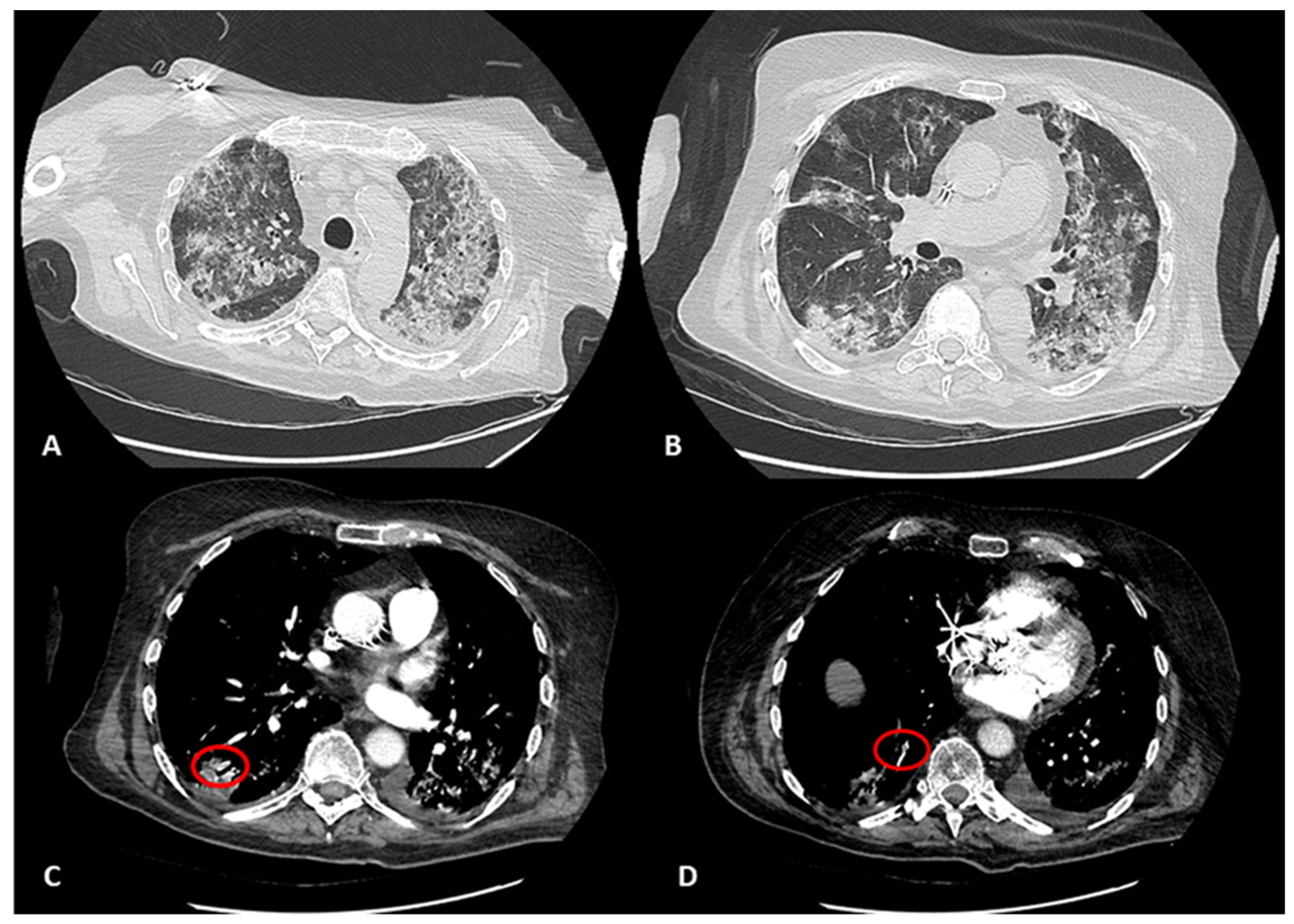
:1. Case Report
2. Ethical Approval
3. Discussion and Conclusions
Author Contributions
Funding
Informed Consent Statement
Acknowledgments
Conflicts of Interest
References
- Li, K.; Fang, Y.; Li, W.; Pan, C.; Qin, P.; Zhong, Y.; Liu, X.; Huang, M.; Liao, Y.; Li, S. CT image visual quantitative evaluation and clinical classification of coronavirus disease (COVID-19). Eur. Radiol. 2020, 30, 4407–4416. [Google Scholar] [CrossRef] [PubMed] [Green Version]
- Prokop, M.; van Everdingen, W.; van Rees Vellinga, T.; van Ufford, J.Q. CO-RADS—A categorical CT assessment scheme for patients with suspected COVID-19: Definition and evaluation. Radiology 2020, 27, 2020. [Google Scholar] [CrossRef] [PubMed]
- Capalbo, C.; Aceti, A.; Simmaco, M.; Bonfini, R.; Rocco, M.; Ricci, A.; Napoli, C.; Rocco, M.; Alfonsi, V.; Teggi, A.; et al. The Exponential Phase of the COVID-19 Pandemic in Central Italy: An Integrated Care Pathway. Int. J. Environ. Res. Public Health 2020, 17, 3792. [Google Scholar] [CrossRef] [PubMed]
- Jin, X.H.; Zheng, K.I.; Pan, K.-H.; Xie, Y.-P.; Zheng, M.-H. COVID-19 in a patient with chronic lymphocytic leukaemia. Lancet Haematol. 2020, 7, e351–e352. [Google Scholar] [CrossRef]
- Paneesha, S.; Pratt, G.; Parry, H.; Moss, P. COVID-19 infection in therapy-naive patients with B-cell chronic lymphocytic leukemia. Leuk. Res. 2020, 93, 106366. [Google Scholar] [CrossRef] [PubMed]
- WHO: Laboratory Biosafety Guidance Related to Coronavirus Disease (COVID-19). Available online: https://apps.who.int/iris/bitstream/handle/10665/331500/WHO-WPE-GIH-2020.2-eng.pdf?sequence=1&isAllowed=y (accessed on 24 April 2020).
- Corman, V.M.; Landt, O.; Kaiser, M.; Molenkamp, R.; Meijer, A.; Chu, D.; Bleiker, T.; Brünink, S.; Schneider, J.; Schmidt, L.; et al. Detection of 2019 novel coronavirus (2019-nCoV) by real-time RT-PCR. Euro Surveill. 2020, 25, 2000045. [Google Scholar] [CrossRef] [PubMed] [Green Version]
- Lou, B.; Li, T.D.; Zheng, S.F.; Su, Y.Y.; Li, Z.Y.; Liu, W.; Yu, F.; Ge, S.X.; Zou, Q.D.; Yuan, Q.; et al. Serology characteristics of SARS-CoV-2 infection since the exposure and post symptoms onset. Eur. Respir. J. 2020, 56, 2000763. [Google Scholar] [CrossRef]
- Niu, A.; Ning, B.; Socola, F.; Safah, H.; Reynolds, T.; Ibrahim, M.; Safa, F.; Alfonso, T.; Luk, A.; Mushatt, D.M.; et al. COVID-19 in Patients with Hematological Malignancies: High False Negative Rate with High Mortality. Blood 2020, 135, 6–7. [Google Scholar] [CrossRef]
- Mihaila, R. Management of patients with chronic lymphocytic leukemia during the SARS-CoV-2 pandemic. Oncol. Lett. 2021, 22, 636. [Google Scholar] [CrossRef]
- Nuccetelli, M.; Pieri, M.; Grelli, S.; Ciotti, M.; Miano, R.; Andreoni, M.; Bernardini, S. SARS-CoV-2 infection serology: A useful tool to overcome lockdown? Cell Death Discov. 2020, 6, 38. [Google Scholar] [CrossRef]
- Lippi, G.; Salvagno, G.L.; Pegoraro, M.; Militello, V.; Caloi, C.; Peretti, A.; Gaino, S.; Bassi, A.; Bovo, C.; Cascio, G.L. Assessment of immune response to SARS-CoV-2 with fully automated 2019-nCoV IgG and IgM chemiluminescence immunoassays. Clin. Chem. Lab. Med. 2020, 58, 1156–1159. [Google Scholar] [CrossRef] [PubMed] [Green Version]
- Padoan, A.; Cosma, C.; Sciacovelli, L.; Faggian, D.; Plebani, M. Analytical performances of a chemiluminescence immunoassay for SARS-CoV-2 IgM/IgG and antibody kinetics. Clin. Chem. Lab. Med. 2020, 58, 1081–1088. [Google Scholar] [CrossRef] [PubMed] [Green Version]
- Available online: https://www.who.int/news-room/commen-taries/detail/immunity-passports (accessed on 24 April 2020).
- Weinstein, M.C.; Freedberg, K.A.; Hyle, E.P.; Paltiel, A.D. Waiting for Certainty on COVID-19 Antibody Tests-At What Cost? N. Engl. J. Med. 2020, 5, e37. [Google Scholar] [CrossRef] [PubMed]
- Cheng, M.P.; Papenburg, J.; Desjardins, M.; Kanjilal, S.; Quach, C.; Libman, M.; Dittrich, S.; Yansouni, C.P. Diagnostic Testing for Severe Acute Respiratory Syndrome–Related Coronavirus 2—A Narrative Review. Ann. Int. Med. 2020, 172, 726–734. [Google Scholar] [CrossRef] [PubMed] [Green Version]
- Available online: http://www.governo.it/sites/new.governo.it/files/CallForTender_20200419_en.pdf (accessed on 24 April 2020).
- Available online: https://www.fda.gov/medical-devices/emergency-situations-medical-devices/eua-authorized-serology-test-performance (accessed on 24 April 2020).
- Safarpour, D.; Srinivasan, K.; Farooqui, M.; Roth, C.; Ghouse, M. A Case of COVID-19-Induced Lymphocytosis in a Patient with Treatment-Naive CLL: Should It Be treated? Clin. Lymphoma Myeloma Leuk. 2021, 21, 69–72. [Google Scholar] [CrossRef] [PubMed]


| FEBRUARY | MARCH | APRIL | MAY | |||||||||||||||||||||||||||||||||||||
|---|---|---|---|---|---|---|---|---|---|---|---|---|---|---|---|---|---|---|---|---|---|---|---|---|---|---|---|---|---|---|---|---|---|---|---|---|---|---|---|---|
| RT-PCR-SARS CoV-2 | - | - | - | - | ||||||||||||||||||||||||||||||||||||
| SARS CoV-2 IgG α -N (a) | - | |||||||||||||||||||||||||||||||||||||||
| SARS CoV-2 IgG α -N (b) | - | |||||||||||||||||||||||||||||||||||||||
| SARS CoV-2 IgG α -N+S | + | |||||||||||||||||||||||||||||||||||||||
| SARS CoV-2 IgG α-S | + | |||||||||||||||||||||||||||||||||||||||
| Blood WBC Count * | 17 | 47 | 62 | 61 | 45 | 42 | 35 | 41 | 25 | 13 | ||||||||||||||||||||||||||||||
| Blood Lympho Count * | 13 | 40 | 54 | 49 | 37 | 36 | 24 | 38 | 22 | 11 | ||||||||||||||||||||||||||||||
| Blood Neutro Count * | 2, 6 | 6 | 7, 2 | 6, 2 | 4, 8 | 4, 6 | 4, 1 | 2, 3 | 1, 8 | 1, 6 | ||||||||||||||||||||||||||||||
| Fever > 37.5° | + | + | ||||||||||||||||||||||||||||||||||||||
| Dyspnea | + | + | + | + | + | + | + | + | + | + | + | + | + | + | + | + | + | + | + | + | + | |||||||||||||||||||
| Cough | + | + | + | + | + | + | + | + | + | + | + | + | + | + | + | + | + | + | + | + | + | + | + | + | + | + | + | + | ||||||||||||
| Emergency Room | ||||||||||||||||||||||||||||||||||||||||
| Intensive Care Unit | ||||||||||||||||||||||||||||||||||||||||
| Pneumology | ||||||||||||||||||||||||||||||||||||||||
| Hospitalization | ||||||||||||||||||||||||||||||||||||||||
Publisher’s Note: MDPI stays neutral with regard to jurisdictional claims in published maps and institutional affiliations. |
© 2022 by the authors. Licensee MDPI, Basel, Switzerland. This article is an open access article distributed under the terms and conditions of the Creative Commons Attribution (CC BY) license (https://creativecommons.org/licenses/by/4.0/).
Share and Cite
Visco, V.; Lippi, M.E.; Salerno, G.; Licata, M.A.V.A.C.; de Dominicis, C.; Antolino, G.; La Verde, G.; Santino, I.; Simmaco, M.; Sciacchitano, S. Challenges in Diagnosis and Clinical Management of COVID-19 in Patient with B-Cell Chronic Lymphocytic Leukemia (CLL): Report of One Case. Hematol. Rep. 2022, 14, 31-37. https://doi.org/10.3390/hematolrep14010006
Visco V, Lippi ME, Salerno G, Licata MAVAC, de Dominicis C, Antolino G, La Verde G, Santino I, Simmaco M, Sciacchitano S. Challenges in Diagnosis and Clinical Management of COVID-19 in Patient with B-Cell Chronic Lymphocytic Leukemia (CLL): Report of One Case. Hematology Reports. 2022; 14(1):31-37. https://doi.org/10.3390/hematolrep14010006
Chicago/Turabian StyleVisco, Vincenzo, Maria Enrichetta Lippi, Gerardo Salerno, Maria Angela Vittoria A. C. Licata, Chiara de Dominicis, Giusy Antolino, Giacinto La Verde, Iolanda Santino, Maurizio Simmaco, and Salvatore Sciacchitano. 2022. "Challenges in Diagnosis and Clinical Management of COVID-19 in Patient with B-Cell Chronic Lymphocytic Leukemia (CLL): Report of One Case" Hematology Reports 14, no. 1: 31-37. https://doi.org/10.3390/hematolrep14010006
APA StyleVisco, V., Lippi, M. E., Salerno, G., Licata, M. A. V. A. C., de Dominicis, C., Antolino, G., La Verde, G., Santino, I., Simmaco, M., & Sciacchitano, S. (2022). Challenges in Diagnosis and Clinical Management of COVID-19 in Patient with B-Cell Chronic Lymphocytic Leukemia (CLL): Report of One Case. Hematology Reports, 14(1), 31-37. https://doi.org/10.3390/hematolrep14010006