Review

Reactive Oxygen Species as Key Molecules in the Pathogenesis of Alcoholic Fatty Liver Disease and Nonalcoholic Fatty Liver Disease: Future Perspectives

1
Department of Hepatobiliary Surgery, The Third Affiliated Hospital, Chongqing Medical University, Yubei, Chongqing 401120, China
2
School of Public Health, Chongqing Medical University, Chongqing 400016, China
*
Authors to whom correspondence should be addressed.
Curr. Issues Mol. Biol. 2025, 47(6), 464; https://doi.org/10.3390/cimb47060464
Submission received: 9 April 2025 / Revised: 6 June 2025 / Accepted: 12 June 2025 / Published: 17 June 2025
(This article belongs to the Special Issue Molecular Research on Free Radicals and Oxidative Stress)

Abstract

Reactive oxygen species (ROS) are central to the progression of alcoholic fatty liver disease (ALD) and nonalcoholic fatty liver disease (NAFLD). In ALD, ROS arise from alcohol metabolism (CYP2E1 and ADH/ALDH2), causing oxidative damage and fibrosis. In NAFLD, mitochondrial dysfunction, ER stress, and lipotoxicity drive ROS overproduction due to metabolic dysregulation. Both diseases share ROS-mediated pathways, including mitochondrial/ER dysfunction, inflammation, and impaired lipid metabolism, accelerating steatosis to cirrhosis and cancer. Antioxidants, ER modulators, and lifestyle changes show therapeutic potential but require further clinical validation. Future research should leverage multi-omics and targeted therapies to optimize ROS-focused interventions for ALD and NAFLD.

1. Introduction

In recent years, significant global increases in the incidence of both alcoholic fatty liver disease (ALD) and nonalcoholic fatty liver disease (NAFLD) have been witnessed, establishing them as leading causes of non-viral liver disease. ALD shows a strong correlation with chronic excessive alcohol consumption, particularly prevalent among heavy drinkers in Western male populations [1]. In parallel, NAFLD has emerged as the hepatic component of metabolic syndrome, closely associated with obesity and type 2 diabetes. With approximately 25% of adults worldwide affected, the prevalence of NAFLD continues to rise in tandem with growing obesity and diabetes rates. Collectively, ALD and NAFLD now surpass viral hepatitis as the most burdensome liver diseases globally [2].
The pathogenesis of ALD involves reactive oxygen species (ROS) generated through multiple pathways of alcohol metabolism. These ROS, along with alcohol-derived metabolites, inflict direct hepatocellular damage while promoting hepatic inflammation and fibrosis [3]. Similarly, NAFLD progression is driven by oxidative stress stemming from insulin resistance and dysregulated lipid metabolism, both hallmark features of the disease [4]. ROS not only cause hepatocyte injury but also initiate and amplify inflammatory signaling pathways, creating a self-perpetuating cycle of oxidative damage and chronic inflammation.
Clinically significant is the increasing population with concurrent NAFLD and alcohol consumption. These patients demonstrate accelerated progression to liver fibrosis, cirrhosis, and hepatocellular carcinoma. Current evidence implicates ROS as a critical mediator in the exacerbated disease progression observed when NAFLD coexists with alcohol use. Therefore, elucidating ROS mechanisms in these overlap syndromes is crucial for understanding their rapid progression and may yield novel therapeutic approaches. This review examines ROS involvement in both ALD and NAFLD progression, compares their sources and mechanistic roles, evaluates antioxidant-based therapies, and explores future research directions targeting ROS in these diseases.

2. ROS Pathogenesis in ALD and NAFLD

ROS represent a class of oxygen-derived free radicals—including superoxide (O₂), hydrogen peroxide (H2O2), hydroxyl radical (OH), and singlet oxygen (1O2)—that play dual roles in liver physiology and pathology. These reactive molecules originate primarily from the following three key sources: (1) mitochondrial electron leakage during oxidative phosphorylation; (2) NOX enzyme activity; and (3) cytochrome P450 metabolism (particularly CYP2E1). While serving as crucial second messengers in normal cell signaling and host defense, ROS become pathogenic when their production overwhelms cellular antioxidant capacity [5].
The liver employs specialized cellular systems for ROS generation and regulation: hepatocytes maintain redox balance through tightly controlled mitochondrial ROS production coupled with efficient scavenging systems; Kupffer cells utilize NOX-derived ROS as defensive weapons during immune activation; and activated hepatic stellate cells (HSCs) leverage ROS signaling to drive fibrotic responses during tissue repair [6].
The pathophysiological impact of specific ROS follows distinct mechanisms: signaling ROS (O2/H2O2): modulate inflammatory cascades at physiological levels; destructive ROS (OH): cause indiscriminate macromolecular damage through membrane lipid peroxidation and DNA strand breaks; and dual-role H2O2: functions as a redox regulator at low concentrations but triggers oxidative injury when accumulated [6].
Additionally, since nitric oxide (NO) acts as an intercellular messenger with high diffusibility in the body, its reaction with O₂⁻ forms a unique type of ROS—peroxynitrite (ONOO−), which also belongs to reactive nitrogen species (RNS). ONOO− can attack all biomolecules, causing not only oxidative damage but also nitrative damage [7]. Among the most destructive oxidant species in cells are ·OH and ONOO− [8].

2.1. ROS Production in NAFLD (Figure 1)

NAFLD represents a dynamic disease continuum, progressing from simple hepatic steatosis to the more aggressive inflammatory condition of nonalcoholic steatohepatitis (NASH). This progression carries significant clinical implications, as NASH-related cirrhosis demonstrates markedly higher HCC incidence compared to other chronic liver diseases [9].
The liver serves as the metabolic command center for lipid homeostasis, uniquely coordinating fatty acid uptake, de novo lipogenesis, β-oxidation, and very-low-density lipoproteins (VLDLs)-mediated lipid export. Hepatocytes maintain lipid balance through three primary pathways: (1) mitochondrial β-oxidation for energy production, (2) VLDL assembly and secretion for peripheral distribution, and (3) lipid droplet storage for temporary buffering [10].
The following two pathophysiological drivers dominate NAFLD development:
(1)
Metabolic dysregulation: Approximately 15% of hepatic fatty acids are derived directly from dietary intake. Chronic overnutrition leads to excessive lipid accumulation in hepatocytes, forming lipid droplets. This stored fat can subsequently be broken down into free fatty acids (FFAs), further contributing to hepatic stress and inflammation [11].
(2)
Insulin resistance: The accumulation of lipid droplets and FFAs from metabolic dysregulation can impair insulin signaling. This weakened insulin response reduces the suppression of adipocyte lipolysis, leading to an excessive release of FFAs into the bloodstream. These FFAs are then transported to the liver, compounding the cycle of fat accumulation and liver dysfunction [12,13].
This metabolic perfect storm triggers multiple hepatotoxic mechanisms: mitochondrial dysfunction from FFA overload, ER stress response activation, and lipotoxicity-mediated cellular damage [14]. Together, these processes create a self-perpetuating cycle of oxidative stress through three well-characterized ROS-generating pathways (Figure 1):
Figure 1. Mechanisms of ROS generation in NAFLD.
Cimb 47 00464 g001

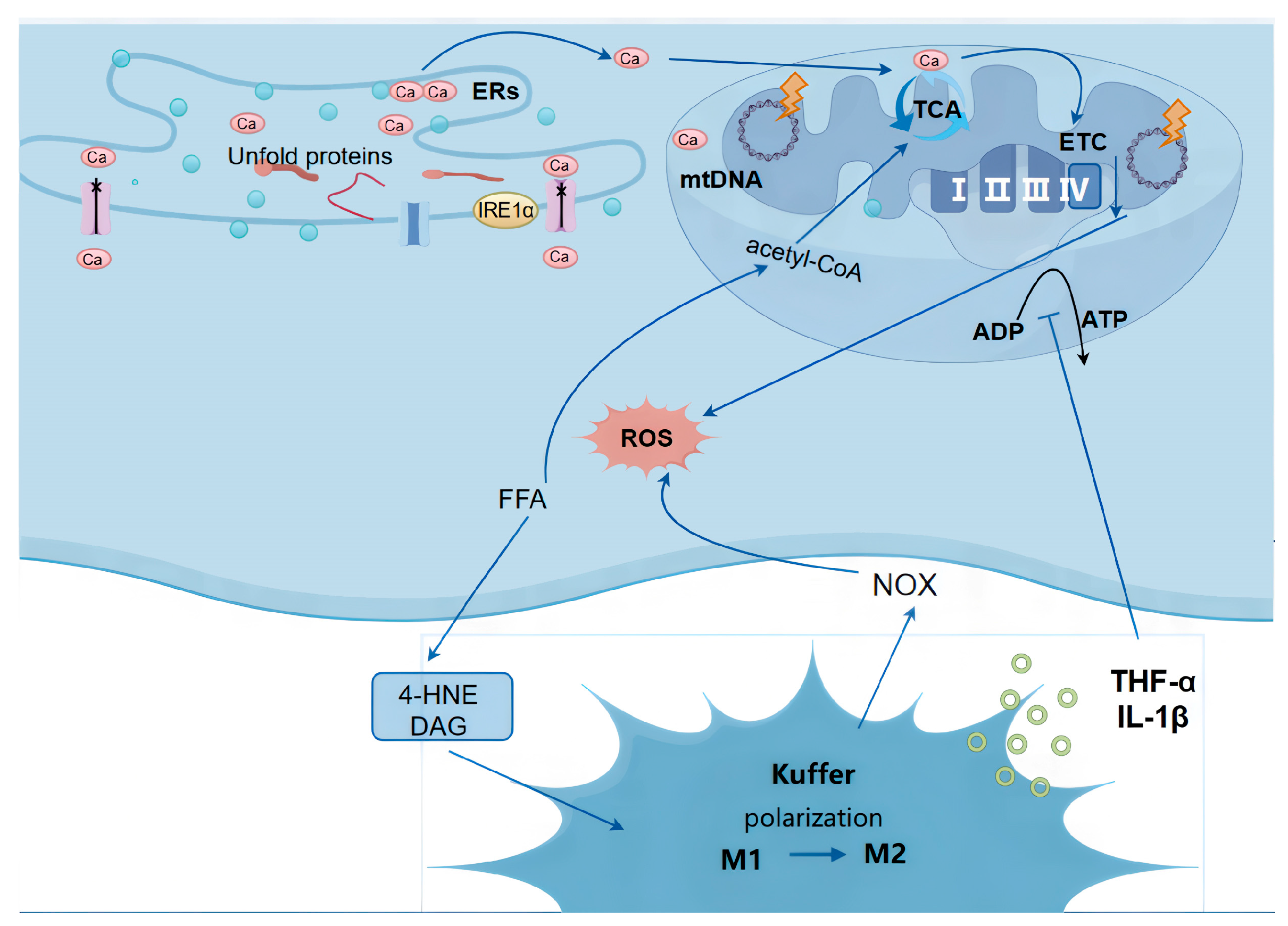
2.1.1. Mitochondrial Dysfunction

Hepatocyte mitochondria serve two critical metabolic functions: ATP production and fatty acid β-oxidation. Under normal physiological conditions, mitochondrial respiration generates ROS as natural byproducts. The electron transport chain (ETC), composed of enzyme complexes and mobile electron carriers (coenzyme Q and cytochrome c), typically leaks only 2–3% of electrons during aerobic respiration, resulting in physiological ROS levels [15].
In NAFLD pathogenesis, lipid metabolism dysregulation leads to excessive intracellular fatty acid accumulation, which significantly impairs mitochondrial function. When the ETC reaches saturation from FFAs β-oxidation, electron leakage increases dramatically, generating pathological ROS levels [16]. This oxidative stress is further exacerbated through multiple mechanisms: FFA overload compromises mitochondrial membrane integrity, while inflammatory cytokines from activated Kupffer cells and HSCs in NASH cause mtDNA damage, impairing mitochondrial protein synthesis and ETC complex function. FFAs induce ER stress that disrupts cellular Ca2+ homeostasis, promoting mitochondrial permeability transition pore (mPTP) opening. The subsequent Ca2+ influx into mitochondria hyperactivates the respiratory chain, tricarboxylic acid cycle (TCA), and oxidative phosphorylation, creating an “electron traffic jam” that increases ROS production [17,18].

2.1.2. Endoplasmic Reticulum Stress (ER Stress)

Endoplasmic reticulum stress (ERS) is a pathological process triggered by the abnormal accumulation of misfolded or unfolded proteins within the ER lumen. Its core inducers include oxidative stress, metabolic disturbances, and toxic insults [19,20]. While ERS initially functions as an adaptive or protective cellular mechanism, persistent or excessive protein misfolding leads to cellular damage and stress. In NAFLD, ERS is primarily driven by lipotoxicity: a high-fat diet or obesity causes excessive accumulation of free fatty acids (FFAs) and cholesterol in hepatocytes. Concurrently, during NASH progression, inflammatory cytokines disrupt ER proteostasis, cross-activating ER stress pathways. When combined with lipid metabolism dysfunction, ethanol-induced suppression of lipophagy further exacerbates FFA accumulation, synergistically promoting ERs.
The unfolded protein response (UPR) constitutes the core mechanism of ERS, mediated by three ER transmembrane sensors: protein kinase R-like endoplasmic reticulum kinase (PERK), inositol-requiring enzyme 1α (IRE1α), and activating transcription factor 6 (ATF6) [21]. Calcium (Ca2+) dysregulation is a key contributor to ERS-induced ROS generation. UPR activation stimulates IRE1α, leading to Ca2+ leakage from the ER. Additionally, sarco/endoplasmic reticulum Ca2+-ATPase (SERCA), responsible for pumping cytosolic Ca2+ back into the ER, becomes impaired during ERS. This results in ER Ca2+ depletion and cytosolic/mitochondrial Ca2+ overload, further driving ROS production [22].
Moreover, ERs suppresses the antioxidant defense system: while PERK activation transiently induces Nrf2 expression, chronic ERs promotes Nrf2 degradation, reducing antioxidant enzymes such as SOD and catalase (CAT) [23]. Additionally, IRE1α upregulates NADPH oxidase 4 (NOX4), further amplifying ROS generation [18].

2.1.3. Lipotoxicity

Lipotoxicity results from excessive accumulation of FFAs and their metabolites (ceramides, diacylglycerols (DAG), and 4-hydroxynonenal(4-HNE)) in hepatocytes, causing direct cellular damage and metabolic dysfunction. This process drives Kupffer cell polarization from anti-inflammatory (M2) to pro-inflammatory (M1) phenotypes, which then generate excessive ROS via NADPH oxidase (NOX) activation while secreting inflammatory cytokines (TNF-α and IL-1β) that exacerbate oxidative stress. These mediators concurrently disrupt mitochondrial membrane potential and impair ATP production, creating a self-amplifying cycle of metabolic dysfunction and ROS overproduction [24,25].
Three core mechanisms of hepatocyte injury in NAFLD: mitochondrial dysfunction (excessive FFAs cause electron leakage in the ETC and ROS burst), endoplasmic reticulum stress (ERS triggers Ca2+ dysregulation and NOX4 upregulation via the UPR pathway), and lipotoxicity (FFA metabolites activate Kupffer cell M1 polarization, exacerbating oxidative stress through NOX). Key molecules labeled in the figure—including the TCA cycle, ATP, ROS, and the M1 → M2 polarization process—collectively illustrate the vicious cycle of oxidative stress and metabolic dysregulation in NAFLD.

2.2. ROS Production in ALD (Figure 2)

ALD is caused by chronic excessive alcohol consumption and is a major cause of chronic liver disease worldwide [26]. While some patients may develop severe ALD, including alcoholic steatohepatitis (ASH), cirrhosis, and HCC, the pathogenesis of ALD remains unclear despite significant progress over the past two decades, and there are currently no FDA-approved drugs for ALD treatment.
The development of ALD is rooted in the disruption of hepatic metabolic homeostasis following alcohol absorption in the intestine and subsequent metabolism in the liver and other organs. Hepatic steatosis occurs in over 90% of heavy drinkers and is characterized by lipid accumulation in hepatocytes [27]. Multiple mechanisms contribute to hepatic steatosis, including impaired mitochondrial fatty acid β-oxidation, lipid migration from extrahepatic tissues to the liver, and alterations in lipid metabolism-related transcription factors. ASH is defined by specific histological features, including marked steatosis, inflammatory cell infiltration, pericellular fibrosis, and hepatocyte ballooning degeneration. Among ASH patients, 8–20% progress to cirrhosis, and 3–10% of those with alcohol-related cirrhosis develop HCC [26].
ALD shares some similarities with NAFLD in terms of pathogenesis, but since the liver is the primary site of alcohol metabolism, the main source of ROS in ALD is derived from alcohol metabolism [28]. There are three major pathways through which alcohol metabolism generates ROS in the liver (Figure 2).
Figure 2. Mechanism of ROS generation in ALD.
Cimb 47 00464 g002

2.2.1. Alcohol Dehydrogenase (ADH) and Aldehyde Dehydrogenase 2 (ALDH2) Pathway

Ethanol is first metabolized to acetaldehyde by ADH, which is then further converted to acetate by ALDH2. Both steps generate reduced nicotinamide adenine dinucleotide (NADH), leading to an increased NADH/NAD+ ratio. This imbalance inhibits the mitochondrial respiratory chain, resulting in enhanced electron leakage during electron transfer and subsequent ROS production.

2.2.2. Microsomal Ethanol Oxidizing System (MEOS)

MEOS represents an alternative pathway for alcohol metabolism, primarily occurring in the endoplasmic reticulum. Cytochrome P450 2E1 (CYP2E1), the key enzyme in MEOS, catalyzes the oxidation of ethanol to acetaldehyde. During this process, CYP2E1 requires NADPH as an electron donor, which is oxidized to NADP+ while generating superoxide anion (O2). These superoxide anions are further converted to hydrogen peroxide (H2O2) and other ROS. This pathway constitutes the most significant source of ROS in alcohol metabolism.

2.2.3. Catalase Pathway

The catalase pathway primarily operates during chronic alcohol consumption and metabolizes ethanol by decomposing hydrogen peroxide. This pathway generates relatively lower levels of ROS compared to the other two mechanisms.
Three major pathways of ethanol metabolism generate ROS in ALD. The first is the ADH- and ALDH2-mediated metabolic pathway, where ethanol is converted to acetaldehyde and then to acetate, simultaneously producing NADH that leads to redox imbalance. The second is the MEOS, catalyzed by the CYP2E1 enzyme, which directly generates ROS intermediates. The third is the catalase pathway, producing relatively lower levels of ROS. Key molecules labeled in the figure include metabolic enzymes, cofactors, and ROS, demonstrating the molecular correlation between alcohol metabolism and oxidative stress.

2.3. Similarities and Differences in ROS Production Between ALD and NAFLD

2.3.1. Common Sources of ROS in ALD and NAFLD

Both ALD and NAFLD involve excessive ROS production due to increased electron leakage in the mitochondrial ETC, driven by alcohol metabolism in ALD and FFA metabolism in NAFLD. Additionally, both diseases can induce endoplasmic reticulum (ER) stress, leading to Ca2+ dyshomeostasis and subsequent mitochondrial ROS generation. When progressing to ASH or NASH, both conditions activate Kupffer cells and promote the release of pro-inflammatory cytokines (e.g., TNF-α and IL-6), further enhancing ROS production.

2.3.2. Key Differences in ROS Generation Between ALD and NAFLD

(1)
Distinct Initial Triggers of ROS Production
ALD: Alcohol metabolism is the primary source of ROS, particularly through the ADH/ALDH2 and CYP2E1 pathways, which directly generate large amounts of ROS.
NAFLD: Insulin resistance and dysregulated lipid metabolism are the main drivers of ROS production. FFA overload leads to mitochondrial dysfunction and ER stress, indirectly increasing ROS levels.
(2)
Different Dominant Pathways of ROS Generation
In ALD:
ADH/ALDH2 Pathway: Alcohol metabolism generates NADH, disrupting the NADH/NAD+ ratio, inhibiting the mitochondrial respiratory chain, and increasing electron leakage.
CYP2E1 Pathway: CYP2E1-mediated alcohol metabolism produces substantial superoxide anion (O₂⁻) and hydrogen peroxide (H2O2), representing the dominant ROS-generating mechanism in ALD.
In NAFLD:
Mitochondrial β-Oxidation: FFA overload accelerates mitochondrial β-oxidation, overloading the ETC and increasing electron leakage.
ER Stress: FFAs induce ER stress, causing Ca2+ leakage and subsequent mitochondrial ROS production.
Kupffer Cells Activation: FFAs and lipid metabolites directly damage hepatocytes and activate Kupffer cells to produce ROS.
See Table 1 for a comparative summary.

3. Mechanisms of ROS Action in ALD and NAFLD

3.1. Shared Mechanisms of ROS in ALD and NAFLD Pathogenesis (Figure 3)

Despite their distinct etiologies, ALD and NAFLD converge through ROS-mediated mechanisms, where oxidative stress damages mitochondria, ER, and lysosomes while simultaneously activating inflammatory pathways and exacerbating lipid metabolic dysfunction, collectively driving disease progression from steatosis to hepatitis, fibrosis, and ultimately hepatocellular carcinoma (Figure 3).
Figure 3. Common mechanisms of ROS in ALD and NAFLD.
Cimb 47 00464 g003

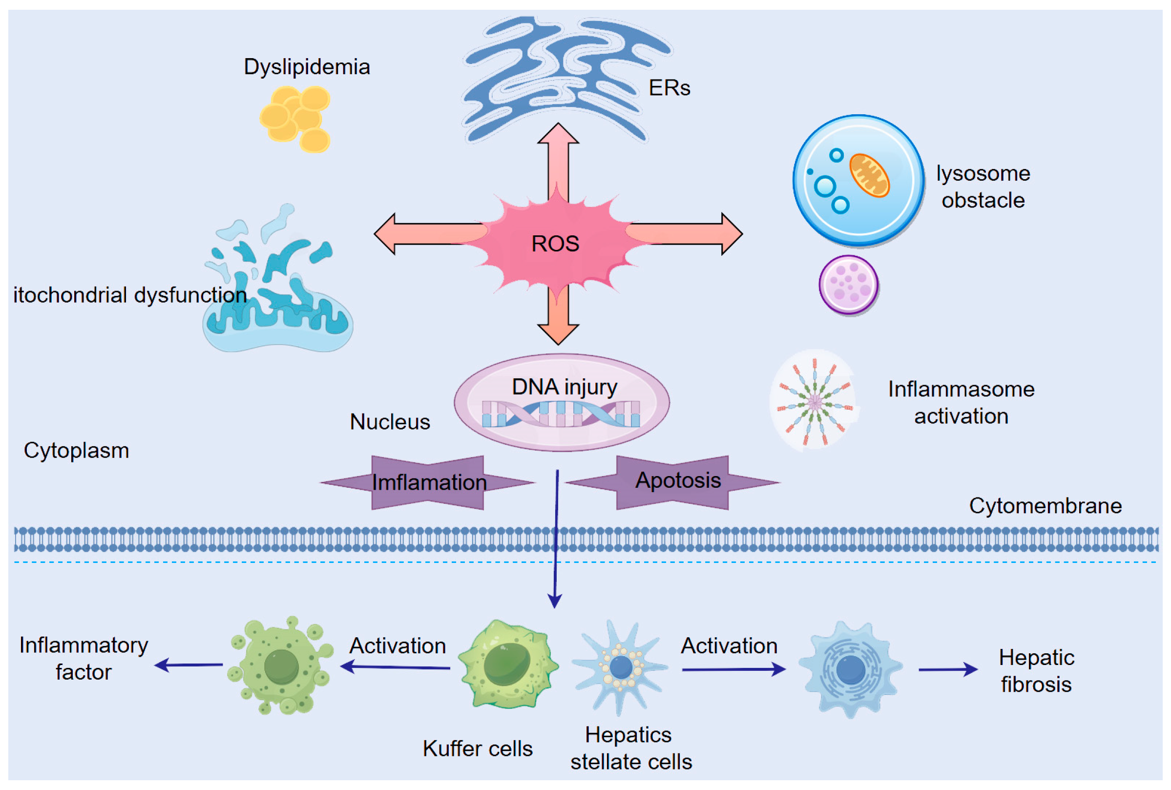
3.1.1. ROS-Induced Mitochondrial Dysfunction

Mitochondria serve as both the primary source and key target of intracellular ROS. Excessive ROS induce mitochondrial membrane lipid peroxidation, disrupting the ETC and impairing oxidative phosphorylation. This oxidative stress reduces levels of PTEN-induced putative kinase 1 (PINK1), a protective mitochondrial serine/threonine–protein kinase, while concurrently causing abnormal mitochondrial morphology, diminished membrane potential, and decreased activities of Complex I and IV—collectively exacerbating ETC dysfunction and electron leakage to generate more ROS [15].
The vulnerability of mtDNA—lacking histone protection and having limited repair capacity—makes it particularly susceptible to ROS-mediated damage. ROS (e.g., O2 and ·OH) directly attack mtDNA, inducing base modifications, strand breaks, and cross-linking, which compromise mtDNA integrity and disrupt mitochondrial protein synthesis. Furthermore, ROS-generated lipid peroxidation products (e.g., 4-HNE) bind to and inactivate mtDNA, while ROS-induced loss of mitochondrial membrane potential promotes Ca2+ influx, aggravating mtDNA damage [29].
ROS also trigger hepatocyte apoptosis through multiple pathways. Oxidative modification of anti-apoptotic Bcl-2 family proteins (e.g., Bcl-2 and Bcl-xL), inhibiting their function and promoting BAX/BAK activation. p53-mediated upregulation of BAX expression, driving BAX/BAK oligomerization to form mitochondrial outer membrane pores, increasing permeability and cytochrome C release—a pivotal step in activating the apoptosome and caspase cascade [30,31].
Additionally, ROS-induced mitochondrial damage and lipid peroxidation activate ferroptosis pathways, further accelerating hepatocyte death [32].

3.1.2. ROS-Induced Endoplasmic Reticulum Stress (ER Stress)

ROS promote ERs by oxidizing amino acid residues (e.g., cysteine, methionine, and histidine) in proteins, leading to structural and functional ER damage. Cysteine residues (-SH) are particularly susceptible to ROS attack, where oxidation by H2O2 or OH generates sulfenic (-SOH), sulfinic (-SO2H), or sulfonic (-SO3H) acid modifications. These oxidative changes disrupt disulfide bonds (-S-S-), inducing protein misfolding, aggregation, or degradation, thereby exacerbating the UPR. This establishes a vicious cycle of ERS amplification, Ca2+ dyshomeostasis, and mitochondrial ROS release [33].
ERS Triggers Pro-Inflammatory Cascades
The three major UPR sensors—PERK, IRE1α, and ATF6—drive pro-inflammatory cytokine release through multiple pathways [34]:
(1)
IRE1α upregulates TNF-α and IL-6 via its downstream target, spliced X-box binding protein 1 (XBP1s) [35].
(2)
ER stress activates the IκB kinase (IKK) complex, inducing NF-κB nuclear translocation and subsequent TNF-α/IL-6 production [36].
(3)
IRE1α recruits TRAF2, activating the JNK pathway to amplify cytokine release [37].
This sustained inflammation promotes HSC activation and fibrotic collagen deposition, accelerating NASH progression and liver fibrosis [19].
ERS Drives Metabolic Dysregulation
UPR intersects with cytosolic stress kinases (JNK, PKR, and IKK), each linked to metabolic disorders [38]:
(1)
Obesity and Insulin Resistance:
ERS activates c-Jun N-terminal kinase (JNK), which phosphorylates IRS1 at serine residues, impairing insulin receptor signaling and inducing insulin resistance [39,40,41]. Global JNK deficiency prevents weight gain, whereas JNK activation promotes obesity [42].
(2)
Lipid Metabolism Dysfunction:
ERS disrupts apolipoprotein B100 (ApoB100) folding, reducing VLDL assembly/secretion and causing hepatic triglyceride (TG) accumulation [43]. These metabolic disturbances—severe obesity, insulin resistance, and dyslipidemia—worsen NAFLD and ALD progression.

3.1.3. ROS-Mediated Lysosomal Dysfunction

ROS exert dual opposing effects on lysosomal function that collectively impair cellular homeostasis: By inducing lipid peroxidation of lysosomal membranes, ROS increase membrane permeability, causing leakage of cathepsins into the cytosol, where they promote proteolysis, while hydroxyl radicals (OH) generate protein carbonyl derivatives through side-chain oxidation, leading to structural alterations, cross-linking, and enhanced proteasomal degradation [37].
Paradoxically, ROS simultaneously suppress autophagic flux by oxidizing critical amino acid residues in lysosomal enzymes to inhibit their catalytic activity [44], while also disrupting microtubule stability and soluble N-ethylmaleimide-sensitive factor (SNARE) protein function vesicle-associated membrane protein 8 (VAMP8)/syntaxin 17 (STX17) to impair autophagosome–lysosome fusion [45]. This autophagic blockade exacerbates hepatic steatosis by preventing lipid droplet clearance [46,47] and triggers Nod-like receptor pyrin-containing protein 3 (NLRP3) inflammasome activation through accumulated damaged organelles/proteins, amplifying IL-1β/IL-18 release [48,49]. Furthermore, oxidative damage to lysosomal membranes activates HSCs, directly promoting fibrogenesis [37,50].

3.1.4. ROS-Mediated Activation of Inflammatory Signaling Pathways

Beyond ER stress and lysosome-mediated autophagy dysfunction, ROS activate inflammatory pathways through multiple mechanisms. As a core component of inflammatory responses, the NLRP3 inflammasome is directly activated by ROS through either oxidation of NLRP3 protein or mitochondrial damage-induced mtDNA release. Activated NLRP3 complexes with ASC (apoptosis-associated speck-like protein) and pro-caspase-1 to form the inflammasome, which cleaves pro-IL-1β and pro-IL-18 into their mature forms, triggering inflammatory cell recruitment and amplifying inflammatory cascades [49].
ROS further activate the MAPK pathway by stimulating upstream kinases (ASK1 and MEKK1), leading to ERK activation and nuclear translocation of transcription factors (AP-1 and ATF2) that upregulate pro-inflammatory cytokines (TNF-α and IL-6) and fibrogenic genes (TGF-β) [51,52]. In IL-6 signaling, ROS promote receptor dimerization and JAK kinase activation, resulting in STAT protein phosphorylation. Phosphorylated STAT dimers then translocate to the nucleus to induce expression of IL-6 and anti-apoptotic genes [53].
ROS-induced cytokines (TNF-α and IL-1β) reduce mitochondrial membrane potential and ATP production, further increasing ROS leakage. This creates a vicious cycle where elevated ROS activate NF-κB and JNK pathways to amplify cytokine production [54,55]. Additionally, ROS-stimulated fibrogenic factors (TGF-β and PDGF) activate quiescent HSCs, transforming them into collagen-producing myofibroblasts. Activated HSCs excessively synthesize extracellular matrix (ECM) components while suppressing matrix metalloproteinase (MMP) activity, collectively exacerbating fibrotic progression [55].
As central regulators, ROS drive hepatic fibrosis through multifaceted inflammatory activation—directly modifying inflammasome components, amplifying cytokine signaling via MAPK/JAK-STAT pathways, and sustaining a self-perpetuating oxidative stress-inflammation cycle that ultimately activates HSCs and disrupts ECM homeostasis.

3.1.5. ROS-Mediated Dysregulation of Lipid Metabolism

ROS induce mitochondrial dysfunction, impairing hepatic β-oxidation of FFAs and leading to lipid metabolism dysregulation, resulting in hepatic steatosis. Both alcohol-induced and metabolic disorder-associated mitochondrial dysfunction exacerbate FFA accumulation, establishing a vicious cycle of lipotoxicity and oxidative stress.
Beyond indirect effects through mitochondrial impairment and inflammatory pathway activation, ROS directly modulate lipid metabolism through multiple mechanisms. In hepatocytes, AMP-activated protein kinase (AMPK) plays a crucial role in maintaining mitochondrial function and promoting autophagy. AMPK activation enhances fatty acid oxidation and reduces lipid accumulation; however, ROS can directly oxidize the α subunit of AMPK, inhibiting fatty acid oxidation while promoting lipogenesis and hepatic steatosis [56].
Sterol regulatory element-binding protein 1c (SREBP-1c), a master transcriptional regulator of hepatic lipogenesis, shows markedly elevated expression (5-fold higher than controls) in steatotic livers [12]. ROS activate SREBP-1c, which upregulates downstream targets including acetyl-CoA carboxylase (ACC) and FFAs, thereby promoting fatty acid and triglyceride synthesis. Additionally, ROS oxidatively modify the DNA-binding domain of peroxisome proliferator-activated receptor α (PPARα), a key regulator of fatty acid oxidation, suppressing its transcriptional activity and contributing to lipid accumulation [57].
Concurrent with these metabolic disturbances, hepatic fibrogenesis progresses through ROS-mediated mechanisms. Hepatocyte inflammation and altered immune microenvironments prompt Kupffer cells to release ROS and TGF-β, activating HSCs. Activated HSCs significantly increase collagen production, ultimately driving liver fibrosis [58].

3.1.6. Additional Mechanisms of ROS Action in Hepatocytes

Beyond mitochondrial DNA damage, ROS directly target nuclear DNA and RNA through multiple oxidative pathways. The nucleobase guanine (G), possessing the lowest oxidation potential, is particularly vulnerable to ROS attack. Hydroxyl radicals (OH) specifically oxidize the C8 position of guanine, forming the mutagenic adduct 8-hydroxy-2′-deoxyguanosine (8-OHdG)—a well-characterized biomarker of oxidative DNA damage [59,60]. Peroxynitrite interacts with both DNA bases and sugar moieties, inducing DNA strand breaks. Due to its low oxidation potential, the most vulnerable DNA base attacked by ROS/RNS is guanine, leading to the formation of 8-nitroguanine [8]. More severely, ·OH can penetrate cellular and nuclear membranes to directly attack DNA backbone structures, inducing both single- and double-strand breaks (DSBs), with DSBs representing the most catastrophic form of genomic damage [61].
ROS also drive extensive lipid peroxidation cascades, oxidizing polyunsaturated fatty acids in cellular membranes and cytoplasm to generate highly reactive aldehyde byproducts, including 4-HNE, 4-hydroperoxy-2-nonenal (HPNE), and MDA. These electrophilic intermediates readily form covalent adducts with cellular proteins and nucleic acids [62,63], establishing a self-perpetuating cycle of macromolecular damage that exacerbates hepatocyte dysfunction and death. ONOO− can cause severe damage to amino acids and proteins by modifying them through the oxidation or nitration of tyrosine, tryptophan, methionine, and other amino acid residues [64].
The core mechanisms of ROS in steatotic liver disease are as follows: (1) mitochondrial dysfunction, (2) endoplasmic reticulum stress, (3) lysosomal impairment, (4) inflammatory pathway activation, (5) lipid metabolism dysregulation, and (6) DNA damage. Key cellular components and molecules are labeled to illustrate the multi-pathway hepatotoxic effects of ROS.

3.2. Differential Mechanisms of ROS in ALD Versus NAFLD

3.2.1. The Role of ROS in ALD

There are significant epidemiological differences in ALD between Eastern and Western populations. Western populations demonstrate substantially higher rates of alcohol-induced cirrhosis and hepatocellular carcinoma, which correlates with their greater prevalence of heavy drinking and higher per capita alcohol consumption. In contrast, the incidence of alcoholic steatohepatitis has increased rapidly in recent years, particularly among young adults aged 19–44 years and women. Studies reveal that while Western populations require daily alcohol intake exceeding 80 g to significantly increase cirrhosis risk, Eastern populations show elevated risk at just 60 g/day, with women being particularly susceptible—cirrhosis may develop with as little as 20 g/day [2]. These differences suggest that beyond drinking patterns, variations in alcohol metabolism may play a key role. Eastern populations have a higher frequency of the ALDH2×2 gene mutation, which impairs acetaldehyde metabolism, along with greater CYP2E1 activity that promotes ROS generation during alcohol metabolism [65].
ALD also exhibits marked gender differences, with women being more vulnerable to alcohol’s toxic effects and more prone to developing ALD. This heightened susceptibility may stem from women’s lower alcohol dehydrogenase (ADH) activity, which shifts alcohol metabolism toward the microsomal ethanol-oxidizing system (MEOS) pathway [66]. The increased sensitivity of Eastern populations and women to alcohol-induced liver damage strongly implicates ROS as a critical mediator in ALD pathogenesis. ROS-induced oxidative stress appears to play a pivotal role in the initiation and progression of ALD. Alcohol consumption induces rapid elevation of ROS levels, while chronic alcohol exposure leads to persistent ROS stimulation combined with the synergistic effects of ethanol and its metabolites, resulting in more acute and severe oxidative damage.
The combined action of alcohol and ROS potently inhibits multiple hepatic enzymes, significantly disrupting lipid metabolism through dual inhibition of AMPK, a critical regulator of fatty acid β-oxidation. Notably, SREBP1c expression is rapidly upregulated by acetaldehyde and ROS following acute alcohol exposure, with this induction occurring more rapidly than the gradual stimulation observed in NAFLD, leading to pronounced hepatic lipid accumulation [67,68]. Epidemiological studies confirm this accelerated pathogenesis, with hepatic steatosis developing within two weeks of heavy drinking and present in 90% of moderate-to-heavy drinkers [26].
Alcohol-derived aldehydes and ROS act synergistically to promote extensive lipid peroxidation. Liver biopsy specimens from chronic drinkers demonstrate the formation of carcinogenic exocyclic DNA adducts through 4-HNE-DNA reactions, which impair DNA function while accelerating fibrotic progression [69,70]. Concurrently, MDA accumulation induces cross-linking of cellular macromolecules, including proteins and nucleic acids. These reactive aldehydes and ROS collectively activate HSCs, driving accelerated fibrogenesis and cirrhosis development [71]. Consequently, chronic alcohol exposure induces hepatic fibrosis more rapidly than NAFLD, with 10–20% of ALD patients developing fibrosis—a significantly higher rate than observed in NAFLD—reflecting the more aggressive oxidative stress and tissue damage in ALD [27].
An intriguing clinical observation is that while ALD progression is rapid during active drinking, abstinence can effectively halt disease advancement. Hepatic steatosis and inflammation typically show significant improvement within weeks to months of cessation, with some patients demonstrating fibrosis regression [72,73]. This phenomenon may relate to unique adaptive mechanisms in alcohol-exposed livers. In vitro studies using ethanol-treated hepatocytes and HepG2 cells reveal that chronic alcohol exposure not only induces CYP2E1 and ROS production but also upregulates glutathione (GSH), glutathione-S-transferase (GST), and heme oxygenase-1 (HO-1) through increased γ-glutamylcysteine synthetase expression, collectively mitigating oxidative damage [74]. Parallel research demonstrates alcohol-induced activation of Nrf2 and subsequent upregulation of CYP2A5, which contributes to ROS clearance [75]. These adaptive responses persist after alcohol withdrawal, enabling continued ROS neutralization and oxidative stress resolution.

3.2.2. The Role of ROS in NAFLD

A strong positive correlation exists between the degree of oxidative stress/ROS elevation and disease severity in NAFLD patients. Longitudinal studies have demonstrated a progressive and significant increase in both serum and hepatic ROS levels during disease progression from simple steatosis to NASH and subsequent fibrosis [76]. Comparative analyses reveal that NASH patients exhibit substantially elevated serum ROS concentrations compared to those with simple steatosis, while hepatic ROS levels show a stage-dependent increase that strongly correlates with fibrosis severity (F2-F4 vs. F0-F1 stages) [65].
The oxidative stress biomarkers malondialdehyde (MDA) and 8-hydroxy-2′-deoxyguanosine (8-OHdG) serve as direct molecular evidence of ROS-induced oxidative damage. Their concentrations demonstrate significant positive correlations with histological grading of steatosis, lobular inflammation, and fibrosis staging. Clinical data indicate that serum MDA levels are significantly elevated in NAFLD patients compared to healthy controls, with NASH patients showing further increases [77]. Similarly, quantitative immunohistochemical analyses reveal that hepatic 8-OHdG levels exhibit a strong positive correlation with the degree of liver fibrosis [77,78].
The development of antibodies targeting nitrotyrosine has significantly advanced research on peroxynitrite. Numerous animal and large-scale clinical studies have detected nitrotyrosine in the tissues of obese and diabetic patients, suggesting its potential role in the diagnosis of NAFLD [79]. More recently, studies have proposed that molecular fluorescent probes for real-time detection of ONOO− levels exhibit higher sensitivity and efficacy than tissue staining or serum biomarker assays in assessing early-stage toxicity and diagnosis of NAFLD [80].
The pathophysiology of ROS accumulation in NAFLD involves both increased production and impaired clearance mechanisms. A critical determinant of ROS homeostasis is the cellular antioxidant defense system, particularly the enzymatic antioxidant capacity. Comparative studies demonstrate significant reductions in the activities of key antioxidant enzymes—including superoxide dismutase (SOD) and glutathione peroxidase (GPx)—in NAFLD patients relative to healthy controls, with the most pronounced deficiencies observed in NASH patients [81]. Importantly, patients with compromised antioxidant defenses show a three-fold higher incidence of liver fibrosis (45% vs. 15%), suggesting that antioxidant depletion may represent a critical driver of disease progression from steatosis to NASH and fibrosis. These findings are supported by multiple clinical studies that consistently demonstrate significant associations between ROS levels, oxidative damage markers, antioxidant capacity, and NAFLD progression [82,83], providing compelling evidence for the central role of oxidative stress in NAFLD pathogenesis.
The development of NAFLD is primarily driven by insulin resistance and dysregulated lipid metabolism, with accumulated ROS subsequently inducing organelle and cellular damage through oxidative stress—representing the “second hit” in NAFLD pathogenesis.
The metabolic disturbances caused by ROS in NAFLD differ significantly from those in ALD. ROS-mediated mechanisms in NAFLD include the following:
(1)
Activation of inflammatory cytokines that interfere with insulin receptor substrate (IRS) phosphorylation, exacerbating insulin resistance [84,85].
(2)
Inhibition of fatty acid oxidation through various inflammatory signaling pathways, promoting lipid accumulation and worsening hepatic steatosis [86].
These parallel processes synergistically accelerate NAFLD progression.
During early NAFLD stages, elevated FFAs stimulate mitochondrial activity, generating excessive ROS that damage mitochondrial structure and function. As disease progresses, accumulated mitochondrial dysfunction suppresses respiratory chain activity, creating an energy imbalance that impairs ROS clearance. This vicious cycle worsens hepatic lipid accumulation and oxidative stress, ultimately promoting hepatocyte necrosis/apoptosis and driving NASH and fibrogenesis [87,88].
NAFLD severity correlates strongly with the degree and duration of metabolic dysfunction: 70% of type 2 diabetes patients develop NAFLD; 90% of NAFLD patients are overweight; and 70% exhibit morbid obesity [89].
Unlike ALD, NAFLD features gradual but persistent ROS production that is difficult to resolve without addressing the underlying metabolic abnormalities. This results in insidious disease onset, with established NASH and fibrosis proving particularly refractory to treatment and carrying a poor prognosis.

3.3. NAFLD with Concurrent ALD (MetALD)

As our understanding of NAFLD and ALD deepens, particularly regarding their shared pathogenic mechanisms, it has become evident that sustained oxidative stress damage driven by both conditions can lead to irreversible histological changes (Figure 4). The latest guidelines on fatty liver disease definitions have introduced the term MetALD (metabolic dysfunction and alcohol-associated liver disease) to describe the overlapping state between NAFLD and ALD [90]. Epidemiological studies demonstrate that MetALD exhibits a prevalence of 5.8% and a unique clinical phenotype (65% of cases), characterized by a greater metabolic disease burden (88% obesity and 62% diabetes/insulin resistance) and more severe hepatic injury (32% elevated ALT/AST, 18.5% FIB-4 ≥ 1.3) compared to NAFLD alone [86]. Although the current MetALD diagnostic framework excludes heavy drinkers (>60 g/day) and does not fully capture metabolism–alcohol interactions, it provides clinically relevant insights by identifying targetable oxidative stress pathways (Nrf2/FXR) and supporting combined therapeutic strategies (alcohol abstinence with metabolic optimization). This classification represents an advancement in precision hepatology, though further research is needed in biomarker development, targeted clinical trials, and mechanistic exploration to optimize its clinical utility [91].
Figure 4. ROS-induced liver damage. Continuous oxidative stress can transform the liver through simple steatohepatitis, steatohepatitis, and even hepatocellular carcinoma.
Cimb 47 00464 g004

4. Prevention and Therapeutic Approaches for ALD and NAFLD

The prevention and treatment of ALD and NAFLD share common features while exhibiting distinct differences, primarily stemming from their divergent sources and mechanisms of ROS generation and action. Fundamentally, antioxidant therapies for both conditions focus on two key aspects: reduction in ROS production and alleviation of oxidative stress damage. Lifestyle modification represents a crucial preventive measure applicable to both diseases.
Current therapeutic strategies under investigation for both ALD and NAFLD include several pharmacological approaches: conventional antioxidants (e.g., vitamin E and N-acetylcysteine); mitochondria-targeted antioxidants (e.g., MitoQ); ER stress modulators (e.g., 4-PBA); and enzyme inhibitors targeting ROS-generating systems (e.g., NOX inhibitors). These interventions either directly suppress ROS-generating pathways or enhance ROS scavenging capacity to mitigate oxidative injury.

4.1. Prevention of ALD and NAFLD

Strict lifestyle management plays an equally important role in both ALD and NAFLD, with many similarities between the two conditions. Dietary modifications, regular exercise, weight management, and smoking cessation have all been proven to effectively ameliorate oxidative stress in patients with either ALD or NAFLD [92].

4.1.1. Abstinence from Alcohol

Alcohol abstinence constitutes the cornerstone intervention for ALD prevention and treatment, while it is equally critical for NAFLD management. Clinical studies demonstrate that six months of abstinence significantly reduces serum oxidative stress markers (MDA and 8-OHdG) and improves hepatic function parameters in ALD patients [93]. Longitudinal follow-up studies reveal substantially improved 5-year survival rates among abstinent ALD patients compared to continued drinkers [94,95]. Notably, even moderate alcohol consumption exacerbates hepatic steatosis and increases fibrosis risk in NAFLD patients, warranting complete alcohol avoidance in this population [96].

4.1.2. Comprehensive Lifestyle Management

(1)
Dietary Intervention
Adoption of a Mediterranean diet rich in antioxidants, monounsaturated fatty acids, and dietary fiber, while restricting refined carbohydrate intake, has been demonstrated to ameliorate hepatic steatosis and oxidative stress in NAFLD patients [97,98].
(2)
Exercise Regimen
Moderate-intensity aerobic exercise (150 min/week) enhances antioxidant enzyme activity in NAFLD patients. Animal studies confirm that exercise attenuates alcohol-induced liver injury through activation of the Nrf2 pathway [94,99].
(3)
Weight Control
Weight reduction of 5–10% represents an established therapeutic strategy for fatty liver disease, significantly improving hepatic steatosis and oxidative stress in NAFLD patients while ameliorating insulin resistance (IR) and reversing NASH histopathology [100,101]. Notably, obesity exacerbates alcohol-induced oxidative stress, rendering weight management equally crucial for ALD patients [102].

4.1.3. Tobacco Control

Tobacco smoke contains abundant free radicals and pro-oxidants, serving as a significant exogenous source of oxidative stress that directly elevates hepatic ROS levels. Smoking cessation therefore represents a crucial intervention for ameliorating oxidative stress in both ALD and NAFLD patients. A large-scale cohort study found that the progression of NAFLD in smokers of all ages was significantly higher than that in non-smokers and was significantly related to the duration and amount of smoking [103]. In addition, smoking can also aggravate alcohol-induced liver injury, and smoking cessation can improve liver function indexes in ALD patients [104].

4.2. Therapeutic Approaches for ALD and NAFLD

The treatment of ALD and NAFLD currently represents both a challenge and a focus in research. As a critical mediator in both diseases’ progression, ROS has emerged as an important therapeutic target. Various antioxidant therapies have demonstrated promising efficacy in studies. However, while monotherapy with antioxidants has shown some benefits in preclinical studies or specific patient subgroups, the clinical results remain limited. With advancing understanding of disease mechanisms and application of novel technologies—including the updated concept of managing multi-morbidity and comorbidities, along with emerging targeted therapies and nano/molecular therapies—new opportunities are emerging for antioxidant treatment of ALD and NAFLD.

4.2.1. Antioxidant Therapy

Vitamin E and its derivatives
Clinical studies demonstrate that vitamin E and its more potent isoforms (α-tocopherol and tocotrienols) significantly improve hepatic inflammation and fibrosis in NAFLD patients [105,106]. The natural antioxidant epigallocatechin gallate (EGCG) shows potential in animal and cellular studies by activating Nrf2 and AMPK pathways, with clinical evidence supporting its preventive and therapeutic benefits in NAFLD [107,108,109]. However, natural antioxidants have shown disappointing outcomes in ALD treatment, though vitamin E supplementation may still benefit ALD patients with nutritional deficiencies [102].
Pentoxifylline (PTX)
This methylxanthine derivative alleviates NASH progression by reducing ROS-induced lipid peroxidation and demonstrates therapeutic effects in both ALD and NAFLD [110,111].
S-Adenosylmethionine (SAM) and Betaine
SAM, a key methyl donor in liver disease, reduces oxidative stress by promoting glutathione synthesis. ALD patients with SAM deficiency show improved outcomes after supplementation (mortality reduction: 12% vs. 29%) [112,113,114]. Betaine enhances SAM levels via homocysteine remethylation, showing hepatoprotective effects in animal models of alcoholic steatosis [115,116,117,118]. Its phase II trial (NCT03073343) is evaluating ALT reduction in NAFLD [117].
Controversies in Antioxidant Therapy
A systematic review of 77 clinical trials found low-quality evidence supporting antioxidant efficacy in NAFLD, highlighting the need for well-designed RCTs [119]. While NOX1/4 inhibitors (e.g., GKT137831) reduce hepatic fibrosis in murine models [120,121], the pan-caspase inhibitor emricasan failed to improve outcomes in NASH-related cirrhosis in large trials [122,123].
Therapeutic Limitations
Although antioxidant enzyme activators may mitigate alcohol-induced oxidative stress in ALD, their efficacy in NAFLD remains limited since oxidative stress is secondary to core metabolic dysfunction. Nrf2 activators and similar targeted approaches show partial potential but require further validation [112,123].

4.2.2. Targeted Therapy

The treatment of NAFLD and ALD is transitioning from traditional antioxidant support to precise targeted interventions. Although conventional antioxidants such as vitamin E demonstrate favorable safety profiles, their therapeutic effects remain limited to modest biochemical improvements without achieving histological reversal.
In March 2024, the FDA approved resmetirom (Rezdiffra™), the first drug specifically indicated for NASH with significant fibrosis (F2–F3), marking a watershed moment in hepatology [124]. Prior to this, no pharmacological treatments for NAFLD/NASH had received FDA approval, leaving clinicians to rely on lifestyle modifications and off-label metabolic drugs. The MAESTRO-NASH trial, a rigorous global phase 3 study, demonstrated that resmetirom alleviates hepatic steatosis, inflammation, and fibrosis through thyroid hormone receptor-β (THR-β) agonism—a novel mechanism distinct from previously unsuccessful candidates. This breakthrough validates decades of research on NASH pathogenesis and establishes THR-β as a viable drug target [125].
However, limitations temper enthusiasm. The trial’s 52-week histological improvement endpoint is a surrogate marker, leaving hard outcomes (e.g., cirrhosis prevention and mortality) unconfirmed. Enrollment biases (e.g., exclusion of advanced cirrhosis and underrepresentation of ethnic minorities) may limit generalizability, while the 25% response rate underscores the need for predictive biomarkers [126]. Long-term safety—particularly thyroid/cardiovascular effects—remains under review [127,128].
In regeneration studies, the FXR agonist OCA exhibited 100-fold greater efficacy than endogenous ligands in treating NASH-related fibrosis, with robust clinical benefits and safety evidence for improving NASH fibrosis. Although not yet FDA-approved, these findings support OCA’s use for NASH-induced fibrosis, particularly in combination therapies [129].
Resmetirom’s approval ushers in a new era of transforming NASH from “untreatable” to “modifiable”. However, its true impact hinges on real-world validation and iterative innovation. The journey has only just begun. Current research limitations that require future resolution include the following: (1) precision medicine: stratifying responders through genomic/proteomic analysis; (2) combination therapy: pairing resmetirom with GLP-1 agonists or FXR modulators to enhance efficacy; and (3) disease modification: extending trials to early NAFLD and compensated cirrhosis (F4).
In addition to the FDA-approved resmetirom therapy, modern antioxidant therapies have evolved from simple free radical scavenging to multidimensional modulation, including:
(1)
Mitochondria-Specific Protection: Novel mitochondria-targeted antioxidants (e.g., MitoQ and SkQ1) selectively accumulate in the mitochondrial matrix, neutralizing ROS leaked from the electron transport chain [130]. Anti-oxCIN4 improves NAFLD phenotypes in WD-fed mice through three primary mechanisms: A) enhancing mitochondrial function (fatty acid oxidation); B) stimulating the antioxidant defense system (enzymatic and non-enzymatic); and C) protecting against impaired autophagy. Collectively, these findings support the potential application of Anti-oxCIN4 in NAFLD prevention/therapy [131].
(2)
Redox Signaling Modulation: Selective regulation of NOX isoforms (e.g., inhibiting NOX4 while preserving NOX2-mediated immune function) maintains host defense while mitigating oxidative stress. Formononetin (FMN), a flavonoid with diverse bioactivities including antioxidant and anti-inflammatory effects, targets NOX4-based NADPH oxidase hyperactivity, enhances NADP/NADPH levels, and thereby promotes ferroptosis in activated HSCs, alleviating liver fibrosis [132].
(3)
Endoplasmic Reticulum-Targeted Therapy: 4-Acetylantroquinonol B (4-AAQB), a natural ubiquinone derivative extracted from Antrodia cinnamomea mycelia, significantly ameliorates ER stress and inflammation in NAFLD mouse models as well as in J774A.1 and RAW264.7 cells [133]. The lipid-lowering drug fenofibrate improves NAFLD in high-cholesterol diet-fed mice by suppressing ERN1 and XBP1 expression, reducing MAPK8 phosphorylation, and alleviating ER stress [134]. Additionally, the insulin sensitizer pioglitazone has been shown to inhibit hepatic ER stress and insulin resistance in diabetic mice [135].
(4)
Metal-Based Nanozyme Therapy: Nanomaterials with enzyme-like activities, termed nanozymes, feature metal-active centers as their key components. These centers effectively mimic catalytic redox processes, enabling them to emulate the activities of enzymes such as SOD and CAT [136]. The nanoparticles demonstrate robust ROS-scavenging capabilities, eliminating O₂⁻, H₂O₂, and ·OH while indirectly suppressing ONOO− formation. Animal studies have shown promising therapeutic outcomes [137].
These innovative approaches demonstrate that precise targeting of key nodes in oxidative stress pathways significantly enhances therapeutic outcomes. Notably, with the advancement of emerging technologies such as targeted therapy and CRISPR-based gene-editing nanocarriers, it is anticipated that related drugs will soon enter clinical trials.

4.2.3. Treatment of Comorbid Metabolic Disorders

Fatty liver disease is not merely hepatic steatosis but represents a complex systemic metabolic dysfunction, necessitating concurrent management of systemic metabolic abnormalities to control associated oxidative stress.
Foremost among recent breakthroughs is this year’s star drug class—GLP-1 receptor agonists—which induce clinically significant weight loss (≥5% in 60% of patients) while reducing hepatic fat content by 30–40% (MRI-PDFF data) [138].
SGLT2 inhibitors exhibit a unique “starvation-mimicking” mechanism that upregulates antioxidant enzyme expression while reducing oxidative stress markers [139]. The phase III EMPEROR trial [140] demonstrated that empagliflozin significantly decreases systemic oxidative stress markers (e.g., TNF-α receptor) through modulation of the circulating proteome, suppresses inflammatory pathways (IL-6/STAT3), and improves myocardial energy metabolism. This study confirmed that this drug class provides multi-target benefits beyond glycemic control, particularly for diabetic comorbidities.
The PPAR-α/γ dual agonist saroglitazar improves hepatic inflammation and insulin sensitivity. A phase II clinical trial showed saroglitazar significantly ameliorated ALT, liver fat content (LFC), insulin resistance, and atherogenic dyslipidemia in NAFLD/NASH patients [141] (ClinicalTrials.gov Identifier: NCT03061721).
However, critical challenges remain to be addressed, i.e., limited efficacy in advanced fibrosis (F4 improvement rate < 15%), insufficient long-term safety data for combination therapies (lack of >5-year follow-up), and imprecise patient stratification (current biomarker accuracy requires improvement) [142].

4.2.4. Treatment of NAFLD with ALD Comorbidity

The MetALD concept has provided important insights into managing NAFLD-ALD overlap, yet it still fails to fully address the therapeutic needs of this patient population. Currently, there are no unified standards for managing NAFLD and ALD overlap.
As with any other disease, the cornerstone of MetALD treatment should address its underlying etiology [90]. For obese patients, first-line therapy includes physical activity and dietary interventions, while diabetic patients typically require glucose-lowering medications. In severe ALD cases, psychosocial interventions for alcohol cessation and pharmacotherapy for alcohol use disorder (AUD) are crucial. However, current treatment strategies have significant limitations—isolated metabolic interventions (e.g., weight or glycemic control) may not fully counteract alcohol-induced hepatotoxicity, while alcohol abstinence alone may be insufficient, as persistent metabolic dysfunction can still drive disease progression in MetALD patients. Consequently, many MetALD patients ultimately require additional therapies [91].
To date, pharmacotherapy for steatohepatitis has primarily focused on NAFLD. Nevertheless, given the shared pathogenic pathways between NAFLD and ALD, drugs developed for NAFLD may also hold promise for ALD and MetALD [143].
Emerging evidence suggests the following two drug classes, which may be particularly beneficial for MetALD:
GLP-1 receptor agonists—Beyond ameliorating steatohepatitis, they modulate the dopamine reward system, reducing alcohol cravings (animal models show a 50% reduction in alcohol consumption). Additionally, by delaying gastric emptying and enhancing satiety, semaglutide may indirectly mitigate alcohol-related harm [144,145]. A recent large retrospective study in obese patients found that semaglutide was associated with a 50–60% reduction in AUD incidence and relapse risk over 50 months compared to other anti-obesity medications, with similar findings in T2D cohorts [146]. Notably, due to its weight-loss effects, caution is warranted in patients with normal BMI [147].
FGF21 analogs—Efruxifermin and pegozafermin, two FGF21 analogs, have shown promising results in NASH resolution and fibrosis regression [148,149]. Given their ability to suppress CYP2E1 activity (thereby reducing oxidative stress from alcohol metabolism), they may also be highly relevant for MetALD. FGF21, a stress-induced hormone secreted by the liver and adipose tissue in response to high fructose or alcohol intake, has been shown to reduce alcohol consumption in animal models. While human data are lacking, FGF21 analogs—once approved for MASLD in coming years—are expected to gain attention for MetALD.
Drugs targeting lipid and/or bile acid metabolism (e.g., obeticholic acid and lanifibranor) may similarly benefit hepatic steatosis, inflammation, and even fibrosis, as dysregulated lipid metabolism triggers HSC activation. This mechanistic rationale may explain the anti-fibrotic effects observed in phase II/III trials [150,151].
Among investigational ALD therapies, larsucosterol (an endogenous oxysterol and epigenetic modulator inhibiting DNA methyltransferases) has shown encouraging results in phase I/IIa trials for alcohol-associated hepatitis, with phase IIb data anticipated soon [152].
In contrast, resmetirom may demonstrate limited efficacy in MetALD. Although thyroid hormone promotes hepatic lipid oxidation and improves insulin sensitivity, no significant dysfunction in this pathway has been reported in ALD. Thus, resmetirom’s benefits for MetALD may be modest, particularly in ALD-dominant cases.

4.2.5. Therapeutic Distinctions Between ALD and NAFLD

Significant differences exist in antioxidant therapy approaches between ALD and NAFLD, primarily due to their divergent ROS generation and action mechanisms.
In ALD, the substantial ROS production from alcohol metabolism—combined with chronic alcohol consumption sustaining ROS generation—results in more severe and recalcitrant oxidative stress. This explains why alcohol abstinence remains the cornerstone of ALD management, as it directly limits ROS generation at its source. Consequently, antioxidant monotherapy has demonstrated limited efficacy in clinical ALD studies.
Conversely, NAFLD antioxidant strategies emphasize comorbid disease management (e.g., T2DM and obesity), which themselves exacerbate oxidative stress. NAFLD-associated ROS arises from multifactorial sources, and oxidative injury typically progresses insidiously. This chronic, low-grade ROS exposure may render antioxidant interventions more effective in NAFLD, as they have broader opportunities to modulate ROS production and clearance. Notably, emerging NAFLD therapies—including resmetirom, GLP-1 receptor agonists, and SGLT2 inhibitors—have demonstrated robust efficacy, partly through indirect antioxidant mechanisms.

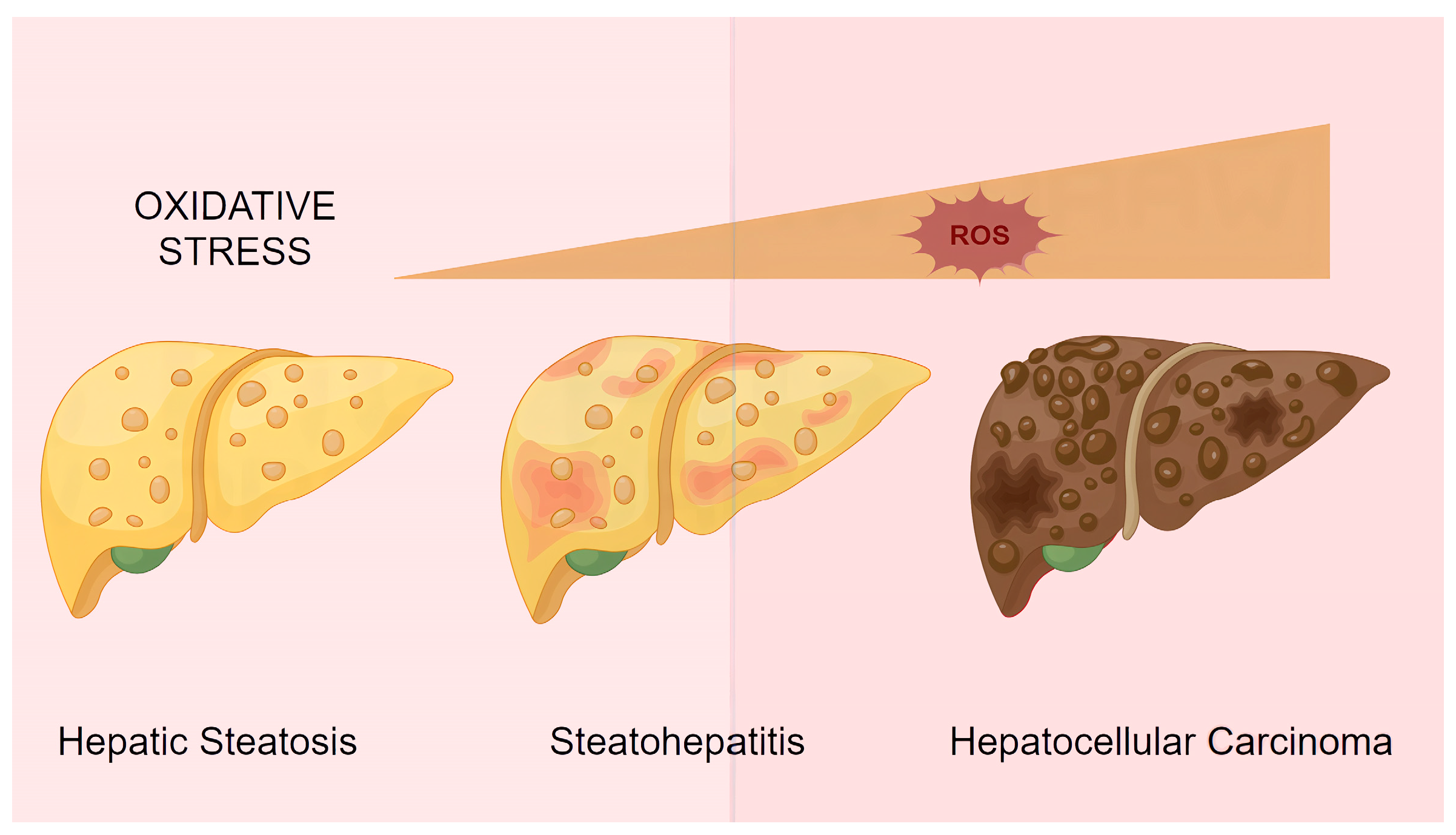
5. Current Challenges and Future Perspectives

ROS play a pivotal role in the pathological progression of both ALD and NAFLD. Excessive ROS generation not only directly induces hepatocyte injury but also drives disease progression by activating inflammatory responses, exacerbating lipid metabolism dysregulation, promoting fibrosis, and triggering apoptosis. Therefore, a deeper understanding of the mechanisms underlying ROS-mediated damage in ALD and NAFLD is critical for developing effective therapeutic strategies.
With advancing technology, the integration of multi-omics signatures—including genomics, metabolomics, and microbiomics—into precision medicine, guided by artificial intelligence (AI) for optimized treatment regimens, presents novel opportunities not only for ALD/NAFLD therapy but also for other oxidative stress-related pathologies (e.g., ischemia–reperfusion injury). With advances in the integration of medicine and engineering, RNS fluorescent probe technology can now be utilized in clinical practice to evaluate the efficacy of therapeutic drugs. This progress foreshadows the emergence of more precise and sustained antioxidant treatment modalities.
Furthermore, improved management of comorbidities—such as incorporating diabetes management guidelines [11] into liver-related outcomes, establishing national NAFLD treatment quality metrics, and aligning real-world evidence with randomized controlled trial data—can enhance outcomes in fatty liver disease. Additionally, AI may help reduce the variability in histological assessments that currently challenges clinical trials. Finally, ongoing technological advancements will enable increasingly personalized approaches to managing NAFLD and ALD patients. These developments may lead to rapid evolution in current diagnostic and therapeutic recommendations for these conditions.

Author Contributions

Conceptualization, P.G. and F.H.; data curation, H.Y. and Z.Z.; writing—original draft preparation, Z.Z.; writing—review and editing, Z.Z. and F.H.; supervision P.G. and F.H.; funding acquisition, Z.Z., P.G. and F.H. contributed equally to the article and are the co-corresponding authors. All authors have read and agreed to the published version of the manuscript.

Funding

This research and the APC was funded by [[Medical Research Project of Science and Health Union of Yubei District of Chongqing] grant number (2023YBKW19)].

Conflicts of Interest

The authors declare no conflicts of interest.

Abbreviations

The following abbreviations are used in this manuscript:
4-HNE4-hydroxynonenal
4-PBA4-phenylbutyric acid
8-OHdGadduct 8-hydroxy-2′-deoxyguanosine
ADHalcohol dehydrogenase
AKGα-ketoglutarate
ALDalcoholic fatty liver disease
ALDH2aldehyde dehydrogenase 2
AMPKAMP-activated protein kinase
ASHalcohol-related hepatitis
ATFactivating transcription factor
DAGdiacylglycerols
ERendoplasmic reticulum
ETCelectron transport chain
FFAsfree fatty acids
GSHglutathione
GSTglutathione-S-transferase
HCChepatocellular carcinoma
HO-1heme oxygenase-1
HSCshepatic stellate cell
IL-1βinterleukin
IRE1αinositol-requiring enzyme 1α
MAPKmitogen-activated protein kinase
MDAmalondialdehyde
MEOSmicrosomal ethanol oxidizing system
mPTPmitochondrial permeability transition pore
NAD+nicotinamide adenine dinucleotide
NAFLDnonalcoholic fatty liver disease
NASHnonalcoholic steatohepatitis
NLRP3Nod-like receptor pyrin-containing protein 3
NOXNADPH oxidase
Nrf2NF-E2 p45-related factor 2
PERKPKR-like ER kinase
PINK1PTEN-induced putative kinase 1
PPARαperoxisome proliferator-activated receptor α
PTXpentoxifylline
ROSreactive oxygen species
SAMS-adenosylmethionine
SERCAsarcoplasmic/endoplasmic reticulum calcium ATPase
SOD/GPxsuperoxide dismutase/glutathione peroxidase
SREBP-1csterol regulatory element-binding protein 1c
TBE-31alkynyltricyclic dicyanenone
TCAtricarboxylic acid cycle
TNF-αtumor necrosis factor
TRAF2TNF receptor-associated factor 2
TUDCAtaurine-conjugated ursodeoxycholic acid
UPRunfolded protein response
VLDLsvery-low-density lipoproteins
XBP1sX-box binding protein 1

References

  1. Seitz, H.K.; Bataller, R.; Cortez-Pinto, H.; Gao, B.; Gual, A.; Lackner, C.; Mathurin, P.; Mueller, S.; Szabo, G.; Tsukamoto, H. Alcoholic liver disease. Nat. Rev. Dis. Prim. 2018, 4, 16. [Google Scholar] [CrossRef] [PubMed]
  2. Devarbhavi, H.; Asrani, S.K.; Arab, J.P.; Nartey, Y.A.; Pose, E.; Kamath, P.S. Global burden of liver disease: 2023 update. J. Hepatol. 2023, 79, 516–537. [Google Scholar] [CrossRef] [PubMed]
  3. Zhou, Y.; Yuan, G.; Zhong, F.; He, S. Roles of the complement system in alcohol-induced liver disease. Clin. Mol. Hepatol. 2020, 26, 677–685. [Google Scholar] [CrossRef]
  4. Rives, C.; Fougerat, A.; Ellero-Simatos, S.; Loiseau, N.; Guillou, H.; Gamet-Payrastre, L.; Wahli, W. Oxidative Stress in NAFLD: Role of Nutrients and Food Contaminants. Biomolecules 2020, 10, 1702. [Google Scholar] [CrossRef]
  5. Granger, D.N.; Kvietys, P.R. Reperfusion injury and reactive oxygen species: The evolution of a concept. Redox Biol. 2015, 6, 524–551. [Google Scholar] [CrossRef]
  6. Sun, T.; Xiao, S.; Wang, M.; Xie, Q.; Zhang, L.; Gong, M.; Zhang, D.; Zhou, C. Reactive Oxygen Species Scavenging Nanozymes: Emerging Therapeutics for Acute Liver Injury Alleviation. Int. J. Nanomed. 2023, 18, 7901–7922. [Google Scholar] [CrossRef] [PubMed]
  7. Azzi, A. Oxidative Stress: What Is It? Can It Be Measured? Where Is It Located? Can It Be Good or Bad? Can It Be Prevented? Can It Be Cured? Antioxidants 2022, 11, 1431. [Google Scholar] [CrossRef]
  8. Jomova, K.; Alomar, S.Y.; Alwasel, S.H.; Nepovimova, E.; Kuca, K.; Valko, M. Several lines of antioxidant defense against oxidative stress: Antioxidant enzymes, nanomaterials with multiple enzyme-mimicking activities, and low-molecular-weight antioxidants. Arch. Toxicol. 2024, 98, 1323–1367. [Google Scholar] [CrossRef]
  9. Mittal, S.; El-Serag, H.B.; Sada, Y.H.; Kanwal, F.; Duan, Z.; Temple, S.; May, S.B.; Kramer, J.R.; Richardson, P.A.; Davila, J.A. Hepatocellular Carcinoma in the Absence of Cirrhosis in United States Veterans is Associated With Nonalcoholic Fatty Liver Disease. Clin. Gastroenterol. Hepatol. 2016, 14, 124–131.e1. [Google Scholar] [CrossRef]
  10. Sinha, R.A.; Singh, B.K.; Yen, P.M. Direct effects of thyroid hormones on hepatic lipid metabolism. Nat. Rev. Endocrinol. 2018, 14, 259–269. [Google Scholar] [CrossRef]
  11. Sakurai, Y.; Kubota, N.; Yamauchi, T.; Kadowaki, T. Role of Insulin Resistance in MAFLD. Int. J. Mol. Sci. 2021, 22, 4156. [Google Scholar] [CrossRef] [PubMed]
  12. Donnelly, K.L.; Smith, C.I.; Schwarzenberg, S.J.; Jessurun, J.; Boldt, M.D.; Parks, E.J. Sources of fatty acids stored in liver and secreted via lipoproteins in patients with nonalcoholic fatty liver disease. J. Clin. Investig. 2005, 115, 1343–1351. [Google Scholar] [CrossRef]
  13. Romero-Gomez, M.; SZelber-Sagi Trenell, M. Treatment of NAFLD with diet, physical activity and exercise. J. Hepatol. 2017, 67, 829–846. [Google Scholar] [CrossRef] [PubMed]
  14. DeBose-Boyd, R.A.; Ye, J. SREBPs in Lipid Metabolism, Insulin Signaling, and Beyond. Trends Biochem. Sci. 2018, 43, 358–368. [Google Scholar] [CrossRef]
  15. Mansouri, A.; Gattolliat, C.H.; Asselah, T. Mitochondrial Dysfunction and Signaling in Chronic Liver Diseases. Gastroenterology 2018, 155, 629–647. [Google Scholar] [CrossRef]
  16. Hirschey, M.D.; Shimazu, T.; Goetzman, E.; Jing, E.; Schwer, B.; Lombard, D.B.; Grueter, C.A.; Harris, C.; Biddinger, S.; Ilkayeva, O.R.; et al. SIRT3 regulates mitochondrial fatty-acid oxidation by reversible enzyme deacetylation. Nature 2010, 464, 121–125. [Google Scholar] [CrossRef]
  17. Lee, K.C.; Wu, P.S.; Lin, H.C. Pathogenesis and treatment of non-alcoholic steatohepatitis and its fibrosis. Clin. Mol. Hepatol. 2023, 29, 77–98. [Google Scholar] [CrossRef] [PubMed]
  18. Tao, Y.; Qiu, T.; Yao, X.; Jiang, L.; Wang, N.; Jiang, J.; Jia, X.; Wei, S.; Zhang, J.; Zhu, Y.; et al. IRE1alpha/NOX4 signaling pathway mediates ROS-dependent activation of hepatic stellate cells in NaAsO(2)-induced liver fibrosis. J. Cell. Physiol. 2021, 236, 1469–1480. [Google Scholar] [CrossRef]
  19. Celik, C.; Lee, S.Y.T.; Yap, W.S.; Thibault, G. Endoplasmic reticulum stress and lipids in health and diseases. Prog. Lipid Res. 2023, 89, 101198. [Google Scholar] [CrossRef]
  20. Lebeaupin, C.; Vallée, D.; Hazari, Y.; Hetz, C.; Chevet, E.; Bailly-Maitre, B. Endoplasmic reticulum stress signalling and the pathogenesis of non-alcoholic fatty liver disease. J. Hepatol. 2018, 69, 927–947. [Google Scholar] [CrossRef]
  21. Fu, S.; Yang, L.; Li, P.; Hofmann, O.; Dicker, L.; Hide, W.; Lin, X.; Watkins, S.M.; Ivanov, A.R.; Hotamisligil, G.S.; et al. Aberrant lipid metabolism disrupts calcium homeostasis causing liver endoplasmic reticulum stress in obesity. Nature 2011, 473, 528–531. [Google Scholar] [CrossRef] [PubMed]
  22. Kopp, M.C.; Larburu, N.; Durairaj, V.; Adams, C.J.; Ali, M.M.U. UPR proteins IRE1 and PERK switch BiP from chaperone to ER stress sensor. Nat. Struct. Mol. Biol. 2019, 26, 1053–1062. [Google Scholar] [CrossRef]
  23. He, F.; Ru, X.; Wen, T. NRF2, a Transcription Factor for Stress Response and Beyond. Int. J. Mol. Sci. 2020, 21, 4777. [Google Scholar] [CrossRef] [PubMed]
  24. Rosso, C.; Kazankov, K.; Younes, R.; Esmaili, S.; Marietti, M.; Sacco, M.; Carli, F.; Gaggini, M.; Salomone, F.; Møller, H.J.; et al. Crosstalk between adipose tissue insulin resistance and liver macrophages in non-alcoholic fatty liver disease. J. Hepatol. 2019, 71, 1012–1021. [Google Scholar] [CrossRef] [PubMed]
  25. Barreby, E.; Chen, P.; Aouadi, M. Macrophage functional diversity in NAFLD—more than inflammation. Nat. Rev. Endocrinol. 2022, 18, 461–472. [Google Scholar] [CrossRef]
  26. Seitz, H.K.; Bataller, R.; Cortez-Pinto, H.; Gao, B.; Gual, A.; Lackner, C.; Mathurin, P.; Mueller, S.; Szabo, G.; Tsukamoto, H. Publisher Correction: Alcoholic liver disease. Nat. Rev. Dis. Prim. 2018, 4, 18. [Google Scholar] [CrossRef]
  27. Matsushita, H.; Takaki, A. Alcohol and hepatocellular carcinoma. BMJ Open Gastroenterol. 2019, 6, e000260. [Google Scholar] [CrossRef] [PubMed]
  28. Hyun, J.; Han, J.; Lee, C.; Yoon, M.; Jung, Y. Pathophysiological Aspects of Alcohol Metabolism in the Liver. Int. J. Mol. Sci. 2021, 22, 5717. [Google Scholar] [CrossRef]
  29. Wang, Y.; Chen, C.; Chen, J.; Sang, T.; Peng, H.; Lin, X.; Zhao, Q.; Chen, S.; Eling, T.; Wang, X. Overexpression of NAG-1/GDF15 prevents hepatic steatosis through inhibiting oxidative stress-mediated dsDNA release and AIM2 inflammasome activation. Redox Biol. 2022, 52, 102322. [Google Scholar] [CrossRef]
  30. Sun, X.; Seidman, J.S.; Zhao, P.; Troutman, T.D.; Spann, N.J.; Que, X.; Zhou, F.; Liao, Z.; Pasillas, M.; Yang, X.; et al. Neutralization of Oxidized Phospholipids Ameliorates Non-alcoholic Steatohepatitis. Cell Metab. 2020, 31, 189–206.e8. [Google Scholar] [CrossRef]
  31. Liu, Y.-N.; Zhu, H.-X.; Li, T.-Y.; Yang, X.; Li, X.-J.; Zhang, W.K. Lipid nanoparticle encapsulated oleic acid induced lipotoxicity to hepatocytes via ROS overload and the DDIT3/BCL2/BAX/Caspases signaling in vitro and in vivo. Free. Radic. Biol. Med. 2024, 222, 361–370. [Google Scholar] [CrossRef] [PubMed]
  32. Wu, S.; Guo, N.; Xu, H.; Li, Y.; Sun, T.; Jiang, X.; Fu, D.; You, T.; Diao, S.; Huang, Y.; et al. Caveolin-1 ameliorates hepatic injury in non-alcoholic fatty liver disease by inhibiting ferroptosis via the NOX4/ROS/GPX4 pathway. Biochem. Pharmacol. 2024, 230, 116594. [Google Scholar] [CrossRef] [PubMed]
  33. Guan, B.-J.; Krokowski, D.; Majumder, M.; Schmotzer, C.L.; Kimball, S.R.; Merrick, W.C.; Koromilas, A.E.; Hatzoglou, M. Translational Control during Endoplasmic Reticulum Stress beyond Phosphorylation of the Translation Initiation Factor eIF2α. J. Biol. Chem. 2014, 289, 12593–12611. [Google Scholar] [CrossRef]
  34. Wang, L.; Wang, C.-C. Oxidative protein folding fidelity and redoxtasis in the endoplasmic reticulum. Trends Biochem. Sci. 2022, 48, 40–52. [Google Scholar] [CrossRef] [PubMed]
  35. Sharma, R.S.; Harrison, D.J.; Kisielewski, D.; Cassidy, D.M.; McNeilly, A.D.; Gallagher, J.R.; Walsh, S.V.; Honda, T.; McCrimmon, R.J.; Dinkova-Kostova, A.T.; et al. Experimental Nonalcoholic Steatohepatitis and Liver Fibrosis Are Ameliorated by Pharmacologic Activation of Nrf2 (NF-E2 p45-Related Factor 2). Cell. Mol. Gastroenterol. Hepatol. 2018, 5, 367–398. [Google Scholar] [CrossRef]
  36. Lee, K.P.; Dey, M.; Neculai, D.; Cao, C.; Dever, T.E.; Sicheri, F. Structure of the Dual Enzyme Ire1 Reveals the Basis for Catalysis and Regulation in Nonconventional RNA Splicing. Cell 2008, 132, 89–100. [Google Scholar] [CrossRef]
  37. Li, Y.; Chen, M.; Xu, Y.; Yu, X.; Xiong, T.; Du, M.; Sun, J.; Liu, L.; Tang, Y.; Yao, P.; et al. Iron-Mediated Lysosomal Membrane Permeabilization in Ethanol-Induced Hepatic Oxidative Damage and Apoptosis: Protective Effects of Quercetin. Oxidative Med. Cell. Longev. 2015, 2016, 4147610. [Google Scholar] [CrossRef]
  38. Tam, A.B.; Mercado, E.L.; Hoffmann, A.; Niwa, M. ER stress activates NF-κB by integrating functions of basal IKK activity, IRE1 and PERK. PLoS ONE 2012, 7, e45078. [Google Scholar] [CrossRef]
  39. Sabio, G.; Das, M.; Mora, A.; Zhang, Z.; Jun, J.Y.; Ko, H.J.; Barrett, T.; Kim, J.K.; Davis, R.J. A Stress Signaling Pathway in Adipose Tissue Regulates Hepatic Insulin Resistance. Science 2008, 322, 1539–1543. [Google Scholar] [CrossRef]
  40. Das, M.; Sabio, G.; Jiang, F.; Rincón, M.; Flavell, R.A.; Davis, R.J. Induction of Hepatitis by JNK-Mediated Expression of TNF-α. Cell 2009, 136, 249–260. [Google Scholar] [CrossRef]
  41. Sabio, G.; Kennedy, N.J.; Cavanagh-Kyros, J.; Jung, D.Y.; Ko, H.J.; Ong, H.; Barrett, T.; Kim, J.K.; Davis, R.J. Role of Muscle c-Jun NH2-Terminal Kinase 1 in Obesity-Induced Insulin Resistance. Mol. Cell. Biol. 2010, 30, 106–115. [Google Scholar] [CrossRef] [PubMed]
  42. Sabio, G.; Cavanagh-Kyros, J.; Ko, H.J.; Jung, D.Y.; Gray, S.; Jun, J.Y.; Barrett, T.; Mora, A.; Kim, J.K.; Davis, R.J. Prevention of Steatosis by Hepatic JNK1. Cell Metab. 2009, 10, 491–498. [Google Scholar] [CrossRef] [PubMed]
  43. Wang, H.; Nikain, C.; Fortounas, K.I.; Amengual, J.; Tufanli, O.; La Forest, M.; Yu, Y.; Wang, M.C.; Watts, R.; Lehner, R.; et al. FITM2 deficiency results in ER lipid accumulation, ER stress, and reduced apolipoprotein B lipidation and VLDL triglyceride secretion in vitro and in mouse liver. Mol. Metab. 2024, 90, 102048. [Google Scholar] [CrossRef] [PubMed]
  44. Carotti, S.; Aquilano, K.; Zalfa, F.; Ruggiero, S.; Valentini, F.; Zingariello, M.; Francesconi, M.; Perrone, G.; Alletto, F.; Antonelli-Incalzi, R.; et al. Lipophagy Impairment Is Associated With Disease Progression in NAFLD. Front. Physiol. 2020, 11, 850. [Google Scholar] [CrossRef]
  45. Koga, H.; Kaushik, S.; Cuervo, A.M. Altered lipid content inhibits autophagic vesicular fusion. FASEB J. 2010, 24, 3052–3065. [Google Scholar] [CrossRef]
  46. Hamasaki, M.; Furuta, N.; Matsuda, A.; Nezu, A.; Yamamoto, A.; Fujita, N.; Oomori, H.; Noda, T.; Haraguchi, T.; Hiraoka, Y.; et al. Autophagosomes form at ER–mitochondria contact sites. Nature 2013, 495, 389–393. [Google Scholar] [CrossRef]
  47. Shen, Q.; Shi, Y.; Liu, J.; Su, H.; Huang, J.; Zhang, Y.; Peng, C.; Zhou, T.; Sun, Q.; Wan, W.; et al. Acetylation of STX17 (syntaxin 17) controls autophagosome maturation. Autophagy 2020, 17, 1157–1169. [Google Scholar] [CrossRef]
  48. Tilg, H.; ARMoschen Szabo, G. Interleukin-1 and inflammasomes in alcoholic liver disease/acute alcoholic hepatitis and nonalcoholic fatty liver disease/nonalcoholic steatohepatitis. Hepatology 2016, 64, 955–965. [Google Scholar] [CrossRef]
  49. Tschopp, J.; Schroder, K. NLRP3 inflammasome activation: The convergence of multiple signalling pathways on ROS production? Nat. Rev. Immunol. 2010, 10, 210–215. [Google Scholar] [CrossRef]
  50. Ahmad, A.; Ahmad, R. Resveratrol mitigate structural changes and hepatic stellate cell activation in N′-nitrosodimethylamine-induced liver fibrosis via restraining oxidative damage. Chem. Interact. 2014, 221, 1–12. [Google Scholar] [CrossRef]
  51. Tsuchida, T.; Friedman, S.L. Mechanisms of hepatic stellate cell activation. Nat. Rev. Gastroenterol. Hepatol. 2017, 14, 397–411. [Google Scholar] [CrossRef] [PubMed]
  52. Zhou, H.; Liu, Y.; Su, Y.; Ji, P.; Kong, L.; Sun, R.; Zhang, D.; Xu, H.; Li, W.; Li, W. Ginsenoside Rg1 attenuates lipopolysaccharide-induced chronic liver damage by activating Nrf2 signaling and inhibiting inflammasomes in hepatic cells. J. Ethnopharmacol. 2024, 324, 117794. [Google Scholar] [CrossRef] [PubMed]
  53. Fu, H.; Ge, Y.; Liu, X.; Deng, S.; Li, J.; Tan, P.; Yang, Y.; Wu, Z. Exposure to the environmental pollutant chlorpyrifos induces hepatic toxicity through activation of the JAK/STAT and MAPK pathways. Sci. Total. Environ. 2024, 928, 171711. [Google Scholar] [CrossRef] [PubMed]
  54. Yang, S.; Li, F.; Lu, S.; Ren, L.; Bian, S.; Liu, M.; Zhao, D.; Wang, S.; Wang, J. Ginseng root extract attenuates inflammation by inhibiting the MAPK/NF-κB signaling pathway and activating autophagy and p62-Nrf2-Keap1 signaling in vitro and in vivo. J. Ethnopharmacol. 2022, 283, 114739. [Google Scholar] [CrossRef]
  55. Iracheta-Vellve, A.; Petrasek, J.; Gyogyosi, B.; Bala, S.; Csak, T.; Kodys, K.; Szabo, G. Interleukin-1 inhibition facilitates recovery from liver injury and promotes regeneration of hepatocytes in alcoholic hepatitis in mice. Liver Int. 2017, 37, 968–973. [Google Scholar] [CrossRef]
  56. Dong, J.; Li, M.; Peng, R.; Zhang, Y.; Qiao, Z.; Sun, N. ACACA reduces lipid accumulation through dual regulation of lipid metabolism and mitochondrial function via AMPK- PPARα- CPT1A axis. J. Transl. Med. 2024, 22, 1–14. [Google Scholar] [CrossRef]
  57. Liu, Y.; Xu, W.; Zhai, T.; You, J.; Chen, Y. Silibinin ameliorates hepatic lipid accumulation and oxidative stress in mice with non-alcoholic steatohepatitis by regulating CFLAR-JNK pathway. Acta Pharm. Sin. B 2019, 9, 745–757. [Google Scholar] [CrossRef]
  58. Ding, X.; Zhu, X.-L.; Xu, D.-H.; Li, S.; Yang, Q.; Feng, X.; Wei, Y.-G.; Li, H.; Yang, L.; Zhang, Y.-J.; et al. NPM promotes hepatotoxin-induced fibrosis by inhibiting ROS-induced apoptosis of hepatic stellate cells and upregulating lncMIAT-induced TGF-β2. Cell Death Dis. 2023, 14, 1–16. [Google Scholar] [CrossRef]
  59. Seki, S.; Kitada, T.; Sakaguchi, H.; Nakatani, K.; Wakasa, K. Pathological significance of oxidative cellular damage in human alcoholic liver disease. Histopathology 2003, 42, 365–371. [Google Scholar] [CrossRef]
  60. Kitada, T.; Seki, S.; Iwai, S.; Yamada, T.; Sakaguchi, H.; Wakasa, K. In situ detection of oxidative DNA damage, 8-hydroxydeoxyguanosine, in chronic human liver disease. J. Hepatol. 2001, 35, 613–618. [Google Scholar] [CrossRef]
  61. Gao, H.; Jiang, Y.; Zeng, G.; Huda, N.; Thoudam, T.; Yang, Z.; Liangpunsakul, S.; Ma, J. Cell-to-cell and organ-to-organ crosstalk in the pathogenesis of alcohol-associated liver disease. eGastroenterology 2024, 2, e100104. [Google Scholar] [CrossRef] [PubMed]
  62. Feng, H.; Stockwell, B.R. Unsolved mysteries: How does lipid peroxidation cause ferroptosis? PLoS biology 2018, 16, e2006203. [Google Scholar] [CrossRef]
  63. Catalá, A.; Díaz, M. Editorial: Impact of Lipid Peroxidation on the Physiology and Pathophysiology of Cell Membranes. Front. Physiol. 2016, 7, 423. [Google Scholar] [CrossRef]
  64. Bartesaghi, S.; Radi, R. Fundamentals on the biochemistry of peroxynitrite and protein tyrosine nitration. Redox Biol. 2018, 14, 618–625. [Google Scholar] [CrossRef] [PubMed]
  65. Ipsen, D.H.; Lykkesfeldt, J.; Tveden-Nyborg, P. Molecular mechanisms of hepatic lipid accumulation in non-alcoholic fatty liver disease. Cell. Mol. Life Sci. 2018, 75, 3313–3327. [Google Scholar] [CrossRef]
  66. Penaloza, C.G.; Cruz, M.; Germain, G.; Jabeen, S.; Javdan, M.; Lockshin, R.A.; Zakeri, Z. Higher sensitivity of female cells to ethanol: Methylation of DNA lowers Cyp2e1, generating more ROS. Cell Commun. Signal. 2020, 18, 1–15. [Google Scholar] [CrossRef]
  67. Zeng, T.; Zhang, C.-L.; Song, F.-Y.; Zhao, X.-L.; Yu, L.-H.; Zhu, Z.-P.; Xie, K.-Q. PI3K/Akt pathway activation was involved in acute ethanol-induced fatty liver in mice. Toxicology 2012, 296, 56–66. [Google Scholar] [CrossRef]
  68. Krycer, J.R.; Sharpe, L.J.; Luu, W.; Brown, A.J. The Akt–SREBP nexus: Cell signaling meets lipid metabolism. Trends Endocrinol. Metab. 2010, 21, 268–276. [Google Scholar] [CrossRef] [PubMed]
  69. Sarsour, E.H.; Kumar, M.G.; Chaudhuri, L.; Kalen, A.L.; Goswami, P.C. Redox Control of the Cell Cycle in Health and Disease. Antioxidants Redox Signal. 2009, 11, 2985–3011. [Google Scholar] [CrossRef]
  70. Goldar, S.; Khaniani, M.S.; Derakhshan, S.M.; Baradaran, B. Molecular Mechanisms of Apoptosis and Roles in Cancer Development and Treatment. Asian Pac. J. Cancer Prev. 2015, 16, 2129–2144. [Google Scholar] [CrossRef]
  71. Taniai, M. Alcohol and hepatocarcinogenesis. Clin. Mol. Hepatol. 2020, 26, 736–741. [Google Scholar] [CrossRef] [PubMed]
  72. Wong, W.J.; Emdin, C.; Bick, A.G.; Zekavat, S.M.; Niroula, A.; Pirruccello, J.P.; Dichtel, L.; Griffin, G.; Uddin, M.; Gibson, C.J.; et al. Clonal haematopoiesis and risk of chronic liver disease. Nature 2023, 616, 747–754. [Google Scholar] [CrossRef] [PubMed]
  73. Huang, D.Q.; Terrault, N.A.; Tacke, F.; Gluud, L.L.; Arrese, M.; Bugianesi, E.; Loomba, R. Global epidemiology of cirrhosis - aetiology, trends and predictions. Nat. Rev. Gastroenterol. Hepatol. 2023, 20, 388–398. [Google Scholar] [CrossRef]
  74. Lin, L.; Yang, S.; Xiao, Z.; Hong, P.; Sun, S.; Zhou, C.; Qian, Z.J. The Inhibition Effect of the Seaweed Polyphenol, 7-Phloro-Eckol from Ecklonia Cava on Alcohol-Induced Oxidative Stress in HepG2/CYP2E1 Cells. Mar. Drugs 2021, 19, 158. [Google Scholar] [CrossRef]
  75. Lu, Y.; Zhang, X.H.; Cederbaum, A.I. Ethanol Induction of CYP2A5: Role of CYP2E1-ROS-Nrf2 Pathway. Toxicol. Sci. 2012, 128, 427–438. [Google Scholar] [CrossRef]
  76. Gonzalez, A.; Huerta-Salgado, C.; Orozco-Aguilar, J.; Aguirre, F.; Tacchi, F.; Simon, F.; Cabello-Verrugio, C.; Gasparovic, A.C. Role of Oxidative Stress in Hepatic and Extrahepatic Dysfunctions during Nonalcoholic Fatty Liver Disease (NAFLD). Oxidative Med. Cell. Longev. 2020, 2020, 1–16. [Google Scholar] [CrossRef]
  77. Seki, S.; Kitada, T.; Yamada, T.; Sakaguchi, H.; Nakatani, K.; Wakasa, K. In situ detection of lipid peroxidation and oxidative DNA damage in non-alcoholic fatty liver diseases. J. Hepatol. 2002, 37, 56–62. [Google Scholar] [CrossRef]
  78. Pirola, C.J.; Garaycoechea, M.; Flichman, D.; Castaño, G.O.; Sookoian, S. Liver mitochondrial DNA damage and genetic variability of Cytochrome b – a key component of the respirasome – drive the severity of fatty liver disease. J. Intern. Med. 2020, 289, 84–96. [Google Scholar] [CrossRef]
  79. Pacher, P.; Beckman, J.S.; Liaudet, L. Nitric Oxide and Peroxynitrite in Health and Disease. Physiol. Rev. 2007, 87, 315–424. [Google Scholar] [CrossRef]
  80. Wang, N.; Lu, X.; Wang, J.; Wang, H.; Zhang, B.; Zhao, W.; Zhang, J. Quasi-LD-Targeted and ONOO-Responsive Fluorescent Probe for Investigating the Interaction of Nonalcoholic Fatty Liver with Drug-Induced Liver Injury. Anal. Chem. 2023, 95, 5967–5975. [Google Scholar] [CrossRef]
  81. Dias, K.A.; Oliveira, L.A.; Pereira, S.M.S.; Abrantes, L.C.S.; Vicente, L.C.O.d.S.; Gonçalves, R.V.; Della Lucia, C.M. Anti-inflammatory and antioxidant effects of anthocyanins in Nonalcoholic fatty liver disease (NAFLD): A systematic review of in vivo studies. Crit. Rev. Food Sci. Nutr. 2025, 1–18. [Google Scholar] [CrossRef]
  82. Greatorex, S.; Kaur, S.; Xirouchaki, C.E.; Goh, P.K.; Wiede, F.; Genders, A.J.; Tran, M.; Jia, Y.; Raajendiran, A.; Brown, W.A.; et al. Mitochondria- and NOX4-dependent antioxidant defense mitigates progression to nonalcoholic steatohepatitis in obesity. J. Clin. Investig. 2023, 134. [Google Scholar] [CrossRef] [PubMed]
  83. Spahis, S.; Delvin, E.; Borys, J.-M.; Levy, E. Oxidative Stress as a Critical Factor in Nonalcoholic Fatty Liver Disease Pathogenesis. Antioxidants Redox Signal. 2017, 26, 519–541. [Google Scholar] [CrossRef]
  84. Zhang, T.; Liu, J.; Shen, S.; Tong, Q.; Ma, X.; Lin, L. SIRT3 promotes lipophagy and chaperon-mediated autophagy to protect hepatocytes against lipotoxicity. Cell Death Differ. 2019, 27, 329–344. [Google Scholar] [CrossRef] [PubMed]
  85. Bartesaghi, S.; Wallenius, K.; Hovdal, D.; Liljeblad, M.; Wallin, S.; Dekker, N.; Barlind, L.; Davies, N.; Seeliger, F.; Winzell, M.S.; et al. Subcutaneous delivery of FGF21 mRNA therapy reverses obesity, insulin resistance, and hepatic steatosis in diet-induced obese mice. Mol. Ther.-Nucleic Acids 2022, 28, 500–513. [Google Scholar] [CrossRef] [PubMed]
  86. Smith, G.I.; Shankaran, M.; Yoshino, M.; Schweitzer, G.G.; Chondronikola, M.; Beals, J.W.; Okunade, A.L.; Patterson, B.W.; Nyangau, E.; Field, T.; et al. Insulin resistance drives hepatic de novo lipogenesis in nonalcoholic fatty liver disease. J. Clin. Investig. 2020, 130, 1453–1460. [Google Scholar] [CrossRef]
  87. Fromenty, B.; Roden, M. Mitochondrial alterations in fatty liver diseases. J. Hepatol. 2022, 78, 415–429. [Google Scholar] [CrossRef]
  88. Loomba, R.; Friedman, S.L.; Shulman, G.I. Mechanisms and disease consequences of nonalcoholic fatty liver disease. Cell 2021, 184, 2537–2564. [Google Scholar] [CrossRef]
  89. Younossi, Z.M. Non-alcoholic fatty liver disease—A global public health perspective. J. Hepatol. 2019, 70, 531–544. [Google Scholar] [CrossRef]
  90. Kalligeros, M.; Vassilopoulos, A.; Vassilopoulos, S.; Victor, D.W.; Mylonakis, E.; Noureddin, M. Prevalence of Steatotic Liver Disease (MASLD, MetALD, and ALD) in the United States: NHANES 2017-2020. Clin. Gastroenterol. Hepatol. Off. Clin. Pract. J. Am. Gastroenterol. Assoc. 2024, 22, 1330–1332.e1334. [Google Scholar] [CrossRef]
  91. Marek, G.W.; Malhi, H. MetALD: Does it require a different therapeutic option? Hepatology 2024, 80, 1424–1440. [Google Scholar] [CrossRef]
  92. Thoma, C.; Day, C.P.; Trenell, M.I. Lifestyle interventions for the treatment of non-alcoholic fatty liver disease in adults: A systematic review. J. Hepatol. 2012, 56, 255–266. [Google Scholar] [CrossRef] [PubMed]
  93. Louvet, A.; Labreuche, J.; Artru, F.; Bouthors, A.; Rolland, B.; Saffers, P.; Lollivier, J.; Lemaître, E.; Dharancy, S.; Lassailly, G.; et al. Main drivers of outcome differ between short term and long term in severe alcoholic hepatitis: A prospective study. Hepatology 2017, 66, 1464–1473. [Google Scholar] [CrossRef]
  94. Shojaee-Moradie, F.; Cuthbertson, D.J.; Barrett, M.; Jackson, N.C.; Herring, R.; Thomas, E.L.; Bell, J.; Kemp, G.J.; Wright, J.; Umpleby, A.M. Exercise Training Reduces Liver Fat and Increases Rates of VLDL Clearance But Not VLDL Production in NAFLD. J. Clin. Endocrinol. Metab. 2016, 101, 4219–4228. [Google Scholar] [CrossRef]
  95. Hofer, B.S.; Simbrunner, B.; Hartl, L.; Jachs, M.; Bauer, D.J.M.; Balcar, L.; Paternostro, R.; Schwabl, P.; Semmler, G.; Scheiner, B.; et al. Alcohol Abstinence Improves Prognosis Across All Stages of Portal Hypertension in Alcohol-Related Cirrhosis. Clin. Gastroenterol. Hepatol. 2022, 21, 2308–2317.e7. [Google Scholar] [CrossRef] [PubMed]
  96. Nobili, V.; Parola, M.; Alisi, A.; Marra, F.; Piemonte, F.; Mombello, C.; Sutti, S.; Povero, D.; Maina, V.; Novo, E.; et al. Oxidative stress parameters in paediatric non-alcoholic fatty liver disease. Int. J. Mol. Med. 2010, 26, 471–476. [Google Scholar] [CrossRef] [PubMed]
  97. Yang, J.; Fernández-Galilea, M.; Martínez-Fernández, L.; González-Muniesa, P.; Pérez-Chávez, A.; Martínez, J.A.; Moreno-Aliaga, M.J. Oxidative Stress and Non-Alcoholic Fatty Liver Disease: Effects of Omega-3 Fatty Acid Supplementation. Nutrients 2019, 11, 872. [Google Scholar] [CrossRef]
  98. Yki-Järvinen, H.; Luukkonen, P.K.; Hodson, L.; Moore, J.B. Dietary carbohydrates and fats in nonalcoholic fatty liver disease. Nat. Rev. Gastroenterol. Hepatol. 2021, 18, 770–786. [Google Scholar] [CrossRef]
  99. Li, S.; Tan, H.-Y.; Wang, N.; Zhang, Z.-J.; Lao, L.; Wong, C.-W.; Feng, Y. The Role of Oxidative Stress and Antioxidants in Liver Diseases. Int. J. Mol. Sci. 2015, 16, 26087–26124. [Google Scholar] [CrossRef]
  100. Vilar-Gomez, E.; Martinez-Perez, Y.; Calzadilla-Bertot, L.; Torres-Gonzalez, A.; Gra-Oramas, B.; Gonzalez-Fabian, L.; Friedman, S.L.; Diago, M.; Romero-Gomez, M. Weight Loss Through Lifestyle Modification Significantly Reduces Features of Nonalcoholic Steatohepatitis. Gastroenterology 2015, 149, 367–378.e5. [Google Scholar] [CrossRef]
  101. Promrat, K.; Kleiner, D.E.; Niemeier, H.M.; Jackvony, E.; Kearns, M.; Wands, J.R.; Fava, J.L.; Wing, R.R. Randomized controlled trial testing the effects of weight loss on nonalcoholic steatohepatitis. Hepatology 2010, 51, 121–129. [Google Scholar] [CrossRef] [PubMed]
  102. Ayares, G.; Idalsoaga, F.; Díaz, L.A.; Arnold, J.; Arab, J.P. Current Medical Treatment for Alcohol-Associated Liver Disease. J. Clin. Exp. Hepatol. 2022, 12, 1333–1348. [Google Scholar] [CrossRef]
  103. Marti-Aguado, D.; Clemente-Sanchez, A.; Bataller, R. Cigarette smoking and liver diseases. J. Hepatol. 2022, 77, 191–205. [Google Scholar] [CrossRef] [PubMed]
  104. Lin, H.-M.; Zhang, J.-R.; Li, M.-X.; Hou, H.; Wang, H.; Huang, Y. Cigarette smoking and alcohol-related liver disease. Liver Res. 2024, 8, 237–245. [Google Scholar] [CrossRef] [PubMed]
  105. Violi, F.; Cangemi, R. ; Cangemi, R. Pioglitazone, vitamin E, or placebo for nonalcoholic steatohepatitis. N. Engl. J. Med. 2010, 363, 1185–1186; author reply 1186. [Google Scholar]
  106. Chin, K.-Y.; Ekeuku, S.O.; Chew, D.C.H.; Trias, A. Tocotrienol in the Management of Nonalcoholic Fatty Liver Disease: A Systematic Review. Nutrients 2023, 15, 834. [Google Scholar] [CrossRef]
  107. Tang, G.; Xu, Y.; Zhang, C.; Wang, N.; Li, H.; Feng, Y. Green Tea and Epigallocatechin Gallate (EGCG) for the Management of Nonalcoholic Fatty Liver Diseases (NAFLD): Insights into the Role of Oxidative Stress and Antioxidant Mechanism. Antioxidants 2021, 10, 1076. [Google Scholar] [CrossRef] [PubMed]
  108. Sakata, R.; Nakamura, T.; Torimura, T.; Ueno, T.; Sata, M. Green tea with high-density catechins improves liver function and fat infiltration in non-alcoholic fatty liver disease (NAFLD) patients: A double-blind placebo-controlled study. Int. J. Mol. Med. 2013, 32, 989–994. [Google Scholar] [CrossRef]
  109. Dey, P.; Kim, J.B.; Chitchumroonchokchai, C.; Li, J.; Sasaki, G.Y.; Olmstead, B.D.; Stock, K.L.; Thomas-Ahner, J.M.; Clinton, S.K.; Bruno, R.S. Green tea extract inhibits early oncogenic responses in mice with nonalcoholic steatohepatitis. Food Funct. 2019, 10, 6351–6361. [Google Scholar] [CrossRef]
  110. Zein, C.O.; Lopez, R.; Fu, X.; Kirwan, J.P.; Yerian, L.M.; McCullough, A.J.; Hazen, S.L.; Feldstein, A.E. Pentoxifylline decreases oxidized lipid products in nonalcoholic steatohepatitis: New evidence on the potential therapeutic mechanism. Hepatology 2012, 56, 1291–1299. [Google Scholar] [CrossRef]
  111. Zein, C.O.; Yerian, L.M.; Gogate, P.; Lopez, R.; Kirwan, J.P.; Feldstein, A.E.; McCullough, A.J. Pentoxifylline improves nonalcoholic steatohepatitis: A randomized placebo-controlled trial. Hepatology 2011, 54, 1610–1619. [Google Scholar] [CrossRef] [PubMed]
  112. Mora, S.I.; García-Román, J.; Gómez-Ñañez, I.; García-Román, R. Chronic liver diseases and the potential use of S-adenosyl-l-methionine as a hepatoprotector. Eur. J. Gastroenterol. Hepatol. 2018, 30, 893–900. [Google Scholar] [CrossRef]
  113. Lieber, C.S. S-Adenosyl-l-methionine and alcoholic liver disease in animal models: Implications for early intervention in human beings. Alcohol 2002, 27, 173–177. [Google Scholar] [CrossRef] [PubMed]
  114. Karaa, A.; Thompson, K.J.; McKillop, I.H.; Clemens, M.G.; Schrum, L.W. S-ADENOSYL-L-METHIONINE ATTENUATES OXIDATIVE STRESS AND HEPATIC STELLATE CELL ACTIVATION IN AN ETHANOL-LPS-INDUCED FIBROTIC RAT MODEL. Shock 2008, 30, 197–205. [Google Scholar] [CrossRef]
  115. Kharbanda, K.K. Alcoholic Liver Disease and Methionine Metabolism. Semin. Liver Dis. 2009, 29, 155–165. [Google Scholar] [CrossRef]
  116. Purohit, V.; Abdelmalek, M.F.; Barve, S.; Benevenga, N.J.; Halsted, C.H.; Kaplowitz, N.; Kharbanda, K.K.; Liu, Q.-Y.; Lu, S.C.; McClain, C.J.; et al. Role of S-adenosylmethionine, folate, and betaine in the treatment of alcoholic liver disease: Summary of a symposium. Am. J. Clin. Nutr. 2007, 86, 14–24. [Google Scholar] [CrossRef]
  117. Kharbanda, K.K.; Mailliard, M.E.; Baldwin, C.R.; Beckenhauer, H.C.; Sorrell, M.F.; Tuma, D.J. Betaine attenuates alcoholic steatosis by restoring phosphatidylcholine generation via the phosphatidylethanolamine methyltransferase pathway. J. Hepatol. 2007, 46, 314–321. [Google Scholar] [CrossRef] [PubMed]
  118. Kim, S.J.; Jung, Y.S.; Kwon, D.Y.; Kim, Y.C. Alleviation of acute ethanol-induced liver injury and impaired metabolomics of S-containing substances by betaine supplementation. Biochem. Biophys. Res. Commun. 2008, 368, 893–898. [Google Scholar] [CrossRef]
  119. Lombardi, R.; Onali, S.; Thorburn, D.; Davidson, B.R.; Gurusamy, K.S.; Tsochatzis, E. Pharmacological interventions for non-alcohol related fatty liver disease (NAFLD): An attempted network meta-analysis. Cochrane Database Syst Rev. 2017, 3, CD011640. [Google Scholar] [CrossRef] [PubMed]
  120. Aoyama, T.; Paik, Y.-H.; Watanabe, S.; Laleu, B.; Gaggini, F.; Fioraso-Cartier, L.; Molango, S.; Heitz, F.; Merlot, C.; Szyndralewiez, C.; et al. Nicotinamide adenine dinucleotide phosphate oxidase in experimental liver fibrosis: GKT137831 as a novel potential therapeutic agent. Hepatology 2012, 56, 2316–2327. [Google Scholar] [CrossRef]
  121. Paik, Y.H.; Iwaisako, K.; Seki, E.; Inokuchi, S.; Schnabl, B.; Osterreicher, C.H.; Kisseleva, T.; Brenner, D.A. The nicotinamide adenine dinucleotide phosphate oxidase (NOX) homologues NOX1 and NOX2/gp91(phox) mediate hepatic fibrosis in mice. Hepatology 2011, 53, 1730–1741. [Google Scholar] [CrossRef]
  122. Frenette, C.; Kayali, Z.; Mena, E.; Mantry, P.S.; Lucas, K.J.; Neff, G.; Rodriguez, M.; Thuluvath, P.J.; Weinberg, E.; Bhandari, B.R.; et al. Emricasan to prevent new decompensation in patients with NASH-related decompensated cirrhosis. J. Hepatol. 2021, 74, 274–282. [Google Scholar] [CrossRef] [PubMed]
  123. Barreyro, F.J.; Holod, S.; Finocchietto, P.V.; Camino, A.M.; Aquino, J.B.; Avagnina, A.; Carreras, M.C.; Poderoso, J.J.; Gores, G.J. The pan-caspase inhibitor Emricasan (IDN-6556) decreases liver injury and fibrosis in a murine model of non-alcoholic steatohepatitis. Liver Int. 2014, 35, 953–966. [Google Scholar] [CrossRef] [PubMed]
  124. Keam, S.J. Resmetirom: First Approval. Drugs 2024, 84, 729–735. [Google Scholar] [CrossRef]
  125. Harrison, S.A.; Bedossa, P.; Guy, C.D.; Schattenberg, J.M.; Loomba, R.; Taub, R.; Labriola, D.; Moussa, S.E.; Neff, G.W.; Rinella, M.E.; et al. A Phase 3, Randomized, Controlled Trial of Resmetirom in NASH with Liver Fibrosis. N. Engl. J. Med. 2024, 390, 497–509. [Google Scholar] [CrossRef] [PubMed]
  126. Unagolla, J.M.; Das, S.; Flanagan, R.; Oehler, M.; Menon, J.U. Targeting chronic liver diseases: Molecular markers, drug delivery strategies and future perspectives. Int. J. Pharm. 2024, 660, 124381. [Google Scholar] [CrossRef]
  127. Sookoian, S.; Pirola, C.J. Resmetirom for treatment of MASH. Cell 2024, 187, 2897.e1. [Google Scholar] [CrossRef]
  128. Ratziu, V.; Scanlan, T.S.; Bruinstroop, E. Thyroid hormone receptor-β analogues for the treatment of metabolic dysfunction-associated steatohepatitis (MASH). J. Hepatol. 2024, 82, 375–387. [Google Scholar] [CrossRef]
  129. Sanyal, A.J.; Ratziu, V.; Loomba, R.; Anstee, Q.M.; Kowdley, K.V.; Rinella, M.E.; Sheikh, M.Y.; Trotter, J.F.; Knapple, W.; Lawitz, E.J.; et al. Results from a new efficacy and safety analysis of the REGENERATE trial of obeticholic acid for treatment of pre-cirrhotic fibrosis due to non-alcoholic steatohepatitis. J. Hepatol. 2023, 79, 1110–1120. [Google Scholar] [CrossRef]
  130. Williamson, J.; Hughes, C.M.; Cobley, J.N.; Davison, G.W. The mitochondria-targeted antioxidant MitoQ, attenuates exercise-induced mitochondrial DNA damage. Redox Biol. 2020, 36, 101673. [Google Scholar] [CrossRef]
  131. Amorim, R.; Simões, I.C.M.; Teixeira, J.; Cagide, F.; Potes, Y.; Soares, P.; Carvalho, A.; Tavares, L.C.; Benfeito, S.; Pereira, S.P.; et al. Mitochondria-targeted anti-oxidant AntiOxCIN(4) improved liver steatosis in Western diet-fed mice by preventing lipid accumulation due to upregulation of fatty acid oxidation, quality control mechanism and antioxidant defense systems. Redox Biol. 2022, 55, 102400. [Google Scholar] [CrossRef]
  132. Liu, M.; Gu, Y.; Nie, W.; Zhu, X.; Qi, M.; Zhao, R.; Zhu, W.; Zhang, X. Formononetin Induces Ferroptosis in Activated Hepatic Stellate Cells to Attenuate Liver Fibrosis by Targeting NADPH Oxidase 4. Phytotherapy Res. 2024, 38, 5988–6003. [Google Scholar] [CrossRef] [PubMed]
  133. Yen, I.-C.; Tu, Q.-W.; Chang, T.-C.; Lin, P.-H.; Li, Y.-F.; Lee, S.-Y. 4-Acetylantroquinonol B ameliorates nonalcoholic steatohepatitis by suppression of ER stress and NLRP3 inflammasome activation. Biomed. Pharmacother. 2021, 138, 111504. [Google Scholar] [CrossRef]
  134. Zhang, N.; Lu, Y.; Shen, X.; Bao, Y.; Cheng, J.; Chen, L.; Li, B.; Zhang, Q. Fenofibrate Treatment Attenuated Chronic Endoplasmic Reticulum Stress in the Liver of Nonalcoholic Fatty Liver Disease Mice. Pharmacology 2015, 95, 173–180. [Google Scholar] [CrossRef] [PubMed]
  135. Yoshiuchi, K.; Kaneto, H.; Matsuoka, T.-A.; Kasami, R.; Kohno, K.; Iwawaki, T.; Nakatani, Y.; Yamasaki, Y.; Shimomura, I.; Matsuhisa, M. Pioglitazone Reduces ER Stress in the Liver: Direct Monitoring of in vivo ER Stress Using ER Stress-activated Indicator Transgenic Mice. Endocr. J. 2009, 56, 1103–1111. [Google Scholar] [CrossRef] [PubMed]
  136. Ren, X.; Chen, D.; Wang, Y.; Li, H.; Zhang, Y.; Chen, H.; Li, X.; Huo, M. Nanozymes-recent development and biomedical applications. J. Nanobiotechnol. 2022, 20, 1–18. [Google Scholar] [CrossRef]
  137. Hao, C.; Qu, A.; Xu, L.; Sun, M.; Zhang, H.; Xu, C.; Kuang, H. Chiral Molecule-mediated Porous CuxO Nanoparticle Clusters with Antioxidation Activity for Ameliorating Parkinson’s Disease. J. Am. Chem. Soc. 2019, 141, 1091–1099. [Google Scholar] [CrossRef]
  138. Armstrong, M.J.; Gaunt, P.; Aithal, G.P.; Barton, D.; Hull, D.; Parker, R.; Hazlehurst, J.M.; Guo, K.; Abouda, G.; A Aldersley, M.; et al. Liraglutide safety and efficacy in patients with non-alcoholic steatohepatitis (LEAN): A multicentre, double-blind, randomised, placebo-controlled phase 2 study. Lancet 2016, 387, 679–690. [Google Scholar] [CrossRef]
  139. Pruett, J.E.; Everman, S.J.; Hoang, N.H.; Salau, F.; Taylor, L.C.; Edwards, K.S.; Hosler, J.P.; Huffman, A.M.; Romero, D.G.; Cardozo, L.L.Y. Mitochondrial function and oxidative stress in white adipose tissue in a rat model of PCOS: Effect of SGLT2 inhibition. Biol. Sex Differ. 2022, 13, 1–17. [Google Scholar] [CrossRef]
  140. Zannad, F.; Ferreira, J.P.; Butler, J.; Filippatos, G.; Januzzi, J.L.; Sumin, M.; Zwick, M.; Saadati, M.; Pocock, S.J.; Sattar, N.; et al. Effect of empagliflozin on circulating proteomics in heart failure: Mechanistic insights into the EMPEROR programme. Eur. Hear. J. 2022, 43, 4991–5002. [Google Scholar] [CrossRef]
  141. Gawrieh, S.; Noureddin, M.; Loo, N.; Mohseni, R.; Awasty, V.; Cusi, K.; Kowdley, K.V.; Lai, M.; Schiff, E.; Parmar, D.; et al. Saroglitazar, a PPAR-α/γ Agonist, for Treatment of NAFLD: A Randomized Controlled Double-Blind Phase 2 Trial. Hepatology (Baltimore, Md.) 2021, 74, 1809–1824. [Google Scholar] [CrossRef]
  142. Kokkorakis, M.; Muzurović, E.; Volčanšek, Š.; Chakhtoura, M.; Hill, M.A.; Mikhailidis, D.P.; Mantzoros, C.S. Steatotic Liver Disease: Pathophysiology and Emerging Pharmacotherapies. Pharmacol. Rev. 2024, 76, 454–499. [Google Scholar] [CrossRef] [PubMed]
  143. Gratacós-Ginès, J.; Ariño, S.; Sancho-Bru, P.; Bataller, R.; Pose, E. MetALD: Clinical aspects, pathophysiology and treatment. JHEP Rep. Innov. Hepatol. 2025, 7, 101250. [Google Scholar] [CrossRef]
  144. Newsome, P.N.; Buchholtz, K.; Cusi, K.; Linder, M.; Okanoue, T.; Ratziu, V.; Sanyal, A.J.; Sejling, A.-S.; Harrison, S.A. A Placebo-Controlled Trial of Subcutaneous Semaglutide in Nonalcoholic Steatohepatitis. N. Engl. J. Med. 2021, 384, 1113–1124. [Google Scholar] [CrossRef] [PubMed]
  145. Aranäs, C.; Edvardsson, C.E.; Shevchouk, O.T.; Zhang, Q.; Witley, S.; Sköldheden, S.B.; Zentveld, L.; Vallöf, D.; Tufvesson-Alm, M.; Jerlhag, E. Semaglutide reduces alcohol intake and relapse-like drinking in male and female rats. EBioMedicine 2023, 93, 104642. [Google Scholar] [CrossRef] [PubMed]
  146. Wang, W.; Volkow, N.D.; Berger, N.A.; Davis, P.B.; Kaelber, D.C.; Xu, R. Associations of semaglutide with incidence and recurrence of alcohol use disorder in real-world population. Nat. Commun. 2024, 15, 1–13. [Google Scholar] [CrossRef]
  147. Volkow, N.D.; Wise, R.A.; Baler, R. The dopamine motive system: Implications for drug and food addiction. Nat. Rev. Neurosci. 2017, 18, 741–752. [Google Scholar] [CrossRef]
  148. A Harrison, S.; Frias, J.P.; Neff, G.; A Abrams, G.; Lucas, K.J.; Sanchez, W.; Gogia, S.; Sheikh, M.Y.; Behling, C.; Bedossa, P.; et al. Safety and efficacy of once-weekly efruxifermin versus placebo in non-alcoholic steatohepatitis (HARMONY): A multicentre, randomised, double-blind, placebo-controlled, phase 2b trial. Lancet Gastroenterol. Hepatol. 2023, 8, 1080–1093. [Google Scholar] [CrossRef]
  149. Loomba, R.; Sanyal, A.J.; Kowdley, K.V.; Bhatt, D.L.; Alkhouri, N.; Frias, J.P.; Bedossa, P.; Harrison, S.A.; Lazas, D.; Barish, R.; et al. Randomized, Controlled Trial of the FGF21 Analogue Pegozafermin in NASH. N. Engl. J. Med. 2023, 389, 998–1008. [Google Scholar] [CrossRef]
  150. Younossi, Z.M.; Ratziu, V.; Loomba, R.; Rinella, M.; Anstee, Q.M.; Goodman, Z.; Bedossa, P.; Geier, A.; Beckebaum, S.; Newsome, P.N.; et al. Obeticholic acid for the treatment of non-alcoholic steatohepatitis: Interim analysis from a multicentre, randomised, placebo-controlled phase 3 trial. Lancet 2019, 394, 2184–2196. [Google Scholar] [CrossRef]
  151. Francque, S.M.; Bedossa, P.; Ratziu, V.; Anstee, Q.M.; Bugianesi, E.; Sanyal, A.J.; Loomba, R.; Harrison, S.A.; Balabanska, R.; Mateva, L.; et al. A Randomized, Controlled Trial of the Pan-PPAR Agonist Lanifibranor in NASH. N. Engl. J. Med. 2021, 385, 1547–1558. [Google Scholar] [CrossRef]
  152. Hassanein, T.; McClain, C.J.; Vatsalya, V.; Stein, L.L.; Flamm, S.L.; Martin, P.; Cave, M.C.; Mitchell, M.; Barton, B.; Nagy, L.; et al. Safety, Pharmacokinetics, and Efficacy Signals of Larsucosterol (DUR-928) in Alcohol-Associated Hepatitis. Am. J. Gastroenterol. 2023, 119, 107–115. [Google Scholar] [CrossRef] [PubMed]
Table 1. Mechanisms of ROS production in ALD and NAFLD.
ALDNAFLD
SimilaritiesMain sources of ROS Endoplasmic reticulum stress Inflammation simulationAlcohol metabolism and FFA metabolism can lead to increased mitochondrial ETC electron leakage and ROS overproduction.
This results in an imbalance of calcium homeostasis and induces mitochondrial ROS production.
The activation of Kupffer cells and the release of inflammatory factors (such as TNF-α and IL-6) promote ROS production.
DifferenceTriggersAlcohol metabolism is the main source of ROS production.Insulin resistance and lipid metabolism disorders are the main drivers of ROS production.
Generation pathwaysCYP2E1 pathway: production of large amounts of O2 and H2O2.Insulin resistance and lipid metabolism disorders are the main drivers of ROS production.
ADH/ALDH2 pathway: Alcohol metabolism produces NADH, which inhibits the mitochondrial respiratory chain and increases electron leakage.Mitochondrial fatty acid beta oxidation: FFA overload leads to overload of the electron transport chain and increased electron leakage.
ERS: FFA-induced Ca2+ leakage and mitochondrial ROS production.
Disclaimer/Publisher’s Note: The statements, opinions and data contained in all publications are solely those of the individual author(s) and contributor(s) and not of MDPI and/or the editor(s). MDPI and/or the editor(s) disclaim responsibility for any injury to people or property resulting from any ideas, methods, instructions or products referred to in the content.

