Article

Comprehensive Pan-Cancer Analysis of ZNF668 Reveals the Prognostic and Immunological Significance of ZNF668

1
School of Clinical Medicine, Guangdong Pharmaceutical University, Guangzhou 510006, China
2
Department of Immuno-Oncology, The First Affiliated Hospital of Guangdong Pharmaceutical University, Guangzhou 510080, China
3
Department of Ultrasound, The First Affiliated Hospital of Guangdong Pharmaceutical University, Guangzhou 510080, China
*
Author to whom correspondence should be addressed.
Curr. Issues Mol. Biol. 2025, 47(12), 997; https://doi.org/10.3390/cimb47120997 (registering DOI)
Submission received: 1 October 2025 / Revised: 21 November 2025 / Accepted: 26 November 2025 / Published: 28 November 2025
(This article belongs to the Section Bioinformatics and Systems Biology)

Abstract

Transcription factors from the Zinc Finger Protein (ZFP) family are extensively implicated in tumorigenesis, yet the roles of many members, such as ZNF668, remain uncharacterized. This study presents a comprehensive pan-cancer analysis of ZNF668, investigating its expression profiles, genetic alterations, functional pathways, association with immune infiltration, and clinical correlations across cancer types from TCGA. Our pan-cancer analysis identifies ZNF668 as a frequently overexpressed gene with significant diagnostic and prognostic value. Its overexpression, often driven by gene amplification, is linked to fundamental cellular processes such as RNA splicing and transcriptional regulation. Critically, ZNF668 is implicated in promoting a state of adaptive immune resistance. While its expression positively correlates with the immunogenic MSI phenotype and suggests T-cell infiltration, this is likely offset by a dual immunosuppressive mechanism comprising a strong association with a cancer-associated fibroblast (CAF)-driven, T-cell-exhausted TME and a concurrent suppression of neutrophil recruitment. Furthermore, molecular docking identified Dasatinib as a potential ZNF668 inhibitor. These findings establish ZNF668 as a key regulator of CAF-mediated immune suppression, presenting it as a novel therapeutic target for restoring effective anti-tumor immunity.

1. Introduction

Cancer remains a leading cause of mortality worldwide, posing a significant global health challenge. According to the International Agency for Research on Cancer (IARC), 2022 saw an estimated 20 million new cancer diagnoses and 9.7 million deaths, with projections indicating new cases could surpass 35 million annually by 2050 [1]. Despite advances in conventional therapies like surgery, radiotherapy, and chemotherapy, their efficacy is often hampered by substantial adverse effects, acquired resistance, and high recurrence rates [2]. The emergence of immunotherapy, particularly immune checkpoint inhibitors, has marked a paradigm shift in oncology, offering durable clinical benefits for patients [3,4]. Nevertheless, the widespread clinical application of this transformative approach is hampered by the fact that a substantial proportion of patients exhibit either primary or acquired resistance, which remains a major clinical obstacle [5,6].
It is increasingly recognized that the tumor microenvironment (TME) is a pivotal determinant of cancer progression and therapeutic response [7]. This complex ecosystem, comprising tumor cells, immune cells, stromal cells, and the extracellular matrix, undermines therapeutic efficacy by promoting immunotherapy resistance through the formation of physical barriers and the release of suppressive signals [8,9]. The state of the tumor immune microenvironment is profoundly shaped by its underlying transcriptional landscape. Therefore, its dysregulation is rooted in the malfunctioning of a complex gene regulatory network that serves as the master blueprint for cellular identity and function. Therefore, elucidating the upstream master transcriptional regulators that govern this network has become a core scientific objective for deepening our understanding of the immunosuppressive microenvironment and identifying novel therapeutic targets to improve clinical outcomes.
Among these regulators, the Zinc Finger Protein (ZFP) family is of paramount importance. As the largest superfamily of transcription factors encoded by the human genome [10], ZFPs utilize zinc ions to form stable finger-like domains that mediate essential functions, including sequence-specific DNA binding, RNA recognition, and protein interactions [11,12]. Notably, ZFPs exhibit a striking functional duality in oncology, acting as both oncogenes and tumor suppressors [13]. For instance, oncogenic ZFPs such as ZEB1, ZKSCAN3, and ZFX drive malignancy by promoting proliferation, migration, epithelial-mesenchymal transition, and chemoresistance [14,15]. Conversely, tumor-suppressive ZFPs like ZNF24 and ZNF545 inhibit tumor growth by inducing cell cycle arrest and apoptosis [16,17].
While the ZFP family has been extensively researched, the specific role of its member, ZNF668, in cancer remains largely uncharacterized. Therefore, this study undertakes a comprehensive pan-cancer analysis to elucidate the functions of ZNF668. We conducted an integrated evaluation of its expression landscape, prognostic significance, genetic alterations, associated functional pathways, its relationship with tumor-infiltrating immune cells and pivotal immunoregulators. Collectively, this comprehensive analysis aims to elucidate the multifaceted role of ZNF668 across various cancers, providing foundational insights into its potential as a prognostic indicator and its relevance in the tumor immune microenvironment.

2. Materials and Methods

2.1. ZNF668 Expression and Subcellular Localization Analysis

We analyzed the expression and localization of ZNF668 using public databases. Pan-cancer gene expression data for normal and tumor tissues were obtained from the harmonized TCGA pan-cancer cohort via UCSC Xena (https://xenabrowser.net/, accessed on 3 August 2025) [18]. ZNF668 protein expression levels were retrieved from the CPTAC module on the cProSite (https://cprosite.ccr.cancer.gov//, accessed on 3 August 2025) [19], while expression data for tumor cell lines were sourced from CCLE (https://portals.broadinstitute.org/ccle//, accessed on 3 August 2025) [20]. Additionally, the subcellular localization of ZNF668 was determined by analyzing immunofluorescence images of three cancer cell lines (U-251MG, A431, and U2OS) from the Human Protein Atlas (https://www.proteinatlas.org//, accessed on 3 August 2025) [21].

2.2. Diagnostic and Prognostic Significance of ZNF668

To evaluate the diagnostic value of ZNF668 in pan-cancer, we constructed receiver operating characteristic (ROC) curves with the standard of area under the curve (AUC) > 0.7. By integrating the expression data of ZNF668 with relevant prognostic information, including overall survival (OS), disease-specific survival (DSS), disease-free interval (DFI), and progression-free interval (PFI), we conducted Cox proportional hazards regression analysis using the coxph function from the “survival” package (Version: 3.8-3).

2.3. Mutation Status of ZNF668

We performed a systematic analysis of somatic mutations in the ZNF668 gene across various cancer types using the cBioPortal database (https://www.cbioportal.org/) [22]. Our analysis focused on assessing the frequency, type, and distribution of these mutations along the protein sequence. A lollipop plot was generated to visualize the precise location and recurrence frequency of each mutation on the ZNF668 protein, detailing their positions relative to annotated functional domains.

2.4. Copy Number Variation (CNV) Analysis of ZNF668

We performed a pan-cancer analysis of ZNF668 copy number variations (CNVs) using data from the GDC Pan-Cancer Atlas (https://gdc.cancer.gov/about-data/publications/pancanatlas). CNVs were categorized into heterozygous and homozygous amplifications and deletions. To assess the functional impact of these alterations, we integrated raw CNV data with corresponding mRNA expression profiles via TCGA barcodes and correlated CNV status of ZNF668 with its expression levels. Statistical significance was determined using p-values adjusted for the False Discovery Rate (FDR).

2.5. Single Nucleotide Variant (SNV) Analysis of ZNF668

We calculated the single nucleotide variant (SNV) percentage (frequency of harmful mutations) for each cancer type using pan-cancer somatic mutation data from the GDC Pan-Cancer Atlas. The frequency was determined by dividing the number of mutated samples by the total number of samples analyzed. Subsequently, we integrated SNV and mRNA expression data via TCGA barcodes to correlate the mutation status of ZNF668 with its expression level, with p-values adjusted for the FDR.

2.6. Functional Enrichment and Protein–Protein Interaction (PPI) Network Analysis of ZNF668 in Cancers

The STRING (https://cn.string-db.org/) database was used to conduct the protein–protein interaction (PPI) network analysis of ZNF668. Based on the “Similar Gene Detection” function of the GEPIA2 database(http://gepia2.cancer-pku.cn/), the top 200 co-expressed genes showing a significant positive correlation with ZNF668 were selected for subsequent enrichment analysis. We then employed the “clusterProfiler” R package (Version 4.6.2) to perform Gene Ontology (GO) analysis, encompassing biological processes (BP), cellular components (CC), and molecular functions (MF). Additionally, the “GSVA” R package (Version 1.46.0) was used to conduct Gene Set Variation Analysis (GSVA), calculating single-sample enrichment scores for Hallmark gene sets from MSigDB (https://www.gsea-msigdb.org/).

2.7. The Role of ZNF668 in the Tumor Immune Microenvironment

To profile immune cell infiltration across pan-cancer samples, we utilized four widely recognized algorithms (EPIC, MCP-counter, xCell, and CIBERSORT) implemented via the “IOBR” R package (Version 0.99.9). Subsequently, correlation analysis was employed to investigate the association between ZNF668 gene expression and specific immune cell subpopulations. Additionally, we also analyzed the correlation of ZNF668 gene expression with tumor mutational burden (TMB) and microsatellite instability (MSI). Furthermore, we examined the correlation of ZNF668 expression with a panel of key immune regulators, including major histocompatibility complex (MHC) molecules, chemokines and their receptors, and various immunosuppressive and immune-activating molecules.

2.8. Drug Sensitivity Analysis

To identify potential inhibitors of ZNF668, we comprehensively analyzed drug sensitivity data obtained from the CellMiner database (https://discover.nci.nih.gov/cellminer/) [23]. Specifically, the correlation between ZNF668 mRNA expression levels and the half-maximal inhibitory concentration (IC50) for compounds was calculated to pinpoint candidate drugs that may suppress ZNF668 expression.

2.9. Docking Validation of Drug Molecule Components and ZNF668

To investigate protein-ligand interactions, we performed molecular docking simulations. The 3D structures of ZNF668 protein were predicted using AlphaFold [24], while the corresponding small molecule structures were retrieved in SDF format from PubChem (https://pubchem.ncbi.nlm.nih.gov/). We utilized the CB-Dock2 web server for blind docking analysis (https://cadd.labshare.cn/cb-dock2/php/blinddock.php). The binding affinity was evaluated by the Vina score, where a more negative score indicates a stronger predicted binding between the ligand and receptor.

2.10. Immunohistochemistry (IHC) and Evaluation

IHC was performed to evaluate the protein expression of ZNF668 on a commercial multi-tumor tissue microarray (TMA) (Cat# ZL-MTU122; Wellbio Biotechnology, Shanghai, China), containing 20 tumor types with two 1.5 mm cores each. Briefly, after deparaffinization and rehydration, slides underwent high-pressure heat-induced epitope retrieval in EDTA buffer (pH 9.0) for 1.5 min. Endogenous peroxidase was quenched with 3% H2O2, and non-specific sites were blocked with 10% normal goat serum (Boster, Wuhan, China, AR1009). Sections were incubated overnight at 4 °C with a rabbit anti-ZNF668 primary antibody (1:300; Bioworld, St. Louis Park, MN, USA, BS62290), followed by an HRP-conjugated goat anti-rabbit secondary antibody (1:2000; Abcam, Cambridge, UK, ab205718) for 45 min at 37 °C. The signal was developed using a DAB kit (Maxim, Fuzhou, China, DAB-4033) and sections were counterstained with Harris hematoxylin. Finally, slides were dehydrated, mounted, and digitized using a NanoZoomer® S360 scanner (Hamamatsu Photonics, Hamamatsu, Japan). ZNF668 expression was semi-quantitatively evaluated using the H-score method and the results were plotted using the ggplot2 package (Version 4.0.0) in R.

2.11. Statistical Analysis

All statistical analyses were conducted using R software (version 4.2.1; R Core Team, Vienna, Austria). Continuous variables were analyzed using either the t-test or the Wilcoxon rank-sum test, while categorical variables were compared using either the chi-squared test or Fisher’s exact test. Associations were assessed using the non-parametric Spearman’s rank correlation test. Statistical significance was defined as a p value < 0.05.

3. Results

3.1. ZNF668 mRNA and Protein Expression in Pan-Cancer

TCGA pan-cancer analysis indicated that ZNF668 is generally highly expressed in the majority of cancer types (Figure 1A). Specifically, its mRNA expression was significantly upregulated in 10 cancer types, including BLCA, BRCA, COAD, ESCA, HNSC, LIHC, LUAD, READ, STAD, and UCEC. Conversely, ZNF668 expression was significantly downregulated in 3 cancer types: KICH, KIRP, and THCA (p < 0.05). In paired samples, ZNF668 mRNA expression was significantly upregulated in 7 cancer types: BRCA, ESCA, HNSC, KIRC, LIHC, LUAD, and STAD, while it remained significantly downregulated in KICH, PRAD, and THCA (Figure 1B). To validate these findings at the protein level, we analyzed ZNF668 protein expression using the CPTAC database. Among the 9 cancer types with proteomics data recorded in CPTAC, ZNF668 protein expression was significantly upregulated in 5 cancers: breast cancer, head and neck cell carcinoma, hepatocellular carcinoma, LUAD, and LUSC (Figure 1C).

3.2. Expression Profiles and Subcellular Localization of ZNF668 in Pan-Cancer Cell Lines

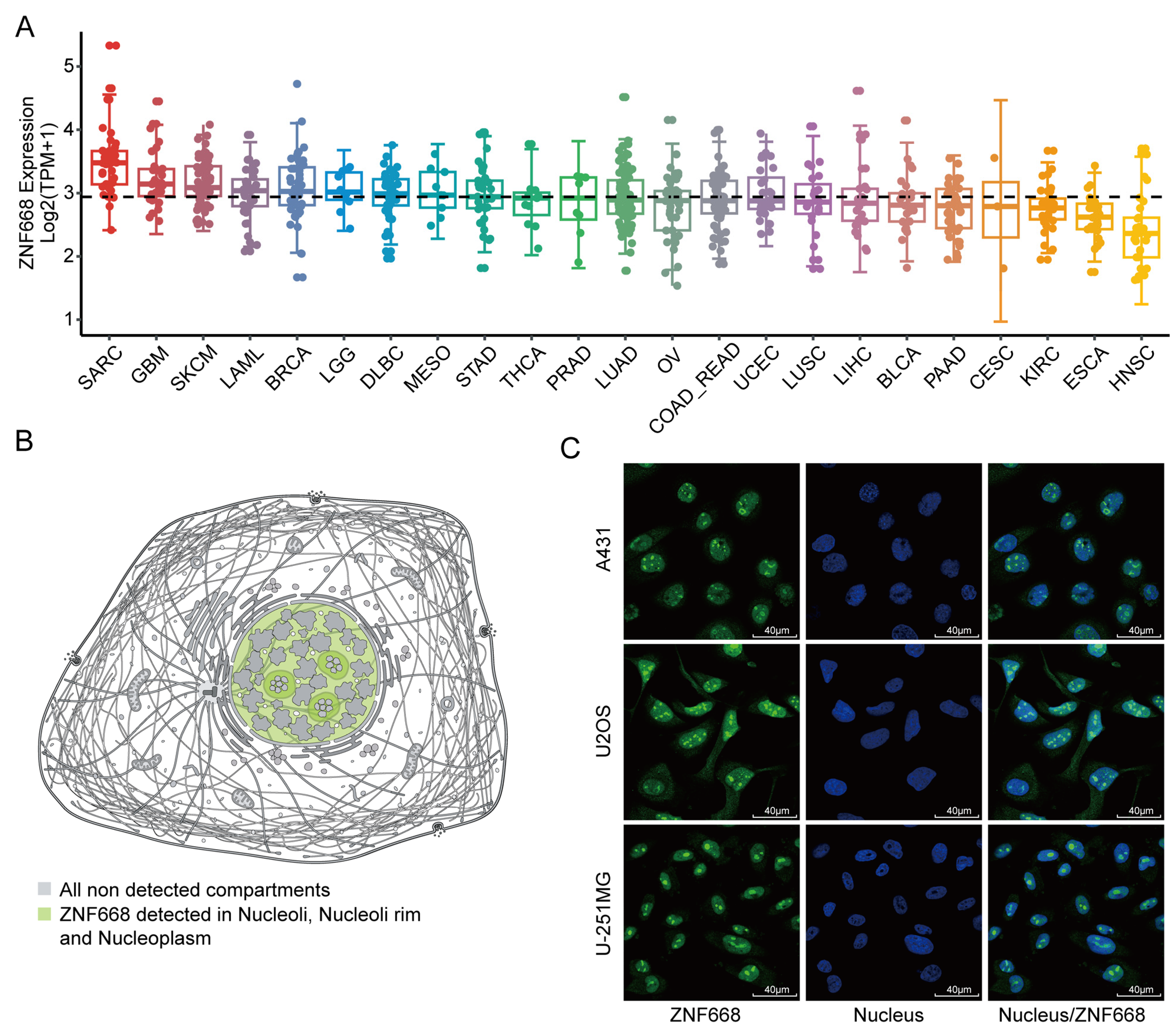
ZNF668 mRNA expression was analyzed across a wide range of cancer cell lines using the CCLE database. The results revealed significant heterogeneity and tissue specificity in its expression pattern (Figure 2A). The top three tumor cell lines showing the highest expression were SARC, GBM, and SKCM. In contrast, ZNF668 expression was generally lower in various cancer cell lines of epithelial origin, with the lowest levels observed in HNSC, ESCA, and KIRC. Furthermore, the predicted subcellular localization of ZNF668 was in the nucleus, and this was consistent with immunofluorescence validation images obtained from the HPA database. (Figure 2B,C).

3.3. Diagnostic Performance of ZNF668

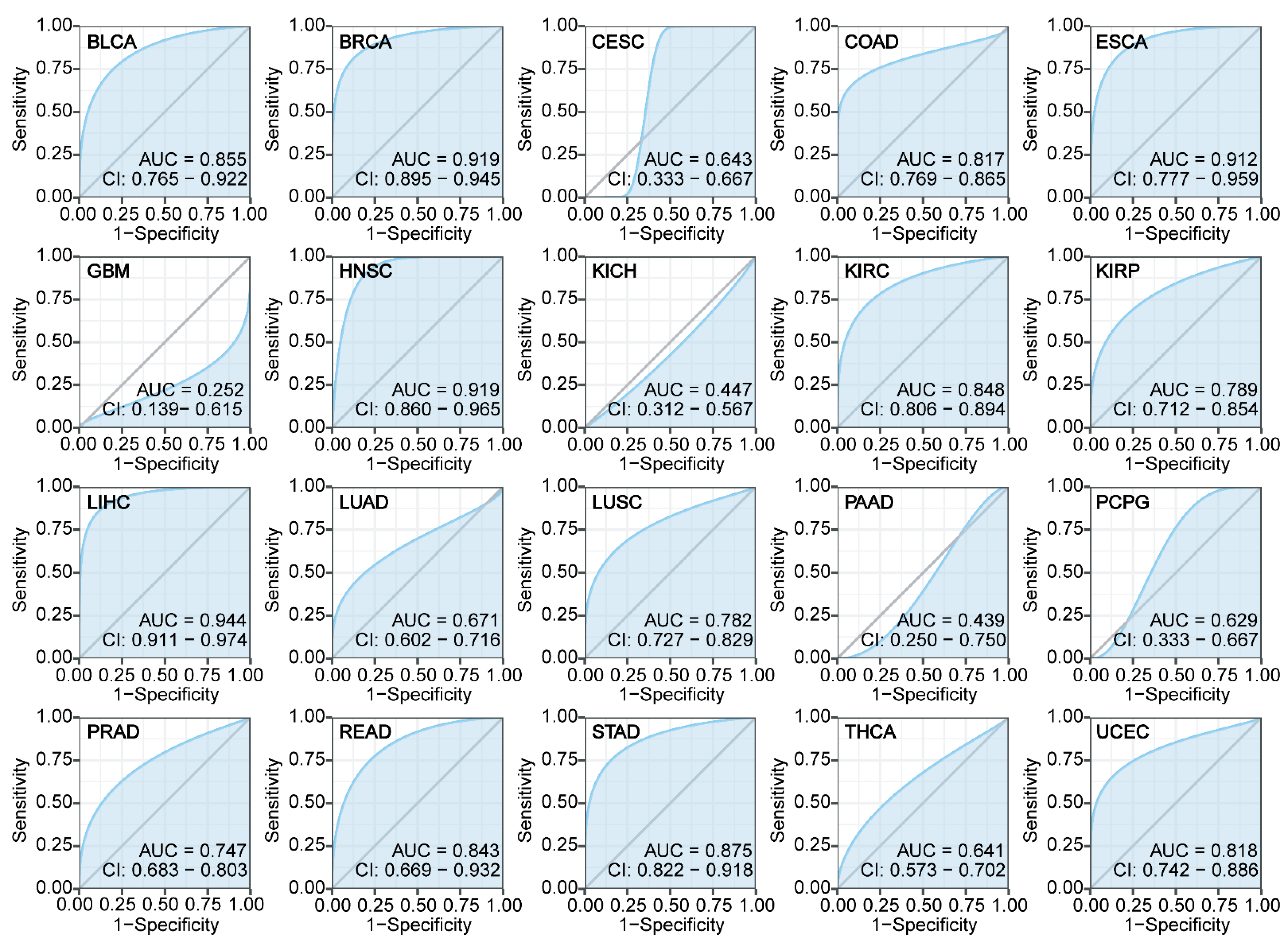
To evaluate the utility of ZNF668 as a potential pan-cancer diagnostic biomarker, we performed ROC curve analysis of its expression levels. The results demonstrated that ZNF668 exhibited high accuracy in distinguishing tumor tissues from normal tissues, showing excellent diagnostic performance in 13 different cancer types with an AUC greater than 0.7 (Figure 3). Specifically, these cancers included BLCA, BRCA, COAD, ESCA, HNSC, KIRC, KIRP, LIHC, LUSC, PRAD, READ, STAD, and UCEC. It is particularly noteworthy that the diagnostic predictive ability of ZNF668 for BRCA, ESCA, HNSC, and LIHC was especially outstanding (AUC > 0.9).

3.4. Prognostic Significance of ZNF668 Expression

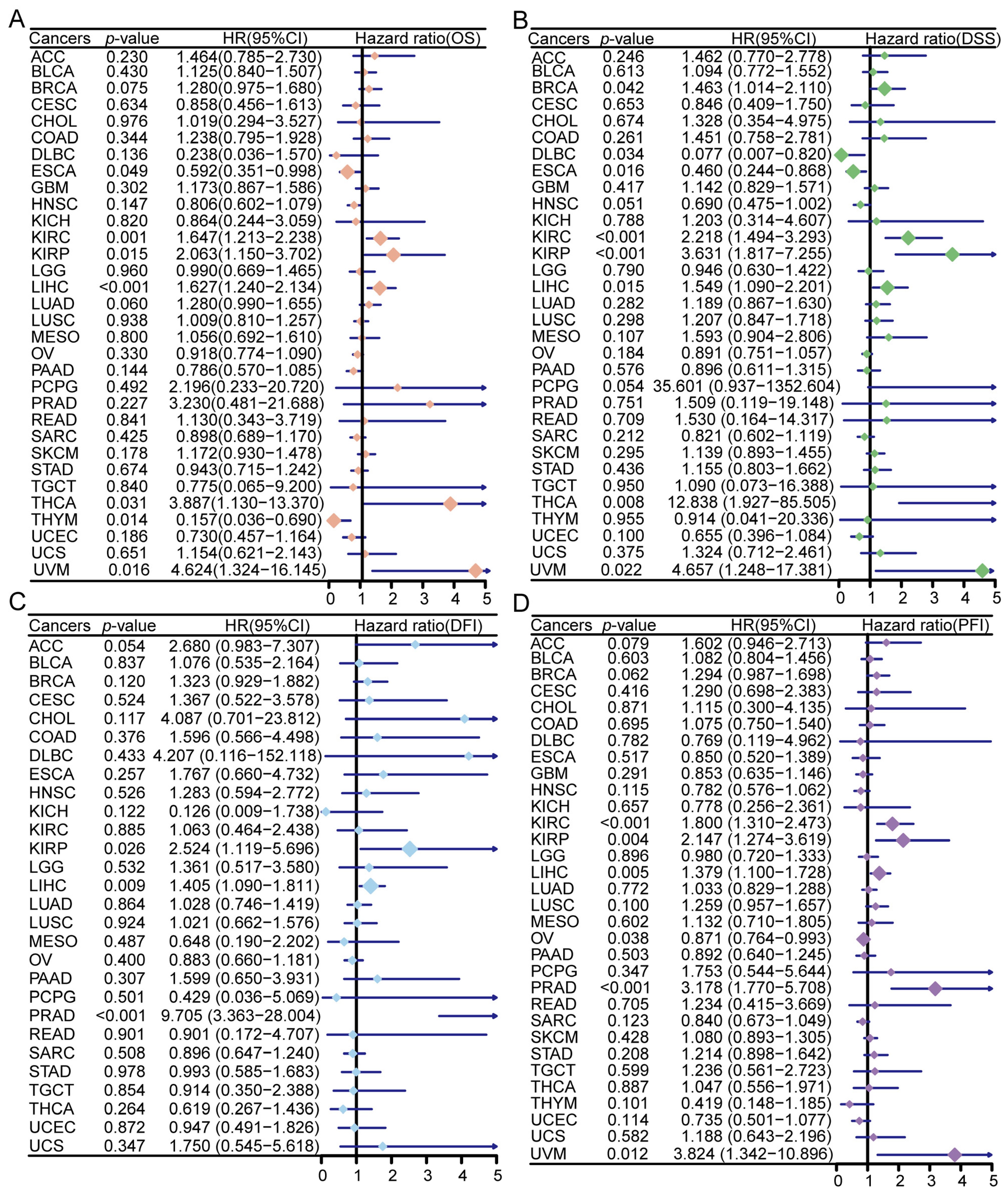
High ZNF668 expression was significantly associated with OS in multiple cancer types (Figure 4A). Specifically, high ZNF668 expression predicted a poorer OS in 5 cancer types: KIRC, KIRP, LIHC, THCA, and UVM, whereas it was associated with a better OS in ESCA and THYM. In terms of DSS, high ZNF668 expression indicated a poor prognosis in 6 cancer types: BRCA, KIRC, KIRP, LIHC, THCA, and UVM, but predicted a better prognosis in DLBC and ESCA (Figure 4B). For DFI, elevated ZNF668 expression was significantly correlated with a worse prognosis in KIRP, LIHC, and PRAD (Figure 4C). In the PFI analysis, high ZNF668 expression predicted a poorer prognosis for 5 cancer types: KIRC, KIRP, LIHC, PRAD, and UVM, while it was associated with a better prognosis in OV (Figure 4D). These results suggest that ZNF668 can serve as a potential tumor-type specific prognostic biomarker, although its significance in some cancers (e.g., DLBC, THCA, THYM) was not independent of clinical covariates in subsequent multivariate Cox analyses (Table S1).

3.5. Genetic Alteration Features of ZNF668

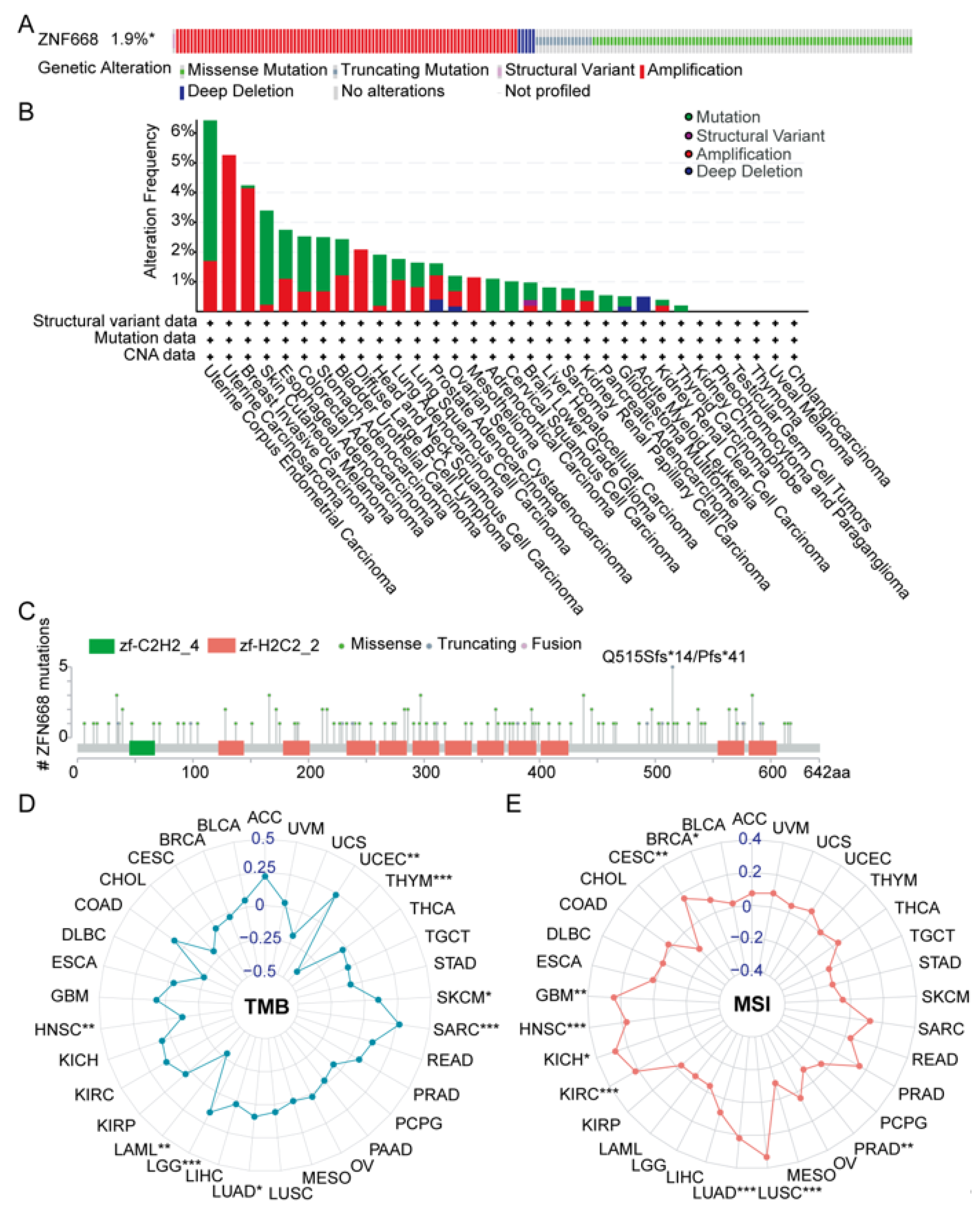
Analysis of the cBioPortal pan-cancer cohort revealed that ZNF668 is genetically altered in 1.9% of all tumors (Figure 5A). We further examined the pan-cancer genetic alterations of ZNF668. UCEC, UCS, and BRCA exhibited the highest mutation rates, at 6.43%, 5.26%, and 4.24%, respectively (Figure 5B). In most cancer types, mutations or amplifications were the predominant types of genetic alterations, while structural variants and deep deletions were relatively less common. The ZNF668 gene encodes a protein of 619 amino acids containing multiple zinc finger domains. Focusing specifically on somatic mutations, their overall prevalence was 1.0%. Among the identified ZNF668 gene variants, a total of 123 variants of unknown significance were detected, including 106 missense mutations, 16 truncating mutations, and 1 fusion variant. Figure 5C illustrates 5 representative mutation events and their patient origins, with the truncating mutation at amino acid position 515 (Q515Sfs14/Pfs41) being relatively frequent among the mutations shown.
Prompted by this evidence of genetic instability, we correlated ZNF668 expression with TMB and MSI. Our findings revealed varying correlations between ZNF668 expression and TMB across different cancer types (Figure 5D). A significant positive correlation was observed in LUAD, SKCM, SARC, UCEC and LGG. Conversely, ZNF668 expression exhibited a significant negative correlation with TMB in HNSC, LAML, and THYM. Notably, a consistent positive correlation was observed between ZNF668 expression and MSI in several cancer types, including BRCA, CESC, GBM, HNSC, KICH, KIRC, LUAD, LUSC, and PRAD (Figure 5E).

3.6. Landscape of Genetic Alterations in ZNF668

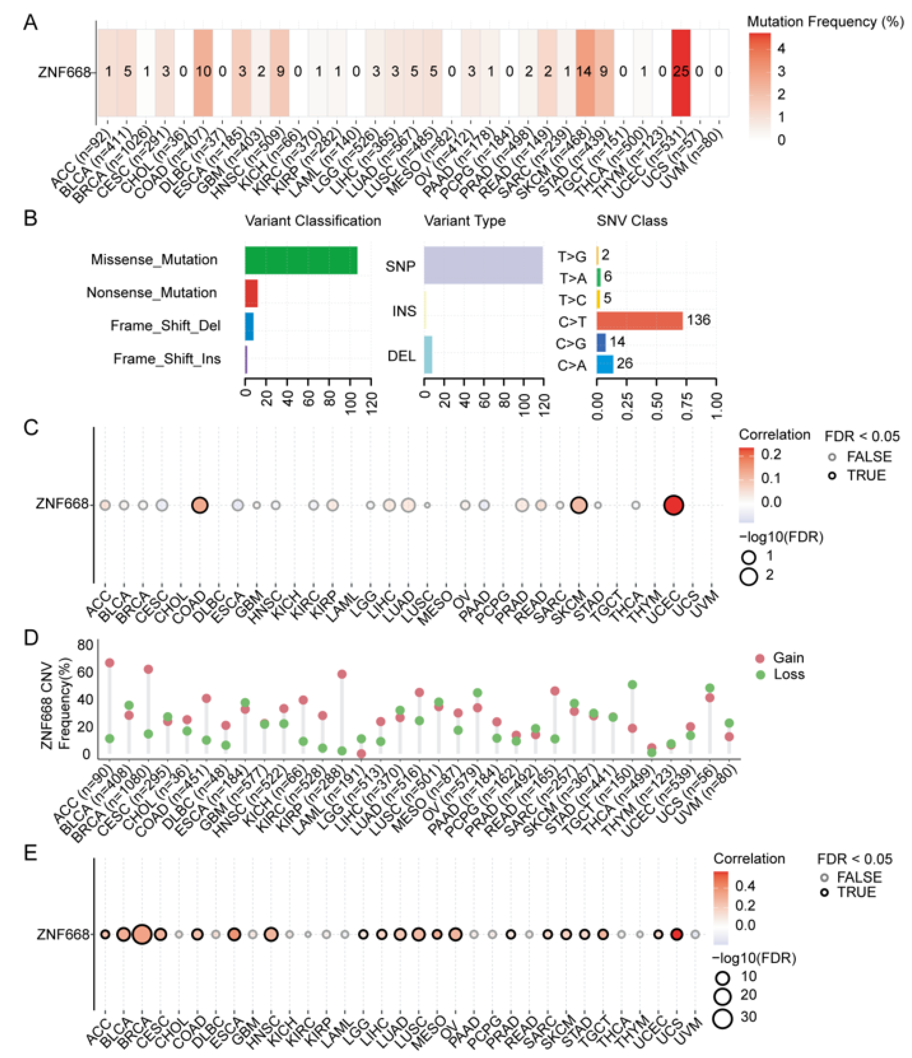
Analysis of SNVs revealed that UCEC exhibited the highest mutation frequency (~4.7%) in the ZNF668 gene, followed by SKCM and COAD (Figure 6A). Missense mutations were the predominant SNV type, and C-to-T transitions were the primary form of single nucleotide mutations (Figure 6B). Notably, in COAD, SKCM, and UCEC, a positive correlation was observed between ZNF668 SNVs and its mRNA expression (Figure 6C). For CNVs, both amplifications and deletions of ZNF668 were observed in most cancer types (Figure 6D). Among them, high-frequency homozygous amplifications were present in ACC, BRCA, and KIRP, while high-frequency heterozygous deletions were found in OV, TGCT, and UCS. Furthermore, in the majority of cancer types, a positive correlation was found between ZNF668 CNV and its mRNA expression (Figure 6E).

3.7. ZNF668 Interaction Network and Functional Enrichment Analysis

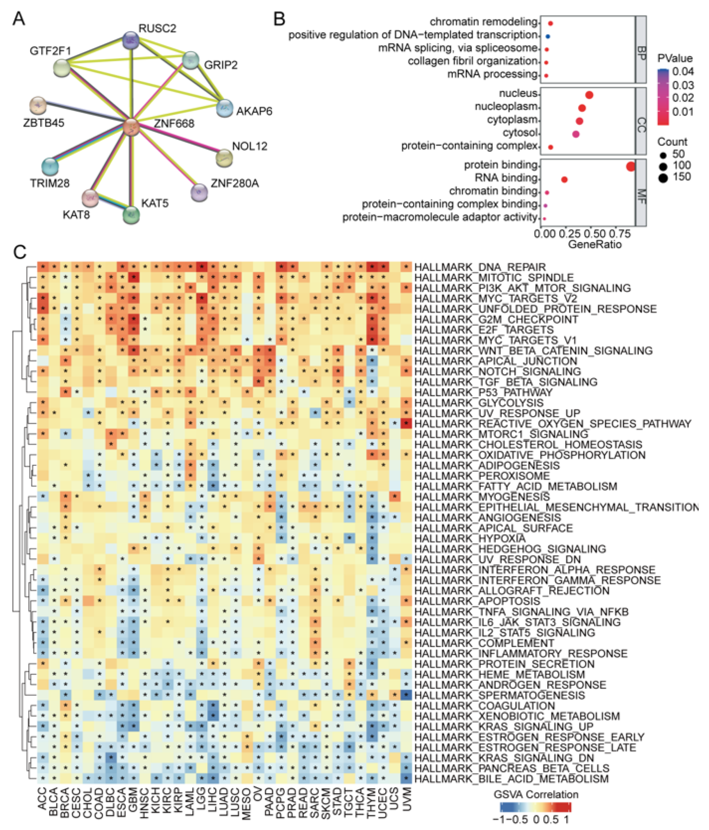
To elucidate the functional context of ZNF668, we constructed a ZNF668-centric PPI network using the STRING database, highlighting its top 10 core interactors (Figure 7A). Subsequently, GO analysis (Figure 7B) showed that in terms of BP, genes co-expressed with ZNF668 were significantly enriched in transcriptional regulation (e.g., “regulation of transcription by RNA polymerase II,” “chromatin remodeling”) and embryonic development. For CC, they were mainly enriched in the nucleus. At the MF level, they were primarily involved in molecular binding functions such as protein, RNA, and metal ion binding. GSVA revealed a robust positive correlation with pathways integral to cell proliferation and survival, including DNA repair, mitotic spindle, G2M checkpoint, PI3K/AKT/MTOR signaling, myc targets, and the unfolded protein response. Conversely, its expression was significantly and negatively correlated with pathways associated with specialized metabolism and signaling pathways, such as xenobiotic metabolism, bile acid metabolism, KRAS signaling, and estrogen response (Figure 7C).

3.8. Association of ZNF668 Expression with Immune Regulatory Molecules

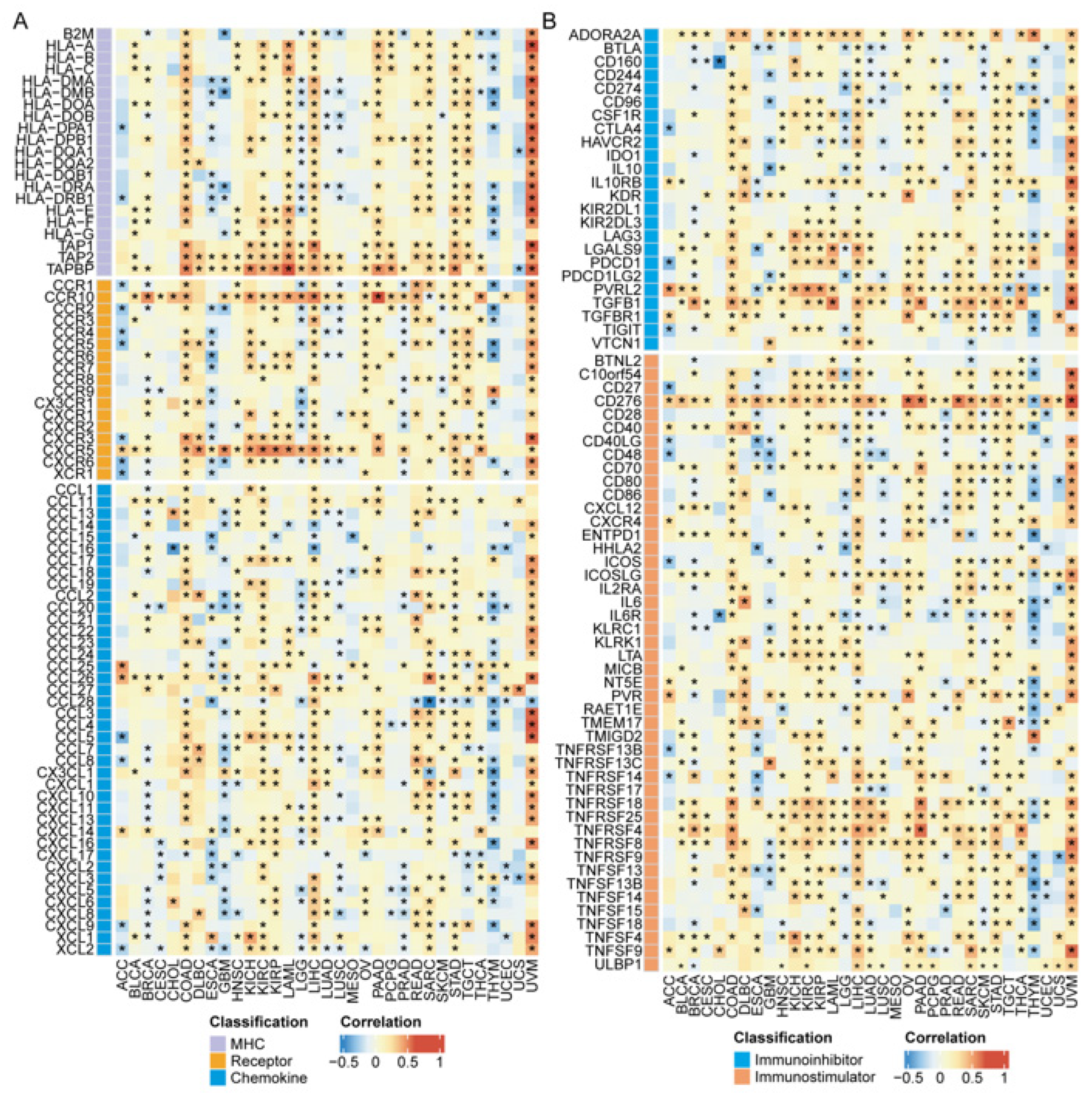
ZNF668 expression exhibited a remarkably consistent positive correlation with a large number of immune regulatory genes across diverse cancer types. This pattern was particularly strong for key costimulatory and coinhibitory molecules. For instance, strong positive associations were consistently observed for antigen presentation machinery components like TAP1, TAP2, and TAPBP (Figure 8A). Similarly, ZNF668 was positively correlated with PVRL2 (Nectin-2), CD276 (B7-H3), TGFB1, and multiple members of the TNF receptor superfamily, including TNFRSF4 (OX40), TNFRSF18 (GITR), and TNFRSF25, in the vast majority of analyzed cancers (Figure 8B). Other key immune molecules that followed this predominantly positive correlation pattern include the immune checkpoint regulators ADORA2A, CTLA4, LAG3, and PDCD1 (PD-1), as well as the costimulatory molecules CD27, ICOSLG, and LTA (Figure 8B). This widespread positive association extended to crucial chemokine receptors such as CXCR5 and CCR10, which were significantly correlated with ZNF668 in over 15 distinct malignancies (Figure 8A).
In contrast to the widespread positive associations, the correlation of ZNF668 with a subset of immune genes was heterogeneous, varying significantly between different cancer types. The association with B2M exemplifies this pattern, showing a positive correlation in tumors like COAD and PCPG, but a significant negative correlation in GBM, LUAD, LUSC, and THCA. This heterogeneity was particularly evident among major immune checkpoints and costimulatory molecules (Figure 8A). For example, CD274 (PD-L1) and TIGIT were positively correlated with ZNF668 in gastrointestinal cancers (COAD, STAD, LIHC) but were negatively correlated in others, such as BRCA, LGG, and LUSC (Figure 8B). Similarly, the costimulatory molecule CD86 showed a positive correlation in COAD and LIHC but a strong negative correlation in GBM, LGG, and LUSC (Figure 8B). This context-dependency also extended to molecules like TNFRSF14 (HVEM), CSF1R, and several HLA genes, which were positively correlated in cancers like COAD and LIHC but negatively correlated in ESCA, LGG, and LUSC (Figure 8A,B). While a consistent negative correlation with a single gene across most cancers was rare, it was a defining feature for a few chemokines, most notably CXCL17 and CCL28. Furthermore, we identified a consistent negative correlation between ZNF668 expression and a specific group of chemokines primarily responsible for neutrophil recruitment, notably CXCL8, CXCL2, and CXCL5 (Figure 8A).

3.9. Correlation of ZNF668 Expression with Immune Cell Infiltration

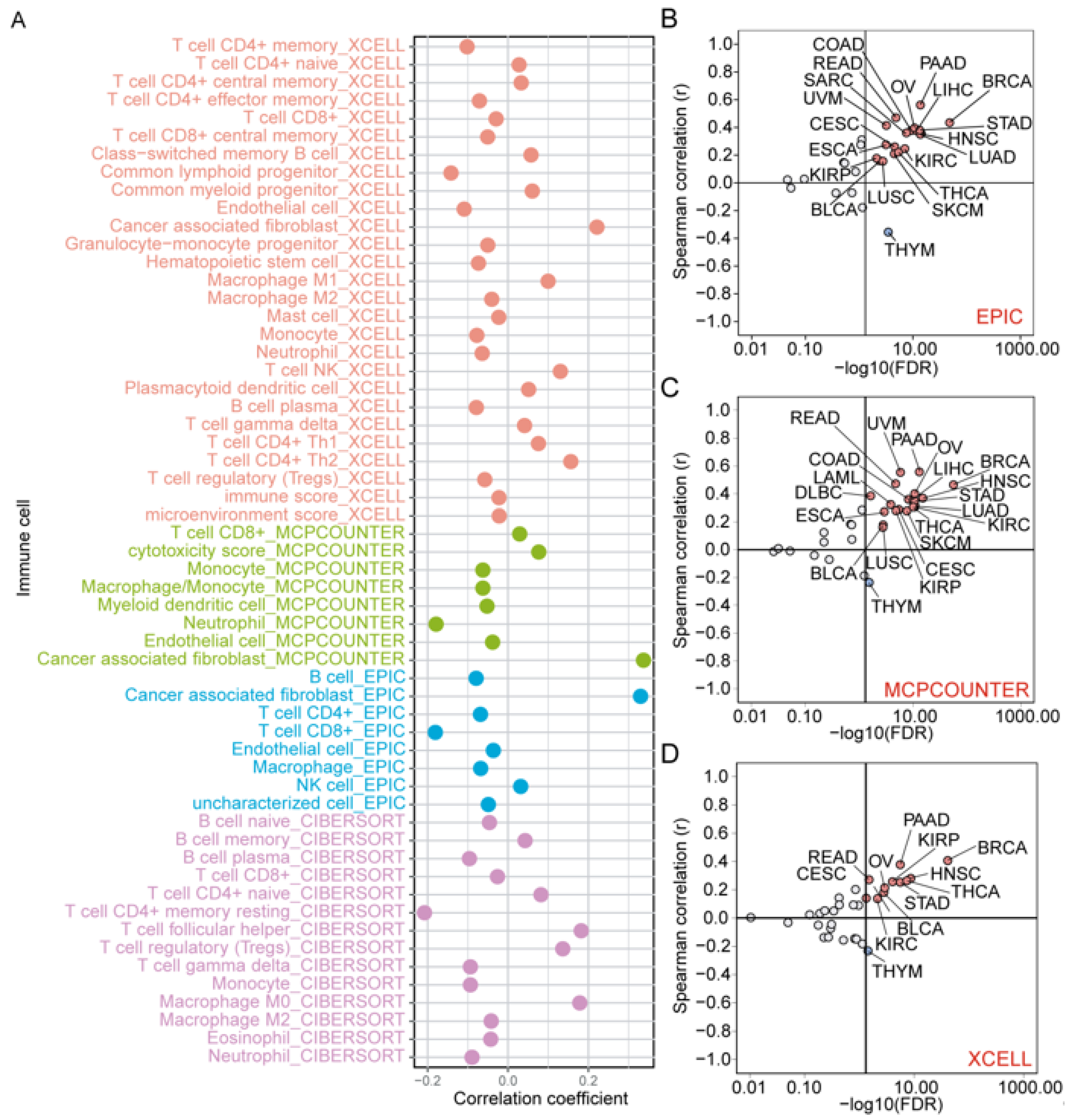
ZNF668 expression broadly correlated with immune cell infiltration across various cancers. Notably, among these correlations, ZNF668 expression showed a consistent and significant positive correlation with the infiltration of CAFs, confirmed by integrating results from three distinct computational algorithms: EPIC, MCP-counter, and xCell (Figure 9A). Further analysis revealed a strong and consistent positive correlation between ZNF668 expression levels and CAF abundance in the vast majority of analyzed cancer types, such as PAAD, READ, and BRCA (Figure 9B–D). In sharp contrast, ZNF668 expression showed a consistent negative correlation with CAF infiltration in THYM.

3.10. Drug Sensitivity Analysis and Molecular Docking

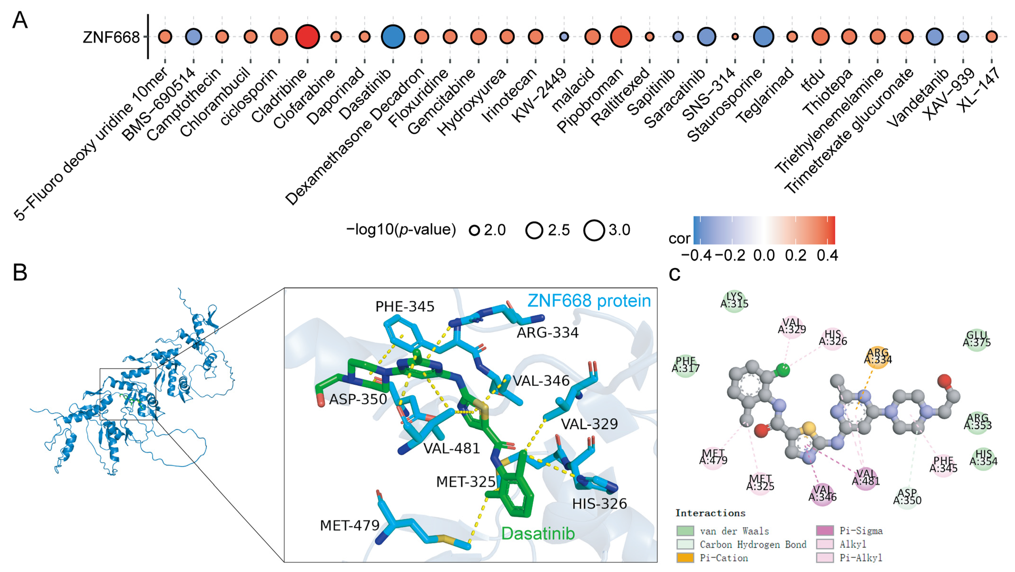
To identify potential inhibitors targeting ZNF668, we evaluated the correlation between ZNF668 mRNA expression and drug sensitivity data obtained from the CellMiner database. Among all screened agents, Dasatinib exhibited the strongest negative correlation with ZNF668 expression (rho= −0.441, p < 0.05) (Figure 10A), suggesting a hypothesis that Dasatinib might exert its effects by potentially interacting with ZNF668 for anti-tumor effects. Molecular docking further demonstrated that Dasatinib stably binds to ZNF668, with a low binding energy of −7.6 kcal/mol, reflecting the high stability of the resulting complex (Figure 10B). The complex is stabilized through hydrogen bonds with PHE-345 and ASP-350, a π-cation interaction with ARG-334, and hydrophobic contributions from MET-325, MET-479, VAL-329, VAL-346, VAL-481, and HIS-326 (Figure 10C).

3.11. Validation of ZNF668 Expression by TMA

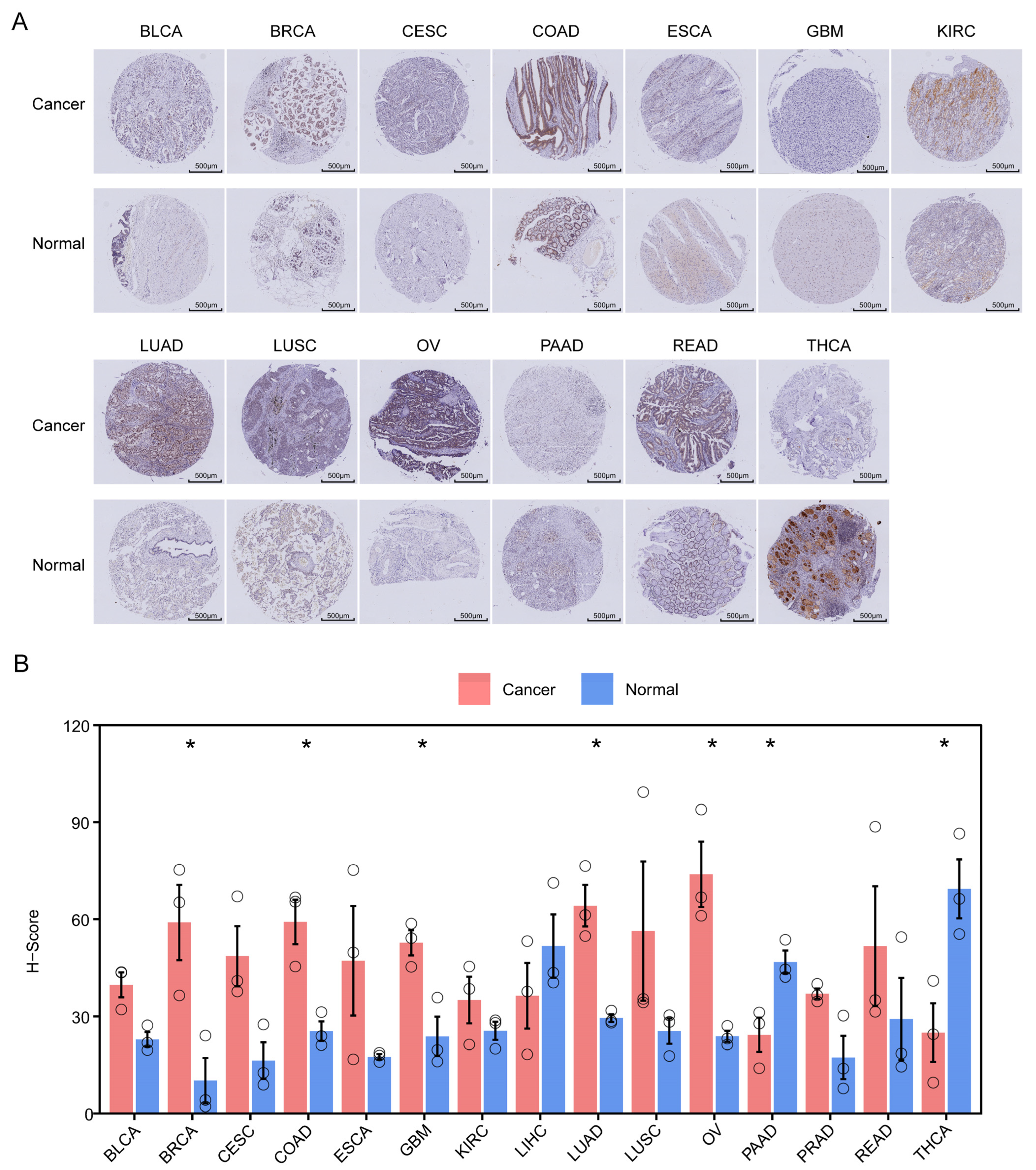
Corroborating our bioinformatic predictions, IHC results confirmed that ZNF668 protein levels were significantly elevated in BRCA, COAD, GBM, LUAD, and OV, while conversely being significantly decreased in PAAD and THCA (Figure 11A,B).

4. Discussion

This study provides a comprehensive pan-cancer analysis of ZNF668, investigating its expression patterns, prognostic value, genomic alterations, functional pathways, and its complex interplay with the tumor immune microenvironment. Furthermore, to explore its therapeutic potential, we employed drug sensitivity correlation and molecular docking to identify potential inhibitors.
We first established the clinical relevance of ZNF668 by analyzing its expression and prognostic value across a wide range of human cancers. ZNF668 was frequently dysregulated, exhibiting prominent overexpression in malignancies such as BRCA, HNSC, LIHC, and LUAD, while showing reduced expression in others, including KICH and THCA. Subsequently, our IHC analysis served to both validate and expand upon our bioinformatic findings. We confirmed the significant ZNF668 upregulation in BRCA and LUAD and downregulation in THCA, which aligned perfectly with the TCGA and CPTAC database analyses. Furthermore, we supplemented the database findings by providing the protein-level evidence of significant ZNF668 overexpression in OV. However, while our IHC showed ZNF668 was significantly upregulated in COAD and GBM, in contrast to the non-significant results from TCGA, the trend of upregulation was consistent.
Beyond its dysregulated expression, ZNF668 showed significant potential as a clinical biomarker, underscored by its high diagnostic accuracy for distinguishing tumors from normal tissue (AUC > 0.9 in BRCA, ESCA, HNSC, and LIHC) and its consistent association with shorter OS, DSS, and PFI in KIRP and LIHC. However, these clinical findings present an apparent paradox when contrasted with previous in vitro research. While the oncogenic role aligns with reports of ZNF668 promoting proliferation in leukemia cells [25], it contradicts studies where ZNF668 acts as a potent inhibitor of migration and invasion in lung and bladder cancer cells [26,27]. This raises the question of why ZNF668 functions as a tumor suppressor in isolated epithelial cancer cells but correlates with poor prognosis in patient tumors of the same origin.
To resolve this discrepancy, our analysis of the CCLE database pointed towards a context-dependent role for ZNF668, as we found its expression was significantly higher in cell lines of mesenchymal origin (e.g., SARC, GBM, LAML) compared to those from many epithelial cancers (e.g., HNSC, ESCA). This suggested a lineage-specific functional switch, wherein it may act as an oncogene in high-expression mesenchymal tumors but as a tumor suppressor in low-expression epithelial cancers. However, our clinical findings challenge this hypothesis, as ZNF668 overexpression in epithelial tumors like LIHC consistently predicts poor survival. This discrepancy reveals the limitations of cell-line models and points to the TME as a critical determinant of ZNF668’s ultimate oncogenic role.
Further analysis revealed that a high-ZNF668 TME presents a concurrently inflamed yet profoundly immunosuppressive state. Specifically, the TME is immunologically “hot,” as ZNF668’s positive correlation with T-cell co-stimulatory molecules (e.g., TNFRSF4, CD27) and antigen presentation machinery (e.g., TAP1, TAPBP) indicates T-cell recruitment and activation [28,29]. However, this is counterbalanced by marked immunosuppression, characterized by upregulated checkpoints like PD-1, CTLA4, and LAG3. This paradox may be mechanistically linked to a strong positive correlation between ZNF668 and CAF abundance. CAFs, through intricate signaling networks often involving TGF-β, are known to drive treatment failure and immune escape [30,31,32]. We thus propose that a CAF-driven, TGF-β-mediated suppressive network underlies this state of adaptive immune resistance, resulting in a T-cell-infiltrated but functionally exhausted TME [33].
While the ZNF668-CAF axis suggests a significant mechanism for immune evasion, its immunomodulatory influence also extends to other aspects of TME. We found that ZNF668 expression exhibited a consistent positive correlation with MSI across cancers. Since MSI is a hallmark of defective DNA mismatch repair (MMR) that results in a hypermutated phenotype and abundant immunogenic neoantigens [34,35], this finding suggests that high ZNF668 expression may either contribute to this MSI-high phenotype or be associated with impaired MMR function. Moreover, we observed that ZNF668 expression was consistently negatively correlated with a specific group of pro-inflammatory chemokines (notably CXCL8, CXCL2, and CXCL5) that are primarily responsible for neutrophil recruitment [36,37]. Despite the above consistent associations, ZNF668’s role exhibits significant context-dependency and complexity at a broader level. For instance, its association with key molecules like CD274 (PD-L1) and B2M is positive in gastrointestinal cancers but shifts to a negative correlation in others, including BRCA, LUSC, and GBM. This heterogeneity also extends to TMB, which is positively correlated with ZNF668 in LUAD and SKCM yet negatively correlated in HNSC and LAML.
GO enrichment analysis revealed that ZNF668 is predominantly enriched in fundamental nuclear processes, including the spliceosome, transcriptional regulation, and chromatin remodeling. These pathways are integral to the establishment and maintenance of malignant phenotypes. The spliceosome, for instance, contributes to oncogenesis not only through its canonical alternative splicing activities that reshape the tumor transcriptome and proteome, but also via non-canonical, splicing-independent mechanisms that influence a wide range of core aspects of cancer progression [38,39]. Dysregulation of transcriptional control reshapes gene expression programs to drive tumor progression [40], while imbalances in chromatin remodeling regulate gene expression and exacerbate splicing abnormalities by altering chromatin accessibility and histone modifications [41]. The functional consequence of this regulation is evident in GSVA, which revealed that its expression was positively correlated with pathways integral to cell proliferation and survival. The convergence of these functions within the nucleus is highly consistent with the established nuclear localization of the ZNF668 protein, reinforcing the hypothesis that it functions as a critical upstream regulatory hub governing key oncogenic processes.
We also investigated the upstream mechanisms of the aberrant expression of ZNF668 by analyzing its genetic alteration features. Amplification is the most predominant type of genetic alteration for ZNF668 in multiple cancers, and its mRNA expression level is significantly positively correlated with its copy number variation level in the vast majority of tumors. This provides a potential molecular explanation for the abnormal expression of ZNF668 and is consistent with the mechanism by which many known oncogenes promote tumorigenesis through copy number amplification [42].
Given the strong association between ZNF668 and CAF abundance revealed by our bioinformatic analysis, we sought to identify its potential inhibitors. Through an analysis of drug sensitivity profiles, we discovered a strong negative correlation between ZNF668 expression and response to Dasatinib. This association was further supported by preliminary molecular docking simulations, which indicated a potential stable binding between the Dasatinib and the ZNF668 protein. Dasatinib, a multi-kinase inhibitor targeting BCR-ABL, SRC, and c-KIT, has been shown not only to inhibit the growth of lung cancer-derived CAFs but also to revert their oncogenic phenotype to that of non-tumorigenic fibroblasts [43]. Furthermore, Dasatinib enhances immunotherapy efficacy by reprogramming CAFs. In models of colorectal and triple-negative breast cancer, Dasatinib selectively inhibits CAFs and remodels the extracellular matrix, thereby promoting the deep infiltration of drugs and CD8+ T cells into the tumor, converting immunologically “cold” tumors into “hot” tumors and significantly enhancing the efficacy of anti-PD-1 immunotherapy [44,45]. While these potent CAF-inhibitory effects of Dasatinib are well-documented, its molecular target remains unclear. Our findings provide a novel mechanistic link between these independent observations, leading us to propose the scientific hypothesis that ZNF668 may be the key molecular mediator through which Dasatinib exerts its CAF-modulating and immune-reprogramming functions. This hypothesis warrants further experimental investigation.
This study has certain limitations. The discrepant associations observed across different tumor types underscore the context-dependent functionality of ZNF668, warranting further investigation to delineate its specific mechanistic roles. Furthermore, while ZNF668 shows promise as a biomarker, its prognostic and diagnostic utility requires robust validation in larger, prospectively collected clinical cohorts. Critically, the proposition of Dasatinib as a putative inhibitor of ZNF668 is currently based solely on in silico predictions; therefore, empirical corroboration via biophysical assays, such as the cellular thermal shift assay or surface plasmon resonance, is required to confirm a direct interaction.

5. Conclusions

In summary, this study redefines ZNF668 as a key mediator of immunosuppression and a biomarker for poor prognosis. Our work reconciles the conflicting reports of ZNF668’s tumor-suppressive in vitro activities with its clear pro-tumorigenic association in patients, identifying the TME as the decisive, context-dependent switch, which is mechanistically underscored by a strong correlation with CAF abundance that cultivates a T-cell-exhausted, immunosuppressive landscape. This association provides a clear rationale for targeting ZNF668, with Dasatinib identified as a potential inhibitor. Our study provides a new framework for restoring effective anti-tumor immunity by targeting ZNF668 to dismantle the immunosuppressive TME.

Supplementary Materials

The following supporting information can be downloaded at: https://www.mdpi.com/article/10.3390/cimb47120997/s1.

Author Contributions

Conceptualization, X.H. and C.H.; Methodology, X.H. and J.G.; Project Administration, S.C.; Resources, X.H.; Data Curation, X.H. and W.H.; Formal Analysis, X.H. and H.Z.; Supervision, C.H.; Writing—Original Draft, All authors; Writing—Review and Editing, All authors. All authors have read and agreed to the published version of the manuscript.

Funding

This research received no external funding.

Institutional Review Board Statement

The study was conducted using publicly available data from The Cancer Genome Atlas (TCGA), the Human Protein Atlas (HPA), and the Clinical Proteomic Tumor Analysis Consortium (CPTAC). According to Article 32 of the “Measures for the Ethical Review of Life Science and Medical Research Involving Humans” (People’s Republic of China, 2023), the research was exempted from ethics committee review as it involved publicly available and non-identifiable human data.

Informed Consent Statement

Patient consent was waived as the study used only publicly available and de-identified datasets.

Data Availability Statement

The original data presented in the study are openly available in TCGA and other databases. The raw data supporting the conclusions of this article will be made available by the corresponding authors on request.

Conflicts of Interest

The authors declare no conflicts of interest.

References

  1. World Health Organization. Global cancer burden growing, amidst mounting need for services. Saudi Med. J. 2024, 45, 326–327. [Google Scholar]
  2. Zafar, A.; Khatoon, S.; Khan, M.J.; Abu, J.; Naeem, A. Advancements and limitations in traditional anti-cancer therapies: A comprehensive review of surgery, chemotherapy, radiation therapy, and hormonal therapy. Discov. Oncol. 2025, 16, 607. [Google Scholar] [CrossRef] [PubMed]
  3. Robert, C. A decade of immune-checkpoint inhibitors in cancer therapy. Nat. Commun. 2020, 11, 3801. [Google Scholar] [CrossRef] [PubMed]
  4. Kong, X.; Zhang, J.; Chen, S.; Wang, X.; Xi, Q.; Shen, H.; Zhang, R. Immune checkpoint inhibitors: Breakthroughs in cancer treatment. Cancer Biol. Med. 2024, 21, 451–472. [Google Scholar] [CrossRef] [PubMed]
  5. Hegde, P.S.; Chen, D.S. Top 10 Challenges in Cancer Immunotherapy. Immunity 2020, 52, 17–35. [Google Scholar] [CrossRef]
  6. Schoenfeld, A.J.; Hellmann, M.D. Acquired Resistance to Immune Checkpoint Inhibitors. Cancer Cell 2020, 37, 443–455. [Google Scholar] [CrossRef]
  7. de Visser, K.E.; Joyce, J.A. The evolving tumor microenvironment: From cancer initiation to metastatic outgrowth. Cancer Cell 2023, 41, 374–403. [Google Scholar] [CrossRef]
  8. Baghban, R.; Roshangar, L.; Jahanban-Esfahlan, R.; Seidi, K.; Ebrahimi-Kalan, A.; Jaymand, M.; Kolahian, S.; Javaheri, T.; Zare, P. Tumor microenvironment complexity and therapeutic implications at a glance. Cell Commun. Signal. 2020, 18, 59. [Google Scholar] [CrossRef]
  9. Kim, S.J.; Khadka, D.; Seo, J.H. Interplay between Solid Tumors and Tumor Microenvironment. Front. Immunol. 2022, 13, 882718. [Google Scholar] [CrossRef]
  10. Cassandri, M.; Smirnov, A.; Novelli, F.; Pitolli, C.; Agostini, M.; Malewicz, M.; Melino, G.; Raschellà, G. Zinc-finger proteins in health and disease. Cell Death Discov. 2017, 3, 17071. [Google Scholar] [CrossRef]
  11. Rakhra, G.; Rakhra, G. Zinc finger proteins: Insights into the transcriptional and post transcriptional regulation of immune response. Mol. Biol. Rep. 2021, 48, 5735–5743. [Google Scholar] [CrossRef] [PubMed]
  12. Qian, Y.; Wu, Q. The Multifaceted Roles of Zinc Finger Proteins in Pluripotency and Reprogramming. Int. J. Mol. Sci. 2025, 26, 5106. [Google Scholar] [CrossRef]
  13. Zhao, J.; Wen, D.; Zhang, S.; Jiang, H.; Di, X. The role of zinc finger proteins in malignant tumors. FASEB J. 2023, 37, e23157. [Google Scholar] [CrossRef]
  14. Jen, J.; Wang, Y.C. Zinc finger proteins in cancer progression. J. Biomed. Sci. 2016, 23, 53. [Google Scholar] [CrossRef]
  15. Liu, S.; Sima, X.; Liu, X.; Chen, H. Zinc Finger Proteins: Functions and Mechanisms in Colon Cancer. Cancers 2022, 14, 5242. [Google Scholar] [CrossRef]
  16. Xiao, Y.; Xiang, T.; Luo, X.; Li, C.; Li, Q.; Peng, W.; Li, L.; Li, S.; Wang, Z.; Tang, L.; et al. Zinc-finger protein 545 inhibits cell proliferation as a tumor suppressor through inducing apoptosis and is disrupted by promoter methylation in breast cancer. PLoS ONE 2014, 9, e110990. [Google Scholar] [CrossRef]
  17. Tian, S.; Chen, X.; Li, J. Zinc finger transcription factor ZNF24 inhibits colorectal cancer growth and metastasis by suppressing MMP2 transcription. Genes Dis. 2025, 12, 101529. [Google Scholar] [CrossRef]
  18. Goldman, M.J.; Craft, B.; Hastie, M.; Repečka, K.; McDade, F.; Kamath, A.; Banerjee, A.; Luo, Y.; Rogers, D.; Brooks, A.N.; et al. Visualizing and interpreting cancer genomics data via the Xena platform. Nat. Biotechnol. 2020, 38, 675–678. [Google Scholar] [CrossRef]
  19. Wang, D.; Qian, X.; Du, Y.N.; Sanchez-Solana, B.; Chen, K.; Kanigicherla, M.; Jenkins, L.M.; Luo, J.; Eng, S.; Park, B.; et al. cProSite: A web based interactive platform for online proteomics, phosphoproteomics, and genomics data analysis. J. Biotechnol. Biomed. 2023, 6, 573–578. [Google Scholar] [CrossRef] [PubMed]
  20. Nusinow, D.P.; Szpyt, J.; Ghandi, M.; Rose, C.M.; McDonald, E.R.; Kalocsay, M.; Jané-Valbuena, J.; Gelfand, E.; Schweppe, D.K.; Jedrychowski, M.; et al. Quantitative Proteomics of the Cancer Cell Line Encyclopedia. Cell 2020, 180, 387–402.e16. [Google Scholar] [CrossRef] [PubMed]
  21. Uhlén, M.; Fagerberg, L.; Hallström, B.M.; Lindskog, C.; Oksvold, P.; Mardinoglu, A.; Sivertsson, Å.; Kampf, C.; Sjöstedt, E.; Asplund, A.; et al. Proteomics. Tissue-based map of the human proteome. Science 2015, 347, 1260419. [Google Scholar] [CrossRef]
  22. Gao, J.; Aksoy, B.A.; Dogrusoz, U.; Dresdner, G.; Gross, B.; Sumer, S.O.; Sun, Y.; Jacobsen, A.; Sinha, R.; Larsson, E.; et al. Integrative analysis of complex cancer genomics and clinical profiles using the cBioPortal. Sci. Signal. 2013, 6, pl1. [Google Scholar] [CrossRef]
  23. Reinhold, W.C.; Sunshine, M.; Liu, H.; Varma, S.; Kohn, K.W.; Morris, J.; Doroshow, J.; Pommier, Y. CellMiner: A web-based suite of genomic and pharmacologic tools to explore transcript and drug patterns in the NCI-60 cell line set. Cancer Res. 2012, 72, 3499–3511. [Google Scholar] [CrossRef]
  24. Jumper, J.; Evans, R.; Pritzel, A.; Green, T.; Figurnov, M.; Ronneberger, O.; Tunyasuvunakool, K.; Bates, R.; Žídek, A.; Potapenko, A.; et al. Highly accurate protein structure prediction with AlphaFold. Nature 2021, 596, 583–589. [Google Scholar] [CrossRef]
  25. Hou, Y.; Ning, W.; Huhe, M.; Shu, C. Genome-Wide Detection of Leukemia Biomarkers from lincRNA-Protein-Coding Gene Interaction Networks in the Three-Dimensional Chromatin Structure. Curr. Issues Mol. Biol. 2025, 47, 384. [Google Scholar] [CrossRef]
  26. Zhang, X.; Jiang, G.; Wu, J.; Zhou, H.; Zhang, Y.; Miao, Y.; Feng, Y.; Yu, J. Zinc finger protein 668 suppresses non-small cell lung cancer invasion and migration by downregulating Snail and upregulating E-cadherin and zonula occludens-1. Oncol. Lett. 2018, 15, 3806–3813. [Google Scholar] [CrossRef]
  27. Okuno, Y.; Hattori-Kato, M.; Tanaka, H.; Tonooka, A.; Takeuchi, T. Relationship between the Reduced Expression of Zinc Finger Protein 668 in Bladder Cancer and Its Invasiveness. Int. J. Mol. Sci. 2023, 24, 8668. [Google Scholar] [CrossRef]
  28. Chen, L.; Flies, D.B. Molecular mechanisms of T cell co-stimulation and co-inhibition. Nat. Rev. Immunol. 2013, 13, 227–242. [Google Scholar] [CrossRef]
  29. Wu, B.; Zhang, B.; Li, B.; Wu, H.; Jiang, M. Cold and hot tumors: From molecular mechanisms to targeted therapy. Signal Transduct. Target. Ther. 2024, 9, 274. [Google Scholar] [CrossRef]
  30. Hauge, A.; Rofstad, E.K. Antifibrotic therapy to normalize the tumor microenvironment. J. Transl. Med. 2020, 18, 207. [Google Scholar] [CrossRef]
  31. Wu, F.; Yang, J.; Liu, J.; Wang, Y.; Mu, J.; Zeng, Q.; Deng, S.; Zhou, H. Signaling pathways in cancer-associated fibroblasts and targeted therapy for cancer. Signal Transduct. Target. Ther. 2021, 6, 218. [Google Scholar] [CrossRef]
  32. Maia, A.; Schöllhorn, A.; Schuhmacher, J.; Gouttefangeas, C. CAF-immune cell crosstalk and its impact in immunotherapy. Semin. Immunopathol. 2023, 45, 203–214. [Google Scholar] [CrossRef]
  33. Joyce, J.A.; Fearon, D.T. T cell exclusion, immune privilege, and the tumor microenvironment. Science 2015, 348, 74–80. [Google Scholar] [CrossRef]
  34. Pećina-Šlaus, N.; Kafka, A.; Salamon, I.; Bukovac, A. Mismatch Repair Pathway, Genome Stability and Cancer. Front. Mol. Biosci. 2020, 7, 122. [Google Scholar] [CrossRef]
  35. Xie, N.; Shen, G.; Gao, W.; Huang, Z.; Huang, C.; Fu, L. Neoantigens: Promising targets for cancer therapy. Signal Transduct. Target. Ther. 2023, 8, 9. [Google Scholar] [CrossRef]
  36. Capucetti, A.; Albano, F.; Bonecchi, R. Multiple Roles for Chemokines in Neutrophil Biology. Front. Immunol. 2020, 11, 1259. [Google Scholar] [CrossRef]
  37. Cambier, S.; Gouwy, M.; Proost, P. The chemokines CXCL8 and CXCL12: Molecular and functional properties, role in disease and efforts towards pharmacological intervention. Cell. Mol. Immunol. 2023, 20, 217–251. [Google Scholar] [CrossRef]
  38. Yang, H.; Beutler, B.; Zhang, D. Emerging roles of spliceosome in cancer and immunity. Protein Cell 2022, 13, 559–579. [Google Scholar] [CrossRef]
  39. Ivanova, O.M.; Anufrieva, K.S.; Kazakova, A.N.; Malyants, I.K.; Shnaider, P.V.; Lukina, M.M.; Shender, V.O. Non-canonical functions of spliceosome components in cancer progression. Cell Death Dis. 2023, 14, 77. [Google Scholar] [CrossRef]
  40. Bradner, J.E.; Hnisz, D.; Young, R.A. Transcriptional Addiction in Cancer. Cell 2017, 168, 629–643. [Google Scholar] [CrossRef]
  41. Nair, S.S.; Kumar, R. Chromatin remodeling in cancer: A gateway to regulate gene transcription. Mol. Oncol. 2012, 6, 611–619. [Google Scholar] [CrossRef] [PubMed]
  42. Matsui, A.; Ihara, T.; Suda, H.; Mikami, H.; Semba, K. Gene amplification: Mechanisms and involvement in cancer. Biomol. Concepts 2013, 4, 567–582. [Google Scholar] [CrossRef]
  43. Haubeiss, S.; Schmid, J.O.; Mürdter, T.E.; Sonnenberg, M.; Friedel, G.; van der Kuip, H.; Aulitzky, W.E. Dasatinib reverses cancer-associated fibroblasts (CAFs) from primary lung carcinomas to a phenotype comparable to that of normal fibroblasts. Mol. Cancer 2010, 9, 168. [Google Scholar] [CrossRef]
  44. Kadota, H.; Yuge, R.; Shimizu, D.; Miyamoto, R.; Otani, R.; Hiyama, Y.; Takigawa, H.; Hayashi, R.; Urabe, Y.; Kitadai, Y.; et al. Anti-Programmed Cell Death-1 Antibody and Dasatinib Combination Therapy Exhibits Efficacy in Metastatic Colorectal Cancer Mouse Models. Cancers 2022, 14, 6146. [Google Scholar] [CrossRef]
  45. Zhang, Y.; Zhou, J.; Wang, Y.; Wu, Y.; Li, Y.; Wang, B.; Liu, G.; Gong, Q.; Luo, K.; Jing, J. Stimuli-responsive polymer-dasatinib prodrug to reprogram cancer-associated fibroblasts for boosted immunotherapy. J. Control. Release 2025, 381, 113606. [Google Scholar] [CrossRef]
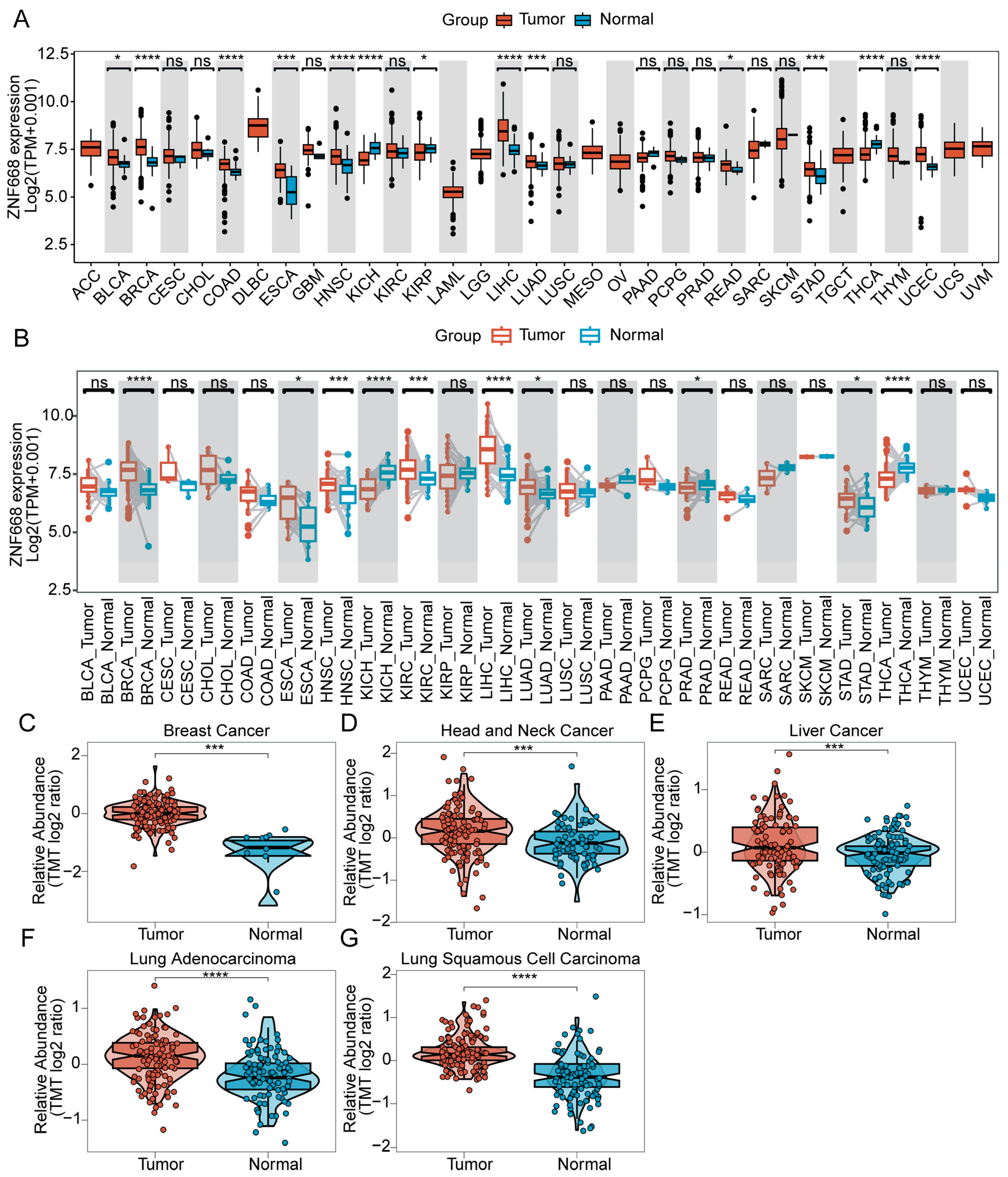
Figure 1. Differential expression of ZNF668 across various cancer types. (A) Comparison of ZNF668 mRNA expression between tumor and normal tissues from TCGA. (B) Paired analysis of ZNF668 mRNA expression in tumor and adjacent normal tissues from TCGA. (CG) Violin plots comparing ZNF668 protein abundance in tumor versus normal tissues from the CPTAC database for (C) Breast Cancer, (D) Head and Neck Cancer, (E) Liver Cancer, (F) Lung Adenocarcinoma, and (G) Lung Squamous Cell Carcinoma. Each dot represents an individual sample. ns, p ≥ 0.05; * p < 0.05; *** p < 0.001; **** p < 0.0001.
Cimb 47 00997 g001
Figure 2. Expression and subcellular localization of ZNF668 in cancer cell lines. (A) ZNF668 mRNA expression in multiple cell lines. (B) Predicted subcellular localization of ZNF668. (C) Immunofluorescence images of ZNF668 subcellular localization in tumor cells from HPA. Image credit: Human Protein Atlas. Image available from v24.0.proteinatlas.org/ENSG00000167394-ZNF668/subcellular.
Cimb 47 00997 g002
Figure 3. Diagnostic potential of ZNF668 expression in pan-cancer. ROC curves illustrating the diagnostic efficacy of ZNF668 expression across various cancer types. The grey line represents the line of no-discrimination (AUC = 0.5).
Cimb 47 00997 g003
Figure 4. Prognostic significance of ZNF668 expression in pan-cancer. Forest plots of univariate Cox regression analysis of ZNF668 expression with (A) OS, (B) DSS, (C) DFI, and (D) PFI across various cancer types. The size of the diamond indicates statistical significance, with larger diamonds representing a significant result.
Cimb 47 00997 g004
Figure 5. Genetic alterations of ZNF668 and its correlation with TMB and MSI. (A) Summary of ZNF668 genetic alterations (including mutations, copy number alterations, and structural variants) across various cancer types from cBioPortal. (B) Landscape of ZNF668 alterations, summarizing mutation types, counts, and frequencies in different cancer types within the TCGA pan-cancer cohort. (C) Lollipop plot depicting the types, numbers, and specific locations of ZNF668 somatic mutations within its protein domains in the TCGA pan-cancer cohort. (D) Radar plot illustrating the correlation between ZNF668 expression and TMB across cancer types. (E) Radar plot illustrating the correlation between ZNF668 expression and MSI across cancer types. * p < 0.05; ** p < 0.01; *** p < 0.001.
Cimb 47 00997 g005
Figure 6. Pan-cancer genetic alteration landscape of ZNF668. (A) SNV frequencies of ZNF668. (B) Classification and mutational spectrum of ZNF668 SNV. (C) Correlation of ZNF668 SNV status with mRNA expression. (D) CNV levels of ZNF668. (E) Correlation of ZNF668 CNV levels with mRNA expression.
Cimb 47 00997 g006
Figure 7. PPI networks and functional enrichment analysis of ZNF668. (A) ZNF668-centric PPI network. (B) GO term enrichment. (C) GSVA pathway enrichment. * p < 0.05.
Cimb 47 00997 g007
Figure 8. Pan-cancer correlation analysis between ZNF668 expression and immune regulatory molecules. (A) Heatmap illustrating the correlation coefficients between ZNF668 expression and MHC molecules, chemokines, and chemokine receptors across various cancer types. (B) Heatmap illustrating the correlation coefficients between ZNF668 expression and immunoinhibitors and immunostimulators across various cancer types. * p < 0.05.
Cimb 47 00997 g008
Figure 9. Pan-Cancer analysis of ZNF668 expression and immune cell infiltration. (A) Correlations between ZNF668 expression and various immune cell subtypes. Correlation analysis between ZNF668 expression and CAF infiltration across pan-cancer cohorts, calculated using the (B) EPIC, (C) MCP-counter, and (D) xCell algorithms. Dots represent cancer types. Color indicates correlation significance (FDR < 0.05): red, positive; blue, negative; white, non-significant.
Cimb 47 00997 g009
Figure 10. Drug sensitivity analysis for ZNF668. (A) Correlation of ZNF668 mRNA expression with drug response (IC50) from the CellMiner database. (B) 3D binding pose of Dasatinib interacting with ZNF668 protein. (C) 2D interaction diagram showing detailed interactions between Dasatinib and ZNF668 protein residues.
Cimb 47 00997 g010
Figure 11. IHC analysis of ZNF668 protein expression in pan-cancer tissues. (A) IHC staining for ZNF668 in various cancer and normal tissues. (B) Quantification of ZNF668 expression using H-scores. Data are shown as mean ± SEM. The circle represents an individual sample. * p < 0.05.
Cimb 47 00997 g011
Disclaimer/Publisher’s Note: The statements, opinions and data contained in all publications are solely those of the individual author(s) and contributor(s) and not of MDPI and/or the editor(s). MDPI and/or the editor(s) disclaim responsibility for any injury to people or property resulting from any ideas, methods, instructions or products referred to in the content.