Case Report

A Case of Non-Small Cell Lung Cancer with Mutually Exclusive EGFR and KRAS Mutations

Department of Pathology and Laboratory Medicine, Temple University Hospital, Philadelphia, PA 19140, USA
*
Author to whom correspondence should be addressed.
Curr. Issues Mol. Biol. 2025, 47(1), 66; https://doi.org/10.3390/cimb47010066
Submission received: 19 December 2024 / Revised: 12 January 2025 / Accepted: 16 January 2025 / Published: 20 January 2025
(This article belongs to the Special Issue Technological Advances Around Next-Generation Sequencing Application)

Abstract

:
Historically, EGFR and KRAS mutations were believed to be mutually exclusive. However, over the past few years, there have been emerging case reports showing the co-existence of both mutations in a single case. The majority of these co-occurring alterations were detected in samples collected from patients with resistance to tyrosine kinase inhibitor (TKI) treatment, indicating a potential functional role in driving resistance to therapy. These co-occurring tumor genomic alterations are not necessarily mutually exclusive, and evidence suggests that multiple clonal and sub-clonal cancer cell populations can co-exist and contribute to EGFR TKI resistance. We have reported such a case of concomitant EGFR and KRAS mutation in a 64-year-old female. This case highlights the importance of continuous molecular testing in managing NSCLC, especially in cases with rare mutation profiles. The emergence of new mutations during treatment can significantly impact the course of therapy and patient outcomes. In this case, the detection of both EGFR and KRAS mutations guided the selection of an appropriate targeted therapeutic strategy, including the use of Amivantamab.

1. Introduction

Non-small cell lung cancer (NSCLC) accounts for 80–85% of all lung cancer cases and is a leading cause of cancer-related deaths worldwide [1,2]. It primarily affects older adults, with smoking being a significant risk factor. However, non-smokers with driver gene mutations, such as EGFR are often diagnosed at advanced stages, which contributes to lower survival rates.
Molecular profiling has transformed NSCLC diagnosis and treatment. The discovery of genetic mutations, especially through next-generation sequencing (NGS), has enabled targeted therapies, improving outcomes for patients [3]. The most common driver mutations are EGFR (present in 45% of Asian and 20% of Caucasian adenocarcinoma patients) and KRAS (found in 25% of adenocarcinomas) [3,4]. EGFR mutations are more common in non-smokers and respond well to tyrosine kinase inhibitors (TKIs), while KRAS mutations, more common in smokers, are resistant to TKI therapy and associated with poorer outcomes [5,6].
While EGFR and KRAS mutations were once thought to be mutually exclusive, recent reports have shown cases where both mutations coexist, particularly in patients who develop resistance to TKIs [7,8]. These co-occurring mutations may drive resistance, but studies indicate no survival benefit from their coexistence. Despite the emergence of KRAS mutations in EGFR (+) NSCLCs at disease progression, their role in the response to EGFR tyrosine kinase inhibitors (TKIs) remains marginally explored [9]. Our case report contributes to the existing literature on coexisting KRAS and EGFR mutations and their impact on clinical outcomes.

2. Case Report

A 64-year-old woman with a past medical history of chronic renal disease, headaches, hypertension, and depression presented to the urgent care clinic in December 2020 with complaints of pain in her left ribs and back that worsened during breathing. On examination, this area was tender. There was no history of injury, shortness of breath, or weight loss. She was a past smoker with no family history of cancer.
A lidocaine patch was prescribed, and a chest X-ray was ordered, which showed a “mass like” density in the left lung base. A chest computer–tomography (CT-scan) was ordered which revealed a 4.4 × 4.6 × 3.3 cm mass in the left lower lobe, highly suspicious for malignancy. In addition, numerous bilateral pulmonary nodules and mildly enlarged mediastinal lymph nodes, suspicious for metastatic disease, were noted. A PET scan confirmed several prominent mediastinal and subcarinal lymph nodes and metabolically active lytic lesions in multiple areas of the spine.
An endobronchial ultrasound-guided fine needle aspiration of the station 11L (interlobar/lobar) lymph node and station 10L (hilar lymph node) were performed. Station 11L was positive for metastatic non-small cell carcinoma. However, the tumor cells in the cell block were insufficient for ancillary and molecular testing. Station 10L had only a few atypical cells and was suboptimal for definitive diagnosis or ancillary testing.
A CT-guided core biopsy of the lung mass was then ordered, which confirmed the diagnosis of pulmonary adenocarcinoma. PDL-1 testing was positive with a >1% TPS (tumor proportion score) score.
Molecular testing was inadequate due to limited tissue. However, a liquid biopsy was performed which revealed an EGFR L858R mutation at a variant allele frequency of 0.44%. The EGFR L858R mutation is a missense alteration located within the kinase domain of the EGFR protein. It is one of the most common mutations in non-small cell lung cancer (NSCLC) and predicts sensitivity to EGFR-targeted therapies.
In January 2021, the patient received a combination of pembrolizumab, pemetrexed, and carboplatin chemotherapy. After four cycles, imaging revealed a significant reduction in the size of the lung mass. Osimertinib was started, which led to further reduction in the tumor size.
The patient was compliant and received her treatment as scheduled. She tolerated her chemotherapy well, except for mild constipation. However, an MRI of the brain was performed in September 2022, which showed two brain lesions: one in the superior right frontal lobe and the other in the right parietal lobe, as well as lesions in the calvarial bone consistent with metastases. The chemotherapy was continued, and a gamma knife surgery was initiated for intracranial metastases.
The patient with T4N2M1c stage IVB lung cancer was responding well to chemotherapy and showed a decrease in the left lower lobe mass, now measuring 2.2 × 1.9 cm, and her CT and MRI showed no evidence of progression of the disease. However, by March 2023, she complained of shortness of breath and a repeat CT-scan showed a small pericardial effusion and multilobulated 4.8 × 4.2 cm left lower lobe perihilar soft tissue mass lesion (Figure 1). The dose of Osimertinib was increased to 80 mg daily, which was well-tolerated by the patient.
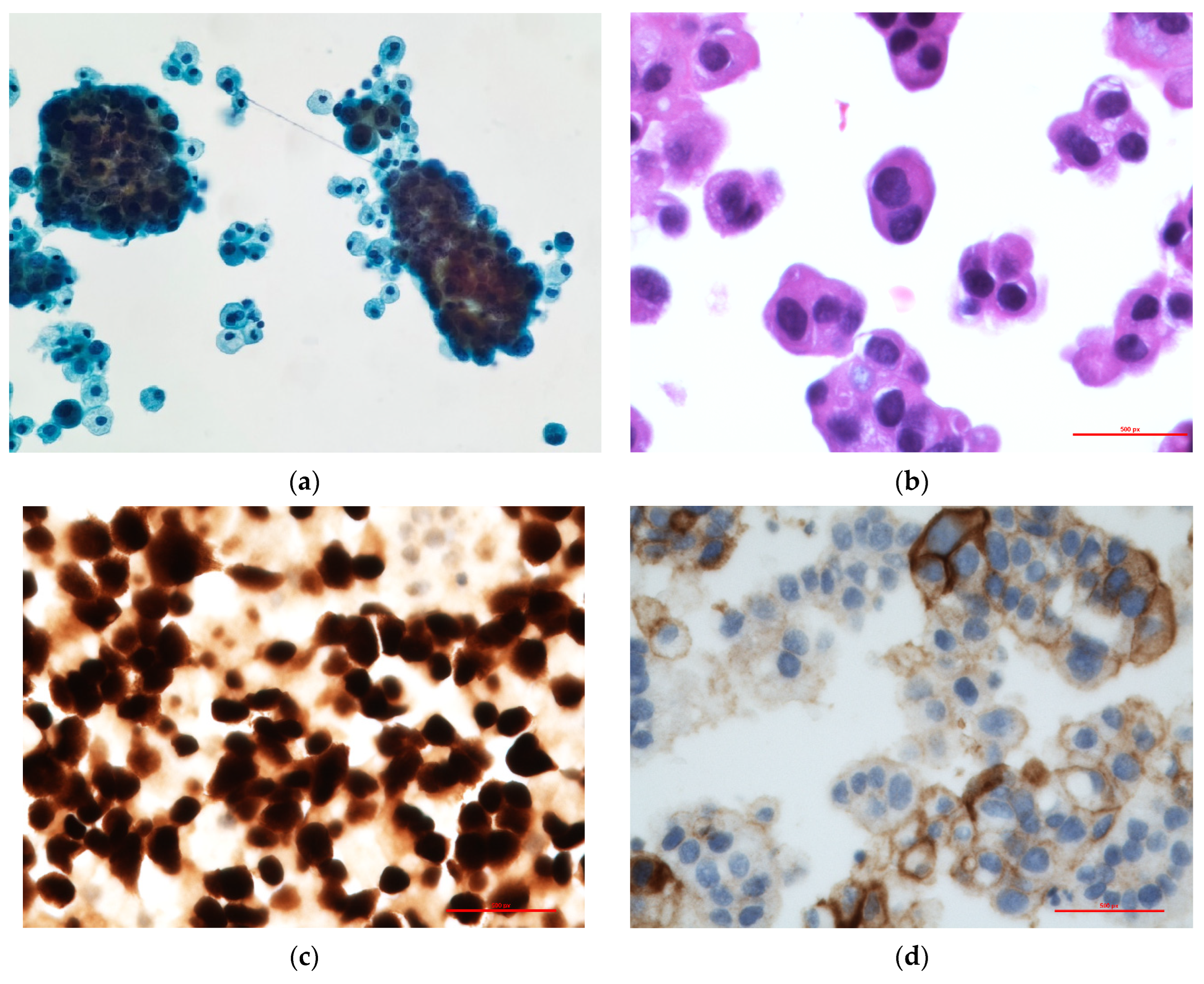
By May 2024, the patient developed moderate pleural effusion and 400 cc of pale-yellow fluid was obtained by thoracentesis that was submitted for cytological analysis. (Figure 2). The cytological examination revealed metastatic adenocarcinoma (Figure 3a,b). The cell block contained adequate tumor cells for ancillary testing and next-generation sequencing. The TTF-1 clone 8G7G3/1 was positive, with strong nuclear staining, confirming metastasis from the lung primary (Figure 3c). The PDL-1 clone 22C3 tumor proportion score was 40% (Figure 3d).
Next-generation sequencing (NGS) revealed both EGFR L858R and KRAS G12D mutations in June 2024. For DNA NGS, tumor DNA was used for multiplex amplification with gene-specific primers targeting the coding exons and exon flanking regions of 275 cancer-related genes using the QIAseq Human Comprehensive Cancer Panel (Qiagen, Inc., Hilden, Germany) NGS was performed using the NextSeq2000 (Illumina, San Diego, CA, USA) and analyzed with the CLC Genomics Workbench (Qiagen). The variants were filtered based on variant allele frequency (VAF, ≥5%), mutation databases, and population-based reference datasets including gnoAD, ClinVar, and COSMIC. NGS confirmed this mutational change, with EGFR L858R having a VAF of 49.50%, and also detected the presence of KRAS G12D at a VAF of 49.27%. The KRAS G12D mutation is an activating alteration within the KRAS gene, a gene commonly mutated in several malignancies including lung adenocarcinoma. KRAS mutations are associated with poor prognosis in NSCLC and are generally mutually exclusive with EGFR mutations. KRAS G12D has been reported in approximately 20% of lung adenocarcinoma cases. Three variants of unknown significance were also detected in the patient specimen. These were ALK R1436H (VAF 23.2%), LRP1B N2814D (40.2%), and KMT2D S2251L (23.2%). ALK R1436H has not been functionally characterized and its effect on protein function is unknown. Although LRP1B N2814D and KMT2D S2251L have not been functionally characterized, mutations in these two genes have been reported in lung adenocarcinomas.
Targeted NGS RNA fusion analysis using a custom Illumina TruSight 527 gene next-generation sequencing panel did not reveal RNA fusions.
Following these findings, standard of care treatment options were discussed, a change in therapy was made, and the patient was started on Amivantamab. The patient was offered enrollment in clinical trials but declined the offer.
Despite the change in therapy, the patient again presented to the hospital within two months of treatment with nausea, vomiting, and bloody diarrhea. She was admitted for pancytopenia and failure to thrive in September 2024. The symptoms were suspected to be related to recent chemotherapy. The clinical condition continued to deteriorate despite all efforts, and the patient passed away while on comfort care.

3. Discussion

Next-generation sequencing (NGS) has significantly advanced the detection of concomitant driver alterations in non-small cell lung cancer (NSCLC) [9,10], uncovering the presence of clonal and sub-clonal populations within tumors that contribute to resistance against tyrosine kinase inhibitor (TKI) therapies. Among these, the coexistence of EGFR and KRAS mutations poses a substantial therapeutic challenge. While KRAS mutations are well-documented drivers of primary resistance to EGFR-TKIs in NSCLC, their role in acquired resistance among EGFR-mutant patients is less well-defined.
Interestingly, studies have shown that some patients with both EGFR and KRAS mutations respond to EGFR-TKI therapy, such as Icotinib, but not to other targeted therapies like crizotinib. This suggests that EGFR may remain the dominant oncogenic driver in certain cases, even in the presence of KRAS mutations. However, tumors are often heterogeneous, with EGFR-mutated cells responding to treatment while KRAS-mutant cells drive resistance, resulting in mixed clinical outcomes.
The prevalence of coexisting KRAS and EGFR mutations appears to increase following EGFR-TKI therapy. For instance, one study reported that the prevalence of dual mutations rose to 3.49% after TKI treatment [11], supporting the hypothesis that dormant tumor cells harboring both mutations may proliferate after EGFR inhibition. Patients with KRAS mutations, particularly at codon 12 (e.g., G12D, G12V, and G12C), exhibit poor responses to treatment, highlighting the aggressive nature of these alterations [12,13].
Several clinical studies have investigated the impact of KRAS mutations on treatment outcomes. Benesova et al. documented limited progression-free survival (PFS) in patients with EGFR/KRAS co-mutations, ranging from 3 to 7 months [14]. Similarly, Ranchiglio et al. reported significantly shorter PFS (2.42 months) and lower objective response rates (ORR) in patients with dominant KRAS mutations compared to those with EGFR mutations alone [15]. Nardo et al. observed poor clinical outcomes in patients with low variant allele frequencies (VAF < 0.2%) of KRAS mutations, with treatment failure times and PFS of approximately 5 months [16].
Other studies have confirmed the complex relationship between EGFR and KRAS co-mutations. For example, Zhuang et al. analyzed 3774 patients and found that EGFR/KRAS co-mutants had an ORR of 62.5% to EGFR-TKI therapy, with no statistically significant difference in PFS compared to patients with single EGFR mutations. However, these findings were inconsistent across studies, with some reporting worse outcomes in co-mutated cases [17].
In cases where KRAS hyper-exchange mutations disrupt the regulatory function of GTPase-activating proteins (GAPs), KRAS remains in a continuously active state, fueling uncontrolled tumor growth and promoting resistance [18]. Dual inhibition strategies targeting both EGFR and downstream pathways, such as the MEK pathway, have been proposed to overcome resistance in EGFR mutant tumors with KRAS mutations.
In the case of our patient, the KRAS G12D mutation likely emerged after prolonged TKI therapy. By mid-2024, the presence of dual EGFR and KRAS mutations led to a shift in treatment to Amivantamab, a dual inhibitor targeting EGFR and MEK pathways. While this provided temporary stabilization, the aggressive nature of the KRAS G12D mutation ultimately contributed to treatment failure and poor outcomes.
This case underscores the complexity of managing NSCLC with coexisting EGFR and KRAS mutations and highlights the importance of continuous molecular monitoring to adapt treatment strategies as the cancer evolves.

4. Conclusions

This case highlights the importance of continuous molecular testing in managing NSCLC, especially in cases with rare mutation profiles. The emergence of new mutations during treatment can significantly impact the course of therapy and patient outcomes. In this case, the detection of both EGFR and KRAS mutations guided the selection of a more personalized targeted therapeutic strategy, including the use of Amivantamab. By utilizing Amivantamab, a bispecific antibody targeting EGFR and MET, the therapy was tailored to address the complexity of the patient’s molecular profile. This highlights the potential for targeted treatments to provide better disease control and improved outcomes in NSCLC patients with co-occurring mutations.
Additionally, this case reinforces the need for clinicians to remain vigilant through regular molecular monitoring, ensuring that therapeutic strategies are continually optimized as the genetic landscape of the tumor evolves over time.

Author Contributions

Conceptualization, A.S.; methodology, A.T.; software, A.S.; validation, A.T. and A.S.; formal analysis, A.S. and I.A.; investigation, A.S.; resources, A.S. and I.A.; data curation, A.T.; writing—original draft preparation, A.T. and A.S.; writing—review and editing, A.S. and I.A.; visualization, A.S. and I.A.; supervision, A.S.; project administration, A.T., funding acquisition, A.S. All authors have read and agreed to the published version of the manuscript.

Funding

This research received no external funding.

Institutional Review Board Statement

Not applicable.

Informed Consent Statement

Not applicable.

Data Availability Statement

Data are contained within the article.

Conflicts of Interest

The authors declare no conflicts of interest.

References

  1. Erratum to “Cancer statistics, 2024”. CA Cancer J. Clin. 2024, 74, 203. [CrossRef] [PubMed]
  2. Xia, C.; Dong, X.; Li, H.; Cao, M.; Sun, D.; He, S.; Yang, F.; Yan, X.; Zhang, S.; Li, N.; et al. Cancer statistics in China and United States, 2022: Profiles, Trends, and Determinants. Chin. Med. J. 2022, 135, 584–590. [Google Scholar] [CrossRef] [PubMed]
  3. Gou, Q.; Gan, X. Novel therapeutic strategies for rare mutations in non-small cell lung cancer. Sci. Rep. 2024, 14, 10317. [Google Scholar] [CrossRef] [PubMed]
  4. Kim, E.S.; Melosky, B.; Park, K.; Yamamoto, N.; Yang, J.C. EGFR tyrosine kinase inhibitors for EGFR mutation-positive non-small-cell lung cancer: Outcomes in Asian populations. Future Oncol. 2021, 17, 2395–2408. [Google Scholar] [CrossRef] [PubMed]
  5. Zhang, X.; Guo, X.; Gao, Q.; Zhang, J.; Zheng, J.; Zhao, G.; Okuda, K.; Tartarone, A.; Jiang, M. Association between cigarette smoking history, metabolic phenotypes, and EGFR mutation status in patients with non-small cell lung cancer. J. Thorac. Dis. 2023, 15, 5689–5699. [Google Scholar] [CrossRef] [PubMed]
  6. Fu, K.; Xie, F.; Wang, F.; Fu, L. Therapeutic strategies for EGFR-mutated non-small cell lung cancer patients with osimertinib resistance. J. Hematol. Oncol. 2022, 15, 173. [Google Scholar] [CrossRef] [PubMed]
  7. Marks, J.L.; Broderick, S.; Zhou, Q.; Chitale, D.; Li, A.R.; Zakowski, M.F.; Kris, M.G.; Rusch, V.W.; Azzoli, C.G.; Seshan, V.E.; et al. Prognostic and therapeutic implications of EGFR and KRAS mutations in resected lung adenocarcinoma. J. Thorac. Oncol. 2008, 3, 111–116. [Google Scholar] [CrossRef] [PubMed]
  8. Jang, T.W.; Oak, C.H.; Chang, H.K.; Suo, S.J.; Jung, M.H. EGFR and KRAS mutations in patients with adenocarcinoma of the lung. Korean J. Intern. Med. 2009, 24, 48–54. [Google Scholar] [CrossRef] [PubMed]
  9. Schwartz, C. P76.33 Concurrent EGFR and KRAS Mutations in Lung Adenocarcinoma: A Single Institution Case Series. J. Thorac. Oncol. 2021, 16, S600–S601. [Google Scholar] [CrossRef]
  10. Ju, L.; Han, M.; Zhao, C.; Li, X. EGFR, KRAS and ROS1 variants coexist in a lung adenocarcinoma patient. Lung Cancer 2016, 95, 94–97. [Google Scholar] [CrossRef] [PubMed]
  11. Choughule, A.; Sharma, R.; Trivedi, V.; Thavamani, A.; Noronha, V.; Joshi, A.; Desai, S.; Chandrani, P.; Sundaram, P.; Utture, S.; et al. Coexistence of KRAS mutation with mutant but not wild-type EGFR predicts response to tyrosine-kinase inhibitors in human lung cancer. Br. J. Cancer 2014, 111, 2203–2204. [Google Scholar] [CrossRef] [PubMed]
  12. Wang, H.; Lin, L.; Liang, C.; Pang, J.; Yin, J.C.; Zhang, J.; Shao, Y.; Sun, C.; Guo, R. Landscape of Concomitant Driver Alterations in Classical EGFR-Mutated Non-Small Cell Lung Cancer. JCO Precis. Oncol. 2024, 8, e2300520. [Google Scholar] [CrossRef] [PubMed]
  13. Gini, B.; Thomas, N.; Blakely, C.M. Impact of concurrent genomic alterations in epidermal growth factor receptor (EGFR)-mutated lung cancer. J. Thorac. Dis. 2020, 12, 2883–2895. [Google Scholar] [CrossRef] [PubMed]
  14. Benesova, L.; Minarik, M.; Jancarikova, D.; Belsanova, B.; Pesek, M. Multiplicity of EGFR and KRAS mutations in non-small cell lung cancer (NSCLC) patients treated with tyrosine kinase inhibitors. Anticancer Res. 2010, 30, 1667–1671. [Google Scholar] [PubMed]
  15. Rachiglio, A.M.; Fenizia, F.; Piccirillo, M.C.; Galetta, D.; Crinò, L.; Vincenzi, B.; Barletta, E.; Pinto, C.; Ferraù, F.; Lambiase, M.; et al. The Presence of Concomitant Mutations Affects the Activity of EGFR Tyrosine Kinase Inhibitors in EGFR-Mutant Non-Small Cell Lung Cancer (NSCLC) Patients. Cancers 2019, 11, 341. [Google Scholar] [CrossRef] [PubMed]
  16. Nardo, G.; Carlet, J.; Marra, L.; Bonanno, L.; Boscolo, A.; Maso, A.D.; Bragadin, A.B.; Indraccolo, S.; Zulato, E. Detection of Low-Frequency KRAS Mutations in cfDNA From EGFR-Mutated NSCLC Patients After First-Line EGFR Tyrosine Kinase Inhibitors. Front. Oncol. 2021, 10, 3055. [Google Scholar] [CrossRef] [PubMed]
  17. Zhuang, X.; Zhao, C.; Li, J.; Su, C.; Chen, X.; Ren, S.; Li, X.; Zhou, C. Clinical features and therapeutic options in non-small cell lung cancer patients with concomitant mutations of EGFR, ALK, ROS1, KRAS or BRAF. Cancer Med. 2019, 8, 2858–2866. [Google Scholar] [CrossRef] [PubMed]
  18. Johnson, C.; Burkhart, D.L.; Haigis, K.M. Classification of KRAS-Activating Mutations and the Implications for Therapeutic Intervention. Cancer Discov. 2022, 12, 913–923. [Google Scholar] [CrossRef] [PubMed]
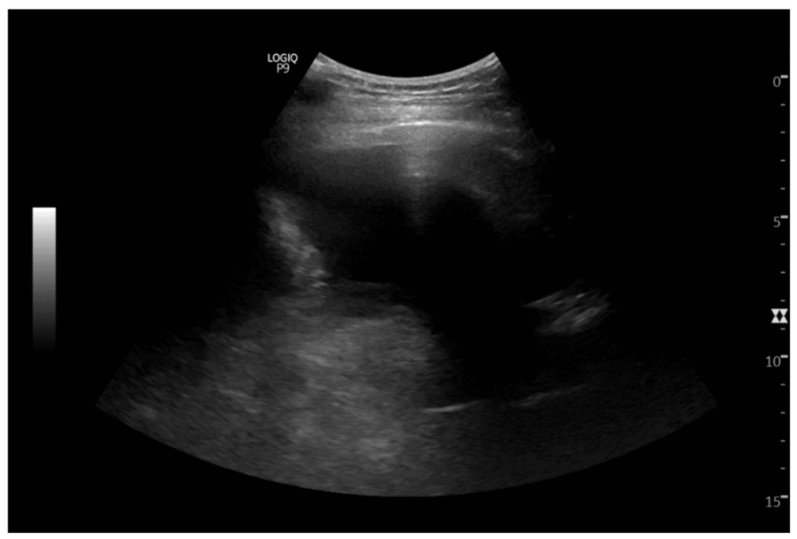
Figure 1. Ultrasound demonstrating a moderate left pleural effusion. Ultrasound-guided thoracocentesis was performed.
Cimb 47 00066 g001
Figure 2. CT-scan at the time of diagnosis showing a left lower lobe perihilar soft tissue mass lesion.
Cimb 47 00066 g002
Figure 3. Pleural fluid sample. (a) Papanicolaou stain 200×, Thin Prep. shows cellular specimen with numerous atypical clusters of cells. (b) Cell block (Hematoxylin and eosin stain) 500×. shows clusters of tumor cells consistent with metastatic adenocarcinoma. (c) TTF-1 stain 400×. Tumor cells stained with TTF-1 confirming metastatic lung adenocarcinoma. (d) PDL-1 immunostain 400×. Membrane staining of a portion of tumor cells in cell block.
Cimb 47 00066 g003
Disclaimer/Publisher’s Note: The statements, opinions and data contained in all publications are solely those of the individual author(s) and contributor(s) and not of MDPI and/or the editor(s). MDPI and/or the editor(s) disclaim responsibility for any injury to people or property resulting from any ideas, methods, instructions or products referred to in the content.