Article

Genetics of 21-OH Deficiency and Genotype–Phenotype Correlation: Experience of the Hellenic National Referral Center

by
Irene Fylaktou
1,*,
Anny Mertzanian
1,
Ioanna Farakla
1,
Alexandros Gryparis
2,
Ioannis Anargyros Vasilakis
1,
Maria Binou
1,
Evangelia Charmandari
1,
Christina Kanaka-Gantenbein
1 and
Amalia Sertedaki
1
1
Division of Endocrinology, Diabetes and Metabolism ‘Aghia Sophia’ Children’s Hospital ENDO-ERN Center for Rare Paediatric Endocrine Diseases, First Department of Pediatrics, Medical School, National and Kapodistrian University of Athens, ‘Aghia Sophia’ Children’s Hospital, 11527 Athens, Greece
2
Department of Speech and Language Therapy, School of Health Sciences, University of Ioannina, 45110 Ioannina, Greece
*
Author to whom correspondence should be addressed.
Curr. Issues Mol. Biol. 2024, 46(10), 10696-10713; https://doi.org/10.3390/cimb46100635
Submission received: 20 August 2024 / Revised: 16 September 2024 / Accepted: 20 September 2024 / Published: 24 September 2024
(This article belongs to the Special Issue Genomic Analysis of Common Disease)

Abstract

:
21-hydroxylase deficiency (21-OHD) represents the most common form of congenital adrenal hyperplasia (CAH) due to CYP21A2 gene pathogenic variants. Τhe aim of this study was the identification of CYP21A2 variants in 500 subjects of Greek origin with a suspicion of 21-OHD and, by using the existing hormonal assessment and genotypes of the 500 subjects tested, to identify a biomarker that could differentiate between the heterozygotes and the cases with no pathogenic variants identified. Five hundred subjects with clinical suspicion of 21-OHD underwent CYP21A2 gene sequencing and Multiplex Ligation Dependent Probe Amplification (MLPA). Genetic diagnosis was achieved in 27.4% of the subjects tested, most of which presented with the non-classic form (NC) of 21-OHD. Heterozygotes accounted for 42.6% of cases, whereas no pathogenic variants were identified in 27% of cases. De novo aberrations, duplications, and five novel variants were also identified. Statistical analysis revealed that the difference between the basal and 60′ post-ACTH stimulation 17-hydroxyprogesterone concentrations (Δ17-OHP60-0) could be a potential biomarker (p < 0.05) distinguishing the heterozygotes from the cases with no pathogenic variants identified, although no clear cut-off value could be set. Further analysis revealed overlapping clinical manifestations among all the subjects tested. The presented phenotypic traits of the subjects tested and the inability to identify a discriminative biochemical marker highlight the importance of comprehensive CYP21A2 genotyping to ascertain the correct genetic diagnosis and proper genetic counselling.

1. Introduction

Congenital adrenal hyperplasia (CAH) is a group of autosomal recessive disorders caused by pathogenic variants identified in seven genes (CYP21A2, CYP11B1, CYP17A1, HSD3B2, StAR, POR, and CYP11A1) involved in the steroidogenesis pathway [1]. Most CAH cases (95%) are the result of pathogenic variants in the CYP21A2 gene encoding for the enzyme 21-hydroxylase [2,3].
The enzyme 21-hydroxylase is responsible for the conversion of 17-hydroxyprogesterone (17-OHP) and progesterone to 11-deoxycortisone and 11-deoxycorticosterone, respectively. Reduced 21-hydroxylase function leads to inadequate cortisol and aldosterone synthesis, resulting in the excessive accumulation of the precursors 17-OHP and progesterone thus directing steroidogenesis towards the uninhibited androgen production, and finally leading to the excess concentration of adrenal androgens, responsible for the clinical picture of affected individuals.
Depending on the degree of enzymatic activity and consequently on the clinical manifestations, CAH is classified in two forms, the non-classic (NC) and classic (C) forms.
The classic form, with an incidence of 1:14,000–1:18,000 live births, based on neonatal screening, is further divided into the classic salt wasting (SW) and the classic simple virilizing (SV) forms [4]. In classic 21-OHD (SW and SV), females present with atypical/ambiguous genitalia at birth due to the intrauterine severe virilization of the external genitalia because of the prenatal excess androgen production and action, while males present with an enlarged penis, that quite often escapes proper medical attention. The SW form is further characterized after birth with severe salt loss, failure to thrive, potentially fatal hypovolemia, and shock due to both cortisol and aldosterone insufficiency. Therefore, in both sexes, the SV form is mainly characterized by the signs of excess androgen production without salt loss since patients exhibit sufficient aldosterone production. The NC or late onset 21-OHD is considered to be the most frequently occurring endocrine disorder, with a wide range of incidence of 1:200 to 1:1000 in various ethnic groups (Caucasians, Askenaki Jews, Yugoslavs, Mediterranean, Hispanics and Anglo-Saxons) [5,6]. The NC form is manifested through a variety of clinical signs ranging from the absence of clinical signs to premature adrenarche, hirsutism, acne, advanced bone age, and menstrual irregularities [7].
However, the boundaries between the different forms of CAH are not strictly defined, and it is therefore advisable to consider CAH as a continuum of phenotypes from asymptomatic to mild and severe [8].
Confirmation of 21-OHD is based on the serum 17-OHP concentrations, both basal and after an ACTH stimulation test [9]. More precisely, basal 17-OHP concentrations of <2 ng/mL (6nM) have been proposed for the exclusion of 21-OHD, with a more accurate measurement for 17-OHP concentrations after the ACTH stimulation test to be >10 ng/mL (30nM) for the diagnosis of 21-OHD [9,10,11]. Although 17-OHP concentration is currently used for 21-OHD diagnosis, the effect of several factors on 17-OHP limits its diagnostic utility [9]. To date, comprehensive CYP21A2 genotyping remains the gold standard for confirming or excluding 21-OHD [12]. The CYP21A2 gene is located on the short arm of chromosome 6 (6p21.31) in a genetic unit known as the RCCX module, whereas its highly homologous nonfunctional pseudogene, CYP21A1P, is located 30 kb upstream [13,14,15,16].
Most pathogenic variants (75%) in the CYP21A2 gene derive from gene conversion events between the pseudogene and the CYP21A2 gene, while 5–10% of cases represent novel or rare variants [17,18]. Furthermore, gene deletions, duplications, and CYP21A1P–CYP21A2 chimeras have also been reported, while de novo aberrations account for 1–2% of the cases [18,19,20,21,22,23,24]. Classification of the CYP21A2 gene pathogenic variants is based on their effect on the 21-hydroxylase enzymatic activity. Null Group variants lead to complete inactivation of the enzyme, whereas Group A variants reduce the enzymatic activity to less than 1%. Both groups are associated with the SW form of the disease. Group B variants reduce the enzymatic activity to 2–5% and are associated with the SV form of the disease, while Group C variants result in an enzymatic activity of 20–50% and are associated with the NC form of the disease [7]. On the contrary, CYP21A2 gene duplications do not have any effect on the 21-hydroxylase enzymatic activity. It has been previously reported that CYP21A2 gene duplications consist of a wild-type CYP21A2 copy and a second CYP21A2 copy carrying one or more pathogenic variants, with the p.Q319* pathogenic variant being more frequently presented [20,21,22,23,24]. CYP21A2 gene duplication with the p.Q319* on the same allele do not affect the enzymatic activity of the 21-hydroxylase thus representing a normal functional allele [25].
Most affected individuals are compound heterozygotes and the patient’s phenotype is defined by the less severe pathogenic variant, with most cases presenting a good genotype–phenotype correlation. However, cases of genotype–phenotype discordance have been reported [26,27,28].
The aim of this study is to present the results of CYP21A2 genotyping in 500 Greek subjects referred with the clinical suspicion of CAH. Furthermore, we aim to identify a biomarker that could differentiate between CYP21A2 heterozygotes and cases with no pathogenic variants identified based on their clinical and hormonal profiles.

2. Materials and Methods

2.1. Subjects

A total of 539 individuals of Greek origin with suspicion of 21-OHD were referred to our center for comprehensive CYP21A2 genotyping during the last 7 years (2017–2023) after clinical and hormonal assessments by their pediatrician/endocrinologist. Clinical assessment was based on CAH-indicative clinical features according to age of presentation as follows: During the neonatal period, the cardinal clinical features were salt-wasting and adrenal crisis, as well as atypical/ambiguous genitalia in females or penile enlargement in males; during childhood and adolescence, premature adrenarche, bone age advancement, hirsutism, acne, and menstrual irregularities were observed; and during adulthood, hirsutism, acne, menstrual irregularities or even subfertility would occur. Hormonal assessments included the basal 17-OHP, cortisol (F), Δ4-Androstenedione (Δ4-A), testosterone (T), dehydroepiandrosterone sulfate (DHEA-S), and adrenocorticotropic hormone (ACTH) levels, while most cases also underwent an ACTH stimulation test.
Out of the 539 subjects referred, 39 were excluded due to insufficient clinical/hormonal data. Those 39 cases were either referred to our center for the exclusion of 21-OHD to confirm the diagnosis of Polycystic Ovary Syndrome (PCOS) or excluded due to the absence of hormonal data (basal 17-OHP, F, Δ4-A, T, DHEA-S) or due to the absence of CAH-indicative clinical features. The remaining 500 subjects were categorized into three groups according to their age at the time of the referral/clinical diagnosis. Age Group I included patients referred during infancy (n = 32/500), Age Group II included subjects referred during childhood/early adolescence (1–13 years, n = 405/500) while Age Group III consisted of subjects in late adolescence (13–18 years, n = 63/500).
The median age of the 500 cases (378 females, 122 males) was 8.5 years (range: 3 days-18 years). A total number of 926 parents were also analyzed.
The study protocol has been approved by the ethical committee of the “Aghia Sophia” Children’s Hospital. Written informed consent was obtained from all participants and/or their legal guardians for the molecular analysis of the CYP21A2 gene.

2.2. DNA Extraction

Genomic DNA was isolated from the peripheral blood samples, employing either the Maxwell® 16 Blood DNA Purification Kit (Promega, Madison, WI, USA) or the QIAamp DNA Blood Mini Kit (Qiagen, Hilden, Germany) according to the manufacturer’s instructions.

2.3. CYP21A2 Gene Sequencing

PCR and bidirectional sequencing of the exons and introns of CYP21A2 gene was carried out as previously described [10]. Purification of the PCR products was performed with the combination of two different enzymes [Exonuclease I/Shrimp Alkaline Phosphatase (rSAP), New England BioLabs, Ipswich, MA, USA].
Sequencing of the CYP21A2 gene was carried out for all cases and their parents (when available). Analysis of the CYP21A2 gene was based on the reference gene NG_007941.2 and transcript NM_000500.9.
In cases where two or more pathogenic variants were identified on the same allele, these were reported as multiple pathogenic variants and their impact on the enzymatic activity of the 21-hydroxylase was defined by the most severe variant. CYP21A1P–CYP21A2 chimeras were also included in this category.
Negatively genotyped cases in the CYP21A2 gene were categorized as cases with no pathogenic variants identified.

2.4. MLPA

Multiplex Ligation Dependent Probe Amplification (MLPA) employing the P050-C1 CAH kit (MRC Holland, Amsterdam, The Netherlands) was carried out in cases with a suspicion of duplication/deletion of the CYP21A2 gene after sequencing analysis was performed.
The cases in which duplication of the gene (with or without the p.Q319* on the same allele) was observed either in heterozygosity or in trans with a pathogenic variant, were not considered as cases with no pathogenic variants identified or as solely heterozygotes but formed a distinct group, referred to as cases with duplication of the CYP21A2 gene.

2.5. Evaluation of Novel Variants

Novel variants were evaluated according to the ACMG guidelines [29]. The frequency of novel variants was also searched in the Genome Aggregation Database [30].

2.6. Statistical Analysis

Subjects carrying novel variants and duplication of the CYP21A2 gene were excluded from the statistical analysis.
Statistical analysis for the 500 cases was performed using descriptive and inferential statistics. Descriptive statistics (including mean, median, SD, minimum, maximum, and IQR) were employed for the available clinical data of all the genotyped cases. Comparisons of the hormonal concentrations [basal 17-OHP, cortisol, Δ4-Androstenedione, DHEA-S, testosterone, and ACTH, as well as the difference between 60′ and basal 17-OHP concentrations after the ACTH stimulation test (Δ17-OHP60-0), and between 60′ and basal cortisol concentrations after the ACTH stimulation test (ΔF60-0)] amongst each of the three different subgroups of heterozygotes (comprising heterozygotes carrying pathogenic variants that are associated with the SW, SV, and NC forms, respectively) and the group of the cases with no pathogenic variants identified was performed using the Mann–Whitney U test (inferential statistics). The comparison was performed in groups of the same sex. Statistical analysis was performed using IBM SPSS v.28 (IBM Corp. Released 2021. IBM SPSS Statistics for Windows, Version 28.0. Armonk, NY, USA: IBM Corp) and RStudio version 2023.06.0 Build 421 [RStudio Team (2020). RStudio: Integrated Development for R. RStudio, PBC, Boston, MA, USA URL http://www.rstudio.com/ (accessed on 15 July 2023)]. p-values < 0.05 were considered statistically significant.

3. Results

The statistical analysis performed for the 500 cases studied using the different hormonal concentrations (17-OHP, Δ17-OHP60-0, F, ΔF60-0, Δ4-A, DHEA-S, testosterone, and ACTH) amongst cases with no pathogenic variants identified and the different subgroups of heterozygotes revealed that the Δ17-OHP60-0 exhibited a statistically significant difference (p < 0.05) in both sexes as depicted in Figure 1. However, the great overlap in the Δ17-OHP60-0 values amongst all the subgroups of heterozygotes when compared to the group of cases with no identified pathogenic variants in both sexes did not allow us to set discriminating cut-off values.
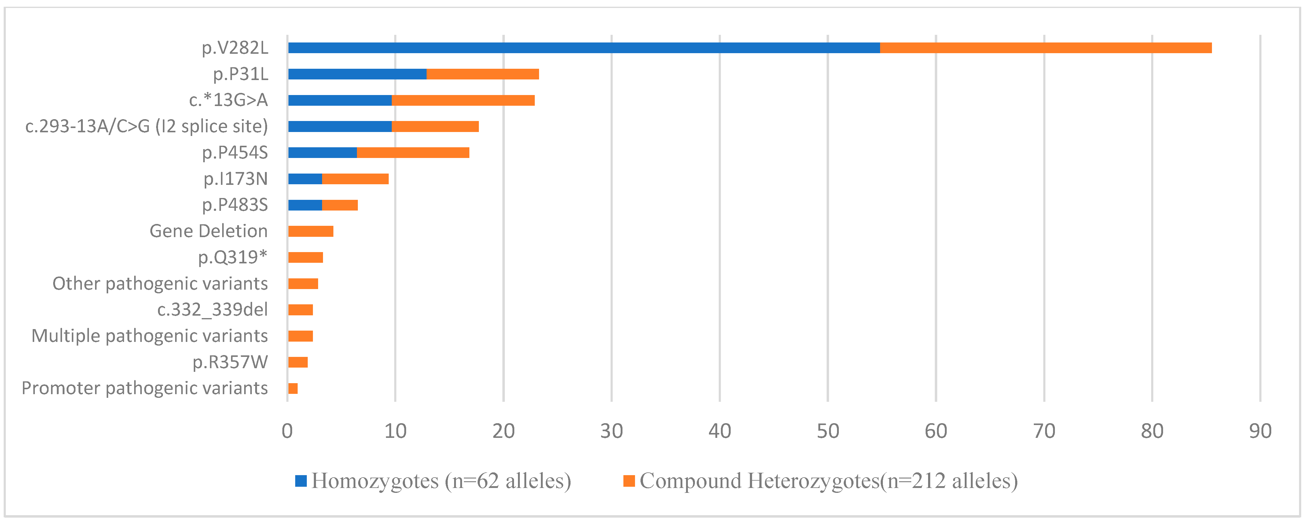
Genetic diagnosis of 21-OHD was achieved in 137 subjects (27.4%), who were genotypically characterized either as compound heterozygotes (n = 106, 21.2%) or homozygotes (n = 31, 6.2%) (Figure 2). Heterozygotes accounted for 42.6% (213/500) of the tested population, while in 27% (135/500), no pathogenic variant was identified. Duplication of the CYP21A2 gene (with or without the p.Q319* in the same allele) was observed in 2.6% (13/500) of the cases.
The classic (SW or SV) form of 21-OHD was diagnosed in 10.2% of cases (14/137)—9.4% (10/106) of the compound heterozygotes and 12.9% (4/31) of the homozygotes. The majority of SW and SV cases carried the c.293-13C>G (I2splice site) pathogenic variant (Group A) and the p.I173N pathogenic variant (Group B) (Table A1) respectively. Most classic 21-OHD patients belonged to Age Group I (64.3%, 9/14).
The NC form was observed in 89% of patients—90.6% (96/106) of the compound heterozygotes and 87.1% (27/31) of the homozygotes (Figure 3). Group C pathogenic variants were identified in 59.2% (81/137) of patients; 24.8% (34/137) harbored a Group C in trans with a Group null/A pathogenic variant and 5.9% (8/137) harbored a Group C in trans with a Group B pathogenic variant. Most cases presenting with the NC form of 21-OHD belonged to Age Groups II and III, with a frequency of 77.3% (106/137) and 11% (15/137), respectively. The age distribution of all patients is presented in Figure 4.
The most common pathogenic variant identified in patients, heterozygotes, and cases with CYP21A2 gene duplication was the p.V282L (Group C variant) with an allele frequency of 33.7% and 19%, respectively (Figure 2 and Figure A1). Most heterozygotes (82.3%, 190/213) belonged to Age Group II.
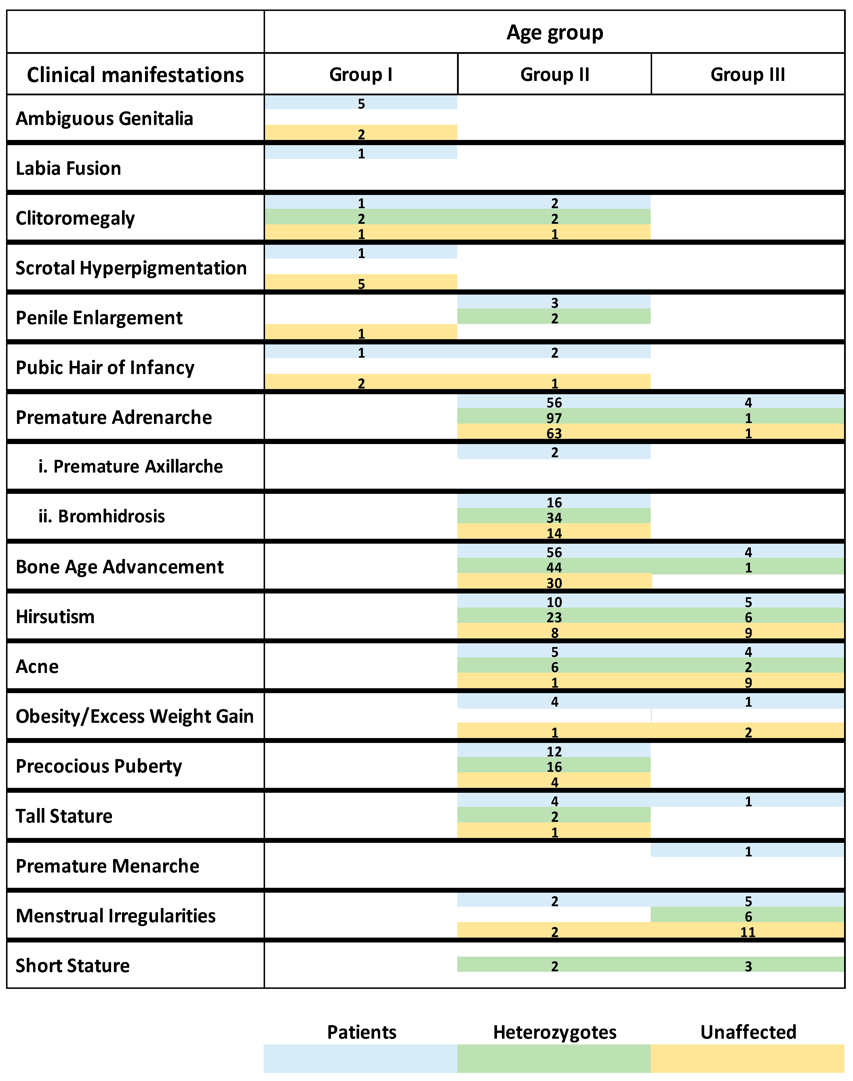
The hormonal profile and clinical manifestations categorized according to age group (Age Group I, Age Group II, and Age Group III) and genotypic group (patients, heterozygotes, and cases with no pathogenic variants identified) are depicted in Table 1, Table 2 and Table 3 and Figure 5.
Moreover, in the study, five novel variants were identified, most located in the gene’s promoter region (Table 4). Two of these novel variants were found in trans with a known pathogenic variant, while the other three were found in heterozygosity (Table 4). The GnomAD frequency of the novel variants (apart from the c.-82C>T) was low or not reported (Table 4).
De novo pathogenic variants/rearrangements were identified in four cases (one compound heterozygote and three heterozygotes), accounting for 0.4% of the alleles tested (4/1000 alleles). The compound heterozygote was found to harbor the maternal p.V282L and a de novo deletion of the CYP21A2 gene whereas the father was carrying the Q319* in cis with duplication of the gene. Among the three heterozygotes, one case harbored a de novo deletion of the CYP21A2 gene, while the other two cases carried the de novo p.I173N and c.293-13C>G pathogenic variants, respectively. The analysis of the heterozygotes’ parents revealed a duplication of the CYP21A2 gene in the father of the heterozygote carrying the de novo deletion of the CYP21A2 gene.
Duplications were observed in 2.6% (13/500) of the cases with 0.8% (4/500) carrying a duplication of the CYP21A2 gene with the p.Q319* in trans with a known pathogenic variant. It is worth mentioning that in one case, a duplication of the gene was observed along with multiple pathogenic variants including the p.Q319* on the same copy of the gene.
In the present study, genetic analysis of the entire CYP21A2 gene was performed in 926 parents, with 2.7% (25/926) of them depicted to be homozygotes/compound heterozygotes of pathogenic, VUS, or novel variants. More precisely, 2.5% (23/926) of the parents were found to harbor pathogenic variants resulting in the NC form (homozygotes/compound heterozygotes), identified solely by genotyping (without any clinical manifestation reported); 0.7% (6/926) harbored a pathogenic variant related to the classical form in trans with a variant related to the NC form; and 0.2% (2/926) carried novel variants or VUS in trans with a known pathogenic variant.

4. Discussion

In the current study, the genetic analysis of 500 subjects with the clinical suspicion of CAH is presented. Patients accounted for 27.4% of cases studied, with most of them presenting with the NC form of 21-OHD, while both the SW and SV forms of 21-OHD were underrepresented. The categorization of 21-OHD patients in different age groups confirmed that the severity of clinical manifestations due to the reduced 21-OH activity and the diagnosis of 21-OHD is directly related to the age at presentation. As expected, the more severe classical form will be diagnosed earlier in life, while the NC form will be diagnosed later, revealing the continuum of 21-OHD through all age groups.
In the current study, different adrenal hormones were assessed to identify a biomarker that could distinguish the heterozygotes of 21-OHD from the cases harboring no pathogenic variants. The Δ17-OHP60-0 in both sexes was found to be a potential biomarker; however, the overlap in its values between the different groups did not allow us to set cut-offs to discriminate them. This overlap in the values of Δ17-OHP60-0 of the group of cases with no pathogenic variants identified and the different groups of heterozygotes shows that this biomarker is not suitable to differentiate heterozygotes from the cases with no pathogenic variants identified, thus it cannot be used in clinical practice. To date, several studies have attempted to distinguish heterozygotes from the cases harboring no pathogenic variants using 17-OHP concentrations as a biomarker with no clear results. This could be attributed to several limitations such as the assay used (RIA) for 17-OHP measurements, the variability of the measurements amongst laboratories, the factors affecting the 17-OHP concentrations, as well as the 17-OHP as a biomarker itself. In this study, high 17-OHP concentrations were observed both in heterozygotes (mean 17-OHP concentrations in Age Group I: 11 ng/mL, Age Group II: 3.1 ng/mL, and Age Group III: 3.8 ng/mL) and cases with no pathogenic variants identified (mean 17-OHP in Age Group I: 6.8ng/mL, Age Group II: 2.7ng/mL, and Age Group III: 4.9 ng/mL), rendering the use of 17-OHP concentrations a non-reliable biomarker and confirming comprehensive CYP21A2 genotyping as the gold standard for their distinction.
As previously mentioned, CYP21A2 genotyping revealed a high percentage of the heterozygotes (42.6%) and the cases without any pathogenic variant (27%), although their hormonal profile and clinical features were indicative of 21-OHD. In this study, the clinical manifestations (ranging from severe to mild) of compound heterozygotes/homozygotes, heterozygotes, and cases with no pathogenic variants identified overlapped in each age group, particularly in Age Group II (comprising 81.2% of all cases).
Specifically, severe clinical manifestations, such as clitoromegaly or atypical/ambiguous genitalia, present in Age Groups I and II were found not only in the compound heterozygotes/homozygotes but also in the heterozygotes and the cases harboring no pathogenic variants. Likewise, the mild clinical manifestations (Age Group II and III) such as premature adrenarche were also present in all genotypic groups. In heterozygotes, the clinical features could be explained by the dominant-negative effect of certain pathogenic variants (such as the p.V282L), which affects the function of the wild-type allele thus reducing the enzymatic activity of 21-hydroxylase [33,34].
The number of cases without any identifiable pathogenic variants reported in this study is not negligible, and this could be explained by the fact that the CAH-related clinical features may not be exclusively indicative of 21-OHD. One of the main clinical manifestations observed in Age Group II and III was premature adrenarche that can also be the result of several other factors such as childhood obesity or being born IUGR (Intrauterine Growth restricted).
Moreover, pathogenic variants in other CAH genes involved in the steroidogenic pathway, the presence of other clinical entities (such as polycystic ovarian syndrome), epigenetic influences as well as environmental factors might also account for the phenotypic characteristics indicative of CAH.
The observed overlap in the clinical manifestations of all the genotypic groups and the inability to fully differentiate the heterozygotes from the cases without any pathogenic variants identified indicate that a more specific scoring system should be established to reduce the need for comprehensive CYP21A2 genotyping.
To date, more than 200 pathogenic variants have been identified in the CYP21A2 gene, with no hot spot regions reported [12,19,35]. The most common pathogenic variant found in our cohort to be related to the NC form of 21-OHD was the p.V282L, which is in concordance with the previous studies in the Greek population [10,36,37]. The frequency of the p.V282L in the Greek population (overall frequency: 24.4% in patients and heterozygotes) is similar to the Italian (23.9%) population but lower than the Cypriot (43.7–60%), Spanish (47.3%), and Portuguese (41.3%) populations [38,39,40,41,42].
The most common pathogenic variant identified in our cohort related to the SW form was the c.293-13C>G [I2splice site] (7.3%) with an incidence that is lower than in the Spanish (12.3%), the Portuguese (14.1%), the Turkish (22–33.2%), and the Croatian (35.5%) population [38,41,43,44,45,46].
Genotyping the CYP21A2 gene led to the identification of five novel variants. Three of them located in the promoter region and which are crucial for the transcriptional activity of the gene, the c.-127G>A and c.-82C>T in heterozygosity and the c.-115G>T in compound heterozygosity with the pathogenic variant p.P31L, were classified as VUS according to the ACMG criteria due to insufficient or conflicting evidence. The other two novel variants were located in exon 7; the p.R255K in heterozygosity was classified as VUS and the p.V282M in compound heterozygosity with p.L308Ffs*6 was classified as Likely Pathogenic. Considering the hormonal data of the patients carrying these novel variants (presented in Table 4), only the p.V282M variant certainly contributes to the patient’s phenotype, and this patient was clinically considered and treated as the non-classic form. It should be noted that the V282 residue is located in a very restricted space at the I-helix, and we can assume that the substitution of Valine by Methionine could have the same effect as the substitution of Valine by Leucine due to the increased chain length of both the Methionine and Leucine when compared to Valine [47]. The patient carrying the c.-115G>T in compound heterozygosity with the p.P31L, presented increased 17-OHP concentrations with normal cortisol values and was therefore considered as heterozygote, since there was no evidence for the effect of the VUS variant. The three remaining VUS variants were only present in heterozygosity and therefore, irrespective of their potential impact on the enzymatic activity, their clinical significance is negligible.
Overall, in vitro studies are required to elucidate the pathogenicity of these variants and to clarify their impact on the enzymatic activity of 21-hydroxylase.
Different copy number variations at the RCCX model have been described due to unequal meiotic crossing-over events. Duplications of the CYP21A2 gene along with the p.Q319* (trimodular haplotype) are known to be present in less than 2% of the general population except for the Spanish population [24,48]. In this study, duplications of the CYP21A2 gene (with or without the p.Q319*) were found in a slightly higher frequency, emphasizing the importance of comprehensive CYP21A2 genotyping, particularly in cases where the p.Q319* is present in order to provide proper genetic diagnosis and counselling [49]. The identification of duplications is also important for family planning since duplications are known to predispose to de novo aberrations in the offspring [50]. De novo aberrations are considered to occur due to unequal crossing over at the RCCX unit [17,18,51,52]. In this study, de novo aberrations were identified in 0.8% of subjects tested. We could speculate that these de novo aberrations probably occurred in the paternal gametic line since the fathers of the subjects tested to be carrying de novo deletion harbored duplications of the CYP21A2 gene. To date, de novo aberrations have only been reported in maternal alleles carrying duplications of the CYP21A2 gene; however, our findings indicate that de novo aberrations might also be the result of unequal crossing over events in the paternal alleles [50].
The identification of parents that harbor pathogenic variants in homozygosity/compound heterozygosity resulting in the NC form of 21-OHD, without any clinical manifestations, has also been previously reported [53,54]. This finding should be strongly considered when parents are only tested for the specific pathogenic variants identified in their offspring.

5. Conclusions

In conclusion, to the best of our knowledge, this is the largest study of the CYP21A2 gene conducted to date in Greece revealing that, in our population, 21-OHD is mainly presented with the NC form. Despite the small sample size of SW and SV patients, the continuum of clinical manifestations in the 21-OHD patients is evident among the different age groups. The identification of four cases with de novo aberrations and parents with the NC form (without reported clinical manifestations), the slightly higher frequency of duplications in the CYP21A2 gene, the high percentage of heterozygotes and cases without any pathogenic variants identified, as well as the inability to identify a discriminative biochemical marker to distinguish the different groups, underline the necessity for comprehensive CYP21A2 genotyping of the family (trios) for proper diagnosis and genetic counselling.

Author Contributions

Conceptualization, A.S. and C.K.-G.; Clinical Investigation, I.F. (Ioanna Farakla), I.A.V. and M.B.; Formal Analysis, A.G.; Methodology, A.S., I.F. (Irene Fylaktou) and C.K.-G.; Project Administration, A.S. and C.K.-G.; Supervision, A.S. and C.K.-G.; Validation, A.M.; Visualization, I.F. (Irene Fylaktou) and A.M.; Writing—Original Draft Preparation, I.F. (Irene Fylaktou) and A.S.; Writing—Review and Editing, A.S., C.K.-G. and E.C. All authors have read and agreed to the published version of the manuscript.

Funding

This research received no external funding.

Institutional Review Board Statement

The study was conducted in accordance with the Declaration of Helsinki and approved by the Institutional Review Board (or Ethics Committee) of ‘Aghia Sophia’ Children’s Hospital (protocol code 1263/15.01.2024 and date of approval 8.02.2024) for studies involving humans.

Informed Consent Statement

Informed consent was obtained from all subjects and/or their legal guardians involved in the study.

Data Availability Statement

The data presented in this study are available on request from the corresponding author due to privacy/ethical reasons.

Acknowledgments

The authors would like to thank the participants, their families, and the following endocrinologists for referring their patients to the Laboratory of Molecular Endocrinology (Division of Endocrinology, Diabetes and Metabolism ‘Aghia Sophia’ Children’s Hospital ENDO-ERN Center for Rare Paediatric Endocrine Diseases, First Department of Pediatrics, Medical School, National and Kapodistrian University of Athens): P. Smyrnaki (59 cases), M. Karantza-Charoni (50 cases), M. Maniati-Christidi (44 cases), M. A. Magiakou (29 cases), A. Galli-Tsinopoulou (26 cases), O. Fafoula (24 cases), S. Leka (20 cases), A. Christoforidis (8 cases), V. Petrou (8 cases), D. Chrisis (7 cases), E. Vlachopapadopoulou (7 cases), A. Voutetakis (6 cases), M. Chatzipsalti (5 cases) and E. Dikaiakou (2 cases). The authors would also like to thank Eleni Golfinopoulou for her expert technical assistance.

Conflicts of Interest

The authors declare no conflicts of interest.

Appendix A

Table A1. Classification of pathogenic variants identified in compound heterozygotes and homozygotes presented with the classical form of 21-OHD. The genotypes identified are also presented.
Classic Form of 21-OHDCompound HeterozygotesHomozygotes
SWGroup Null Variants in Both Alleles (2 Cases)Group A Variant (3 Cases)
c.[1069C>T];[949C>T], c.[955C>T;1360C>T];[(?_-163)_(939 + 39_?)del]c.[293-13C>G];[293-13C>G]
Group Null variant in trans with a Group A variant (2 cases)
c.[1069C>T];[293-13C>G], c.[293-13C>G];[(?_-163)_(939 + 39_?)del]
SVGroup B variant in trans with a Group A variant (3 cases)Group B variant (1 case)
c.[518T>A];[293-13C>G]c.[518T>A];[518T>A]
Group B variant in trans with a null variant (3 cases)
c.[518T>A];[332_339del], c.[332_339del];[1451G>C], c.[518T>A];[(?_-163)_(939 + 39_?)del]
All variants are described herein at the DNA level. The corresponding description at the protein level is presented below. c.1069C>T=p.R357W, c.581T>A=p.I173N, c.332_339del=p.Gly111Valfs*21, c.949C>T=p.Q317* c.955C>T=p.Q319*, c.1451G>C=p.R484P c.1360C>T=p.P454S. The (?_-163)_(939 + 39_?)del corresponds to deletion of the CYP21A2 gene based on the genomic coordinates of the MLPA probes. The c.293-13C>G corresponds to the I2splice site pathogenic variant.
Figure A1. Frequency of CYP21A2 gene pathogenic variants in heterozygotes and cases with duplication of the CYP21A2 gene.
Cimb 46 00635 g0a1

References

  1. El-Maouche, D.; Arlt, W.; Merke, D.P. Congenital adrenal hyperplasia. Lancet 2017, 390, 2194–2210. [Google Scholar] [CrossRef]
  2. White, P.C.; New, M.I.; Dupont, B. HLA-linked congenital adrenal hyperplasia results from a defective gene encoding a cytochrome P-450 specific for steroid 21-hydroxylation. Proc. Natl. Acad. Sci. USA 1984, 81, 7505–7509. [Google Scholar] [CrossRef]
  3. Krone, N.; Dhir, V.; Ivison, H.E.; Arlt, W. Congenital adrenal hyperplasia and P450 oxidoreductase deficiency. Clin. Endocrinol. 2007, 66, 162–172. [Google Scholar] [CrossRef]
  4. Claahsen-van der Grinten, H.L.; Speiser, P.W.; Ahmed, S.F.; Arlt, W.; Auchus, R.J.; Falhammar, H.; Fluck, C.E.; Guasti, L.; Huebner, A.; Kortmann, B.B.M.; et al. Congenital Adrenal Hyperplasia-Current Insights in Pathophysiology, Diagnostics, and Management. Endocr. Rev. 2022, 43, 91–159. [Google Scholar] [CrossRef]
  5. Hannah-Shmouni, F.; Morissette, R.; Sinaii, N.; Elman, M.; Prezant, T.R.; Chen, W.; Pulver, A.; Merke, D.P. Revisiting the prevalence of nonclassic congenital adrenal hyperplasia in US Ashkenazi Jews and Caucasians. Genet. Med. 2017, 19, 1276–1279. [Google Scholar] [CrossRef]
  6. Speiser, P.W.; Dupont, B.; Rubinstein, P.; Piazza, A.; Kastelan, A.; New, M.I. High frequency of nonclassical steroid 21-hydroxylase deficiency. Am. J. Hum. Genet. 1985, 37, 650–667. [Google Scholar] [CrossRef]
  7. New, M.I.; Abraham, M.; Gonzalez, B.; Dumic, M.; Razzaghy-Azar, M.; Chitayat, D.; Sun, L.; Zaidi, M.; Wilson, R.C.; Yuen, T. Genotype-phenotype correlation in 1,507 families with congenital adrenal hyperplasia owing to 21-hydroxylase deficiency. Proc. Natl. Acad. Sci. USA 2013, 110, 2611–2616. [Google Scholar] [CrossRef]
  8. Livadas, S.; Bothou, C. Management of the Female With Non-classical Congenital Adrenal Hyperplasia (NCCAH): A Patient-Oriented Approach. Front. Endocrinol. 2019, 10, 366. [Google Scholar] [CrossRef]
  9. Speiser, P.W.; Arlt, W.; Auchus, R.J.; Baskin, L.S.; Conway, G.S.; Merke, D.P.; Meyer-Bahlburg, H.F.L.; Miller, W.L.; Murad, M.H.; Oberfield, S.E.; et al. Congenital Adrenal Hyperplasia Due to Steroid 21-Hydroxylase Deficiency: An Endocrine Society Clinical Practice Guideline. J. Clin. Endocrinol. Metab. 2018, 103, 4043–4088. [Google Scholar] [CrossRef]
  10. Livadas, S.; Dracopoulou, M.; Dastamani, A.; Sertedaki, A.; Maniati-Christidi, M.; Magiakou, A.M.; Kanaka-Gantenbein, C.; Chrousos, G.P.; Dacou-Voutetakis, C. The spectrum of clinical, hormonal and molecular findings in 280 individuals with nonclassical congenital adrenal hyperplasia caused by mutations of the CYP21A2 gene. Clin. Endocrinol. 2015, 82, 543–549. [Google Scholar] [CrossRef]
  11. Azziz, R.; Hincapie, L.A.; Knochenhauer, E.S.; Dewailly, D.; Fox, L.; Boots, L.R. Screening for 21-hydroxylase-deficient nonclassic adrenal hyperplasia among hyperandrogenic women: A prospective study. Fertil. Steril. 1999, 72, 915–925. [Google Scholar] [CrossRef] [PubMed]
  12. Baumgartner-Parzer, S.; Witsch-Baumgartner, M.; Hoeppner, W. EMQN best practice guidelines for molecular genetic testing and reporting of 21-hydroxylase deficiency. Eur. J. Hum. Genet. 2020, 28, 1341–1367. [Google Scholar] [CrossRef] [PubMed]
  13. Yang, Z.; Mendoza, A.R.; Welch, T.R.; Zipf, W.B.; Yu, C.Y. Modular variations of the human major histocompatibility complex class III genes for serine/threonine kinase RP, complement component C4, steroid 21-hydroxylase CYP21, and tenascin TNX (the RCCX module). A mechanism for gene deletions and disease associations. J. Biol. Chem. 1999, 274, 12147–12156. [Google Scholar] [CrossRef]
  14. Yang, S.Y.; Levine, L.S.; Zachmann, M.; New, M.I.; Prader, A.; Oberfield, S.E.; O'Neill, G.J.; Pollack, M.S.; Dupont, B. Mapping of the 21-hydroxylase deficiency gene within the HLA linkage group. Transplant. Proc. 1978, 10, 753–755. [Google Scholar] [PubMed]
  15. White, P.C.; New, M.I.; Dupont, B. Structure of human steroid 21-hydroxylase genes. Proc. Natl. Acad. Sci. USA 1986, 83, 5111–5115. [Google Scholar] [CrossRef] [PubMed]
  16. Levine, L.S.; Zachmann, M.; New, M.I.; Prader, A.; Pollack, M.S.; O'Neill, G.J.; Yang, S.Y.; Oberfield, S.E.; Dupont, B. Genetic mapping of the 21-hydroxylase-deficiency gene within the HLA linkage group. N. Engl. J. Med. 1978, 299, 911–915. [Google Scholar] [CrossRef]
  17. Tusie-Luna, M.T.; White, P.C. Gene conversions and unequal crossovers between CYP21 (steroid 21-hydroxylase gene) and CYP21P involve different mechanisms. Proc. Natl. Acad. Sci. USA 1995, 92, 10796–10800. [Google Scholar] [CrossRef]
  18. White, P.C.; Speiser, P.W. Congenital Adrenal Hyperplasia due to 21-Hydroxylase Deficiency. Endocr. Rev. 2000, 21, 245–291. [Google Scholar] [CrossRef]
  19. Concolino, P.; Costella, A. Congenital Adrenal Hyperplasia (CAH) due to 21-Hydroxylase Deficiency: A Comprehensive Focus on 233 Pathogenic Variants of CYP21A2 Gene. Mol. Diagn. Ther. 2018, 22, 261–280. [Google Scholar] [CrossRef]
  20. Wedell, A.; Stengler, B.; Luthman, H. Characterization of mutations on the rare duplicated C4/CYP21 haplotype in steroid 21-hydroxylase deficiency. Hum. Genet. 1994, 94, 50–54. [Google Scholar] [CrossRef]
  21. Koppens, P.F.; Hoogenboezem, T.; Degenhart, H.J. CYP21 and CYP21P variability in steroid 21-hydroxylase deficiency patients and in the general population in the Netherlands. Eur. J. Hum. Genet. 2000, 8, 827–836. [Google Scholar] [CrossRef] [PubMed]
  22. Kleinle, S.; Lang, R.; Fischer, G.F.; Vierhapper, H.; Waldhauser, F.; Fodinger, M.; Baumgartner-Parzer, S.M. Duplications of the functional CYP21A2 gene are primarily restricted to Q318X alleles: Evidence for a founder effect. J. Clin. Endocrinol. Metab. 2009, 94, 3954–3958. [Google Scholar] [CrossRef] [PubMed]
  23. Kharrat, M.; Riahi, A.; Maazoul, F.; M'Rad, R.; Chaabouni, H. Detection of a frequent duplicated CYP21A2 gene carrying a Q318X mutation in a general population with quantitative PCR methods. Diagn. Mol. Pathol. 2011, 20, 123–127. [Google Scholar] [CrossRef] [PubMed]
  24. Parajes, S.; Quinteiro, C.; Dominguez, F.; Loidi, L. High frequency of copy number variations and sequence variants at CYP21A2 locus: Implication for the genetic diagnosis of 21-hydroxylase deficiency. PLoS ONE 2008, 3, e2138. [Google Scholar] [CrossRef]
  25. Ezquieta, B.; Beneyto, M.; Munoz-Pacheco, R.; Barrio, R.; Oyarzabal, M.; Lechuga, J.L.; Luzuriaga, C.; Hermoso, F.; Quinteiro, S.; Martinez, S. Gene duplications in 21-hydroxylase deficiency: The importance of accurate molecular diagnosis in carrier detection and prenatal diagnosis. Prenat. Diagn. 2006, 26, 1172–1178. [Google Scholar] [CrossRef]
  26. Wilson, R.C.; Mercado, A.B.; Cheng, K.C.; New, M.I. Steroid 21-hydroxylase deficiency: Genotype may not predict phenotype. J. Clin. Endocrinol. Metab. 1995, 80, 2322–2329. [Google Scholar] [CrossRef]
  27. Nimkarn, S.; Cerame, B.I.; Wei, J.Q.; Dumic, M.; Zunec, R.; Brkljacic, L.; Skrabic, V.; New, M.I.; Wilson, R.C. Congenital adrenal hyperplasia (21-hydroxylase deficiency) without demonstrable genetic mutations. J. Clin. Endocrinol. Metab. 1999, 84, 378–381. [Google Scholar] [CrossRef]
  28. Krone, N.; Braun, A.; Roscher, A.A.; Knorr, D.; Schwarz, H.P. Predicting phenotype in steroid 21-hydroxylase deficiency? Comprehensive genotyping in 155 unrelated, well defined patients from southern Germany. J. Clin. Endocrinol. Metab. 2000, 85, 1059–1065. [Google Scholar] [CrossRef]
  29. Richards, S.; Aziz, N.; Bale, S.; Bick, D.; Das, S.; Gastier-Foster, J.; Grody, W.W.; Hegde, M.; Lyon, E.; Spector, E.; et al. Standards and guidelines for the interpretation of sequence variants: A joint consensus recommendation of the American College of Medical Genetics and Genomics and the Association for Molecular Pathology. Genet. Med. 2015, 17, 405–424. [Google Scholar] [CrossRef]
  30. Karczewski, K.J.; Francioli, L.C.; Tiao, G.; Cummings, B.B.; Alfoldi, J.; Wang, Q.; Collins, R.L.; Laricchia, K.M.; Ganna, A.; Birnbaum, D.P.; et al. The mutational constraint spectrum quantified from variation in 141,456 humans. Nature 2020, 581, 434–443. [Google Scholar] [CrossRef]
  31. Bruque, C.D.; Delea, M.; Fernandez, C.S.; Orza, J.V.; Taboas, M.; Buzzalino, N.; Espeche, L.D.; Solari, A.; Luccerini, V.; Alba, L.; et al. Structure-based activity prediction of CYP21A2 stability variants: A survey of available gene variations. Sci. Rep. 2016, 6, 39082. [Google Scholar] [CrossRef] [PubMed]
  32. Menassa, R.E.M. Functional and Structural Studies of cyp21a2 gene mutants in Congenital Adrenal Hyperplasia (Études Fonctionnelles et Structurales des Mutants du gène CYP21A2 dans l’Hyperplasie Congénitale des Surrénales). Ph.D. Thesis, Université Claude Bernard—Lyon I, Villeurbanne, France, 2009. [Google Scholar]
  33. Felix-Lopez, X.; Riba, L.; Ordonez-Sanchez, M.L.; Ramirez-Jimenez, S.; Ventura-Gallegos, J.L.; Zentella-Dehesa, A.; Tusie-Luna, M.T. Steroid 21-hydroxylase (P450c21) naturally occurring mutants I172N, V281L and I236n/V237E/M239K exert a dominant negative effect on enzymatic activity when co-expressed with the wild-type protein. J. Pediatr. Endocrinol. Metab 2003, 16, 1017–1024. [Google Scholar] [CrossRef] [PubMed]
  34. Neocleous, V.; Shammas, C.; Phedonos, A.A.; Phylactou, L.A.; Skordis, N. Phenotypic variability of hyperandrogenemia in females heterozygous for CYP21A2 mutations. Indian J. Endocrinol. Metab. 2014, 18, S72–S79. [Google Scholar] [CrossRef] [PubMed]
  35. Simonetti, L.; Bruque, C.D.; Fernandez, C.S.; Benavides-Mori, B.; Delea, M.; Kolomenski, J.E.; Espeche, L.D.; Buzzalino, N.D.; Nadra, A.D.; Dain, L. CYP21A2 mutation update: Comprehensive analysis of databases and published genetic variants. Hum. Mutat. 2018, 39, 5–22. [Google Scholar] [CrossRef]
  36. Dacou-Voutetakis, C.; Dracopoulou, M. High incidence of molecular defects of the CYP21 gene in patients with premature adrenarche. J. Clin. Endocrinol. Metab. 1999, 84, 1570–1574. [Google Scholar] [CrossRef]
  37. Dracopoulou-Vabouli, M.; Maniati-Christidi, M.; Dacou-Voutetakis, C. The spectrum of molecular defects of the CYP21 gene in the Hellenic population: Variable concordance between genotype and phenotype in the different forms of congenital adrenal hyperplasia. J. Clin. Endocrinol. Metab. 2001, 86, 2845–2848. [Google Scholar] [CrossRef]
  38. Nan, M.N.; Roig, R.; Martinez, S.; Rives, J.; Urgell, E.; Espinos, J.J.; Tirado, M.; Carreras, G.; Aulinas, A.; Webb, S.M.; et al. Comprehensive Genetic Testing of CYP21A2: A Retrospective Analysis in Patients with Suspected Congenital Adrenal Hyperplasia. J. Clin. Med. 2021, 10, 1183. [Google Scholar] [CrossRef]
  39. Phedonos, A.A.; Shammas, C.; Skordis, N.; Kyriakides, T.C.; Neocleous, V.; Phylactou, L.A. High carrier frequency of 21-hydroxylase deficiency in Cyprus. Clin. Genet. 2013, 84, 585–588. [Google Scholar] [CrossRef]
  40. Neocleous, V.; Fanis, P.; Toumba, M.; Stylianou, C.; Picolos, M.; Andreou, E.; Kyriakou, A.; Iasonides, M.; Nicolaou, S.; Kyriakides, T.C.; et al. The Spectrum of Genetic Defects in Congenital Adrenal Hyperplasia in the Population of Cyprus: A Retrospective Analysis. Horm. Metab. Res. 2019, 51, 586–594. [Google Scholar] [CrossRef]
  41. Santos-Silva, R.; Cardoso, R.; Lopes, L.; Fonseca, M.; Espada, F.; Sampaio, L.; Brandao, C.; Antunes, A.; Braganca, G.; Coelho, R.; et al. CYP21A2 Gene Pathogenic Variants: A Multicenter Study on Genotype-Phenotype Correlation from a Portuguese Pediatric Cohort. Horm. Res. Paediatr. 2019, 91, 33–45. [Google Scholar] [CrossRef]
  42. Concolino, P.; Perrucci, A.; Carrozza, C.; Urbani, A. Genetic Characterization of a Cohort of Italian Patients with Congenital Adrenal Hyperplasia Due to 21-Hydroxylase Deficiency. Mol. Diagn. Ther. 2023, 27, 621–630. [Google Scholar] [CrossRef] [PubMed]
  43. Sadeghi, F.; Yurur-Kutlay, N.; Berberoglu, M.; Cetinkaya, E.; Aycan, Z.; Kara, C.; Ilgin Ruhi, H.; Ocal, G.; Siklar, Z.; Elhan, A.; et al. Identification of frequency and distribution of the nine most frequent mutations among patients with 21-hydroxylase deficiency in Turkey. J. Pediatr. Endocrinol. Metab. 2008, 21, 781–787. [Google Scholar] [CrossRef] [PubMed]
  44. Karaoglan, M. The distribution of intrafamilial CYP21A2 mutant alleles and investigation of clinical features in Turkish children and their siblings in Southeastern Anatolia. J. Pediatr. Endocrinol. Metab. 2019, 32, 1311–1320. [Google Scholar] [CrossRef] [PubMed]
  45. Karaoglan, M.; Nacarkahya, G.; Aytac, E.H.; Keskin, M. Challenges of CYP21A2 genotyping in children with 21-hydroxylase deficiency: Determination of genotype-phenotype correlation using next generation sequencing in Southeastern Anatolia. J. Endocrinol. Investig. 2021, 44, 2395–2405. [Google Scholar] [CrossRef]
  46. Dumic, K.K.; Grubic, Z.; Yuen, T.; Wilson, R.C.; Kusec, V.; Barisic, I.; Stingl, K.; Sansovic, I.; Skrabic, V.; Dumic, M.; et al. Molecular genetic analysis in 93 patients and 193 family members with classical congenital adrenal hyperplasia due to 21-hydroxylase deficiency in Croatia. J. Steroid Biochem. Mol. Biol. 2017, 165, 51–56. [Google Scholar] [CrossRef]
  47. Haider, S.; Islam, B.; D'Atri, V.; Sgobba, M.; Poojari, C.; Sun, L.; Yuen, T.; Zaidi, M.; New, M.I. Structure-phenotype correlations of human CYP21A2 mutations in congenital adrenal hyperplasia. Proc. Natl. Acad. Sci. USA 2013, 110, 2605–2610. [Google Scholar] [CrossRef]
  48. Carvalho, B.; Marques, C.J.; Santos-Silva, R.; Fontoura, M.; Carvalho, D.; Carvalho, F. Congenital Adrenal Hyperplasia Due to 21-Hydroxylase Deficiency: An Update on Genetic Analysis of CYP21A2 Gene. Exp. Clin. Endocrinol. Diabetes 2021, 129, 477–481. [Google Scholar] [CrossRef]
  49. Koppens, P.F.; Hoogenboezem, T.; Degenhart, H.J. Duplication of the CYP21A2 gene complicates mutation analysis of steroid 21-hydroxylase deficiency: Characteristics of three unusual haplotypes. Hum. Genet. 2002, 111, 405–410. [Google Scholar] [CrossRef]
  50. Baumgartner-Parzer, S.M.; Fischer, G.; Vierhapper, H. Predisposition for de novo gene aberrations in the offspring of mothers with a duplicated CYP21A2 gene. J. Clin. Endocrinol. Metab. 2007, 92, 1164–1167. [Google Scholar] [CrossRef]
  51. Speiser, P.W.; White, P.C. Congenital adrenal hyperplasia. N. Engl. J. Med. 2003, 349, 776–788. [Google Scholar] [CrossRef]
  52. Baumgartner-Parzer, S.M.; Nowotny, P.; Waldhausl, W.; Vierhapper, H. A rare duplicated 21-hydroxylase haplotype and a de novo mutation: A family analysis. J. Clin. Endocrinol. Metab. 2003, 88, 2794–2796. [Google Scholar] [CrossRef] [PubMed]
  53. Nandagopal, R.; Sinaii, N.; Avila, N.A.; Van Ryzin, C.; Chen, W.; Finkielstain, G.P.; Mehta, S.P.; McDonnell, N.B.; Merke, D.P. Phenotypic profiling of parents with cryptic nonclassic congenital adrenal hyperplasia: Findings in 145 unrelated families. Eur. J. Endocrinol. 2011, 164, 977–984. [Google Scholar] [CrossRef] [PubMed]
  54. Carrera, P.; Bordone, L.; Azzani, T.; Brunelli, V.; Garancini, M.P.; Chiumello, G.; Ferrari, M. Point mutations in Italian patients with classic, non-classic, and cryptic forms of steroid 21-hydroxylase deficiency. Hum. Genet. 1996, 98, 662–665. [Google Scholar] [CrossRef] [PubMed]
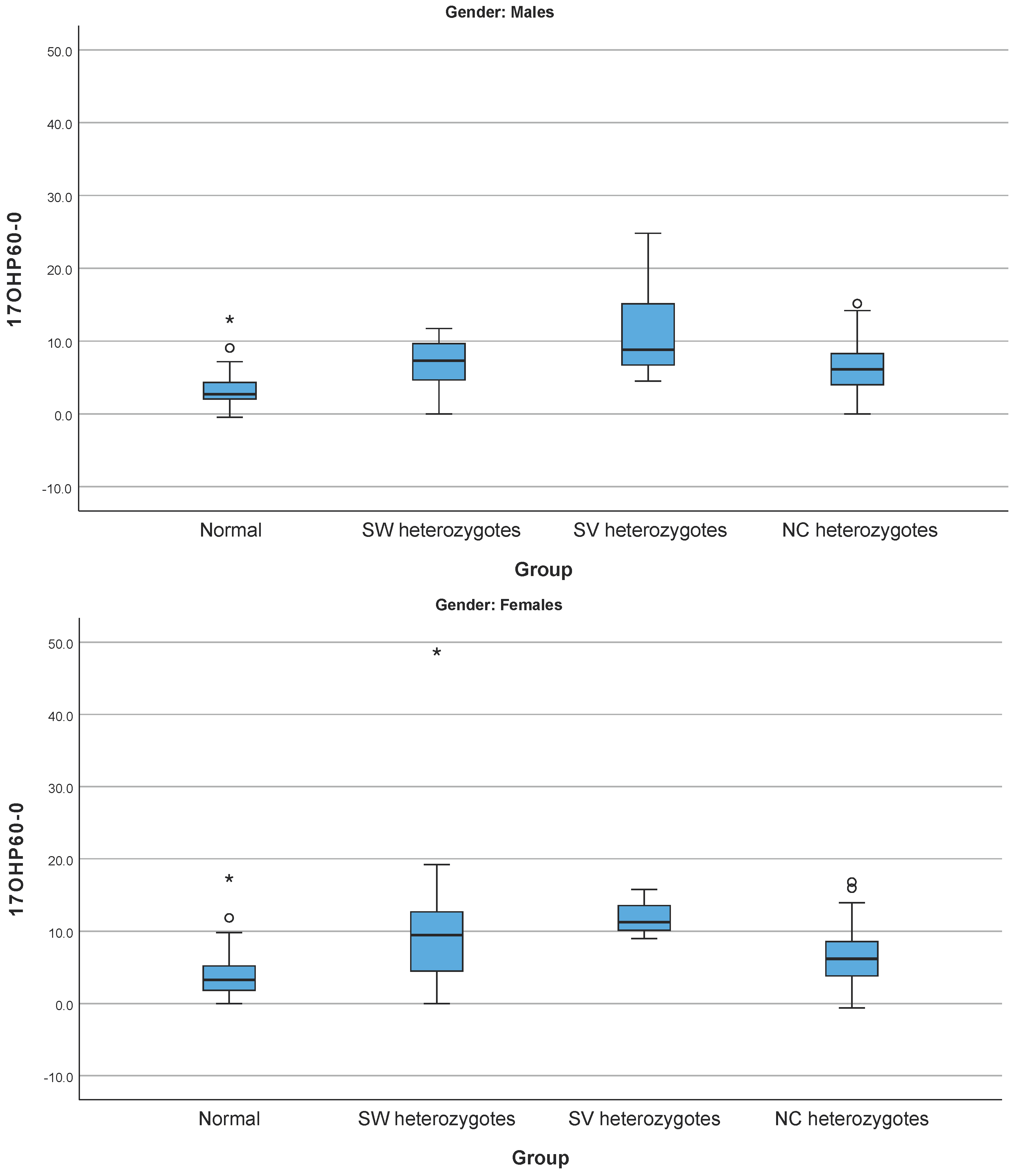
Figure 1. Box plots of Δ17-OHP60-0 in the group of normal (cases with no pathogenic variants identified) and the three groups of heterozygotes for pathogenic variants related to SW, SV, and NC forms of 21-OHD in males and females. Cycles represent extreme values while asterisks represent very extreme values. Normal indicates the cases with no pathogenic variants identified. The horizontal black lines in every group represent the corresponding median values. As shown, although there is a statistically significant differences between the median values among the groups, there is an overlap in the range of the Δ17-OHP60-0 values in the cases with no pathogenic variants identified with each group of the heterozygotes.
Cimb 46 00635 g001
Figure 2. Frequency of CYP21A2 gene pathogenic variants identified in 21-OHD patients. The blue label represents the homozygotes, and the orange label represents the compound heterozygotes.
Cimb 46 00635 g002
Figure 3. The distribution of 21-OHD forms in homozygotes and compound heterozygotes.
Cimb 46 00635 g003
Figure 4. Patients’ age (years) at diagnosis in the different forms of 21-OHD.
Cimb 46 00635 g004
Figure 5. Clinical manifestations of 21-OHD patients, heterozygotes and cases with no pathogenic variants identified (unaffected) distributed according to their age group.
Cimb 46 00635 g005
Table 1. Hormonal data presented among patients, heterozygotes, and cases with no pathogenic variants identified in Age Group I.
Age Group I PatientsAge Group I HeterozygotesAge Group I Cases with No Pathogenic Variants Identified
TotalMalesFemalesTotalMalesFemalesTotalMalesFemales
17-OHP concentrations(ng/mL)
Valid %
Median [min–max]
IQR
Mean ± SD
n = 7
63.6
35.9 [0.8–1.793]
64.9
287.7 ± 664.9
n = 1
25
23 [23–23]
0
23
n = 6
85.7
38.1 [0.8–1.793]
85.5
331.8 ± 717
n = 2
50
11 [4.3–17.6]
6.6
11 ± 9.4
n = 0


n = 2
66.7
11 [4.3–17.6]
6.6
11 ± 9.4
n = 13
100
3.8 [1.7–19]
8.2
6.8 ± 5.7
n = 9
100
4.8 [1.7–19]
8.5
8.1 ± 6.4
n = 4
100
3.7 [3.3–4.3]
0.4
3.8 ± 0.4
F concentrations (μg/dL)
Valid %
Median [min–max]
IQR
Mean ± SD
n = 3
27.3
5.4 [3.3–19.4]
8.1
9.4 ± 8.8
n = 0


n = 3
42.9
5.4 [3.3–19.4]
8.1
9.4 ± 8.8
n = 3
75
15.9 [14.6–18.9]
2.2
16.5 ± 2.2
n = 0


n = 3
100
15.9 [14.6–18.9]
2.2
16.5 ± 2.2
n = 13
100
8 [0.9–23.1]
10.2
9.4 ± 7.3
n = 9
100
6.5 [0.9–20.2]
7.9
8.5 ± 6.8
n = 4
100
10.6 [1.7–23.1]
9.3
11.5 ± 9
Δ4-A (ng/mL)
Valid %
Median [min–max]
IQR
Mean ± SD
n = 4
36.4
6 [0.4–12.5]
5.6
6.2 ± 5.1
n = 0


n = 4
57.1
6 [0.4–12.5]
5.6
6.2 ± 5.1
n = 2
50
2.6 [0.1–5.2]
2.5
2.6 ± 3.6
n = 0


n = 2
66.7
2.6 [0.1–5.2]
2.5
2.6 ± 3.6
n = 8
61.5
0.4 [0–2.9]
2
1 ± 1.2
n = 4
44.4
0.2 [0–2.8]
0.9
0.8 ± 1.4
n = 4
100
1.1 [0.1–2.9]
1.6
1.3 ± 1.3
Τ (ng/dL)
Valid %
Median [min–max]
IQR
Mean ± SD
n = 3
27.3
36.5 [0.1–500]
249.9
178.9 ± 278.7
n = 0


n = 3
42.9
36.5 [0.1–500]
249.9
178.9 ± 278.7
n = 1
25
1.1 [1.1–1.1]
0
1.1
n = 0


n = 1
33.3
1.1 [1.1–1.1]
0
1.1
n = 10
76.9
62 [1.1–193]
74.4
69.2 ± 62.3
n = 6
66.7
68 [1.1–193]
108.6
78.8 ± 76.3
n = 4
100
61.5 [2.5–94]
31.1
54.9 ± 38.4
ACTH (pg/mL)
Valid %
Median [min–max]
IQR
Mean ± SD
n = 4
36.4
34.5 [2.5–163]
65.2
58.6 ± 72.5
n = 0


n = 4
57.1
34.5 [2.5–163]
65.2
58.6 ± 72.5
n = 1
25
130.9 [130.9–130.9]
0
130.9
n = 0


n = 1
33.3
130.9 [130.9–130.9]
0
130.9
n = 8
61.5
53.1 [9.9–91.4]
42.3
49.8 ± 29.8
n = 4
44.4
60.6 [17–91.4]
21.8
57.4 ± 30.7
n = 4
100
38 [9.9–82.8]
32.9
42.1 ± 31.2
DHEAS (μg/dL)
Valid %
Median [min–max]
IQR
Mean ± SD
n = 4
36.4
809 [2–2208.6]
1588.2
957.2 ± 1076.6
n = 0


n = 4
57.1
809 [2–2208.6]
1588.2
957.2 ± 1076.6
n = 2
50
61 [4.1–118]
57.0
61 ± 80.6
n = 0


n = 2
66.7
61 [4.1–118]
57
61 ± 80.6
n = 9
69.2
24 [0.2–2458.5]
978.5
563.4 ± 894.9
n = 6
66.7
5.2 [0.2–980]
74.1
181.4 ± 393.1
n = 3
75
1500 [24–2458.5]
1.217.2
1327.5 ± 1226.4
In patients, in Age Group I there were two female patients that exhibited values that could not be precisely determined by the methodology applied. More precisely, there was one female patient with DHEAS >1500 μg/dL that was set at 1500 μg/dL and one female patient that had Δ4-A > 12.5 ng/mL and was set at 12.5 ng/mL in order to proceed with the statistical analysis. Respectively, in cases with no pathogenic variants identified, there were two females and one male that exhibited higher or lower values. More precisely, in one female, the DHEAS was >1500 μg/dL and was set at 1500 μg/dL, the other female had Δ4-A < 0.1 ng/mL and T < 2.5 ng/dL and was set at 0.1 ng/mL and 2.5 ng/dL, respectively, and the male had Δ4-A < 0.006 ng/mL and was set at 0 ng/mL.
Table 2. Hormonal data presented among patients, heterozygotes, and cases with no pathogenic variants identified cases in Age Group II.
Age Group II PatientsAge Group II HeterozygotesAge Group II Cases with No Pathogenic Variants Identified
TotalMalesFemalesTotalMalesFemalesTotalMalesFemales
17-OHP concentrations (ng/mL)
Valid %
Median [min–max]
IQR
Mean ± SD
n = 100
90.9
11 [0.8–200]
18.8
25.7 ± 40.1
n = 30
88.2
13.6 [1.9–200]
39.6
37.7 ± 52.3
n = 70
92.1
9.6 [0.8–200]
18.2
20.5 ± 32.7
n = 183
98.9
2.6 [0.3–13.7]
2.4
3.1 ± 2.1
n = 47
97.9
2.1 [0.4–7.5]
1.6
2.5 ± 1.5
n = 136
99.3
2.7 [0.3–13.7]
2.6
3.4 ± 2.3
n = 94
97.9
2.1 [0.5–25]
1.7
2.7 ± 2.8
n = 10
100
1.5 [0.5–25]
2.7
4.2 ± 7.5
n = 84
97.7
2.1 [0.5–8.9]
1.6
2.5 ± 1.5
F concentrations (μg/dL)
Valid %
Median [min–max]
IQR
Mean ± SD
n = 91
82.7
12.3 [4.2–26.5]
6.7
12.9 ± 4.9
n = 30
88.2
12.2 [4.2–91.5]
6.7
14.8 ± 15.5
n = 61
80.3
12.5 [5.2–26.5]
6.7
13.3 ± 4.6
n = 170
91.9
13.8 [3.9–29.5]
7.7
14.5 ± 5.6
n = 43
89.6
14 [5.3–23.8]
8.3
13.8 ± 5.3
n = 127
92.7
13.7 [3.9–29.5]
7.6
14.8 ± 5.6
n = 90
93.8
14.8 [0.6–31.5]
9.8
15.5 ± 6.5
n = 10
100
16.6 [2.9–26.7]
10.1
15.7 ± 7.9
n = 80
93
14.8 [0.6–31.5]
9.5
15.5 ± 6.4
Δ4-A (ng/mL)
Valid %
Median [min–max]
IQR
Mean ± SD
n = 71
64.5
0.8 [0–35.3]
1.4
1.7 ± 4.3
n = 21
61.8
0.9 [0.1–35.3]
1.7
3.1 ± 7.6
n = 50
65.8
0.8 [0–6.7]
1.1
1.2 ± 1.3
n = 117
63.2
0.4 [0–76]
0.6
1.2 ± 7
n = 29
60.4
0.4 [0–1.4]
0.6
0.5 ± 0.4
n = 88
64.2
0.5 [0–76]
0.6
1.5 ± 8.1
n = 73
76
0.4 [0.1–22]
0.5
0.9 ± 2.6
n = 7
70
0.3 [0.1–0.7]
0.3
0.4 ± 0.2
n = 66
76.7
0.4 [0.1–22]
0.6
1 ± 2.7
Τ (ng/dL)
Valid %
Median [min–max]
IQR
Mean ± SD
n = 88
80
8 [0–309]
16.5
20.6 ± 49.1
n = 31
91.2
3 [0–309]
22.6
36.0 ± 79.0
n = 57
75
8 [0–78.9]
15
12.3 ± 14.3
n = 144
77.8
5 [0–126]
12
10.6 ± 18.2
n = 41
85.4
7 [0–126]
16
18.2 ± 30.1
n = 103
75.2
3 [0–47]
11
7.6 ± 8.7
n = 83
86.5
2.5 [0–250]
5
9.5 ± 29.1
n = 10
100
2 [0–19]
2.2
3.5 ± 5.6
n = 73
84.9
2.5 [0–250]
5
10.4 ± 30.9
ACTH (pg/mL)
Valid %
Median [min–max]
IQR
Mean ± SD
n = 51
46.4
38.9 [2.3–357]
64.8
73.7 ± 80.6
n = 16
47.1
45.6 [2.3–357]
109.4
101 ± 104.8
n = 35
46.1
36.5 [10–310]
36.5
61.2 ± 64.7
n = 80
43.2
28.2 [0.6–400]
24.8
42.2 ± 49.8
n = 18
37.5
27.9 [8.8–120]
9.5
32.9 ± 24.7
n = 62
45.3
29.4 [0.6–400]
30
44.9 ± 54.9
n = 55
57.3
33 [0.7–229]
35.3
46.8 ± 44.3
n = 8
80
34.5 [13.8–153]
39.4
53.3 ± 46.4
n = 47
54.7
33 [0.7–229]
30.8
45.7 ± 44.4
DHEAS (μg/dL)
Valid %
Median [min–max]
IQR
Mean ± SD
n = 94
85.5
79.9 [0.1–3,742]
118.1
142.1 ± 390.1
n = 30
88.2
83.8 [0.4–448]
195.2
130.4 ± 135.6
n = 64
84.2
79.9 [0.1–3,742]
109.6
147.6 ± 464.9
n = 153
82.7
76.3 [0–964]
82
82.6 ± 93
n = 37
71.1
78 [1.2–252]
102
95.1 ± 68.9
n = 116
84.7
73.4 [0–964]
74.5
78.6 ± 99.4
n = 85
88.5
64.9 [0.3–341]
70.5
80.9 ± 60.8
n = 9
90
65 [0.5–195]
88.8
84.3 ± 63.6
n = 76
88.4
64.4 [0.3–341]
69.4
80.5 ± 60.9
In patients in Age Group II, 2 males were found to have concentrations that could not be precisely determined by the methodology applied. More precisely, there was one female and one male patient that exhibited concentrations of 17-OHP >200 ng/mL and the concentrations were set at 200 ng/mL. In heterozygotes, 5 females had concentrations that could not be precisely determined. Specifically, there were two females that were found to have T < 2.5 ng/dL and was set at 2.5 ng/dL, one female had T < 0.2 ng/dL and was set at 0.2 ng/dL, the fourth female had Δ4-A < 0.1 ng/mL and was set at 0.1 ng/mL, and the fifth female had Δ4-A < 0.3 ng/mL, T < 20 ng/dL and DHEAS < 15 μg/dL and were set at 0.3 ng/mL, 19 ng/dL, and 14 μg/dL, respectively. In cases with no pathogenic variants identified, 13 females and 3 males had concentrations that could not be precisely determined. Specifically, eight females and one male had T < 2.5 ng/dL and was set at 2.5 ng/dL, one female and one male had Δ4-A < 0.1 ng/mL and was set at 0.1 ng/mL, one male had T < 20 ng/dL and was set at 19 ng/dL, one female had Δ4-A < 0.3 ng/mL and T < 20 ng/dL and was set at 0.3 ng/mL and 19 ng/dL, respectively, two females had T < 0.025 ng/dL and was set at 0.02 ng/dL, and finally one female had Δ4-A < 0.1 ng/mL and T < 2.5 ng/dL and was set at 0.1 ng/mL and 2.5 ng/dL, respectively.
Table 3. Hormonal data presented among patients, heterozygotes, and cases with no pathogenic variants identified in Age Group III.
Age Group III PatientsAge Group III HeterozygotesAge Group III Cases with No Pathogenic Variants Identified
TotalMalesFemalesTotalMalesFemalesTotalMalesFemales
17-OHP concentrations (ng/mL)
Valid %
Median [min–max]
IQR
Mean ± SD
n = 14
93.3
13.1 [1.7–119.5]
10.8
23.3 ± 31.8
n = 3
100
12 [1.7–20.6]
9.4
11.4 ± 9.4
n = 11
91.7
13.5 [3.4–119.5]
10
26.6 ± 35.2
n = 18
100
3.6 [0.8–7.8]
1.7
3.8 ± 1.7
n = 4
100
4.3 [3.3–5.2]
1.3
4.2 ± 0.9
n = 14
100
3.3 [0.8–7.8]
1.4
3.7 ± 1.9
n = 24
96
3.6 [0.6–20.9]
2.8
4.9 ± 4.5
n = 6
100
4.1 [2.6–13.1]
1.5
5.4 ± 3.9
n = 18
94.7
3.2 [0.6–20.9]
3
4.8 ± 4.8
F concentrations (μg/dL)
Valid %
Median [min–max]
IQR
Mean ± SD
n = 11
73.3
16.2 [4.6–27.9]
5.9
15 ± 6.4
n = 3
100
16.2 [6.5–17.3]
5.4
13.3 ± 5.9
n = 8
66.7
15.4 [4.6–27.9]
6
15.6 ± 6.8
n = 15
83.3
14.9 [9.8–23.2]
4.8
15.5 ± 4.3
n = 4
100
15.9 [9.8–23.2]
4.8
16.2 ± 5.5
n = 11
78.6
14.9 [10–23.1]
4.4
15.2 ± 4.1
n = 22
84.6
18.5 [5.4–29.5]
8.4
17.2 ± 6.1
n = 5
83.3
12.9 [11.1–18.6]
6.4
14.4 ± 3.6
n = 18
90
18.7 [5.4–29.5]
8.3
18 ± 6.4
Δ4-A (ng/mL)
Valid %
Median [min–max]
IQR
Mean ± SD
n = 9
60
3 [1.1–7.9]
1.7
3.4 ± 2
n = 3
100
1.3 [1.1–2.4]
0.6
1.6 ± 0.7
n = 6
50
3.9 [2.7–7.9]
1.1
4.3 ± 1.9
n = 10
55.6
2.3 [0.9–6.2]
1.6
2.8 ± 1.9
n = 2
50
2.1 [1.9–2.3]
0.2
2.1 ± 0.3
n = 8
57.1
2.6 [0.9–6.2]
2.6
3 ± 2.1
n = 19
76
2.4 [0.2–6.8]
2
2.8 ± 1.7
n = 4
66.7
0.7 [0.2–2.3]
0.6
1 ± 0.9
n = 15
78.9
2.6 [1.2–6.8]
2.4
3.3 ± 1.6
Τ (ng/dL)
Valid %
Median [min–max]
IQR
Mean ± SD
n = 13
86.7
18 [0.5–173]
70.3
47.3 ± 57.3
n = 3
100
133 [18–173]
77.5
108 ± 80.5
n = 10
83.3
15.5 [0.5–110]
38.8
29 ± 36.7
n = 16
88.9
30 [0.1–621]
55.5
126.6 ± 218.3
n = 4
100
531 [11.4–621]
228.9
423.6 ± 281.1
n = 12
85.7
26 [0.1–80]
38
27.6 ± 26
n = 18
72
26.5 [0.1–560.1]
42.6
78.7 ± 157.9
n = 3
50
452 [6.8–560.1]
276.6
339.6 ± 293.3
n = 15
78.9
25 [0.1–68]
36.2
26.5 ± 22.1
ACTH (pg/mL)
Valid %
Median [min–max]
IQR
Mean ± SD
n = 8
53.3
30.4 [15.4–142]
21.4
45.6 ± 40.8
n = 0


n = 8
66.7
30.4 [15.4–142]
21.4
45.6 ± 40.8
n = 10
55.6
28.6 [16.6–59.2]
14.9
32.3 ± 12.6
n = 1
25
31.6 [31.6–31.6]
0
31.6
n = 9
64.3
26 [16.6–59.2]
17.9
32.4 ± 13.3
n = 9
36
26.2 [10–84.8]
29.5
34.3 ± 24.6
n = 0


n = 9
47.4
26.2 [10–84.8]
29.5
34.3 ± 24.6
DHEAS (μg/dL)
Valid %
Median [min–max]
IQR
Mean ± SD
n = 13
86.7
243 [0.6–704]
275.6
280.6 ± 212.3
n = 3
100
243 [113.8–260]
73.1
205.6 ± 80
n = 10
83.3
246.5 [0.6–704]
274.6
303.1 ± 237.1
n = 16
88.9
209 [1.3–453]
164.5
189 ± 123.2
n = 3
75
255 [212–298]
43
255 ± 43
n = 13
92.9
131 [1.3–453]
159.5
173.8 ± 131.6
n = 22
88
196.5 [0.8–702]
247
227.9 ± 184.1
n = 4
66.7
139 [72–702]
219
263 ± 296.3
n = 18
94.7
229.5 [0.8–573.5]
241
220.1 ± 161.3
Table 4. Novel variants identified in the CYP21A2 gene: clinical manifestations, hormonal profiles, and genotypes.
VariantLocation CYP21A2GenotypeClinical ManifestationSexHormonal ProfileFrequency (GnomAD)ACMG ClassificationRemarks
AgeHormonal Findings (before Treatment, if Required)ACTH Stimulation Test
0′30′60′
c.-127G>A5’UTRc.[-127G>A];[=]PAf7 yrs17-OHP: 4.98 ng/mL T: <0.025 ng/dL, DHEA-S: 61 μg/dL Δ4-A: 0.4 ng/mL17-OHP (ng/mL)4.9111.88-(not efficiently covered)VUS (PM2, BP7)Promoter region (c.-76 to c.-126)
F (μg/dL)18.326
c.-115G>T5’UTRc.[-115G>T];[92C>T]PAf6 yrs-17-OHP
(ng/mL)
9.2233.60.00003188VUS (PM2, PM3, BP7)Promoter region (c.-76 to c.-126)
F(μg/dL)12.9923.43
c.-82C>T5’UTRc.[-82C>T];[=]PAf7 yrsΔ4-A: 0.96 ng/mL, T: 6 ng/dL, DHEA-S: 178 μg/dL17-OHP
(ng/mL)
3.644.135.690.00223VUS (BS1, BP7)Promoter region (c.-76 to c.-126)
F (μg/dL)31.137.739.9
c.764G>A# (p.R255K)Exon 7c.[764G>A];[=]PHIm6 mos17-OHP: 4 ng/mL, Cortisol: 14.7 μg/dL, ACTH: 99 pg/mL, PRA: 4.18 ng/mL/h, T: 0.88 ng/dL, DHEA-S: 246.8 μg/dL, Δ4-A: 0.76, Aldosterone: 71.5 ng/dL17-OHP (ng/mL)4.399.030.000004032VUS (PM2)[31] *
F (μg/dL)10.224.8
c.844G>A (p.V282M)Exon 7p.[V282M];[L308Ffs*6]PAf11 yrs17-OHP: 17.1 ng/mL, DHEA-S: 121 μg/dL, Δ4-A: 1.68 ng/mL17-OHP (ng/mL)6.557.671.60.00001238LP (PM2, PP2, PM3, PM5)[32]
F (μg/dL)6.5611.713.64
PA: Premature Adrenarche, PHI: Pubic Hair of Infancy, PRA: Plasma Renin Activity, T: Testosterone, f: Female, m: Male, yrs: years, mos: months. Reference range: 17-OHP (ng/mL): 0.25–1.45 (males 6–11 months), 0.2–0.5 (females 6–9 years), 0.2–0.7 (females 10–11 years), 0.15–0.5 (males 10–11 years), 0.25–1.9 (females 12–13); Cortisol(μg/dL): 6.2–19.4; Testosterone(ng/dL): 2–7 (males 6–12 months), 2–20 (females 6–9 years), 5–25 (females 10–11 years), 10–80 (females 12–14 years); DHEA-S(μg/dL):3.4–123.6 (1–12 months), 2.8–85.2 (5–10 years), 33.9–280 (female 11–14 years), 24.4–247 (male 11–14 years); Δ4-A (ng/mL):0.05–0.3 (6–12 months), 0.05–0.55 (3–9 years), 0.1–0.8 (10–11 years), 0.15–2.5 (females 12–17 years); ACTH (pg/mL):7–64, PRA (ng/mL/h):0.5–4.7 (upright posture); Aldosterone (ng/dL): 5–132 (<1 year), 4–76 (5–8 years), 3–28 (9–12 years). The c.92C>T corresponds to p.P31L. According to the Human Genome Variation Society recommendations, the protein sequence number should not be stated in a genotype in which 1 of the 2 variants is present in a noncoding sequence. # The p.R255K has been previously reported in the literature (retrieved from dbSNP) but never reported in patients.
Disclaimer/Publisher’s Note: The statements, opinions and data contained in all publications are solely those of the individual author(s) and contributor(s) and not of MDPI and/or the editor(s). MDPI and/or the editor(s) disclaim responsibility for any injury to people or property resulting from any ideas, methods, instructions or products referred to in the content.