Article

Artemisiae argyi Water Extract Alleviates Obesity-Induced Metabolic Disorder

1
Department of Food Science and Nutrition, Kyungpook National University, 1370 San-Kyuk Dong Puk-Ku, Daegu 41566, Republic of Korea
2
Center for Food and Nutritional Genomics Research, Kyungpook National University, 1370 San-Kyuk Dong Puk-Ku, Daegu 41566, Republic of Korea
3
Center for Beautiful Aging, Kyungpook National University, 1370 San-Kyuk Dong Puk-Ku, Daegu 41566, Republic of Korea
4
Raydel Research Institute, 76, Dongnae-ro, Dong-gu, Daegu 41061, Republic of Korea
5
Bio Convergence Testing Center, Daegu Haany University, 1 Haanydaero, Gyeongsan-si 38610, Republic of Korea
6
Department of Herbology, College of Korean Medicine, Daegu Haany University, 64 Gil, 136, sinsincheondo-ro, Suseong-gu, Daegu 42158, Republic of Korea
*
Authors to whom correspondence should be addressed.
These authors contributed equally to this work.
Curr. Issues Mol. Biol. 2022, 44(12), 6158-6171; https://doi.org/10.3390/cimb44120420
Submission received: 6 October 2022 / Revised: 28 November 2022 / Accepted: 2 December 2022 / Published: 7 December 2022

Abstract

:
Artemisiae argyi is a well-known traditional herbal medicine used in East Asia. Although the antibacterial and anti-inflammatory effects of A. argyi have been reported, its efficacy in improving obesity has not been yet evaluated. In this study, mice were fed a normal diet (AIN-93), a high-fat diet (HFD, 60% of kcal from fat), and an HFD with 0.1% of A. argyi water extract for 16 weeks. The body weight and body fat in A. argyi-fed mice significantly decreased via upregulation of the mRNA expression of fatty acid oxidation-related genes, with a simultaneous decrease in plasma lipid content and leptin levels. A. argyi water extract also ameliorated hepatic steatosis by restricting lipogenesis via lowering the activities of fatty acid synthase and phosphatidic acid phosphatase. Consistently, hepatic histological analysis indicated that A. argyi water extract decreased hepatic lipid accumulation in accordance with the hepatic H, E and Oil Red O-stained area. Additionally, A. argyi ameliorated the impaired glucose homeostasis by increasing the mRNA expression of AMP-activated kinase and glycolysis-related genes. In conclusion, our results indicate that A. argyi can be used to treat obesity-related metabolic conditions.

1. Introduction

As interest in improving quality of life has increased, the efficacy and safety of pharmaceuticals have also garnered increasing attention [1]. Developed countries have previously evaluated the economic value of resource plants distributed globally and are now focusing on securing more diverse plant species and systematically developing new functional pharmaceutical materials from them [2]. Natural products are basic resources for novel drug development, and research using natural product extracts is in the spotlight [3].
Artemisia is a herbaceous plant belonging to the Asteraceae family. It is estimated that 300 of about 400 species of this genus are present in Korea, but only about 40 species have been reported thus far [4]. Artemisiae argyi—widely consumed in Korea—is rich in flavonoids, polysaccharides, and volatile oils, such as isocoumarin, coumarin, diterpenlactone, flavonoid, phellandrene, couprol, cadinene, cineol, artemisinin, and euphatrin [5,6]. Previous studies have investigated A. argyi’s health-promoting properties, concluding the prevention of oxidative stress, cancer, inflammation, osteoporosis, and immunomodulatory and its neuroprotective activities [5,7,8,9,10,11]. However, studies on the efficacy of A. argyi water extract in diet-induced obesity are lacking. Therefore, in this study, we explored the potential of A. argyi as a dietary or supplemental source to modulate obesity-associated metabolic disorders in diet-induced obese mice. This study highlights the potential application of A. argyi water extract as a modulator of obesity-associated metabolic disregulation in functional foods.

2. Materials and Methods

2.1. Extract Preparation

Artemisiae argyi was purchased from Bonchowon (Yeongcheon, Korea) and a voucher herbarium specimen (DHU-KM-2020-07) was verified at the College of Korean Medicine in Daegu Haany University. The above-ground part of dried Artemisiae argyi (300 g) was extracted by 10× volume of distilled water (100 °C) for 2 h. After filtering the extract with qualitative filter paper (Hyundai Micro, No. 22, 285 mm), the solvent was evaporated in vacuo to obtain powders (with a yield rate of 13.2%). Powder was stored at −80 °C until experimentation. Table 1 shows the chemical composition of the A. argyi extract. The total polyphenol content of A. argyi was measured with reference to the method of Rama et al. [12]. Amounts of 100 μL of diluted sample solution, 500 μL of Folin–Ciocalteu’s phenol reagent (diluted to 10×), and 400 μL of 7.5% sodium carbonate were mixed and left in the dark for 30 min, and then absorbance was measured using a multi-function microplate reader (765 nm) (infinite M200 Pro, Tecan, Männedorf, Switzerland). Gallic acid (G7384, Sigma-Aldrich, Saint Louis, MO, USA; ≥97.5% purity) was used to plot a standard calibration curve and calculate the total polyphenol content of the sample. The total flavonoid content of A. argyi was measured with reference to the method of Jiao et al. [13]. Amounts of 100 μL of diluted sample solution, 300 μL of methanol, 20 μL of 10% aluminum chloride solution, 20 μL of 1 M potassium acetate solution, and 560 μL of distilled water were mixed and left in the dark for 30 min, and then absorbance was measured using a multi-function microplate reader (415 nm). Quercetin (Q4951, Sigma-Aldrich, Saint Louis, MO, USA; ≥95% purity) was used to plot a standard calibration curve and calculate the total flavonoid content of the sample. The eupatilin (SML1689, Sigma-Aldrich, Saint Louis, MO, USA; ≥98% purity) contents of the A. argyi was measured with reference to the method of Xia et al. [14]. The A. argyi water extract was dissolved with 50% methanol to 4000 mg/mL, and filtrated through a 0.2 μm PTFE syringe filter (Advantec DIS-MIC-13HP, Toyo Roshi Kaisha, Ltd., Tokyo, Japan). The LC–MS analysis was performed on an Vanquish Horizon UPLC system connected to an Orbitrap Exploris 120 mass spectrometer (Thermo Fisher scientific, Cleveland, OH, USA).

2.2. Experimental Animals and Diet

Thirty male C57BL/6J mice (4-weeks-old) were purchased from Jackson Laboratory (Bar Harbor, ME, USA). The animals were maintained in a temperature- (20–23 °C) and light-controlled (12/12-h light–dark cycle) room and fed a pelletized, commercial, non-purified diet for one week after arrival. The mice were randomly divided into three groups (n = 10 per group) and fed the respective experimental diets for 16 weeks: normal diet control (ND, American Institute of Nutrition [AIN]-93 semi-synthetic diet), high-fat diet control (HFD, 60% of kcal from fat, based on the AIN-93G diet), and 0.1% A. argyi water extract (AA; HFD with 0.1% A. argyi water extract, w/w) (Table 2). The dose of the A. argyi water extract was determined by previous studies [15,16,17]. The mice had free access to the experimental diet and water during the experimental period. Their food intake was recorded daily, and body weight was monitored biweekly. All animal procedures were approved by the Ethics Committee for Animal Studies at Kyungpook National University, Daegu, Republic of Korea (approval no. KNU-2020-0090).

2.3. Sample Preparation

At the end of the experimental period, all the mice were anesthetized with isoflurane (Baxter, United States) Isoflurane was administered in a mixture with oxygen at a constant flow of 0.5–0.7 L/min. The animals were placed in the induction chamber, a plastic container of 20 cm in diameter and 10 cm in height, connected with a polyurethane polyether tube to let in the anesthetic gas mixture. Induction of the anesthesia was achieved by using 4% vaporized isoflurane (Laboratorios Esteve S.A., Barcelona, Spain) in oxygen.
After sacrificed with a 12-h fast, blood samples were collected from the inferior vena cava into a heparin-coated tube for the measurement of plasma parameters. The blood was centrifuged at 1000× g for 15 min at 4 °C, and the plasma was separated. After blood collection, epididymal white adipose tissue (WAT), perirenal WAT, retroperitoneal WAT, mesentery WAT, subcutaneous WAT, and liver were promptly removed, rinsed with physiological saline, and weighed. Among them, epididymal WAT and liver were immediately frozen in liquid nitrogen and stored at −70 °C until the analyses of the enzyme activity and RNA.

2.4. Plasma and Hepatic Lipid Profile

Plasma triglyceride (TG), total cholesterol (TC), and high-density lipoprotein cholesterol (HDL-C) concentrations were determined using commercial kits (Asan, Seoul, Republic of Korea). Plasma free fatty acid (FFA) content was measured using an enzymatic kit (Wako Chemicals, Richmond, VA, USA). Plasma apolipoprotein (Apo) A-I and Apo B levels were also determined using an enzymatic kit (Eiken, Tokyo, Japan). Hepatic lipids were extracted according to Folch’s method [18], and the content was determined using the same commercial kits used for the plasma lipid profile analysis.

2.5. White Adipose Tissue (WAT) and Hepatic Morphology

WAT and liver samples were fixed in 10% buffered formalin, embedded in paraffin, and semi-serial sections at 4 μm-thickness were cut at 1 mm intervals, stained with hematoxylin and eosin (H and E). Frozen hepatic tissue was semi-serial sectioned at 7 μm-thickness with 1 mm intervals stained with Oil Red O, and observed under an optical microscope (Nikon, Tokyo, Japan) under 200× magnification [19,20]. All morphological quantification was performed based on image J using 10 images per group for analysis [21,22].

2.6. Plasma Adipokine Content

Plasma leptin, adiponectin, and resistin levels were measured using a multiplex detection kit (Bio-Rad Laboratories Inc., Hercules, CA, USA). The Luminex 200 LabMAP system (Luminex, Austin, TX, USA) and Bio-Plex Manager software (version 4.1.1; Bio-Rad Laboratories, Inc.) were used for the analysis.

2.7. Fasting Blood Glucose Level, Intraperitoneal Glucose Tolerance Test, and Homeostatic Index of Insulin Resistance (HOMA-IR)

Animals were fasted for 12 h before undergoing a fasting blood glucose (FBG) determination test and an intraperitoneal glucose tolerance test (IPGTT). The FBG concentration in the blood from the tail veins was measured using a OneTouch Select Plus ® meter glucose analyzer (LifeScan, Milpitas, CA, USA). The IPGTT was performed at the 11th week. Glucose was intraperitoneally injected at 0.5 g·per·kg of body weight, and blood glucose concentrations were determined at 0, 30, 60, and 120 min. HOMA-IR was calculated using the following formula:
HOMA-IR = (fasting glucose (mmol/L) × fasting insulin (µL·U/mL))/22.5 [23].

2.8. mRNA Expression Analysis

mRNA extraction was performed as previously described [24]. Total mRNA was reverse transcribed into cDNA using the QuantiTect Reverse Transcription Kit (Qiagen, Hilden, Germany). mRNA expression was quantified via real-time quantitative PCR using the QuantiTect SYBR Green PCR kit (Qiagen) and the SDS7000 sequence-detection system (Applied Biosystems, Foster City, CA, USA). Sequences of the primers used are listed in Supplementary Table S1.

2.9. Enzyme Activities in the Liver and Epididymal WAT

Liver and epididymal WAT samples were prepared and analyzed using the method developed by Hulcher and Oleson [25]. Fatty acid synthase (FAS) activity was analyzed as described by Nepokroeff et al. [26]. The activities of glucose-6-phosphate dehydrogenase (G6PD), malic enzyme (ME), carnitine palmitoyl transferase (CPT), and fatty acid oxidation were measured using previously described methods [27,28,29,30].

2.10. Statistical Analysis

Data are presented as the mean ± standard error of the mean (SE). All statistical analyses were performed using SPSS version 23.0 (IBM, Chicago, IL, USA). Significant differences between the ND and HFD groups and the HFD and AA groups were determined using the Student’s t-test. Differences were considered significant at p < 0.05.

3. Results

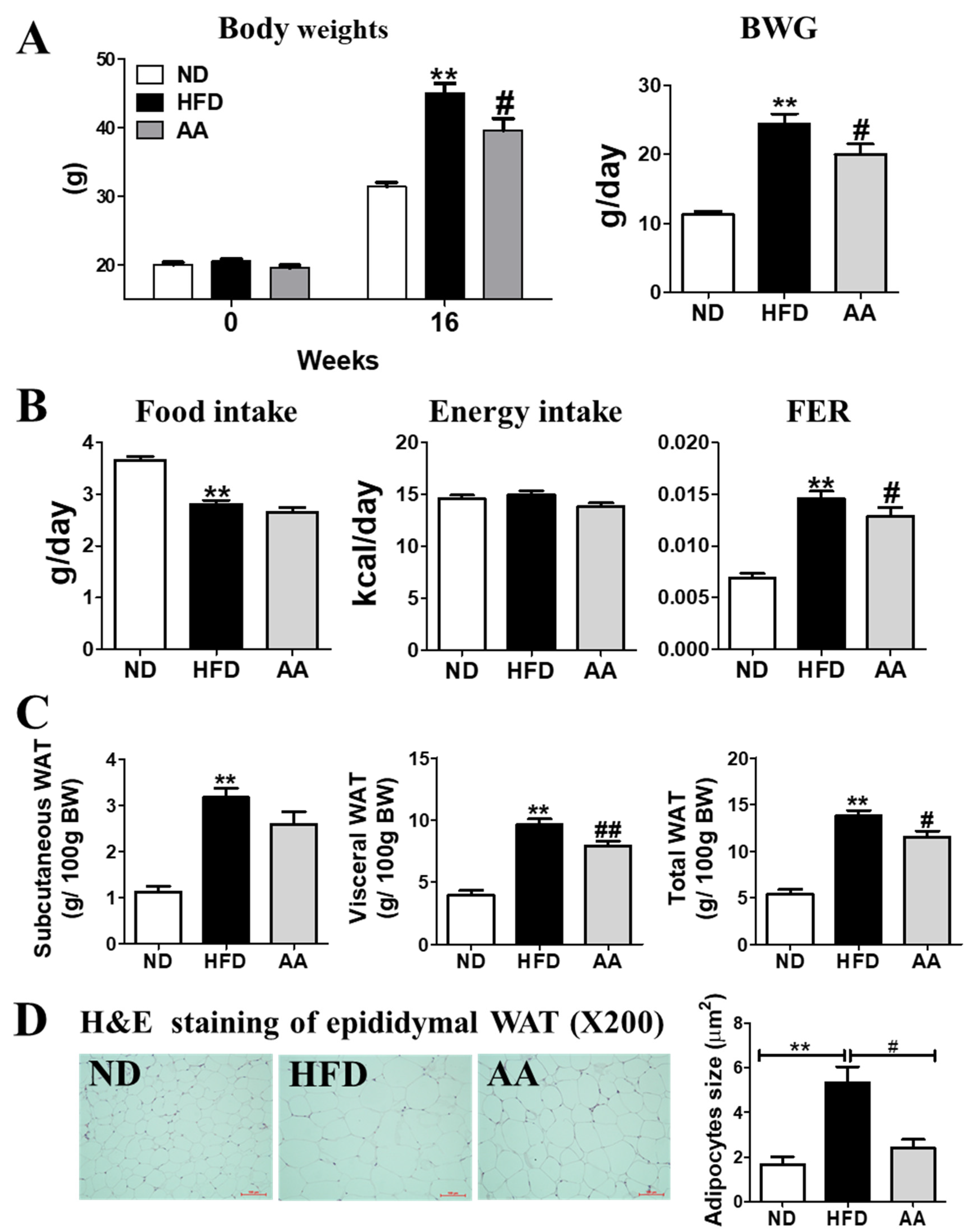
3.1. A. argyi Water Extract Supplementation Reduces Body Weight and Body Fat Mass

The initial body weights of the mice were not significantly different among the three groups. However, at the end of the experimental period, the HFD-fed mice showed a drastic increase in body weight compared with the ND-fed mice ( 1A). The AA-fed mice had lower body weights than the HFD-fed mice because of the suppression of total body weight gain. No significant differences in food and energy intake between the HFD and AA groups were observed; however, the food efficiency ratio was significantly lower in the AA group than in the HFD group (Figure 1B). Subcutaneous, visceral, and total WAT were significantly increased by HFD consumption (Figure 1C). Consistent with these results, morphological observations revealed that epididymal adipocyte size in the AA group was the smallest of all the three groups (Figure 1D).

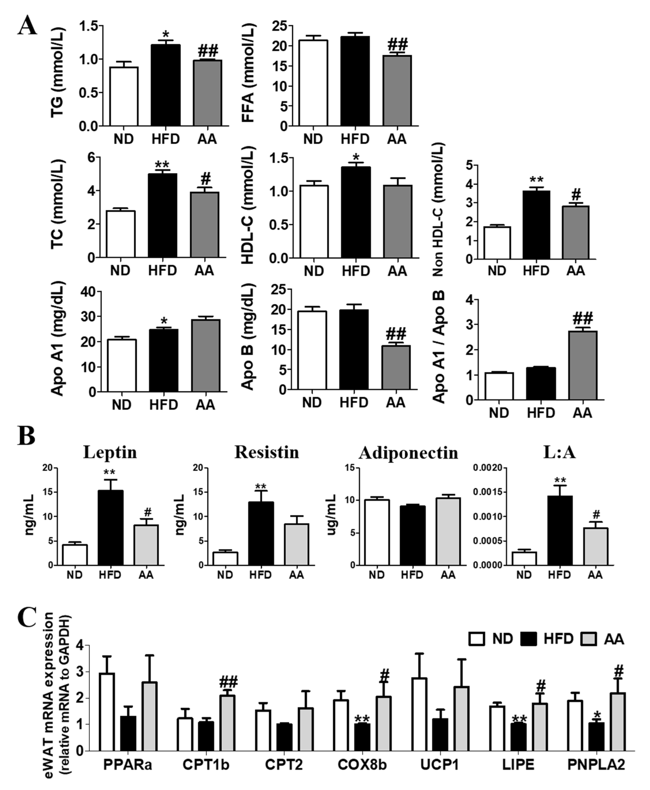
3.2. A. argyi Water Extract Supplementation Improves Plasma Lipid Profiles and Adipokine Levels by Regulating the Adipocyte mRNA Expression involved in Fatty Acid Oxidation

The lipid profiles of the plasma obtained after a 24-h fast are shown in Figure 2A. Plasma TG, FFA, TC, and non-HDL-C levels were significantly lower in the AA group than in the HFD group. In addition, A. argyi water extract supplementation significantly decreased the Apo B level and increased the Apo A1-to-Apo B ratio. The AA group showed significant reductions in visceral and total WAT weights compared with the HFD group. The plasma leptin level and leptin: adiponectin (L:A) ratio were significantly reduced in the AA group (Figure 2B). Moreover, the adipocyte mRNA expression of molecules related to fatty acid oxidation (CPT1b, COX8b, LIPE, and PNPLA2) was significantly lower in the AA group than in the HFD group (Figure 2C).

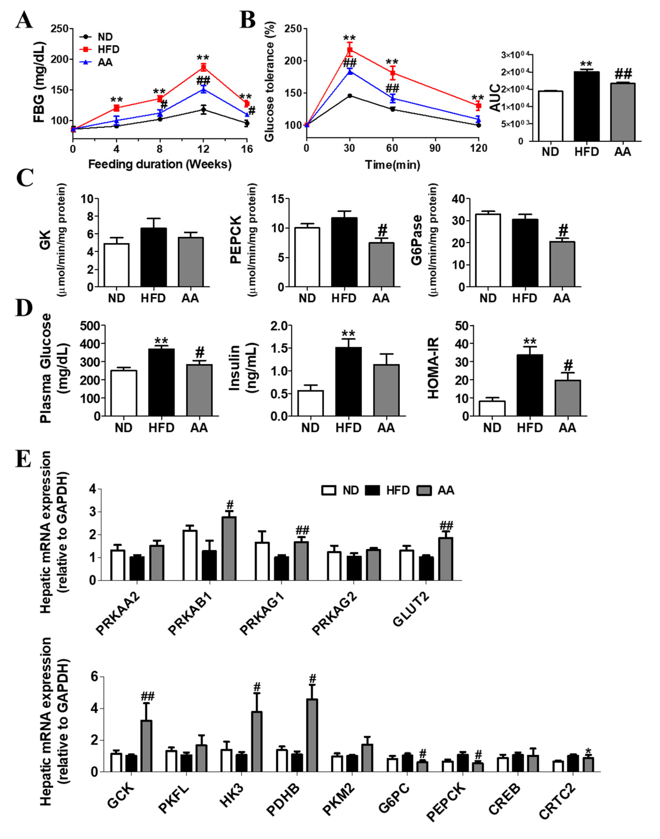
3.3. A. argyi Water Extract Supplementation Alleviates Impaired Glucose Metabolism-Related Obesity

At the beginning of the 4th week of feeding, FBG levels in the AA group were significantly lower than those in the HFD group (Figure 3A). IPGTT and area under the curve (AUC) results indicated that AA ameliorated glucose intolerance (Figure 3B). Regarding hepatic enzyme activities related to glucose metabolism, A. argyi water extract supplementation significantly decreased PEPCK and G6pase activities (Figure 3C). There was no significant difference in plasma insulin levels between the HFD and AA groups; however, plasma glucose levels and HOMA-IR were significantly decreased in the AA group (Figure 3D). The hepatic mRNA expression results are shown in Figure 3E. Regarding the hepatic mRNA expression of AMPK-related genes (PRKAA2, PRKAB1, PRKAG1, PRKAG2, and GLUT2), A. argyi water extract supplementation significantly increased the mRNA expression of PRKAB1, PRKAG1, and GLUT2. Furthermore, A. argyi water extract supplementation significantly upregulated the mRNA expression of genes related to glycolysis (GCK, HK3, and PDHB) and downregulated the expression of those related to gluconeogenesis (G6PC and PEPCK).

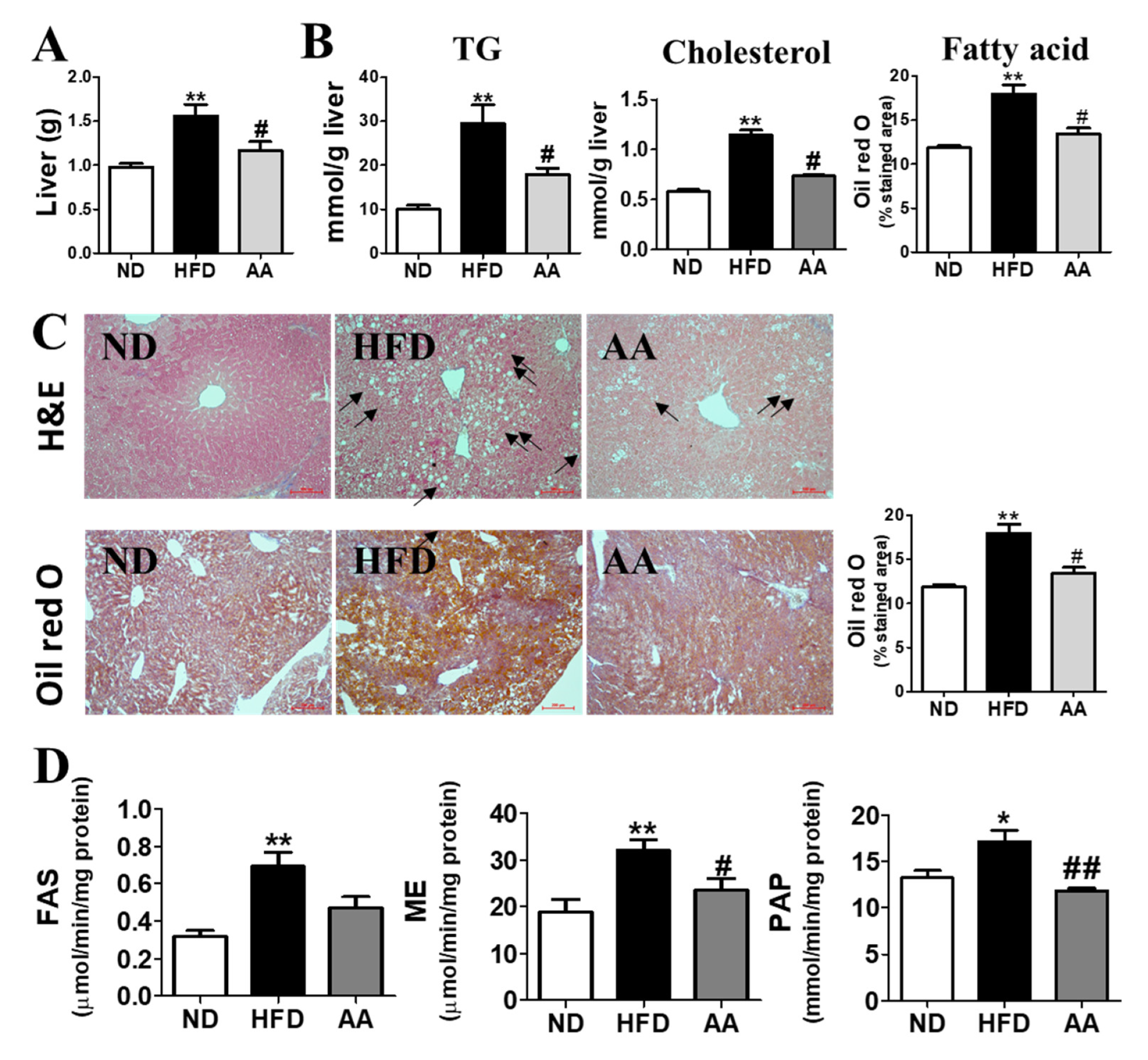
3.4. A. argyi Water Extract Supplementation Alleviates Hepatic Steatosis

Artemisiae argyi water extract supplementation significantly decreased liver weight and lowered hepatic TG and hepatic cholesterol levels (Figure 4A,B). Hepatic morphological observations and Oil Red O staining revealed reduced lipid formation and accumulation in the AA group compared with that the HFD group (Figure 4C). In particular, the Oil Red O-stained area, which can clearly indicate the presence of lipid droplets, was significantly increased by HFD consumption; however, in the AA group there was a significant decrease in the Oil Red O-stained area compared with the values of the HFD group. Moreover, A. argyi water extract supplementation significantly decreased hepatic ME and PAP activities (Figure 4D).

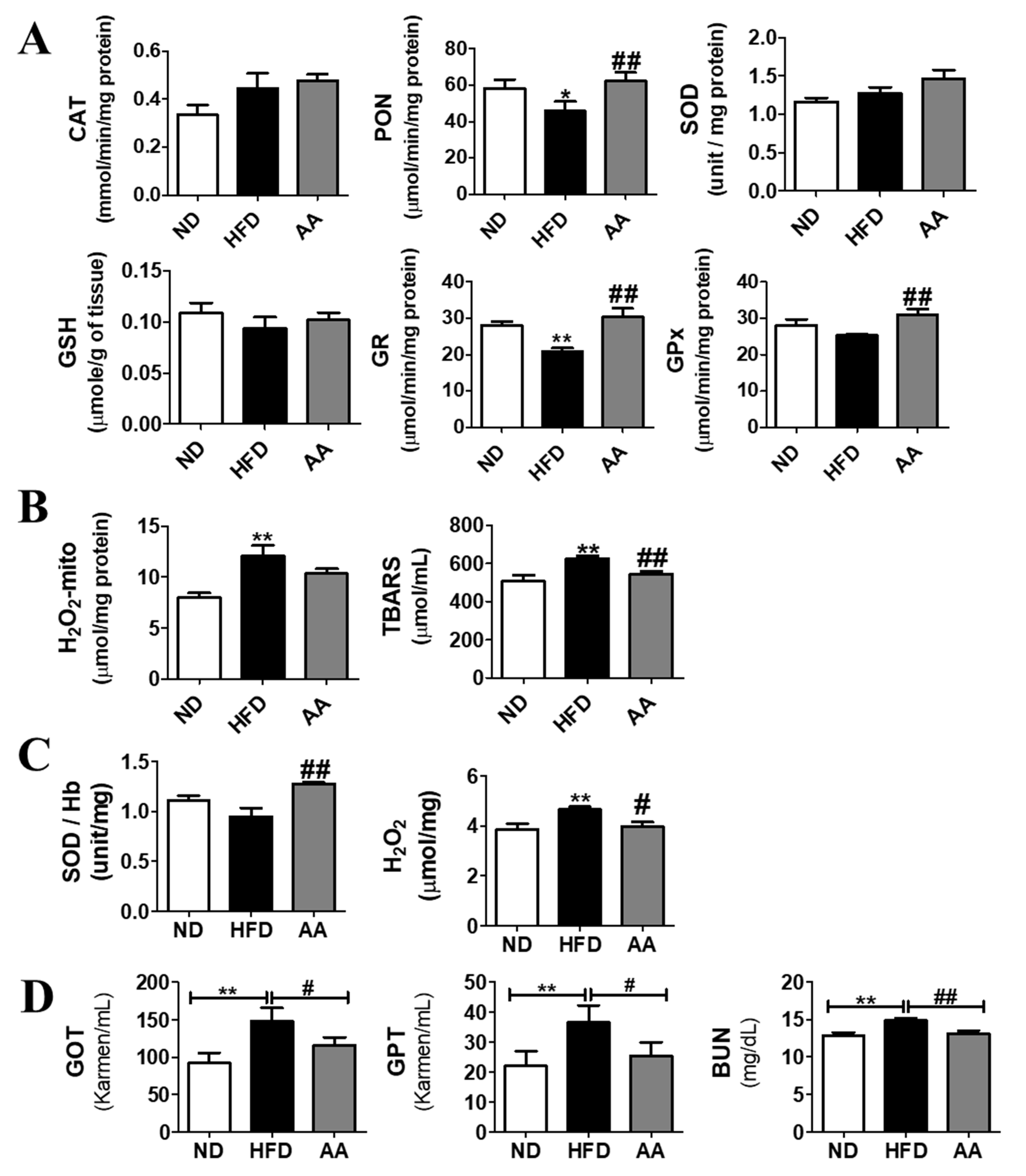
3.5. A. argyi Water Extract Supplementation Ameliorates Oxidative Stress

The hepatic antioxidant activity is shown in Figure 5A. Hepatic PON, GR, and GPx activities were significantly higher in the AA group than in the HFD group. In addition, hepatic TBARS levels were significantly lower in the AA group (Figure 5B). Furthermore, SOD activity was significantly increased and endogenous H2O2 levels in the erythrocytes were significantly decreased in the AA group than in the HFD group (Figure 5C). Plasma concentration of GOT, GPT, and BUN, which are an index of hepatic function, increased in the HFD group compared to the ND group; however, the AA supplement significantly decreased compared to the values of the HFD.

4. Discussion

This study investigated the effects of the A. argyi water extract on metabolic disorders caused by obesity and its complications. Our findings suggest that A. argyi supplementation reduces body weight and body fat without affecting food and energy intake. As shown in Figure 1, the food intake in the AA group was approximately 2.65g, of which the amount of A. argyi water extract intake per day was estimated to be 2.65 mg. This animal dose could be translated to a human dose. Based on previous study, the dose of A. argyi water extract for adults with a mean body weight of 60 kg was 158 mg, in order to be an effective dose of A. argyi water extract. This indicates that the A. argyi water extract might reduce body fat through metabolic regulation rather than appetite suppression. Excessive caloric intake and the availability of an energy-dense diet are the main contributors to obesity [31]. HFD has high energy density, thus elevating body weight owing to increased adiposity in various rodent models [32]. We found that body weight, body fat, and adipocyte size were dramatically increased in the HFD group; however, A. argyi water extract supplementation significantly reduced these biomarkers in such mice than in those fed only HFD. Furthermore, A. argyi water extract significantly increased the mRNA expression of CPT1b, COX8b, LIPE, and PNPLA2 in the epididymal WAT. Fatty acid oxidation inhibits lipid accumulation by inhibiting the re-esterification of fatty acids to TGs and by using acetyl-CoA from the β-oxidation of fatty acids as respiratory fuel [33]. The anti-adiposity effect of A. argyi water extract was supported by the diminished plasma leptin levels and L:A ratio in the present study, as adipokine levels are positively correlated with body fat [34]. Therefore, A. argyi water extract can suppress lipid accumulation via activated fatty acid oxidation in the epididymal WAT.
Our findings also suggest that A. argyi water extract ameliorates hyperglycemia and insulin resistance caused by HFD-induced obesity, which can be interpreted from two perspectives. First, A. argyi water extract ameliorated hepatic steatosis, which could in turn alleviate the impaired blood glucose regulation. Hepatic insulin resistance is a principal component of type 2 diabetes (T2DM) [35]. Impaired hepatic insulin sensitivity induces to increased hepatic gluconeogenesis, hyperinsulinemia, β-cell hypertrophy, and hyperglycemia [36]. Hepatic steatosis is a symptom in patients commonly with T2DM [37] and is closely associated with the long-term consumption of HFD [38]. In the present study, the HFD-fed mice showed significantly increased liver weights and hepatic lipid levels compared with the ND group, whereas they were decreased in the AA group compared with that in the HFD group. Based on previous studies, hepatic lipid accumulation has a positive correlation with liver weights [24,39]. Moreover, Hepatic H and E and Oil Red O staining showed that hepatic lipid accumulation was suppressed in the AA group than in the HFD group. We also observed that the AA group had a significantly decreased hepatic lipid content, as well as hepatic FAS and PAP activities, compared with the HFD group. Thus, A. argyi water extract exerts beneficial effects on the initiation and progression of hepatic steatosis. The marked improvements in hepatic steatosis were associated with the decreases in plasma glucose and insulin levels, which is a reflection of ameliorated hepatic insulin sensitivity [40], as evidenced by a reduced AUC for the IPGTT. Additionally, A. argyi water extract induced a decrease in hepatic lipid content, along with improved hepatic insulin sensitivity. Moreover, insulin suppressed the gluconeogenesis enzymes activities [41]. Thus, decreased gluconeogenesis and the results in decreased hepatic G6pase and PEPCK activities were suggested to be associated with the improved hepatic insulin sensitivity observed in A. argyi water extract-fed, diet-induced obese mice.
In the progression of T2DM, the low-grade inflammation-originated obesity hat is a principal pathophysiological factor related to hyperglycemia and insulin resistance [42,43]. Many studies have reported that various extracts of A. argyi possess antioxidant, antibacterial, and immunomodulatory properties [7,44,45]. A previous study suggested that A. argyi water extract plays a positive role in lipopolysaccharide-induced oxidative stress by restoring the activities of GPx and SOD, and preventing the increase in nitric oxide concentration caused by the over-activation of total nitric oxide synthase [46]. Consistent with these results, the present study showed that A. argyi water extract significantly increased hepatic PON, GR, and GPx activity and decreased hepatic TBARS levels. In addition, hemoglobin SOD activity was elevated, whereas endogenous H2O2 levels in the erythrocytes were decreased by A. argyi water extract supplementation. Furthermore, based on previous studies, a natural product supplement could induce hepatic injury through the interaction with the different cytochrome P-450 isoforms; inflammatory and oxidative activities seem to be the main damage pathway involved in the liver [47,48]. In the present study, A. argyi water extract significantly diminished the plasma GOT, GPT and BUN levels. These findings indicate that A. argyi water extract ameliorated the oxidative stress caused by obesity by regulating the enzymatic antioxidant system without any hepatic toxicity. In addition, the antioxidant property of A. argyi water extract suggests that it can improve glucose homeostasis in diet-induced obese mice.
A. argyi is rich in flavonoids, concluding flavones, flavonoids, flavonols, and chalcones [5]. Especially, it has various flavones including jaceosidin, eupatilin, luteolin, apigenin, and neaptin, and so on [49]. Eupatilin is pharmacologically active flavonoid and considered an index component as well as an active compound of A. argyi. Many studies supported that eupatilin isolated from A. argyi has great antioxidant and anti-inflammation properties in vitro and in vivo and inhibits adipogenesis in 3T3-L1 through the suppression of PPARγ expression [15,50,51,52]. Eupatilin is reported to be contained in A. argyi leaves of 0.46 to 1.22 mg/g [53]. Our experimental material, A. argyi water extract, has 0.82 ± 0.01 mg/ g eupatilin. These results suggested that water extraction of A. argyi could extract the active component of A. argyi. Although further studies are needed to elucidate the exact mode of action of eupatilin in detail, it is clear that A. argyi water extract including eupatilin can exert an inhibitory action on body fat accumulation and the initiation of hepatic steatosis or its progression.

5. Conclusions

A. argyi water extract supplementation has a significant effect on HFD-induced obesity and its complications. The A. argyi water extract improved adiposity, suppressed fatty liver caused by diet-induced obesity, and contributed to the recovery of impaired glucose homeostasis and ameliorated inflammation. Therefore, A. argyi water extract may be used as a functional food for the prevention of obesity and obesity-related diseases. This compound has no toxic effect in vivo; human trials are expected with the appropriate dosage in the near future.

Supplementary Materials

The following supporting information can be downloaded at: https://www.mdpi.com/article/10.3390/cimb44120420/s1, Table S1: Primer list for rt-PCR.

Author Contributions

M.-K.H., Y.H. and H.-J.P. performed the experiments, analyzed the data, M.-R.S. analyzed the data and wrote/edited the manuscript; S.-S.R. and E.-Y.K. supervised the work and had full access to all the data and, therefore, took full responsibility for the integrity of the results and accuracy of the data analysis. All authors have read and agreed to the published version of the manuscript.

Funding

This work was supported by the National Research Foundation of Korea (NRF) grant funded by the Korean Government (MSIT) (Nos. 2018R1A5A2025272, 2020R1I1A1A01067942 and 2020R1I1A3074694) and "Cooperative Research Program for Agriculture Science and Technology Development (Project No. PJ015272042022)" Rural Development Administration, Republic of Korea.

Institutional Review Board Statement

The animal protocols were approved by the Ethics Committee of Kyungpook National University on August 24 2022 (approval no. KNU-2020-0090).

Informed Consent Statement

Not applicable.

Data Availability Statement

Not applicable.

Acknowledgments

We wish to thank all individuals who kindly participated in this study.

Conflicts of Interest

The authors declare no conflict of interest.

References

  1. Scherer, F.M. The pharmaceutical industry. Handb. Health Econ. 2000, 1, 1297–1336. [Google Scholar]
  2. Parganiha, R.; Tripathi, A.K.; Prathyusha, S.; Baghel, P.; Lanjhiyana, S.; Lanjhiyana, S.; Katiyar, D.; Tyagi, S.; Sharma, P.P.; Sarkar, D. A Review of Plants for Hepatic Disorders. J. Complement. Med. Res. 2022, 13, 46. [Google Scholar] [CrossRef]
  3. Nayak, Y.; Veerapur, V.P.; Nagappa, A.N.; Unnikrishnan, M. Perspectives in efficacy, safety and clinical evaluation of bioactive natural products. In Compendium of Bioactive Natural Products, M/S; Studium Press LLC: Huston, Texas, USA, 2009; Volume 2. [Google Scholar]
  4. Mahmood, A.R.; Abdullah, M.R. The effect of active compounds and trace elements extracted from Artemisia fruit on some liver enzymes in humans. Arch. Razi Inst. 2022. [Google Scholar] [CrossRef]
  5. Song, X.; Wen, X.; He, J.; Zhao, H.; Li, S.; Wang, M. Phytochemical components and biological activities of Artemisia argyi. J. Funct. Foods 2019, 52, 648–662. [Google Scholar] [CrossRef]
  6. Yang, S.; Zhang, J.; Jiang, Y.; Xu, Y.Q.; Jin, X.; Yan, S.M.; Shi, B.L. Effects of Artemisia argyi flavonoids on growth performance and immune function in broilers challenged with lipopolysaccharide. Anim. Biosci. 2021, 34, 1169. [Google Scholar] [CrossRef]
  7. Zhang, P.; Shi, B.; Li, T.; Xu, Y.; Jin, X.; Guo, X.; Yan, S. Immunomodulatory effect of Artemisia argyi polysaccharide on peripheral blood leucocyte of broiler chickens. J. Anim. Physiol. Anim. Nutr. 2018, 102, 939–946. [Google Scholar] [CrossRef]
  8. Yun, C.; Jung, Y.; Chun, W.; Yang, B.; Ryu, J.; Lim, C.; Kim, J.-H.; Kim, H.; Cho, S.-I. Anti-inflammatory effects of Artemisia leaf extract in mice with contact dermatitis in vitro and in vivo. Mediat. Inflamm. 2016, 2016, 8027537. [Google Scholar] [CrossRef] [Green Version]
  9. Kim, J.-Y.; Lee, M.S.; Baek, J.M.; Park, J.; Youn, B.-S.; Oh, J. Massive elimination of multinucleated osteoclasts by eupatilin is due to dual inhibition of transcription and cytoskeletal rearrangement. Bone Rep. 2015, 3, 83–94. [Google Scholar] [CrossRef] [PubMed] [Green Version]
  10. Kim, J.K.; Shin, E.-C.; Lim, H.-J.; Choi, S.J.; Kim, C.R.; Suh, S.H.; Kim, C.-J.; Park, G.G.; Park, C.-S.; Kim, H.K. Characterization of nutritional composition, antioxidative capacity, and sensory attributes of Seomae mugwort, a native Korean variety of Artemisia argyi H. Lev. & Vaniot. J. Anal. Methods Chem. 2015, 2015, 916346. [Google Scholar]
  11. Zeng, K.-W.; Wang, S.; Dong, X.; Jiang, Y.; Tu, P.-F. Sesquiterpene dimer (DSF-52) from Artemisia argyi inhibits microglia-mediated neuroinflammation via suppression of NF-κB, JNK/p38 MAPKs and Jak2/Stat3 signaling pathways. Phytomedicine 2014, 21, 298–306. [Google Scholar] [CrossRef]
  12. Rama, P.; Vignesh, A.; Lakshmanan, G.; Murugesan, K. In vitro antioxidant activity of Achyranthes aspera Linn. Int. J. Med. Pharm. Sci. 2013, 3, 67–78. [Google Scholar]
  13. Jiao, H.; Wang, S.Y. Correlation of antioxidant capacities to oxygen radical scavenging enzyme activities in blackberry. J. Agric. Food Chem. 2000, 48, 5672–5676. [Google Scholar] [CrossRef]
  14. Liu, R.; Zhao, J.; He, K.; Zhang, X.; Chang, L.; Xiang, G. Determination of Eupatilin in Folium artemisiae Argyi and its inhibitory effect on hepatoma cells. Pharmacogn. Mag. 2018, 14, 129. [Google Scholar] [PubMed]
  15. Li, S.; Zhou, S.; Yang, W.; Meng, D. Gastro-protective effect of edible plant Artemisia argyi in ethanol-induced rats via normalizing inflammatory responses and oxidative stress. J. Ethnopharmacol. 2018, 214, 207–217. [Google Scholar] [CrossRef] [PubMed]
  16. Shin, N.-R.; Ryu, H.-W.; Ko, J.-W.; Park, S.-H.; Yuk, H.-J.; Kim, H.-J.; Kim, J.-C.; Jeong, S.-H.; Shin, I.-S. Artemisia argyi attenuates airway inflammation in ovalbumin-induced asthmatic animals. J. Ethnopharmacol. 2017, 209, 108–115. [Google Scholar] [CrossRef]
  17. Shin, N.-R.; Park, S.-H.; Ko, J.-W.; Ryu, H.-W.; Jeong, S.-H.; Kim, J.-C.; Shin, D.-H.; Lee, H.-S.; Shin, I.-S. Artemisia argyi attenuates airway inflammation in lipopolysaccharide induced acute lung injury model. Lab. Anim. Res. 2017, 33, 209–215. [Google Scholar] [CrossRef] [Green Version]
  18. Folch, J.; Lees, M.; Sloane Stanley, G.H. A simple method for the isolation and purification of total lipids from animal tissues. J. Biol. Chem. 1957, 226, 497–509. [Google Scholar] [CrossRef]
  19. Fischer, A.H.; Jacobson, K.A.; Rose, J.; Zeller, R. Hematoxylin and eosin staining of tissue and cell sections. Cold Spring Harb. Protoc. 2008, 2008, pdb.prot4986. [Google Scholar] [CrossRef]
  20. Ramirez-Zacarias, J.; Castro-Munozledo, F.; Kuri-Harcuch, W. Quantitation of adipose conversion and triglycerides by staining intracytoplasmic lipids with Oil red O. Histochemistry 1992, 97, 493–497. [Google Scholar] [CrossRef] [PubMed]
  21. Hu, Y.; Yu, J.; Cui, X.; Zhang, Z.; Li, Q.; Guo, W.; Zhao, C.; Chen, X.; Meng, M.; Li, Y. Combination usage of adipocount and image-pro plus/imagej software for quantification of adipocyte sizes. Front. Endocrinol. 2021, 12, 642000. [Google Scholar] [CrossRef] [PubMed]
  22. Mehlem, A.; Hagberg, C.E.; Muhl, L.; Eriksson, U.; Falkevall, A. Imaging of neutral lipids by oil red O for analyzing the metabolic status in health and disease. Nat. Protoc. 2013, 8, 1149–1154. [Google Scholar] [CrossRef] [PubMed] [Green Version]
  23. Matthews, D.R.; Hosker, J.; Rudenski, A.; Naylor, B.; Treacher, D.; Turner, R. Homeostasis model assessment: Insulin resistance and β-cell function from fasting plasma glucose and insulin concentrations in man. Diabetologia 1985, 28, 412–419. [Google Scholar] [CrossRef] [PubMed] [Green Version]
  24. Han, Y.; Yoon, J.; Choi, M.S. Tracing the Anti-Inflammatory Mechanism/Triggers of d-Allulose: A Profile Study of Microbiome Composition and mRNA Expression in Diet-Induced Obese Mice. Mol. Nutr. Food Res. 2020, 64, 1900982. [Google Scholar] [CrossRef]
  25. Hulcher, F.H.; Oleson, W.H. Simplified spectrophotometric assay for microsomal 3-hydroxy-3-methylglutaryl CoA reductase by measurement of coenzyme A. J. Lipid Res. 1973, 14, 625–631. [Google Scholar] [CrossRef] [PubMed]
  26. Nepokroeff, C.M.; Lakshmanan, M.; Porter, J.W. Fatty acid synthase from rat liver. In Methods in Enzymology; Elsevier: Amsterdam, The Netherlands, 1975; Volume 35, pp. 37–44. [Google Scholar]
  27. Rudack, D.; Chisholm, E.M.; Holten, D. Rat liver glucose 6-phosphate dehydrogenase: Regulation by carbohydrate diet and insulin. J. Biol. Chem. 1971, 246, 1249–1254. [Google Scholar] [CrossRef]
  28. Ochoa, S. Malic enzyme: Malic enzymes from pigeon and wheat germ. Methods Enzymol. 1955, 1, 323–326. [Google Scholar]
  29. Markwell, M.A.K.; McGroarty, E.J.; Bieber, L.L.; Tolbert, N. The subcellular distribution of carnitine acyltransferases in mammalian liver and kidney: A new peroxisomal enzyme. J. Biol. Chem. 1973, 248, 3426–3432. [Google Scholar] [CrossRef]
  30. Lazarow, P.B. [19] Assay of peroxisomal β-oxidation of fatty acids. In Methods in Enzymology; Elsevier: Amsterdam, The Netherlands, 1981; Volume 72, pp. 315–319. [Google Scholar]
  31. Drewnowski, A.; Specter, S.E. Poverty and obesity: The role of energy density and energy costs. Am. J. Clin. Nutr. 2004, 79, 6–16. [Google Scholar] [CrossRef] [Green Version]
  32. Coccurello, R.; Romano, A.; Giacovazzo, G.; Tempesta, B.; Fiore, M.; Giudetti, A.M.; Marrocco, I.; Altieri, F.; Moles, A.; Gaetani, S. Increased intake of energy-dense diet and negative energy balance in a mouse model of chronic psychosocial defeat. Eur. J. Nutr. 2018, 57, 1485–1498. [Google Scholar] [CrossRef]
  33. Jaswal, J.S.; Keung, W.; Wang, W.; Ussher, J.R.; Lopaschuk, G.D. Targeting fatty acid and carbohydrate oxidation—A novel therapeutic intervention in the ischemic and failing heart. Biochim. Et Biophys. Acta (BBA)-Mol. Cell Res. 2011, 1813, 1333–1350. [Google Scholar] [CrossRef] [Green Version]
  34. Lubkowska, A.; Radecka, A.; Bryczkowska, I.; Rotter, I.; Laszczyńska, M.; Dudzińska, W. Serum adiponectin and leptin concentrations in relation to body fat distribution, hematological indices and lipid profile in humans. Int. J. Environ. Res. Public Health 2015, 12, 11528–11548. [Google Scholar] [CrossRef] [PubMed] [Green Version]
  35. Savage, D.B.; Petersen, K.F.; Shulman, G.I. Disordered lipid metabolism and the pathogenesis of insulin resistance. Physiol. Rev. 2007, 87, 507–520. [Google Scholar] [CrossRef]
  36. Michael, M.D.; Kulkarni, R.N.; Postic, C.; Previs, S.F.; Shulman, G.I.; Magnuson, M.A.; Kahn, C.R. Loss of insulin signaling in hepatocytes leads to severe insulin resistance and progressive hepatic dysfunction. Mol. Cell 2000, 6, 87–97. [Google Scholar] [CrossRef] [PubMed]
  37. Collaboration, E.R.F. Diabetes mellitus, fasting glucose, and risk of cause-specific death. N. Engl. J. Med. 2011, 364, 829–841. [Google Scholar] [CrossRef] [PubMed]
  38. Bellentani, S.; Marino, M. Epidemiology and natural history of non-alcoholic fatty liver disease (NAFLD). Ann. Hepatol. 2009, 8, S4–S8. [Google Scholar] [CrossRef]
  39. Han, Y.; Park, H.; Choi, B.-R.; Ji, Y.; Kwon, E.-Y.; Choi, M.-S. Alteration of microbiome profile by d-allulose in amelioration of high-fat-diet-induced obesity in mice. Nutrients 2020, 12, 352. [Google Scholar] [CrossRef] [PubMed] [Green Version]
  40. Yu, X.X.; Pandey, S.K.; Booten, S.L.; Murray, S.F.; Monia, B.P.; Bhanot, S. Reduced adiposity and improved insulin sensitivity in obese mice with antisense suppression of 4E-BP2 expression. Am. J. Physiol.-Endocrinol. Metab. 2008, 294, E530–E539. [Google Scholar] [CrossRef] [Green Version]
  41. Hatting, M.; Tavares, C.D.; Sharabi, K.; Rines, A.K.; Puigserver, P. Insulin regulation of gluconeogenesis. Ann. N. Y. Acad. Sci. 2018, 1411, 21–35. [Google Scholar] [CrossRef]
  42. Lontchi-Yimagou, E.; Sobngwi, E.; Matsha, T.E.; Kengne, A.P. Diabetes mellitus and inflammation. Curr. Diabetes Rep. 2013, 13, 435–444. [Google Scholar] [CrossRef] [PubMed]
  43. Hotamisligil, G.S. Inflammation and metabolic disorders. Nature 2006, 444, 860–867. [Google Scholar] [CrossRef] [PubMed]
  44. Xiang, F.; Bai, J.; Tan, X.; Chen, T.; Yang, W.; He, F. Antimicrobial activities and mechanism of the essential oil from Artemisia argyi Levl. et Van. var. argyi cv. Qiai. Ind. Crops Prod. 2018, 125, 582–587. [Google Scholar] [CrossRef]
  45. Huang, H.-C.; Wang, H.-F.; Yih, K.-H.; Chang, L.-Z.; Chang, T.-M. Dual bioactivities of essential oil extracted from the leaves of Artemisia argyi as an antimelanogenic versus antioxidant agent and chemical composition analysis by GC/MS. Int. J. Mol. Sci. 2012, 13, 14679–14697. [Google Scholar] [CrossRef] [Green Version]
  46. Ma, Q.; Wei, Y.; Meng, Z.; Chen, Y.; Zhao, G. Effects of Water Extract from Artemisia argyi Leaves on LPS-Induced Mastitis in Mice. Animals 2022, 12, 907. [Google Scholar] [CrossRef] [PubMed]
  47. Lucena, M.I.; Salmerón, J.; Planas, R.; de Catalunya, G.; Andrade, R.J. Liver injury induced by “natural remedies”: An analysis of cases submitted to the Spanish Liver Toxicity Registry. Rev. Esp. Enferm. Dig. 2008, 100, 688–695. [Google Scholar]
  48. López-Gil, S.; Nuño-Lámbarri, N.; Chávez-Tapia, N.; Uribe, M.; Barbero-Becerra, V.J. Liver toxicity mechanisms of herbs commonly used in Latin America. Drug Metab. Rev. 2017, 49, 338–356. [Google Scholar] [CrossRef]
  49. Nakasugi, T.; Nakashima, M.; Komai, K. Antimutagens in Gaiyou (Artemisia a rgyi Levl. et Vant.). J. Agric. Food Chem. 2000, 48, 3256–3266. [Google Scholar] [CrossRef] [PubMed]
  50. Min, S.-W.; Kim, N.-J.; Baek, N.-I.; Kim, D.-H. Inhibitory effect of eupatilin and jaceosidin isolated from Artemisia princeps on carrageenan-induced inflammation in mice. J. Ethnopharmacol. 2009, 125, 497–500. [Google Scholar] [CrossRef]
  51. Lim, J.C.; Park, S.Y.; Nam, Y.; Nguyen, T.T.; Sohn, U.D. The protective effect of eupatilin against hydrogen peroxide-induced injury involving 5-lipoxygenase in feline esophageal epithelial cells. Korean J. Physiol. Pharmacol. 2012, 16, 313–320. [Google Scholar] [CrossRef] [Green Version]
  52. Kim, J.S.; Lee, S.G.; Min, K.; Kwon, T.K.; Kim, H.-J.; Nam, J.-O. Eupatilin inhibits adipogenesis through suppression of PPARγ activity in 3T3-L1 cells. Biomed. Pharmacother. 2018, 103, 135–139. [Google Scholar] [CrossRef]
  53. Guo, L.; Jiao, Q.; Zhang, D.; Liu, A.-P.; Wang, Q.; Zheng, Y.-G. Quality evaluation of Artemisiae Argyi Folium based on fingerprint analysis and quantitative analysis of multicomponents. Zhongguo Zhong Yao Za Zhi/Zhongguo Zhongyao Zazhi/China J. Chin. Mater. Med. 2018, 43, 977–984. [Google Scholar]
Figure 1. Effect of Artemisiae argyi water extract supplementation on diet-induced obesity. (A) Body weight and body weight gain; (B) food intake, energy intake, and FER; (C) adipose tissue weight; and (D) adipocyte morphology of the study groups. Data are presented as the mean ± standard error of the mean. Significant differences between HFD and ND are indicated as ** p < 0.01. Significant differences between the HFD and AA groups are indicated as # p < 0.05, ## p < 0.01. ND, normal diet (AIN-93G, n = 10); HFD, high-fat diet (60% kcal from fat, n = 10); AA, HFD + A. argyi water extract (1.5%, w/w, n= 10); BWG, body weight gain; FER, food efficiency ratio: body weight gain/energy intake per day; WAT, white adipose tissue.
Cimb 44 00420 g001
Figure 2. Effect of Artemisiae argyi water extract supplementation on diet-induced obesity. (A) Plasma lipid profiles, (B) plasma adipokine levels, and (C) mRNA expression of the epididymal WAT. Data are presented as the mean ± standard error of the mean. Significant differences between HFD and ND are indicated as * p < 0.05, ** p < 0.01. Significant differences between the HFD and AA groups are indicated as # p < 0.05, ## p < 0.01. ND, normal diet (AIN-93G, n = 10); HFD, high-fat diet (60% kcal from fat, n = 10); AA, HFD + A. argyi water extract (1.5%, w/w, n = 10); TG, triglyceride; FFA, free fatty acid; TC, total cholesterol; HDL-C, high density lipoprotein cholesterol; Apo A1, apolipoprotein A-1; Apo B apolipoprotein B; Apo A1/Apo B, Apo A1 and Apo B ratio; L:A, leptin and adiponectin ratio; PPARa, peroxisome proliferator activated receptor alpha; CPT1A, carnitine palmitoyltransferase 1A; CPT2, carnitine palmitoyltransferase 2; COX8b, cytochrome c oxidase subunit 8B; UCP1, uncoupling Protein 1; LIPE, hormone-sensitive lipase; PNPLA2, patatin-like phospholipase domain containing 2.
Cimb 44 00420 g002
Figure 3. Effect of Artemisiae argyi water extract supplementation on diet-induced obesity. (A) Fasting blood glucose level; (B) intraperitoneal glucose tolerance test and area under the curve; (C) activities of hepatic enzymes related to glucose metabolism; (D) plasma glucose and insulin levels and homeostatic index of insulin resistance; and (E) hepatic mRNA expression of genes related to glucose metabolism. Data are presented as the mean ± standard error of the mean. Significant differences between HFD versus ND are indicated as * p < 0.05, ** p < 0.01. Significant differences between the HFD and AA groups are indicated as # p < 0.05, ## p < 0.01. ND, normal diet (AIN-93G, n = 10); HFD, high-fat diet (60% kcal from fat, n = 10); AA, HFD + A. argyi water extract (1.5%, w/w, n = 10); IPGTT, intraperitoneal glucose tolerance test; FBG, fasting blood glucose; AUC, area under the curve; GK, glucokinase; PEPCK, phosphoenolpyruvate carboxykinase; G6Pase, glucose 6-phosphate; HOMA-IR, homeostatic index of insulin resistance; PRKAA2, 5'-AMP-activated protein kinase subunit alpha-2; PRKAB1, 5'-AMP-activated protein kinase subunit beta-1; PRKAG1, 5'-AMP-activated protein kinase subunit gamma-1; PRKAG2, 5'-AMP-activated protein kinase subunit gamma-2; GLUT2, glucose transporter 2; GCK, glucokinase; PFKL, 6-phosphofructokinase, liver type; HK3, hexokinase 3; PDHB, pyruvate dehydrogenase (lipoamide) beta; PKM2, enzyme pyruvate kinase M2; G6PC, glucose-6-phosphatase, catalytic subunit; GAPDH, glyceraldehyde-3-phosphate dehydrogenase; PEPCK, phosphoenolpyruvate carboxykinase; CREB, cAMP response element-binding protein; CRTC2, CREB regulated transcription coactivator 2.
Cimb 44 00420 g003
Figure 4. Effect of Artemisiae argyi water extract supplementation on diet-induced obesity. (A) Liver weight, (B) hepatic lipid content, (C) hepatic morphology (200× magnification), and (D) activities of hepatic enzymes related to fatty acid synthesis. Data are presented as the mean ± standard error of the mean. Significant differences between HFD versus ND are indicated as * p < 0.05, ** p < 0.01. Significant differences between the HFD and AA groups are indicated as # p < 0.05, ## p < 0.01. ND, normal diet (AIN-93G, n = 10); HFD, high-fat diet (60% kcal from fat, n = 10); AA, HFD + A. argyi water extract (1.5%, w/w, n = 10); H and E, hematoxylin and eosin. TG, triglyceride; FAS, fatty acid synthase; ME, malic enzyme; PAP, phosphatidic acid phosphatase.
Cimb 44 00420 g004
Figure 5. Effect of Artemisiae argyi water extract supplementation on diet-induced obesity. (A) Activities of hepatic antioxidant enzymes, (B) hepatic H2O2 and TBARS levels, (C) erythrocyte SOD and H2O2 levels and (D) plasma liver function index. Data are presented as the mean ± standard error of the mean. Significant differences between HFD versus ND are indicated as * p < 0.05, ** p < 0.01. Significant differences between the HFD and AA groups are indicated as # p < 0.05, ## p < 0.01. ND, normal diet (AIN-93G, n = 10); HFD, high-fat diet (60% kcal from fat, n = 10); AA, HFD + A. argyi water extract (1.5%, w/w, n = 10). CAT, catalase; PON, paraoxonase; SOD, superoxide dismutase; TBARS, thiobarbituric acid reactive substances; GOT, glutamic oxaloacetic transaminase; GPT, glutamic oxaloacetic transaminase; BUN, Blood Urea Nitrogen.
Cimb 44 00420 g005
Table 1. Total phenolic, flavonoid, and eupatilin content in the Artemisiae argyi water extract.
Amount
Total polyphenolic content143.94 ± 1.20 (GAE)/g
Total flavonoid content23.42 ± 0.01 (QE)/g
Eupatilin content0.82 ± 0.01 (mg eupatilin/g)
Values represent the mean ± standard error of the mean; GAE, gallic acid equivalents; QE, quercetin equivalents.
Table 2. Diet composition for animal experiment.
Ingredient (g)ND
(AIN-93G)
HFD
(60 kcal% Fat)
AA
(0.1% A. argyi)
Casein200267267
Corn starch397.48663.38163.381
Sucrose10000
Dextrose132176176
Cellulose506767
Soybean oil703333
Lard0327327
Mineral mixture 1354747
Vitamin mixture 2101313
TBHQ, antioxidant0.0140.0190.019
L-cystine344
Choline bitartrate2.533
Artemisiae argyi--1
Total (g)1000.001000.001001.00
Calorie (kcal/g)40005332.625332.62
1 AIN-93G-mineral mixture (Harlan Teklad Co., Madison, WI, USA). 2 AIN-93G-vitamin mixture (Harlan Teklad Co., Madison, WI, USA). ND, normal diet (AIN-93G 16 kcal% fat); HFD, high-fat diet (60 kcal% fat); AA (Artemisiae argyi), HFD + A. argyi water extract 0.1%. TBHQ; tert-butylhydroquinone.
Publisher’s Note: MDPI stays neutral with regard to jurisdictional claims in published maps and institutional affiliations.

