Article

Resveratrol Analog 4-Bromo-Resveratrol Inhibits Gastric Cancer Stemness through the SIRT3-c-Jun N-Terminal Kinase Signaling Pathway

1
Department of Surgery, China Medical University, An-Nan Hospital, Tainan 70965, Taiwan
2
Department of Surgery, E-Da Hospital, Kaohsiung 82445, Taiwan
3
Institute of Medicine, Chung Shan Medical University, Taichung 40201, Taiwan
4
Department of Chinese Medicine, E-Da Hospital/E-Da Cancer Hospital/E-Da Dachang Hospital, Kaohsiung 82445, Taiwan
5
The School of Chinese Medicine for Post-Baccalaureate, I-Shou University, Kaohsiung 82445, Taiwan
6
Department of Biological Science, National Sun Yat-sen University, Kaohsiung 80424, Taiwan
7
Department of Medical Research, E-Da Hospital/E-Da Cancer Hospital, Kaohsiung 82445, Taiwan
8
Department of Internal Medicine, Kaohsiung Municipal Ta-Tung Hospital, Kaohsiung 80145, Taiwan
9
Department of Nursing, College of Medicine, I-Shou University, Kaohsiung 82445, Taiwan
*
Authors to whom correspondence should be addressed.
These authors contributed equally to this work.
Curr. Issues Mol. Biol. 2022, 44(1), 63-72; https://doi.org/10.3390/cimb44010005
Submission received: 23 November 2021 / Revised: 16 December 2021 / Accepted: 17 December 2021 / Published: 22 December 2021
(This article belongs to the Topic Cancer Biology and Therapy)

Abstract

:
Chemotherapy is the treatment of choice for gastric cancer, but the currently available therapeutic drugs have limited efficacy. Studies have suggested that gastric cancer stem cells may play a key role in drug resistance in chemotherapy. Therefore, new agents that selectively target gastric cancer stem cells in gastric tumors are urgently required. Sirtuin-3 (SIRT3) is a deacetylase that regulates mitochondrial metabolic homeostasis to maintain stemness in glioma stem cells. Targeting the mitochondrial protein SIRT3 may provide a novel therapeutic option for gastric cancer treatment. However, the mechanism by which stemness is regulated through SIRT3 inhibition in gastric cancer remains unknown. We evaluated the stemness inhibition ability of the SIRT3 inhibitor 4′-bromo-resveratrol (4-BR), an analog of resveratrol in human gastric cancer cells. Our results suggested that 4-BR inhibited gastric cancer cell stemness through the SIRT3-c-Jun N-terminal kinase pathway and may aid in gastric cancer stem-cell–targeted therapy.

1. Introduction

Gastric cancer is the most common cause of malignancy and cancer-related mortalities worldwide [1]. Systemic chemotherapy with multiple drugs may be an effective strategy for patients with recurrent gastric cancer. Numerous studies have reported that chemotherapeutic resistance in gastric solid tumors results from genetic heterogeneity in tumor cells. Studies have suggested that cancer stem cells (CSCs) contribute to chemotherapy drug resistance [2]. CSCs are a subpopulation of tumor masses with the ability to self-renew, maintain stemness, and cause cancer recurrence [3]. CSCs may be a novel target for anticancer strategies and gastric cancer therapy in precision medicine [2].
CSCs in gastric cancer were first described in 2007 [4]. The most common way to identify gastric CSCs is through investigating the expression of cell surface markers, including CD24 and LGR5. CD24 is a cell surface protein that is expressed in putative stem cells and is overexpressed in various malignant tumors. It was reported to stimulate metastasis and trigger epithelial-to-mesenchymal transition through Notch1 signaling [5]. Leucine-rich repeat-containing G-protein coupled Receptor 5 (LGR5) is overexpressed in stem cell after Helicobacter pylori infection in the gastrointestinal tract [6], which is reported to be able to initiate tumors [7,8], and has been defined as a gastric cancer stem cell marker recently [9,10]. Nanog is a transcription factor widely expressed in human cancers [11,12,13]. It drives self-renewal, metastasis, and chemoresistance [14,15]. The transcription factor Sox2 is involved in the maintenance of an undifferentiated cellular phenotype [16]. Its aberrant expression in cancers often leads to increased chemotherapy resistance [17]. Oct-4 is also a transcriptional factor to regulate pluripotency in the mammalian stem cell population. It is upregulated in several cancers and promotes chemoresistance, metastasis, and recurrence [18,19,20]. Overexpression of these transcription factors, Nanog and Oct-4, promoted the formation of tumor spheroids in vitro [21], and correlated to a poor prognosis of the patient [22,23,24]. The other functional marker, aldehyde dehydrogenase 1 (ALDH1) is widely used to characterize cancer stemness in many studies [25,26,27]. ALDH1 is a detoxifying enzyme responsible for oxidation. Elevation of its activity may protect CSCs against cell death caused by ROS [28]. Increased ALDH1 activity has been found in malignant cancer cells, and served as an indicator for poor prognosis [29].
Sirtuin-3 (SIRT3) is a deacetylase in mitochondria. SIRT3 regulates the mitochondrial respiratory capacity and reduces production of reactive oxygen species, facilitating adaptation to stress. In glioma stem cells, SIRT3 was reported to regulate glioma CSCs with metabolic plasticity and maintain cancer stemness [30]. Its enzyme activity increases chemoresistance. Inactivation of SIRT3 leads to metabolic alterations, loss of stemness, and suppression of tumor formation by CSCs [31]. Ma et al. demonstrated that SIRT3 contributes to chemoresistance through mitochondrial metabolism reprograming and reactive oxygen species downgeneration [32]. SIRT3 is associated with enhanced gastric cancer risk and can thus be a potential prognostic marker [33]. However, the inhibition of SIRT3 activity involved in gastric cancer stemness and chemoresistance has not been thoroughly explored.
The resveratrol analog 4′-bromo-resveratrol (4-BR) has been reported to inhibit SIRT3 activity considerably [34] and suppress melanoma cell growth through mitochondrial metabolic regulation [35]. We evaluated the effects of 4-BR on cancer stemness and chemoresistance inhibition in gastric cancer cells. We postulated that 4-BR would use the SIRT3-mediated pathway to downregulate gastric cancer stemness and increase chemosensitivity.

2. Materials and Methods

2.1. Cell Culture and Reagents

Human gastric cancer cell lines MKN45 (DSMZ; ACC-409) and AGS (ATCC; CRL-1739) were purchased from DSMZ (Braunschweig, Germany) and ATCC (Manassas, VA, USA), respectively, and cultured using RPMI1640 (Gibco, Waltham, MA, USA) medium with 10% fetal bovine serum (Gibco, Waltham, MA, USA) under 5% CO2 at 37 °C. The 4-BR, SP600125, and 5-fluorouracil (5-FU) (Sigma-Aldrich) were purchased from Sigma-Aldrich (St. Louis, MO, USA).

2.2. Cell Viability Analysis

Human gastric cancer cells were seeded in 96-well dishes in quadruplicate at 6000 cells/well and cultured for 24 h before treatment. The cell viability of AGS or MKN45 was evaluated with 5-FU (0.5 μM), 4-BR (25 μM), or a combination of 5-FU and 4-BR for 48 h. Cell viability was analyzed using Cell Counting Kit-8 (Sigma–Aldrich, St. Louis, MO, USA), and absorbance was measured at 450 nm by using a microplate reader.

2.3. Flow Cytometry Analysis

After treatment, the cells were washed with cold phosphate buffer saline and stained with surface marker CD24 (BD Biosciences, San Jose, CA, USA) before being analyzed through flow cytometry. For the ALDH1 activity evaluation, the cells were stained using the AldeRed ALDH Detection Assay kit (Sigma–Aldrich, St. Louis, MO, USA) and analyzed through flow cytometry.

2.4. Western Blotting Analysis

The protein expressions of MKN45 were evaluated through Western blotting after 4-BR (vehicle or 12.5 μM or 25 μM) or JNK inhibitor SP600125 (20 μM) treatment for 48 h. After 4-BR or SP600125 treatment, the human gastric cancer cells were washed with phosphate buffer saline. The total protein samples were extracted, and protein concentrations were measured using Bio-Rad Bradford Protein Assays (Bio-Rad, Hercules, CA, USA). Total protein was separated using sodium dodecyl sulfate–polyacrylamide gel electrophoresis and transferred onto polyvinylidene fluoride membranes. The membranes were incubated with blocking buffer (Bio-Rad, Hercules, CA, USA) for 30 min at room temperature and with primary antibodies: SOX2 (1:1000; #23064; Cell Signaling, Beverly, MA, USA), OCT4(1:1000; #2750; Cell Signaling, Beverly, MA, USA), β-actin (1:5000; #4967; Cell Signaling, Beverly, MA, USA), Notch1 (1:1000; #3608; Cell signaling, Beverly, MA, USA), Sirt3(1:1000; #5490; Cell Signaling, Beverly, MA, USA), JNK (1:1000; #9252; Cell Signaling, Beverly, MA, USA), and p-JNK (1:1000; #9251; Cell Signaling, Beverly, MA, USA) at 4 °C overnight. After washing with phosphate-buffered saline with Tween, the membranes were developed using an enhanced chemiluminescence detection system under MultiGel-21 system (TOP BIO Co., New Taipei City, Taiwan) after incubation with secondary antibodies.

2.5. Soft Agar Colony Formation Analysis

For the colony formation assay, MKN45 cells (2.5 × 10⁴ cells) were seeded in 6-well dishes and coated in 0.3% agar. After 2 weeks, the colonies were stained with 0.005% crystal violet, and the area was measured using ImageJ.

2.6. Sphere Formation Analysis

For the sphere formation analysis, MKN45 cells (1 × 10⁴ cells) were seeded in a NanoCulture Plate (SCIVAX, Kanagawa, Japan) and incubated with 4-BR for 7 days; the spheres were evaluated using ImageJ.

2.7. Statistical Analysis

All data were analyzed using GraphPad Prism (version 8). All graphs in the figures present mean ± standard deviations. For statistical analysis, Student’s t test was performed to compare data between the two groups. Statistical significance was set at p < 0.05. Statistical results were labeled in each figure as follows: * p < 0.05, ** p < 0.01, and *** p < 0.001.

3. Results

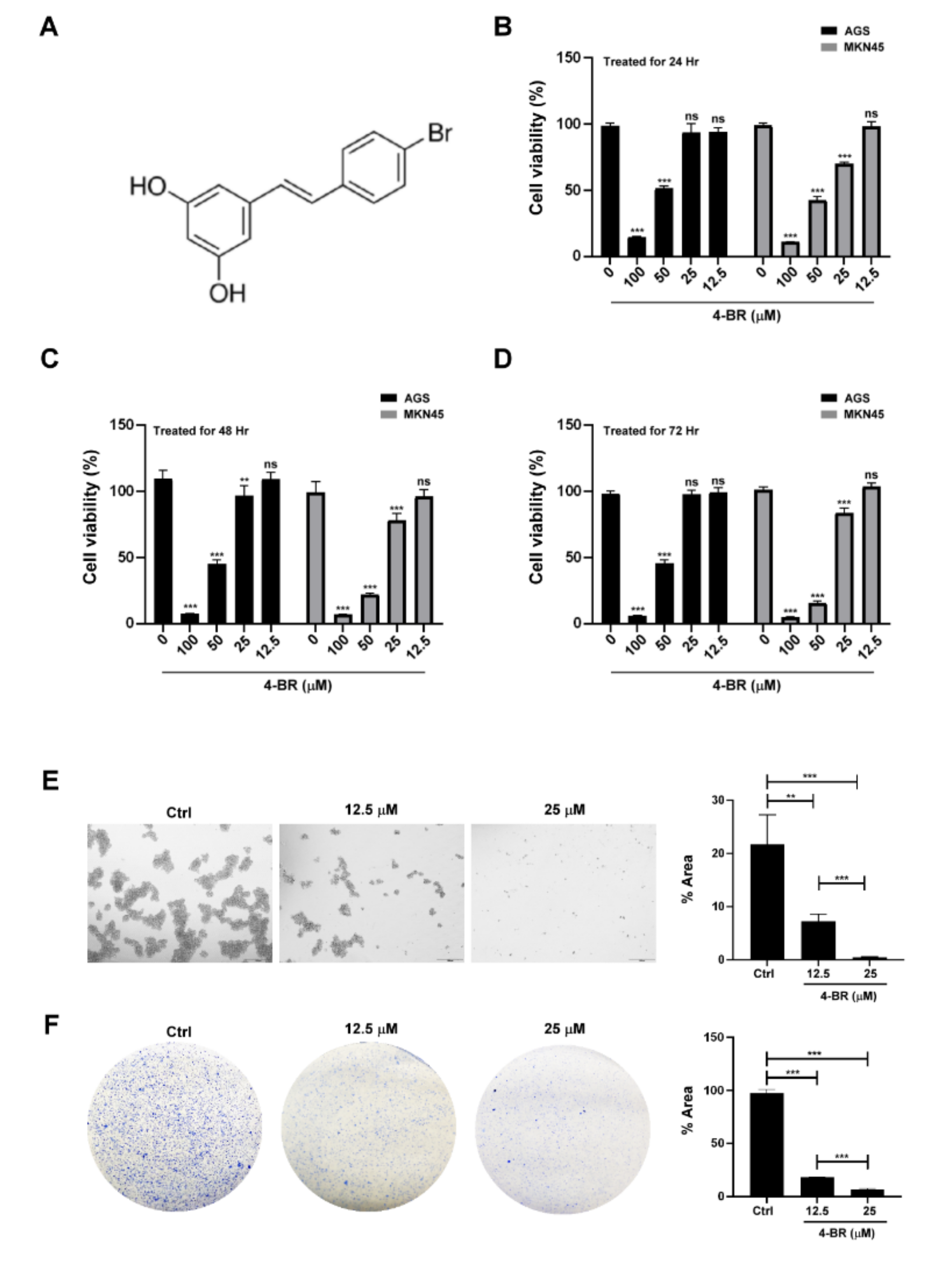
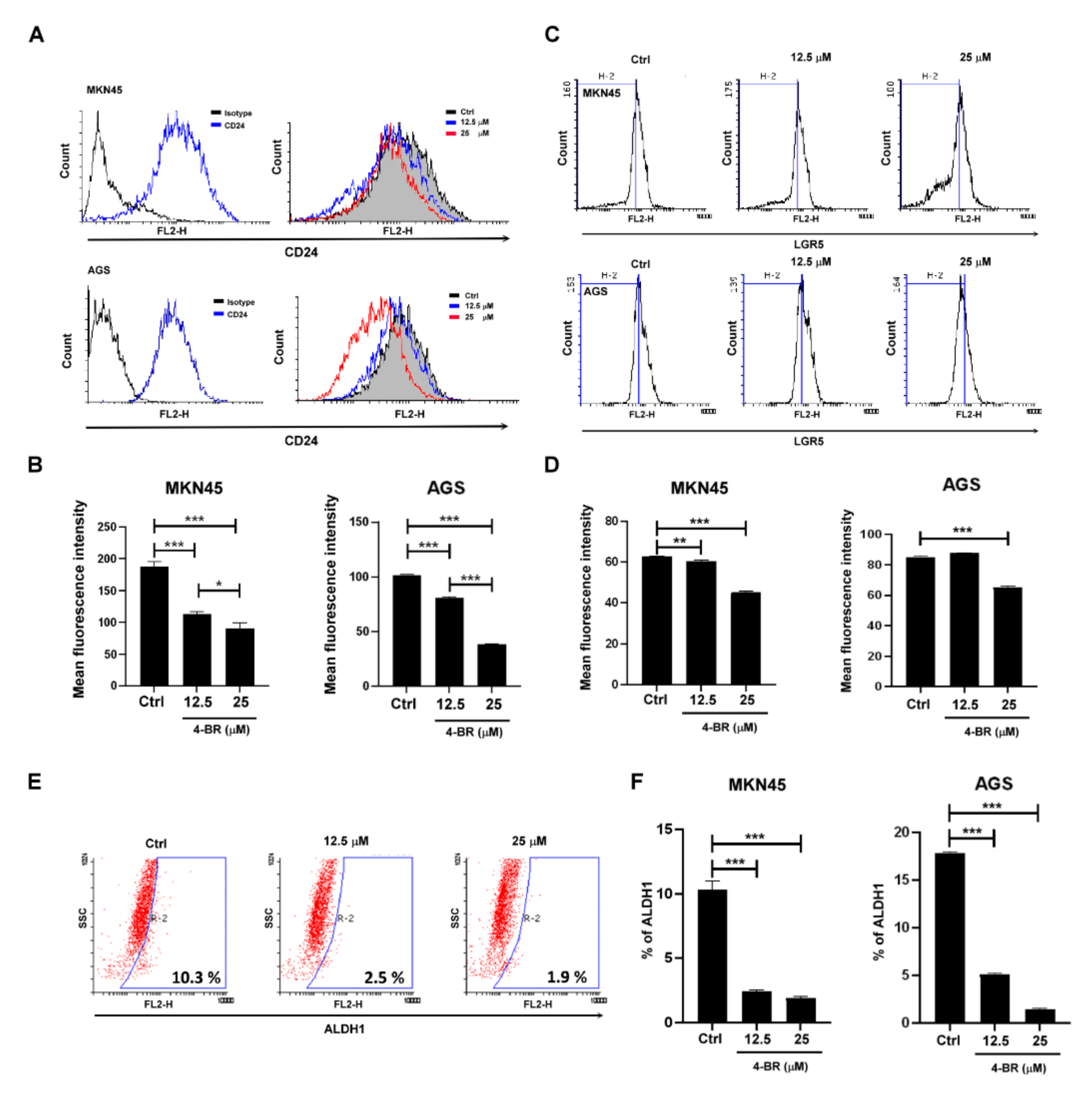
3.1. 4-BR Reduced Gastric Cancer Viability and Cancer Stemness Capacity

To evaluate the cytotoxicity of 4-BR to human gastric cancer cells, the viability of MKN45 and AGS was evaluated through a Cell Counting Kit 8 assay after the treatment of various doses of 4-BR. The results indicated that 4-BR inhibited gastric cancer growth in a dose-dependent manner (Figure 1B–D). The stemness properties of the human gastric cancer cells were then evaluated using a sphere formation and colony formation assay after the low-cytotoxicity 4-BR (25 μM) treatment. The results indicated that 4-BR inhibited the stemness properties of the MKN45 cells (Figure 1E,F).

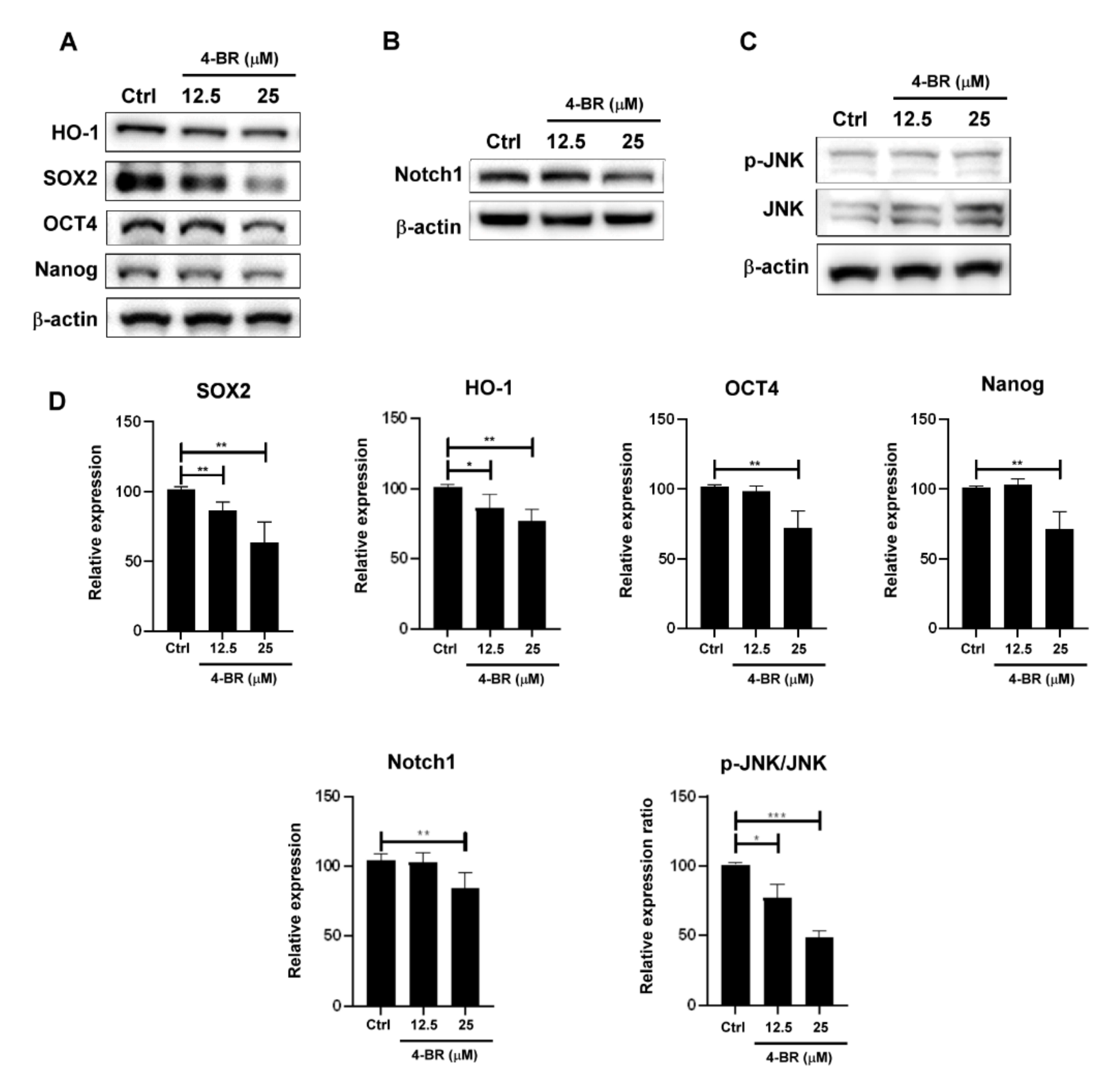
3.2. 4-BR Downregulated Stemness-Related Protein Expressions in Human Gastric Cancer Cells

To evaluate stemness-related protein expression, CD24, LGR5, and ALDH1 activity was measured through flow cytometry (Figure 2), and HO-1, SOX2, Nanog, and Notch1 were evaluated using Western blotting (Figure 3A,B) after a 4-BR (25 μM) treatment for 48 h in MKN45. Activated c-Jun N-terminal kinase (JNK) has been reported to regulate Notch1 expression and the self-renewal of stem cells [36,37]. The results indicated that the 4-BR treatment reduced the expression of markers related to gastric cancer stemness, including Notch, resulting in JNK dephosphorylation and reduced downstream signaling.

3.3. 4-BR Contributed to JNK-Mediated Gastric Cancer Stemness Inhibition and Increased Chemosensitivity to 5-FU

To determine whether 4-BR contributed to JNK-mediated gastric cancer stemness inhibition, we used the JNK inhibitor SP600125 to evaluate the stemness-related transcriptional factor expression of the human gastric cancer cells. The data revealed (Figure 4) that inhibition of JNK activity reduced SOX2, Oct4, and Notch1 expression. To determine whether 4-BR increased chemosensitivity after the inhibition of cancer stemness properties, combined treatment with 4-BR and the clinical chemotherapy drug 5-FU was administered to MKN45 and AGS cells. The results indicated that 4-BR reduced human gastric cancer cell chemoresistance, suggesting that 4-BR increases chemosensitivity by inhibiting stemness and the SIRT3-JNK pathway.

4. Discussion

CSCs are the distinct subpopulation of cells which have the ability to self-renew and are not destroyed by conventional treatments. The failure of anticancer treatment has been attributed to tumor recurrence due to CSCs [38]. Targeting CSCs may prevent or delay tumor development, metastasis chemoresistance, and recurrence. Therefore, it is important to target CSC specific markers for CSCs elimination.
Knockdown of SIRT3 decreases the expression of antioxidant enzymes [39]. Suppression of HO-1 through the knockdown of antioxidant transcription factor nuclear factor increases the sensitivity of 5-FU-resistant pancreatic cancer cells [40]. SIRT3 promotes cellular proliferation and may be involved in 5-FU-induced autophagy, epithelial-mesenchymal transition, and chemoresistance in gastric cancer cells [41]. Our results revealed that inactivation of SIRT3 by 4-BR reduced the expression of the antioxidant enzyme HO-1 and stemness-related proteins and increased chemosensitivity to 5-FU. Aberrant SIRT activity affects metastatic and oncogenic progression. Studies have proposed SIRT3 as a prognostic predictor for breast cancer and ovarian cancer [42,43,44]. SIRT3 may provide a novel therapeutic option for gastric cancer. Synthetic SIRT inhibitors are crucial, and 4-BR is a promising candidate for clinical therapeutic use.
Notch1 signaling is a major cell communication system during organ development and plays a crucial role in oncogenesis and stem regulation of cells [45,46]. It also frequently upregulates in aggressive tumors [47]. The Notch1 pathway controls cancer stem cell proliferation and fate [48,49,50]. Notch1 inhibition may possess potential benefits in clinical cancer therapy [51]. JNKs are members of the mitogen-activated protein kinase family. Activated JNKs catalyze the phosphorylation of numerous substrates, resulting in the alteration of gene expression programs [36]. Suppression of JNK signaling results in the downregulation of Notch1, which is crucial to regulating self-renewal and determining the fate of mammary stem cells [36,37]. Our data demonstrated that 4-BR and the JNK inhibitor inhibited Notch1 expression, suggesting that 4-BR may contribute to JNK-mediated gastric cancer stemness regulation.
In this study, we performed a functional and molecular characterization of human gastric CSCs, and demonstrated the cancer stemness inhibition by 4-BR by using different functional approaches and stem-cell-related markers characterization, including stem cell surface markers, CD24 and LGR5; and stemness-related transcription factors, SOX2, NANOG, Oct-4, and detoxifying enzyme, ALDH1. Our results provided evidence that 4-BR not only inhibited gastric CSC stemness markers through SIRT3-JNK-mediated pathway but also enhanced chemosensitivity to 5-FU.
There is an increasing awareness of the importance of natural products for human health. Dietary phytochemicals are the candidates for anticancer research, and represent an important strategy to target CSCs. Natural products are a source of bioactive compounds. The 4-BR analog, resveratrol, which is primarily found in red grape, was also reported to enhance the chemotherapeutic response and reverse the cancer stemness in several cancer types [52,53]. Unlike 4-BR modulating SIRT3, resveratrol stimulates SIRT1 activity and expression to inhibit cancer stemness [54]. The different cancer stemness inhibition mechanisms of these natural compounds may be introduced into potential strategies in personal precision medicine in cancer therapy.

5. Conclusions

We demonstrated the ability of 4-BR to reduce cancer stemness and chemoresistance in gastric cancer cells. 4-BR may utilize the SIRT3-JNK-mediated pathway to downregulate gastric cancer stemness and increase chemosensitivity. 4-BR is a promising candidate for use in clinical chemotherapy.

Author Contributions

Study design, M.-W.L. and C.-Y.C.; conducting experiments, H.-Y.T. and C.-L.C.; statistical analysis and data interpretation, M.-W.L., C.-Y.C., C.-C.T. and M.-C.W.; manuscript preparation, M.-W.L., C.-Y.C., Y.-S.T. and Y.-S.M. All authors have read and agreed to the published version of the manuscript.

Funding

This work was supported by the E-Da hospital/E-Da cancer hospital (EDCHP109011), Kaohsiung Municipal Ta-Tung Hospital (kmtth-106-031; kmtth-107-030), and the Tainan Municipal An-Nan Hospital, China. Medical University (ANHRF109-09).

Institutional Review Board Statement

Not applicable.

Informed Consent Statement

Not applicable.

Data Availability Statement

All data sets generated or analyzed in this study were included in the published article. Detailed data sets supporting the current study are available from the corresponding author upon request. This study did not generate new codes.

Conflicts of Interest

The authors declare no conflict of interest.

Sample Availability

Samples of the compounds are available from the authors.

References

  1. Bray, F.; Ferlay, J.; Soerjomataram, I.; Siegel, R.L.; Torre, L.A.; Jemal, A. Global cancer statistics 2018: GLOBOCAN estimates of incidence and mortality worldwide for 36 cancers in 185 countries. CA Cancer J. Clin. 2018, 68, 394–424. [Google Scholar] [CrossRef] [PubMed] [Green Version]
  2. Walcher, L.; Kistenmacher, A.K.; Suo, H.; Kitte, R.; Dluczek, S.; Strauß, A.; Blaudszun, A.R.; Yevsa, T.; Fricke, S.; Kossatz-Boehlert, U. Cancer Stem Cells-Origins and Biomarkers: Perspectives for Targeted Personalized Therapies. Front. Immunol. 2020, 11, 1280. [Google Scholar] [CrossRef] [PubMed]
  3. Desai, A.; Yan, Y.; Gerson, S.L. Concise Reviews: Cancer Stem Cell Targeted Therapies: Toward Clinical Success. Stem. Cells Transl. Med. 2019, 8, 75–81. [Google Scholar] [CrossRef] [PubMed] [Green Version]
  4. Yang, Y.C.; Wang, S.W.; Hung, H.Y.; Chang, C.C.; Wu, I.C.; Huang, Y.L.; Lin, T.M.; Tsai, J.L.; Chen, A.; Kuo, F.C.; et al. Isolation and characterization of human gastric cell lines with stem cell phenotypes. J. Gastroenterol. Hepatol. 2007, 22, 1460–1468. [Google Scholar] [CrossRef]
  5. Lim, J.; Lee, K.M.; Shim, J.; Shin, I. CD24 regulates stemness and the epithelial to mesenchymal transition through modulation of Notch1 mRNA stability by p38MAPK. Arch. Biochem. Biophys. 2014, 558, 120–126. [Google Scholar] [CrossRef] [PubMed]
  6. Sigal, M.; Rothenberg, M.E.; Logan, C.Y.; Lee, J.Y.; Honaker, R.W.; Cooper, R.L.; Passarelli, B.; Camorlinga, M.; Bouley, D.M.; Alvarez, G.; et al. Helicobacter pylori Activates and Expands Lgr5(+) Stem Cells Through Direct Colonization of the Gastric Glands. Gastroenterology 2015, 148, 1392–1404.e21. [Google Scholar] [CrossRef]
  7. Barker, N.; Ridgway, R.A.; van Es, J.H.; van de Wetering, M.; Begthel, H.; van den Born, M.; Danenberg, E.; Clarke, A.R.; Sansom, O.J.; Clevers, H. Crypt stem cells as the cells-of-origin of intestinal cancer. Nature 2009, 457, 608–611. [Google Scholar] [CrossRef]
  8. Fatehullah, A.; Terakado, Y.; Sagiraju, S.; Tan, T.L.; Sheng, T.; Tan, S.H.; Murakami, K.; Swathi, Y.; Ang, N.; Rajarethinam, R.; et al. A tumour-resident Lgr5 + stem-cell-like pool drives the establishment and progression of advanced gastric cancers. Nat. Cell Biol. 2021, 23, 1299–1313. [Google Scholar] [CrossRef]
  9. Wang, Z.; Liu, C. Lgr5-Positive Cells are Cancer-Stem-Cell-Like Cells in Gastric Cancer. Cell. Physiol. Biochem. 2015, 36, 2447–2455. [Google Scholar] [CrossRef] [Green Version]
  10. Wang, B.; Chen, Q.; Cao, Y.; Ma, X.; Yin, C.; Jia, Y.; Zang, A.; Fan, W. LGR5 Is a Gastric Cancer Stem Cell Marker Associated with Stemness and the EMT Signature Genes NANOG.; NANOGP8, PRRX1, TWIST1, and BMI1. PLoS ONE 2016, 11, e0168904. [Google Scholar] [CrossRef] [Green Version]
  11. Huang, G.; Zhang, J.; Wang, X.; Chen, Y.; Liu, D.; Guo, S. Clinicopathological and prognostic significance of Nanog expression in non-small cell lung cancer: A meta-analysis. OncoTargets Ther. 2019, 12, 3609–3617. [Google Scholar] [CrossRef] [Green Version]
  12. Liang, C.; Zhao, T.; Ge, H.; Xu, Y.; Ren, S.; Yue, C.; Li, G.; Wu, J. The clinicopathological and prognostic value of Nanog in human gastrointestinal luminal cancer: A meta-analysis. Int. J. Surg. 2018, 53, 193–200. [Google Scholar] [CrossRef]
  13. Wong, O.G.; Cheung, A.N. Stem cell transcription factor NANOG in cancers--is eternal youth a curse? Expert Opin. Ther. Targets 2016, 20, 407–417. [Google Scholar] [CrossRef]
  14. Fatma, H.; Siddique, H.R.; Maurya, S.K. The multiple faces of NANOG in cancer: A therapeutic target to chemosensitize therapy-resistant cancers. Epigenomics 2021, 13, 1885–1900. [Google Scholar] [CrossRef]
  15. Serej, Z.A.; Ebrahimi, A.; Kazemi, T.; Najafi, S.; Amini, M.; Nastarin, P.; Baghbani, E.; Baradaran, B. NANOG gene suppression and replacement of let-7 modulate the stemness, invasion, and apoptosis in breast cancer. Gene 2021, 801, 145844. [Google Scholar] [CrossRef]
  16. Takeda, K.; Mizushima, T.; Yokoyama, Y.; Hirose, H.; Wu, X.; Qian, Y.; Ikehata, K.; Miyoshi, N.; Takahashi, H.; Haraguchi, N.; et al. Sox2 is associated with cancer stem-like properties in colorectal cancer. Sci. Rep. 2018, 8, 17639. [Google Scholar] [CrossRef]
  17. Jia, Y.; Gu, D.; Wan, J.; Yu, B.; Zhang, X.; Chiorean, E.G.; Wang, Y.; Xie, J. The role of GLI-SOX2 signaling axis for gemcitabine resistance in pancreatic cancer. Oncogene 2019, 38, 1764–1777. [Google Scholar] [CrossRef] [Green Version]
  18. Zhang, Q.; Han, Z.; Zhu, Y.; Chen, J.; Li, W. The Role and Specific Mechanism of OCT4 in Cancer Stem Cells: A Review. Int. J. Stem. Cells 2020, 13, 312–325. [Google Scholar] [CrossRef]
  19. Takayama, K.I.; Kosaka, T.; Suzuki, T.; Hongo, H.; Oya, M.; Fujimura, T.; Suzuki, Y.; Inoue, S. Subtype-specific collaborative transcription factor networks are promoted by OCT4 in the progression of prostate cancer. Nat. Commun. 2021, 12, 3766. [Google Scholar] [CrossRef]
  20. Liu, L.; Zhu, H.; Liao, Y.; Wu, W.; Liu, L.; Liu, L.; Wu, Y.; Sun, F.; Lin, H.W. Inhibition of Wnt/β-catenin pathway reverses multi-drug resistance and EMT in Oct4 +/Nanog + NSCLC cells. Biomed. Pharmacother. 2020, 127, 110225. [Google Scholar] [CrossRef]
  21. Kumar, S.M.; Liu, S.; Lu, H.; Zhang, H.; Zhang, P.J.; Gimotty, P.A.; Guerra, M.; Guo, W.; Xu, X. Acquired cancer stem cell phenotypes through Oct4-mediated dedifferentiation. Oncogene 2012, 31, 4898–4911. [Google Scholar] [CrossRef] [PubMed] [Green Version]
  22. Razmi, M.; Ghods, R.; Vafaei, S.; Sahlolbei, M.; Saeednejad Zanjani, L.; Madjd, Z. Clinical and prognostic significances of cancer stem cell markers in gastric cancer patients: A systematic review and meta-analysis. Cancer Cell Int. 2021, 21, 139. [Google Scholar] [CrossRef] [PubMed]
  23. Jiang, W.L.; Zhang, P.F.; Li, G.F.; Dong, J.H.; Wang, X.S.; Wang, Y.Y. Oct-4 is associated with gastric cancer progression and prognosis. Onco Targets Ther. 2016, 9, 517–522. [Google Scholar] [PubMed] [Green Version]
  24. Li, N.; Deng, W.; Ma, J.; Wei, B.; Guo, K.; Shen, W.; Zhang, Y.; Luo, S. Prognostic evaluation of Nanog, Oct4, Sox2, PCNA.; Ki67 and E-cadherin expression in gastric cancer. Med. Oncol. 2015, 32, 433. [Google Scholar] [CrossRef] [PubMed]
  25. Yu, C.C.; Lo, W.L.; Chen, Y.W.; Huang, P.I.; Hsu, H.S.; Tseng, L.M.; Hung, S.C.; Kao, S.Y.; Chang, C.J.; Chiou, S.H. Bmi-1 Regulates Snail Expression and Promotes Metastasis Ability in Head and Neck Squamous Cancer-Derived ALDH1 Positive Cells. J. Oncol. 2011, 2011, 609259. [Google Scholar] [CrossRef] [PubMed]
  26. Dhumal, S.N.; Choudhari, S.K.; Patankar, S.; Ghule, S.S.; Jadhav, Y.B.; Masne, S. Cancer Stem Cell Markers, CD44 and ALDH1, for Assessment of Cancer Risk in OPMDs and Lymph Node Metastasis in Oral Squamous Cell Carcinoma. Head Neck Pathol. 2021. [Google Scholar] [CrossRef] [PubMed]
  27. Wakamatsu, Y.; Sakamoto, N.; Oo, H.Z.; Naito, Y.; Uraoka, N.; Anami, K.; Sentani, K.; Oue, N.; Yasui, W. Expression of cancer stem cell markers ALDH1, CD44 and CD133 in primary tumor and lymph node metastasis of gastric cancer. Pathol. Int. 2012, 62, 112–119. [Google Scholar] [CrossRef]
  28. Lei, H.M.; Zhang, K.R.; Wang, C.H.; Wang, Y.; Zhuang, G.L.; Lu, L.M.; Zhang, J.; Shen, Y.; Chen, H.Z.; Zhu, L. Aldehyde dehydrogenase 1A1 confers erlotinib resistance via facilitating the reactive oxygen species-reactive carbonyl species metabolic pathway in lung adenocarcinomas. Theranostics 2019, 9, 7122–7139. [Google Scholar] [CrossRef]
  29. Li, K.; Guo, X.; Wang, Z.; Li, X.; Bu, Y.; Bai, X.; Zheng, L.; Huang, Y. The prognostic roles of ALDH1 isoenzymes in gastric cancer. Onco Targets Ther. 2016, 9, 3405–3414. [Google Scholar]
  30. Park, H.K.; Hong, J.H.; Oh, Y.T.; Kim, S.S.; Yin, J.; Lee, A.J.; Chae, Y.C.; Kim, J.H.; Park, S.H.; Park, C.K.; et al. Interplay between TRAP1 and Sirtuin-3 Modulates Mitochondrial Respiration and Oxidative Stress to Maintain Stemness of Glioma Stem Cells. Cancer Res. 2019, 79, 1369–1382. [Google Scholar] [CrossRef] [Green Version]
  31. Jaiswal, A.; Xudong, Z.; Zhenyu, J.; Saretzki, G. Mitochondrial sirtuins in stem cells and cancer. FEBS J. 2021, 1–23. [Google Scholar] [CrossRef]
  32. Ma, J.; Liu, B.; Yu, D.; Zuo, Y.; Cai, R.; Yang, J.; Cheng, J. SIRT3 deacetylase activity confers chemoresistance in AML via regulation of mitochondrial oxidative phosphorylation. Br. J. Haematol. 2019, 187, 49–64. [Google Scholar] [CrossRef]
  33. Shen, X.; Li, P.; Xu, Y.; Chen, X.; Sun, H.; Zhao, Y.; Liu, M.; Zhang, W. Association of sirtuins with clinicopathological parameters and overall survival in gastric cancer. Oncotarget 2017, 8, 74359–74370. [Google Scholar] [CrossRef] [Green Version]
  34. Nguyen, G.T.; Gertz, M.; Steegborn, C. Crystal structures of Sirt3 complexes with 4′-bromo-resveratrol reveal binding sites and inhibition mechanism. Chem. Biol. 2013, 20, 1375–1385. [Google Scholar] [CrossRef] [Green Version]
  35. George, J.; Nihal, M.; Singh, C.K.; Ahmad, N. 4′-Bromo-resveratrol, a dual Sirtuin-1 and Sirtuin-3 inhibitor, inhibits melanoma cell growth through mitochondrial metabolic reprogramming. Mol. Carcinog. 2019, 58, 1876–1885. [Google Scholar] [CrossRef]
  36. Xie, X.; Kaoud, T.S.; Edupuganti, R.; Zhang, T.; Kogawa, T.; Zhao, Y.; Chauhan, G.B.; Giannoukos, D.N.; Qi, Y.; Tripathy, D.; et al. c-Jun N-terminal kinase promotes stem cell phenotype in triple-negative breast cancer through upregulation of Notch1 via activation of c-Jun. Oncogene 2017, 36, 2599–2608. [Google Scholar] [CrossRef]
  37. Semba, T.; Sammons, R.; Wang, X.; Xie, X.; Dalby, K.N.; Ueno, N.T. JNK Signaling in Stem Cell Self-Renewal and Differentiation. Int. J. Mol. Sci. 2020, 21, 2613. [Google Scholar] [CrossRef] [Green Version]
  38. Huang, B.; Yan, X.; Li, Y. Cancer Stem Cell for Tumor Therapy. Cancers 2021, 13, 4814. [Google Scholar] [CrossRef]
  39. Torrens-Mas, M.; Hernández-López, R.; Oliver, J.; Roca, P.; Sastre-Serra, J. Sirtuin 3 silencing improves oxaliplatin efficacy through acetylation of MnSOD in colon cancer. J. Cell. Physiol. 2018, 233, 6067–6076. [Google Scholar] [CrossRef]
  40. Kim, E.J.; Kim, Y.J.; Lee, H.I.; Jeong, S.H.; Nam, H.J.; Cho, J.H. NRF2 Knockdown Resensitizes 5-Fluorouracil-Resistant Pancreatic Cancer Cells by Suppressing HO-1 and ABCG2 Expression. Int. J. Mol. Sci. 2020, 21, 4646. [Google Scholar] [CrossRef]
  41. He, X.X.; Huang, C.K.; Xie, B.S. Autophagy inhibition enhanced 5-FU-induced cell death in human gastric carcinoma BGC-823 cells. Mol. Med. Rep. 2018, 17, 6768–6776. [Google Scholar] [CrossRef] [PubMed]
  42. Neeli, P.K.; Gollavilli, P.N.; Mallappa, S.; Hari, S.G.; Kotamraju, S. A novel metadherinΔ7 splice variant enhances triple negative breast cancer aggressiveness by modulating mitochondrial function via NFĸB-SIRT3 axis. Oncogene 2020, 39, 2088–2102. [Google Scholar] [CrossRef] [PubMed]
  43. He, Q.; Chen, K.; Ye, R.; Dai, N.; Guo, P.; Wang, L. Associations of sirtuins with clinicopathological variables and prognosis in human ovarian cancer. Oncol. Lett. 2020, 19, 3278–3288. [Google Scholar] [CrossRef] [PubMed] [Green Version]
  44. Cui, Y.; Qin, L.; Wu, J.; Qu, X.; Hou, C.; Sun, W.; Li, S.; Vaughan, A.T.; Li, J.J.; Liu, J. SIRT3 Enhances Glycolysis and Proliferation in SIRT3-Expressing Gastric Cancer Cells. PLoS ONE 2015, 10, e0129834. [Google Scholar] [CrossRef] [Green Version]
  45. Capaccione, K.M.; Pine, S.R. The Notch signaling pathway as a mediator of tumor survival. Carcinogenesis. 2013, 34, 1420–1430. [Google Scholar] [CrossRef] [Green Version]
  46. O’Brien, R.; Marignol, L. The Notch-1 receptor in prostate tumorigenesis. Cancer Treat. Rev. 2017, 56, 36–46. [Google Scholar] [CrossRef]
  47. Yang, L.; Bai, Y.; Zhang, C.; Du, J.; Cheng, Y.; Wang, Q.; Zhang, B.; Yang, Y. Overexpression of BMP9 promotes ovarian cancer progression via Notch1 signaling. Neoplasma 2021, 65, 1190–1200. [Google Scholar] [CrossRef]
  48. Pal, D.; Kolluru, V.; Chandrasekaran, B.; Baby, B.V.; Aman, M.; Suman, S.; Sirimulla, S.; Sanders, M.A.; Alatassi, H.; Ankem, M.K.; et al. Targeting aberrant expression of Notch-1 in ALDH + cancer stem cells in breast cancer. Mol. Carcinog. 2017, 56, 1127–1136. [Google Scholar] [CrossRef]
  49. Guelfi, S.; Orsetti, B.; Deleuze, V.; Rigau, V.; Bauchet, L.; Duffau, H.; Rothhut, B.; Hugnot, J.P. SLUG and Truncated TAL1 Reduce Glioblastoma Stem Cell Growth Downstream of Notch1 and Define Distinct Vascular Subpopulations in Glioblastoma Multiforme. Cancers 2021, 13, 5393. [Google Scholar] [CrossRef]
  50. Kohlhaas, V.; Blakemore, S.J.; Al-Maarri, M.; Nickel, N.; Pal, M.; Roth, A.; Hövelmeyer, N.; Schäfer, S.C.; Knittel, G.; Lohneis, P.; et al. Active Akt signaling triggers CLL toward Richter transformation via overactivation of Notch1. Blood 2021, 137, 646–660. [Google Scholar] [CrossRef]
  51. Gharaibeh, L.; Elmadany, N.; Alwosaibai, K.; Alshaer, W. Notch1 in Cancer Therapy: Possible Clinical Implications and Challenges. Mol. Pharmacol. 2020, 98, 559–576. [Google Scholar] [CrossRef]
  52. Suh, J.; Kim, D.H.; Surh, Y.J. Resveratrol suppresses migration, invasion and stemness of human breast cancer cells by interfering with tumor-stromal cross-talk. Arch. Biochem. Biophys. 2018, 643, 62–71. [Google Scholar] [CrossRef]
  53. Zhou, C.; Qian, W.; Ma, J.; Cheng, L.; Jiang, Z.; Yan, B.; Li, J.; Duan, W.; Sun, L.; Cao, J.; et al. Resveratrol enhances the chemotherapeutic response and reverses the stemness induced by gemcitabine in pancreatic cancer cells via targeting SREBP1. Cell Prolif. 2019, 52, e12514. [Google Scholar] [CrossRef]
  54. Jin, X.; Wei, Y.; Liu, Y.; Lu, X.; Ding, F.; Wang, J.; Yang, S. Resveratrol promotes sensitization to Doxorubicin by inhibiting epithelial-mesenchymal transition and modulating SIRT1/β-catenin signaling pathway in breast cancer. Cancer Med. 2019, 8, 1246–1257. [Google Scholar] [CrossRef] [Green Version]
Figure 1. 4-BR reduced cell viability and stemness capacity in human gastric cancer cells. (A) Structural formula of 4-BR. (B) MKN45 and AGS were treated with various doses of 4-BR (0, 12.5, 25, 50, and 100 μM) for 24, (C) 48, or (D) 72 h. (E) The 4-BR treatment limited the sphere formation ability in MKN45 (with light microscopy at ×40). (F) The 4-BR treatment suppressed the colony formation ability in MKN45 (with light microscopy at ×10). Data are presented as the means ± standard error; n ≥ 3 independent experiments. Student’s t test: ** p < 0.01, *** p < 0.001.
Cimb 44 00005 g001
Figure 2. Gastric cancer stemness-related markers were inhibited by 4-BR. (A) CD24 expression was analyzed through flow cytometry after 4-BR (12.5 or 25 μM) treatment for 48 h in MKN45 and AGS cells. (B) Quantification of CD24 expression. (C) LGR5 expression was analyzed through flow cytometry after 4-BR (12.5 or 25 μM) treatment for 48 h in MKN45 and AGS cells. (D) Quantification of LGR5 expression. (E) ALDH1+ cells were analyzed through flow cytometry after 4-BR (12.5 or 25 μM) treatment in MKN45 and AGS cells. (F) Quantification of ALDH1+ cells. Data are presented as means ± standard error; n ≥ 3 independent experiments. Student’s t test: * p < 0.05, ** p < 0.01, *** p < 0.001.
Cimb 44 00005 g002
Figure 3. 4-BR inhibited cancer stemness-related transcriptional factors in human gastric cancer cells. (A) HO-1, cancer stemness-related transcriptional factor proteins, (B) Notch1, (C) and JNK phosphorylation in MKN45 cells were analyzed through Western blotting after 4-BR treatment in MKN45 cells for 48 h. (D) Quantification of protein expressions. Data are presented as means ± standard error; n ≥ 3 independent experiments. Student’s t test: * p < 0.05, ** p < 0.01, *** p < 0.001.
Cimb 44 00005 g003
Figure 4. 4-BR contributes to the inhibition of JNK-mediated cancer stemness and increases chemosensitivity to 5-FU. (A) JNK inhibitor SP600125 (SP; 20 μM) inhibited stemness transcriptional factors and Notch1 in MKN45. (B) Quantification of stemness transcriptional factors and Notch1 after JNK inhibitor treatment for 48 h. (C) The cell viability of AGS or MKN45 was evaluated through a Cell Counting Kit assay with 5-FU (0.5 μM), 4-BR (25 μM), or a combination of 5-FU and 4-BR. (D) Role of 4-BR in gastric cancer stemness inhibition. Data are presented as means ± standard error; n ≥ 3 independent experiments. Student’s t test: * p < 0.05, ** p < 0.01, *** p < 0.001, ## p < 0.01, ### p < 0.001.
Cimb 44 00005 g004
Publisher’s Note: MDPI stays neutral with regard to jurisdictional claims in published maps and institutional affiliations.

