
Mixture Containing 5% Polysaccharide Extract of Cerioporus squamosus (Huds.) Quélet, 5% Dexpanthenol, and 0.2% Hyaluronic Acid Shows In Vitro and In Vivo Wound Healing Properties

 , ,
, ,  ,
,
Abstract

1. Introduction
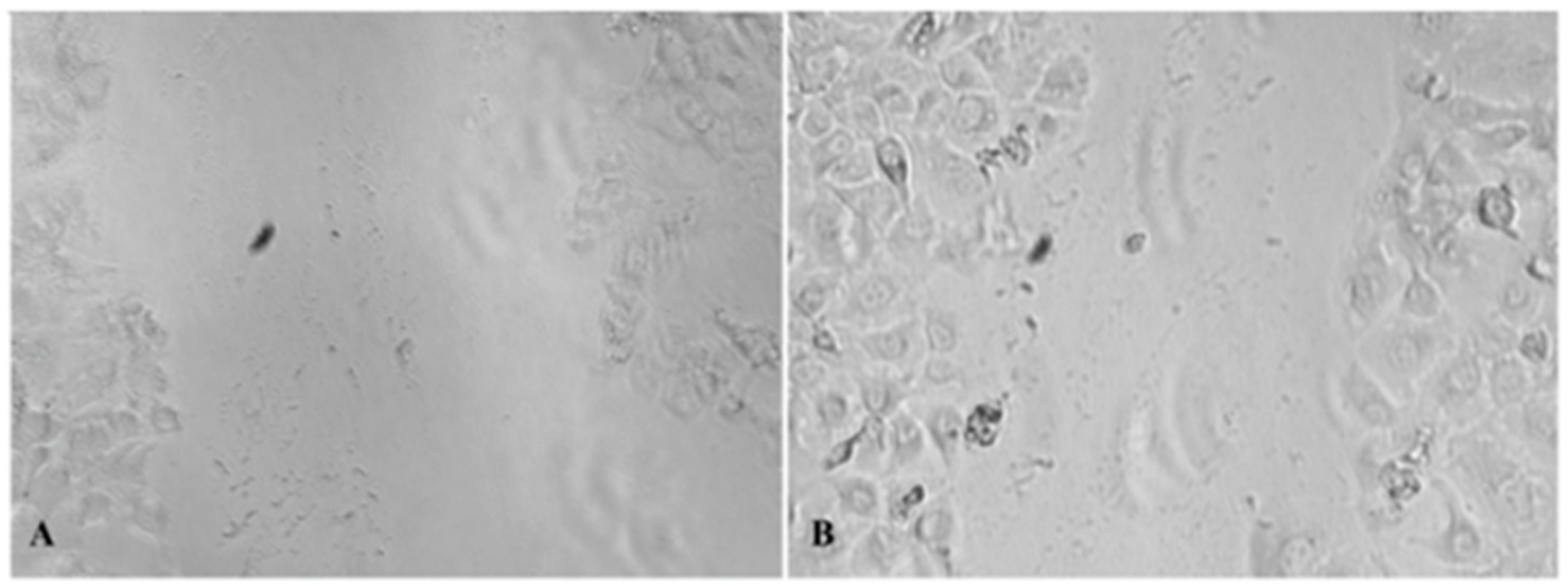
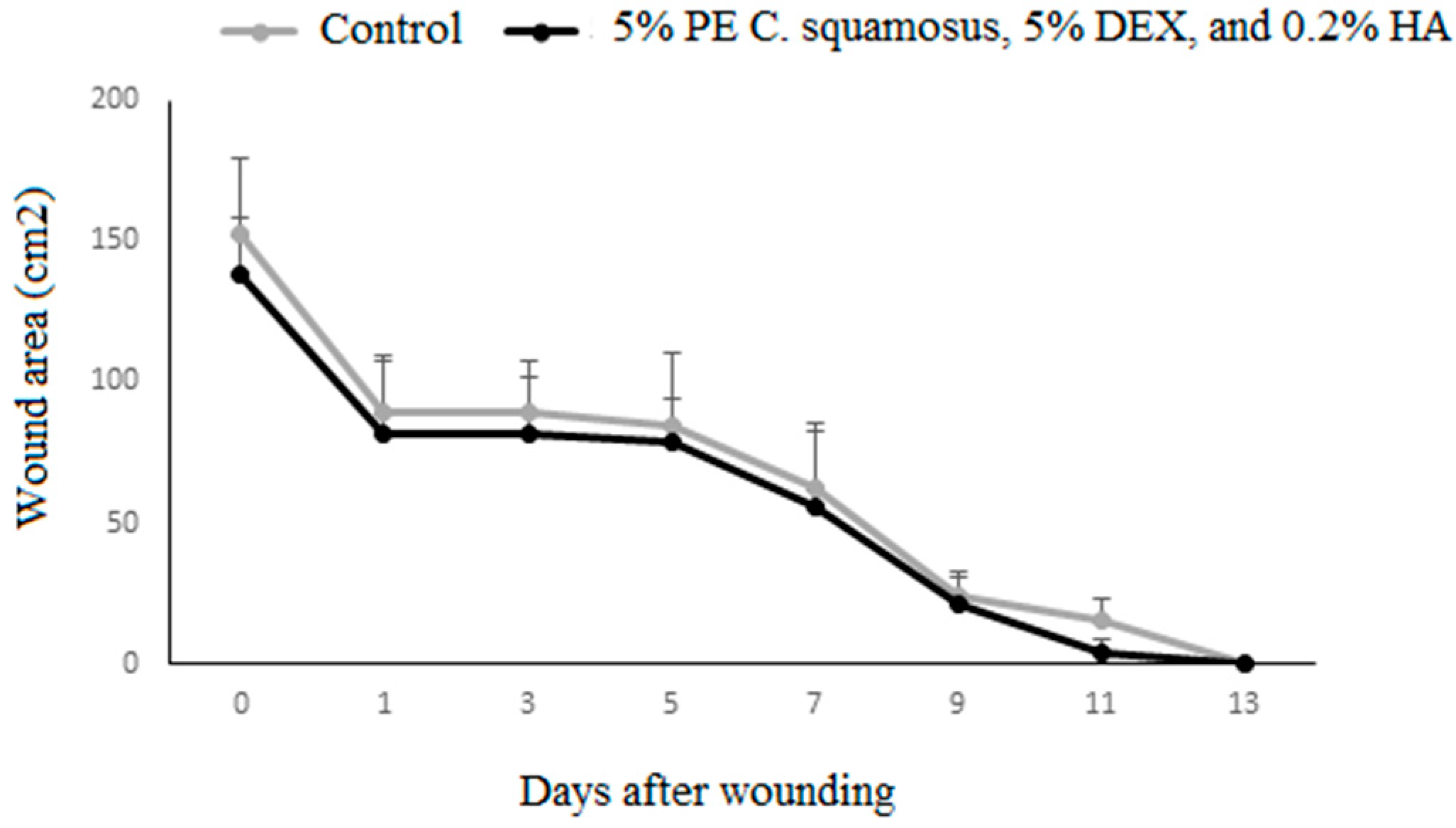
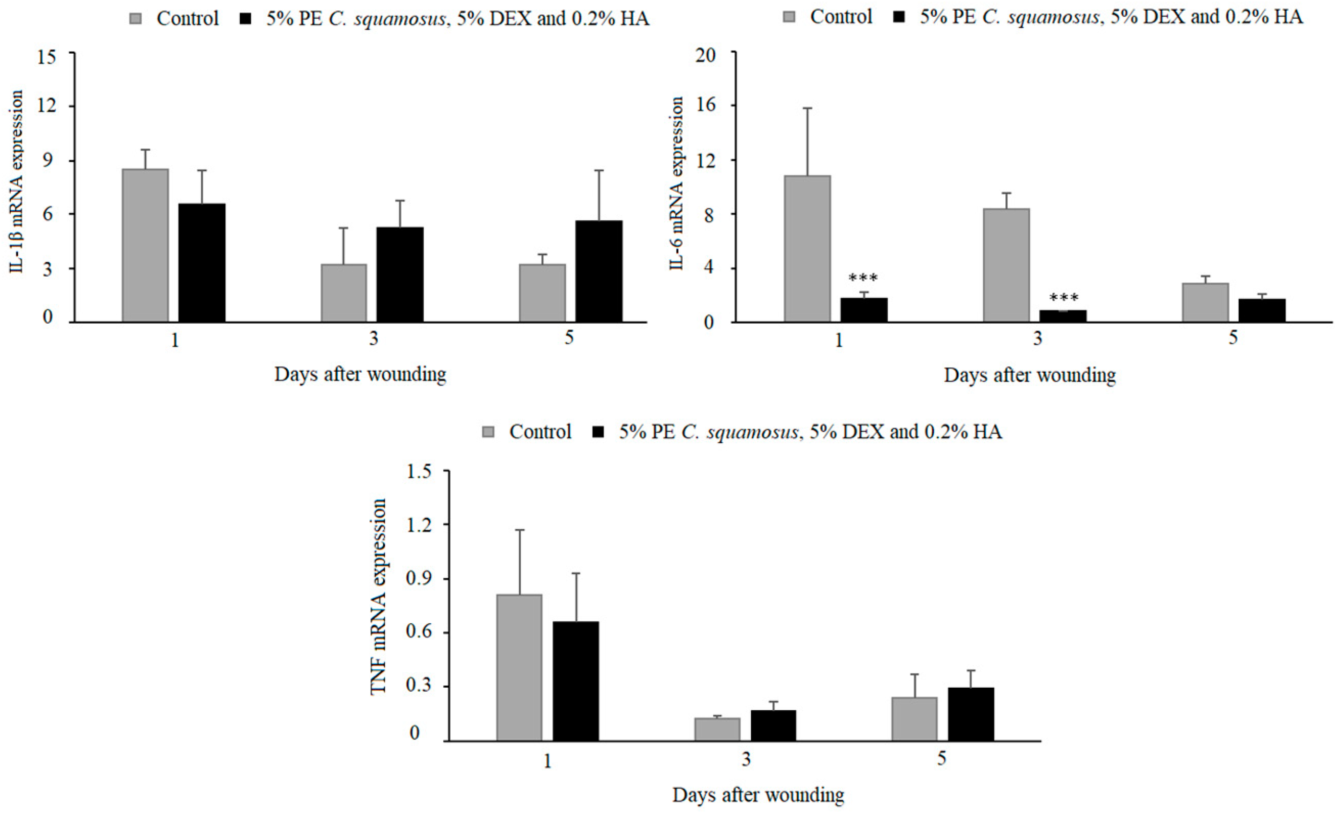
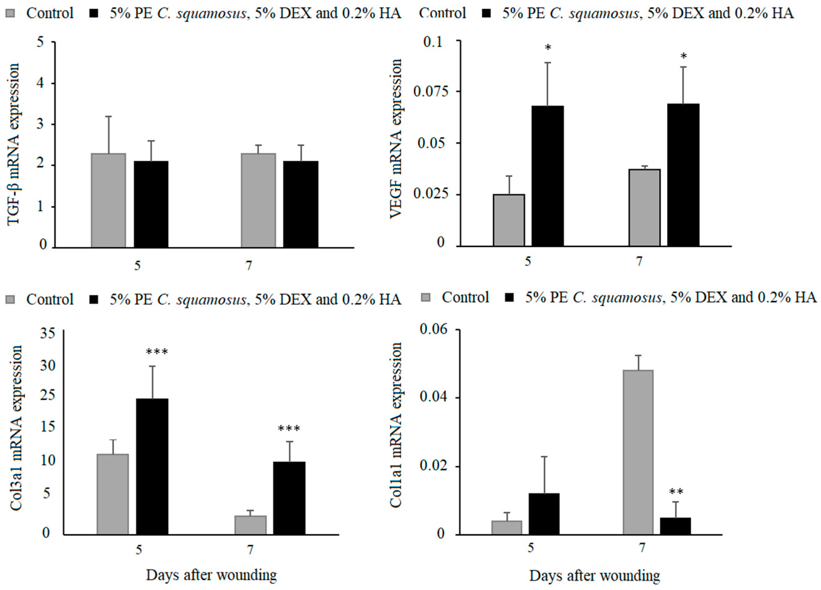
2. Results
3. Discussion
4. Materials and Methods
4.1. Collection of C. squamosus Fruiting Bodies
4.2. Preparation of C. squamosus Polysaccharide Extract
4.3. Measurement of Total, α- and β-Glucan Content in PE of C. squamosus
4.3.1. Measurement of Total Glucan Content in PE of C. squamosus
4.3.2. Measurement of α- and β-Glucan Content in PE of C. squamosus
4.4. Antibacterial and Anticandidal Activity
4.5. Cytotoxicity Towards HaCaT Cell Line
4.6. In Vitro Wound Healing Assay
4.7. In Vivo Experiments
4.7.1. Experimental Animals Protocol
4.7.2. Reverse Transcription and Real-Time Polymerase Chain Reaction (RT-PCR)
4.8. Statistical Analysis
5. Conclusions
6. Patents
Author Contributions
Funding
Institutional Review Board Statement
Data Availability Statement
Conflicts of Interest
Abbreviations
| PE | polysaccharide extract |
| MIC | minimum inhibitory concentration |
| MBC | minimum bactericidal concentration |
| MFC | minimum fungicidal concentration |
| DEX | dexpanthenol |
| HA | hyaluronic acid |
| IL-1β | interleukin-1 beta |
| IL-6 | interleukin-6 |
| TNF | tumor necrosis factor |
| TGF-β | transforming growth factor-β |
| VEGF | vascular endothelial growth factor |
References
- Mapoung, S.; Umsumarng, S.; Semmarath, W.; Arjsri, P.; Thippraphan, P.; Yodkeeree, S.; Limtrakul Dejkriengkraikul, P. Skin wound-healing potential of polysaccharides from medicinal mushroom Auricularia auricula-judae (Bull.). J. Fungi 2021, 7, 247. [Google Scholar] [CrossRef] [PubMed]
- Schultz, G.S.; Chin, G.A.; Moldawer, L.; Diegelmann, R.F. Principles of wound healing. In Mechanisms of Vascular Disease: A Reference Book for Vascular Specialists; Fitridge, R., Thompson, M., Eds.; University of Adelaide Press: Adelaide, Australia, 2011. [Google Scholar]
- Harding, K.; Queen, D. Estimating the cost of wounds for the United Kingdom and its countries. Int. Wound J. 2023, 21, e14616. [Google Scholar] [CrossRef] [PubMed]
- El-Sherbeni, S.; Negm, W.A. The wound healing effect of botanicals and pure natural substances used in in vivo models. Inflammopharmacology 2023, 31, 755–772. [Google Scholar] [CrossRef] [PubMed]
- Wu, Y.; Choi, M.H.; Li, J.; Yang, H.; Shin, H.J. Mushroom cosmetics: The present and future. Cosmetics 2016, 3, 22. [Google Scholar] [CrossRef]
- Sharifi-Rad, J.; Butnariu, M.; Ezzat, S.M.; Adetunji, C.O.; Imran, M.; Sobhani, S.R.; Tufail, T.; Hosseinabadi, T.; Ramírez-Alarcón, K.; Martorell, M.; et al. Mushrooms-rich preparations on wound healing: From nutritional to medicinal attributes. Front. Pharmacol. 2020, 11, 567518. [Google Scholar] [CrossRef]
- Elkhateeb, W.; Daba, G.; Elnahas, M.; Thomas, P.; Emam, M. Metabolic profile and skin-related bioactivities of Cerioporus squamosus hydromethanolic extract. Biodivers. J. 2020, 21, 4732–4740. [Google Scholar] [CrossRef]
- Xu, C.; Wang, F.; Guan, S.; Wang, L. β-Glucans obtained from fungus for wound healing: A review. Carbohydr Polym. 2024, 327, 121662. [Google Scholar] [CrossRef]
- Najafiasl, M.; Osfouri, S.; Azin, R.; Zaeri, S. Fabrication, characterization and in vivo evaluation of dexpanthenol sustained-release nanofibers for wound healing. Polym. Test. 2020, 91, 106827. [Google Scholar] [CrossRef]
- Gorski, J.; Proksch, E.; Baron, J.M.; Schmid, D.; Zhang, L. Dexpanthenol in wound healing after medical and cosmetic interventions (postprocedure wound healing). Pharmaceuticals 2020, 13, 138. [Google Scholar] [CrossRef]
- Juncan, A.M.; Moisă, D.G.; Santini, A.; Morgovan, C.; Rus, L.L.; Vonica-Țincu, A.L.; Loghin, F. Advantages of hyaluronic acid and its combination with other bioactive ingredients in cosmeceuticals. Molecules 2021, 26, 4429. [Google Scholar] [CrossRef]
- Cosmetic Ingredient Review. Safety Assessment of Panthenol, Pantothenic Acid, and Derivatives as Used in Cosmetics. 2017. Available online: https://www.cir-safety.org/sites/default/files/panthenol_0.pdf (accessed on 3 March 2025).
- Cosmetic Ingredient Review. Safety Assessment of Hyaluronates as Used in Cosmetics. 2022. Available online: https://www.cir-safety.org/sites/default/files/Hyaluronates_1.pdf (accessed on 3 March 2025).
- Liu, J.K. Natural products in cosmetics. Nat. Prod. Bioprospect 2022, 12, 40. [Google Scholar] [CrossRef] [PubMed]
- Natakankitkul, S.; Homdok, P.; Wandee, P.; Krisdaphong, T.; Toida. Development of skincare cosmetic from yeast beta-glucans. TJPS 2016, 40, 9–12. [Google Scholar]
- Cosmetic Ingredient Review. Safety Assessment of Yeast-Derived Ingredients as Used in Cosmetics. 2021. Available online: https://cir-safety.org/sites/default/files/yeast092021SLR.pdf (accessed on 3 March 2025).
- Bessa, L.J.; Fazii, P.; Di Giulio, M.; Cellini, L. Bacterial Isolates from Infected Wounds and Their Antibiotic Susceptibility Pattern: Some Remarks about Wound Infection. Int. Wound J. 2015, 12, 47–52. [Google Scholar] [CrossRef] [PubMed]
- Ge, Y.; Wang, Q. Current Research on Fungi in Chronic Wounds. Front. Mol. Biosci. 2023, 9, 1057766. [Google Scholar] [CrossRef]
- Severn, M.M.; Horswill, A.R. Staphylococcus epidermidis and its dual lifestyle in skin health and infection. Nat. Rev. Microbiol. 2023, 21, 97–111. [Google Scholar] [CrossRef]
- Mordi, R.; Momoh, M. Incidence of Proteus Species in Wound Infections and Their Sensitivity Pattern in the University of Benin Teaching Hospital. Afr. J. Biotechnol. 2009, 8, 725–730. [Google Scholar]
- Papapetropoulos, N.; Papapetropoulou, M.; Vantarakis, A. Abscesses and wound infections due to Staphylococcus lugdunensis: Report of 16 cases. Infection 2013, 41, 525–528. [Google Scholar] [CrossRef]
- Patra, S.; Maity, P.; Chakraborty, I.; Sen, I.; Ghosh, D.; Rout, D.; Bhanja, S. Structural studies of immunomodulatory (1 → 3)-, (1 → 4)- glucan from an edible mushroom Polyporus grammocephalus. Int. J. Biol. Macromol. 2020, 168, 649–655. [Google Scholar] [CrossRef]
- Mirończuk-Chodakowska, I.; Witkowska, A. Evaluation of Polish wild mushrooms as beta-glucan sources. Int. J. Environ. Res. Public Health 2020, 17, 7299. [Google Scholar] [CrossRef]
- Doskocil, I.; Havlik, J.; Verlotta, R.; Tauchen, J.; Vesela, L.; Macakova, K.; Opletal, L.; Kokoska, L.; Rada, V. In Vitro immunomodulatory activity, cytotoxicity and chemistry of some Central European polypores. Pharm. Biol. 2016, 54, 2369–2376. [Google Scholar] [CrossRef]
- Liu, Y.S.; Lai, M.C.; Tzeng, Y.C.; Liu, I.M. Polyphenolic Hispolon Derived from Medicinal Mushrooms of the Inonotus and Phellinus Genera Promotes Wound Healing in Hyperglycemia-Induced Impairments. Nutrients 2025, 17, 266. [Google Scholar] [CrossRef] [PubMed]
- Zamboni, F.; Wong, C.K.; Collins, M.N. Hyaluronic acid association with bacterial, fungal and viral infections: Can hyaluronic acid be used as an antimicrobial polymer for biomedical and pharmaceutical applications? Bioact. Mater. 2022, 19, 458–473. [Google Scholar] [CrossRef] [PubMed]
- Helaly, G.F.; El-Aziz, A.A.A.; Sonbol, F.; El-Banna, T.; Lotfy, N. Dexpanthenol and propolis extract in combination with local antibiotics for treatment of Staphylococcal and Pseudomonal wound infections. Arch. Clin. Microbiol. 2011, 2. [Google Scholar]
- Abbas, J.; Bodey, G.P.; Hanna, H.A.; Mardani, M.; Girgawy, E.; Abi-Said, D.; Whimbey, E.; Hachem, R.; Raad, I. Candida krusei fungemia: An escalating serious infection in immunocompromised patients. Arch. Intern. Med. 2000, 160, 2659–2664. [Google Scholar] [CrossRef]
- Fernandes, A.; Petrović, J.; Stojković, D.; Barros, L.; Glamočlija, J.; Soković, M.; Martins, A.; Ferreira, I.C.F.R. Polyporus squamosus (Huds.) Fr from different origins: Chemical characterization, screening of the bioactive properties and specific antimicrobial effects against Pseudomonas aeruginosa. LWT Food Sci. Technol. 2016, 69, 91–97. [Google Scholar] [CrossRef]
- Alves, M.J.; Ferreira, I.C.; Dias, J.; Teixeira, V.; Martins, A.; Pintado, M. A Review on Antimicrobial Activity of Mushroom (Basidiomycetes) Extracts and Isolated Compounds. Planta Med. 2012, 78, 1707–1718. [Google Scholar] [CrossRef]
- Matijašević, D.; Pantić, M.; Rašković, B.; Pavlović, V.; Duvnjak, D.; Sknepnek, A.; Nikšić, M. The Antibacterial Activity of Coriolus versicolor Methanol Extract and Its Effect on Ultrastructural Changes of Staphylococcus aureus and Salmonella Enteritidis. Front Microbiol. 2016, 7, 1226. [Google Scholar] [CrossRef]
- Mohan, S.P.; Palaniappan, A.; Nawaz, M.K.K.; Kripamol, R.; Seenuvasan, R.; Kumar, P.R.A. In vitro cytotoxicity evaluation of flowable hyaluronic acid-acellular stromal vascular fraction (HA-aSVF) mixture for tissue engineering applications. J. Pharm. Bioallied Sci. 2023, 15 (Suppl. S1), S677–S682. [Google Scholar] [CrossRef]
- Boeckel, D.G.; Shinkai, R.S.; Grossi, M.L.; Teixeira, E.R. In Vtro evaluation of cytotoxicity of hyaluronic acid as an extracellular matrix on OFCOL II cells by the MTT assay. Oral Surg. Oral Med. Oral Pathol. Oral Radiol. 2014, 117, e423–e428. [Google Scholar] [CrossRef]
- Klöcker, N.; Rudolph, P.; Verse, T. Evaluation of protective and therapeutic effects of dexpanthenol on nasal decongestants and preservatives: Results of cytotoxic studies in vitro. Am. J. Rhinol. 2004, 18, 315–320. [Google Scholar] [CrossRef]
- Choi, S.Y.; Seop, S.Y.; Hyun, M.Y.; Yoo, K.H.; Kim, B.J.; Kim, M.N.; Cho, J.W. Safety evaluation of topical valproate application. Toxicol. Res. 2013, 29, 87–90. [Google Scholar] [CrossRef] [PubMed]
- Vaou, N.; Stavropoulou, E.; Voidarou, C.; Tsakris, Z.; Rozos, G.; Tsigalou, C.; Bezirtzoglou, E. Interactions between Medical Plant-Derived Bioactive Compounds: Focus on Antimicrobial Combination Effects. Antibiotics 2022, 11, 1014. [Google Scholar] [CrossRef] [PubMed]
- El-Seddawy, F.; Abdel-Maboud, M.; Barakat, N.; Hassaan, M. Dexpanthenol: New Insights on Wound Healing, a Review. J. Adv. Vet. Res. 2023, 13, 1474–1478. [Google Scholar]
- Riessen, R.; Wight, T.N.; Pastore, C.; Henley, C.; Isner, J.M. Distribution of hyaluronan during extracellular matrix remodeling in human restenotic arteries and balloon-injured rat carotid arteries. Circulation 1996, 93, 1141–1147. [Google Scholar] [CrossRef]
- Wei, D.; Zhang, L.; Williams, D.; Browder, I. Glucan stimulates human dermal fibroblast collagen biosynthesis through nuclear factor-1 dependent mechanism. Wound Repair Regen. 2002, 10, 161–168. [Google Scholar] [CrossRef]
- Minqi, Q.; Bing, L.; Dezhi, G.; Qi, X.; Yanjiao, X.; Qiang, D.; Shunqing, T. Aminated β-Glucan with immunostimulating activities and collagen composite sponge for wound repair. Int. J. Biol. Macromol. 2022, 221, 193–203. [Google Scholar] [CrossRef]
- Majtan, J.; Jesenak, M. β-Glucans: Multi-functional modulator of wound healing. Molecules 2018, 23, 806. [Google Scholar] [CrossRef]
- Frenkel, J.S. The role of hyaluronan in wound healing. Int. Wound J. 2014, 11, 159–163. [Google Scholar] [CrossRef]
- Litwiniuk, M.; Krejner, A.; Speyrer, M.S.; Gauto, A.R.; Grzela, T. Hyaluronic Acid in Inflammation and Tissue Regeneration. Wounds 2016, 28, 78–88. [Google Scholar]
- Chen, W.Y.; Abatangelo, G. Functions of hyaluronan in wound repair. Wound Repair Regen. 1999, 7, 79–89. [Google Scholar] [CrossRef]
- Della Sala, F.; Longobardo, G.; Fabozzi, A.; di Gennaro, M.; Borzacchiello, A. Hyaluronic Acid-Based Wound Dressing with Antimicrobial Properties for Wound Healing Application. Appl. Sci. 2022, 12, 3091. [Google Scholar] [CrossRef]
- Lin, Z.Q.; Kondo, T.; Ishida, Y.; Takayasu, T.; Mukaida, N. Essential involvement of IL-6 in the skin wound-healing process as evidenced by delayed wound healing in IL-6-deficient mice. J. Leukoc. Biol. 2003, 73, 713–721. [Google Scholar] [CrossRef] [PubMed]
- Choudhary, V.; Choudhary, M.; Bollag, W.B. Exploring skin wound healing models and the impact of natural lipids on the healing process. Int. J. Mol. Sci. 2024, 25, 3790. [Google Scholar] [CrossRef] [PubMed]
- Gajbhiye, S.; Wairkar, S. Collagen fabricated delivery systems for wound healing: A new roadmap. Biomater. Adv. 2022, 142, 213152. [Google Scholar] [CrossRef]
- Pastar, I.; Stojadinovic, O.; Yin, N.C.; Ramirez, H.; Nusbaum, A.G.; Sawaya, A.; Patel, S.B.; Khalid, L.; Isseroff, R.R.; Tomic-Canic, M. Epithelialization in wound healing. Adv. Wound Care 2014, 3, 445–464. [Google Scholar] [CrossRef]
- Raffetto, J.D. Pathophysiology of wound healing and alterations in venous leg ulcers-review. Phlebology 2016, 31 (Suppl. S1), 56–62. [Google Scholar] [CrossRef]
- Ray, P.; Singh, S.; Gupta, S. Topical antimicrobial therapy: Current status and challenges. Indian J. Med. Microbiol. 2019, 37, 299–308. [Google Scholar] [CrossRef]
- Breijyeh, Z.; Karaman, R. Antibacterial activity of medicinal plants and their role in wound healing. Futur. J. Pharm. Sci. 2024, 10, 68. [Google Scholar] [CrossRef]
- Liu, E.; Gao, H.; Zhao, Y.; Pang, Y.; Yao, Y.; Yang, Z.; Zhang, X.; Wang, Y.; Yang, S.; Ma, X.; et al. The Potential Application of Natural Products in Cutaneous Wound Healing: A Review of Preclinical Evidence. Front. Pharmacol. 2022, 13, 900439. [Google Scholar] [CrossRef]
- Cheng, Y.C.; Li, T.S.; Su, H.L.; Lee, P.C.; Wang, H.D. Transdermal delivery systems of natural products applied to skin therapy and care. Molecules 2020, 25, 5051. [Google Scholar] [CrossRef]
- Lagoa, T.; Queiroga, M.C.; Martins, L. An overview of wound dressing materials. Pharmaceuticals 2024, 17, 1110. [Google Scholar] [CrossRef] [PubMed]
- Criollo-Mendoza, M.S.; Contreras-Angulo, L.A.; Leyva-López, N.; Gutiérrez-Grijalva, E.P.; Jiménez-Ortega, L.A.; Heredia, J.B. Wound Healing Properties of Natural Products: Mechanisms of Action. Molecules 2023, 28, 598. [Google Scholar] [CrossRef] [PubMed]
- Abreu, H.; Zavadinack, M.; Smiderle, F.R.; Cipriani, T.R.; Cordeiro, L.M.C.; Iacomini, M. Polysaccharides from Pleurotus eryngii: Selective extraction methodologies and their modulatory effects on THP-1 macrophages. Carbohydr. Polym. 2021, 252, 117177. [Google Scholar] [CrossRef] [PubMed]
- Đorđevski, N.; Abdullahi, I.U.; Zengin, G.; Božunović, J.; Gašić, U.; Ristanović, E.; Ćirić, A.; Nikolić, B.; Stojković, D. Chemical and biological investigations of Allium scorodoprasum L. flower extracts. Pharmaceuticals 2023, 1, 21. [Google Scholar] [CrossRef]
- Petrović, J.; Kovalenko, V.; Svirid, A.; Stojković, D.; Ivanov, M.; Kostić, M. Individual stereoisomers of verbenol and verbenone express bioactive features. J. Mol. Struct. 2021, 1251, 131999. [Google Scholar] [CrossRef]
- Petrović, J.; Glamočlija, J.; Milinčić, D.D.; Doroški, A.; Lević, S.; Stanojević, S.P.; Kostić, A.Ž.; Minić, D.A.P.; Vidović, B.B.; Plećić, A. Comparative Chemical Analysis and Bioactive Properties of Aqueous and Glucan-Rich Extracts of Three Widely Appreciated Mushrooms: Agaricus bisporus (JE Lange) Imbach, Laetiporus sulphureus (Bull.) Murill and Agrocybe aegerita (V. Brig.) Vizzini. Pharmaceuticals 2024, 17, 1153. [Google Scholar] [CrossRef]

| Microorganisms | PE C. squamosus | HA | DEX | Mixture | Streptomycin Ketoconazole | |
|---|---|---|---|---|---|---|
| P. vulgaris (B44) (clinical isolate) * | MIC | 3.50 | 0.44 | 1.75 | 1.75 | 0.003 |
| MBC | 7.00 | 0.88 | 3.50 | 3.50 | 0.006 | |
| S. lungdunensis (B43) (clinical isolate) * | MIC | 3.50 | >3.50 | 1.75 | 1.75 | 0.003 |
| MBC | 7.00 | >3.50 | 3.50 | 3.50 | 0.006 | |
| S. epidermidis (B45) (clinical isolate) * | MIC | 1.75 | 0.44 | 0.88 | 0.44 | 0.10 |
| MBC | 3.50 | 0.88 | 1.75 | 0.88 | 0.20 | |
| C. kefyr (Y289) (clinical isolate) * | MIC | 0.44 | 0.22 | 0.44 | 0.22 | 0.015 |
| MFC | 0.88 | 0.44 | 0.88 | 0.44 | 0.030 | |
| C. krusei (Y454) (clinical isolate) * | MIC | 0.44 | 1.75 | 0.44 | 0.44 | 0.015 |
| MFC | 0.88 | 3.50 | 0.88 | 0.88 | 0.030 | |
| C. albicans (Y177) (clinical isolate) * | MIC | 0.44 | 0.22 | 0.44 | 0.22 | 0.015 |
| MFC | 0.88 | 0.44 | 0.88 | 0.44 | 0.030 |
| Sample | IC50 (μg/mL) |
|---|---|
| PE C. squamosus | >400 |
| HA | >400 |
| DEX | >400 |
| K2Cr2O7 | 83.40 |
| Tested Sample | Wound Closure After 24 h (%) | Comparison to Control | Comparison to Combination |
|---|---|---|---|
| Control | 16.86 ± 3.17 | ||
| PE C. squamosus | 25.35 ± 4.24 | Not statistically significant | * |
| HA | 8.51 ± 1.11 | * | **** |
| DEX | 31.26 ± 0.97 | ** | * |
| Mixture | 35.81 ± 2.14 | ** |
Disclaimer/Publisher’s Note: The statements, opinions and data contained in all publications are solely those of the individual author(s) and contributor(s) and not of MDPI and/or the editor(s). MDPI and/or the editor(s) disclaim responsibility for any injury to people or property resulting from any ideas, methods, instructions or products referred to in the content. |
© 2025 by the authors. Licensee MDPI, Basel, Switzerland. This article is an open access article distributed under the terms and conditions of the Creative Commons Attribution (CC BY) license (https://creativecommons.org/licenses/by/4.0/).
Share and Cite
Petrović, J.D.; Carević Milićević, T.A.; Glamočlija, J.M.; Kulaš, J.B.; Mirkov, I.I. Mixture Containing 5% Polysaccharide Extract of Cerioporus squamosus (Huds.) Quélet, 5% Dexpanthenol, and 0.2% Hyaluronic Acid Shows In Vitro and In Vivo Wound Healing Properties. Pharmaceuticals 2025, 18, 416. https://doi.org/10.3390/ph18030416
Petrović JD, Carević Milićević TA, Glamočlija JM, Kulaš JB, Mirkov II. Mixture Containing 5% Polysaccharide Extract of Cerioporus squamosus (Huds.) Quélet, 5% Dexpanthenol, and 0.2% Hyaluronic Acid Shows In Vitro and In Vivo Wound Healing Properties. Pharmaceuticals. 2025; 18(3):416. https://doi.org/10.3390/ph18030416
Chicago/Turabian StylePetrović, Jovana D., Tamara A. Carević Milićević, Jasmina M. Glamočlija, Jelena B. Kulaš, and Ivana I. Mirkov. 2025. "Mixture Containing 5% Polysaccharide Extract of Cerioporus squamosus (Huds.) Quélet, 5% Dexpanthenol, and 0.2% Hyaluronic Acid Shows In Vitro and In Vivo Wound Healing Properties" Pharmaceuticals 18, no. 3: 416. https://doi.org/10.3390/ph18030416
APA StylePetrović, J. D., Carević Milićević, T. A., Glamočlija, J. M., Kulaš, J. B., & Mirkov, I. I. (2025). Mixture Containing 5% Polysaccharide Extract of Cerioporus squamosus (Huds.) Quélet, 5% Dexpanthenol, and 0.2% Hyaluronic Acid Shows In Vitro and In Vivo Wound Healing Properties. Pharmaceuticals, 18(3), 416. https://doi.org/10.3390/ph18030416