
Aggregation-Caused Quenching Dyes as Potent Tools to Track the Integrity of Antitumor Nanocarriers: A Mini-Review


Abstract
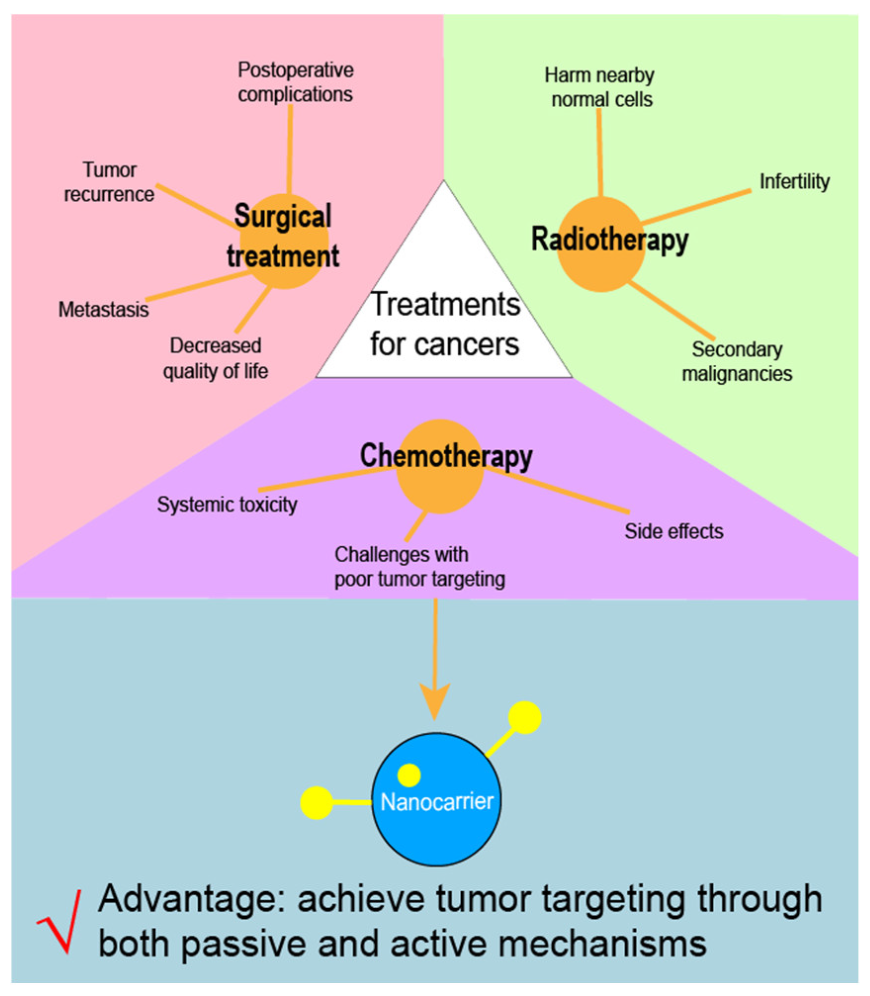
1. Introduction
2. Major Strategies for Developing Antitumor Nanocarriers
2.1. Controlling Drug Release
2.1.1. Applying External Stimuli
2.1.2. Taking Advantage of Nanocarrier Nature
2.2. Increasing the Permeability into Cancer Cells
2.2.1. Applying External Stimuli
2.2.2. Taking Advantage of Nanocarrier Nature
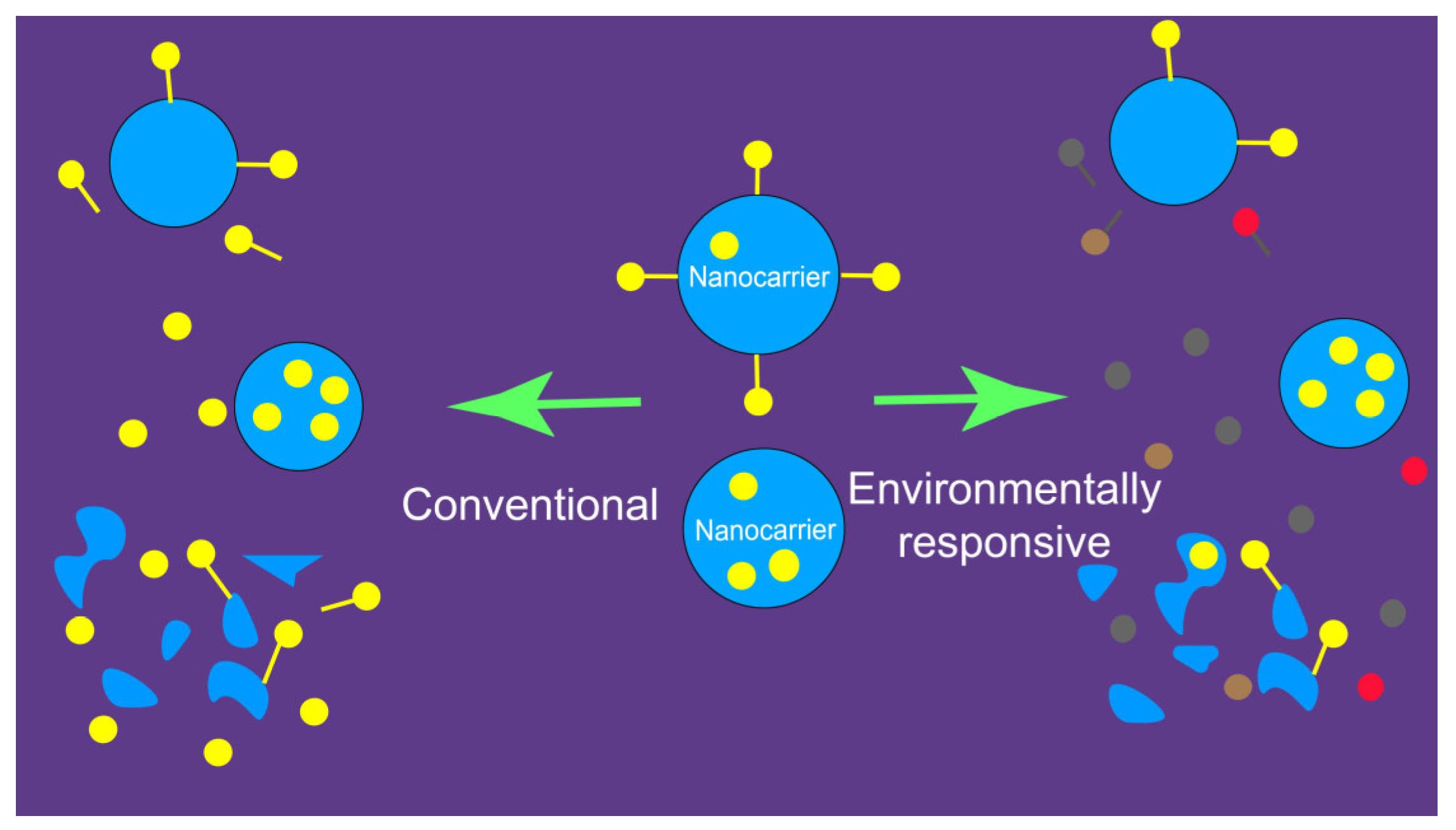
3. The Developmental Bottleneck for Antitumor Nanocarriers
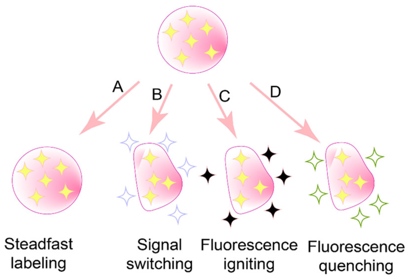
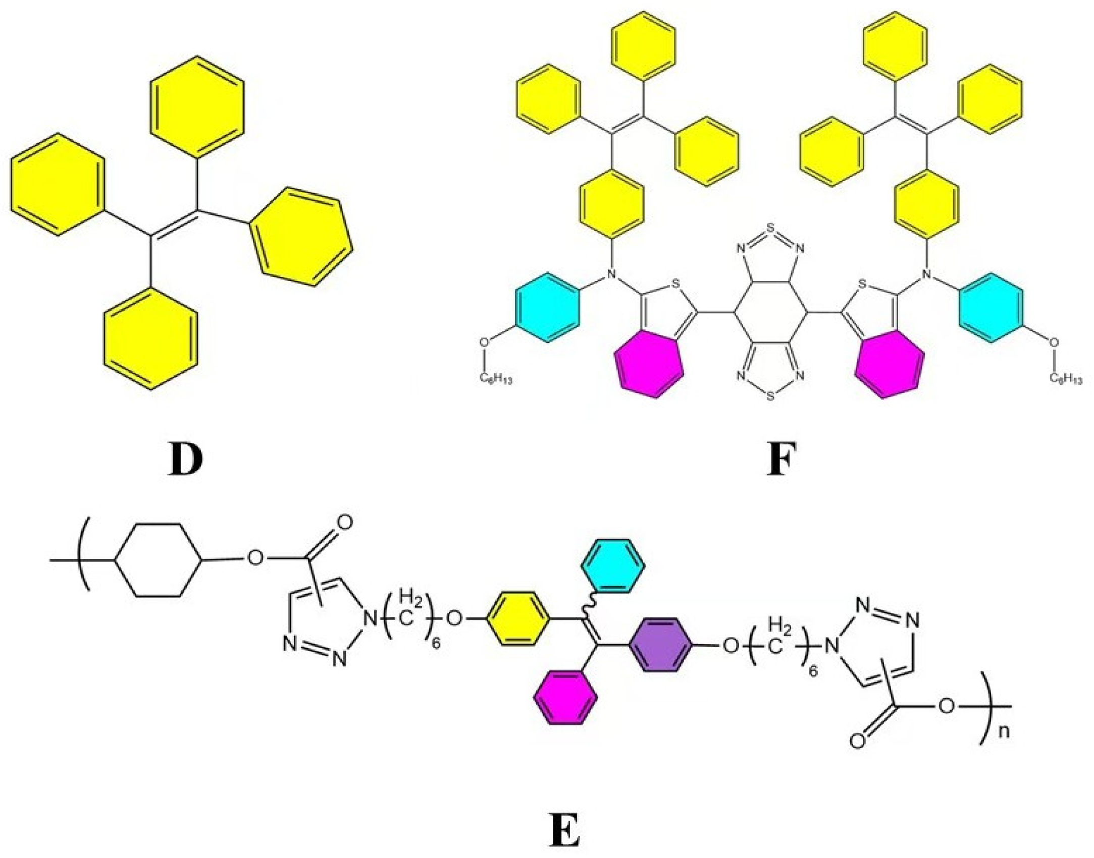
4. Aggregation-Caused Quenching (ACQ) Dyes: A Promising Solution
5. Concluding Remarks
Author Contributions
Funding
Institutional Review Board Statement
Informed Consent Statement
Data Availability Statement
Conflicts of Interest
References
- Bray, F.; Laversanne, M.; Sung, H.; Ferlay, J.; Siegel, R.L.; Soerjomataram, I.; Jemal, A. Global cancer statistics 2022: GLOBOCAN estimates of incidence and mortality worldwide for 36 cancers in 185 countries. CA A Cancer J. Clin. 2024, 74, 229–263. [Google Scholar] [CrossRef] [PubMed]
- Lakdawalla, D.N.; Sun, E.C.; Jena, A.B.; Reyes, C.M.; Goldman, D.P.; Philipson, T.J. An economic evaluation of the war on cancer. J. Health Econ. 2010, 29, 333–346. [Google Scholar] [CrossRef] [PubMed]
- Bu, L.-L.; Yan, J.; Wang, Z.; Ruan, H.; Chen, Q.; Gunadhi, V.; Bell, R.B.; Gu, Z. Advances in drug delivery for post-surgical cancer treatment. Biomaterials 2019, 219, 119182. [Google Scholar] [CrossRef] [PubMed]
- Gomathi, M.; Ayisha Hamna, T.P.; Jijo, A.J.; Saradha Devi, K.M.; Arul, N.; Balachandar, V. Recent advances in radiotherapy and its associated side effects in cancer—A review. J. Basic Appl. Zool. 2019, 80, 14. [Google Scholar]
- De Vita, V.T., Jr.; Chu, E. A history of cancer chemotherapy. Cancer Res. 2008, 68, 8643–8653. [Google Scholar] [CrossRef]
- Xiong, Z.; Tong, T.; Xie, Z.; Yu, S.; Zhuang, R.; Jia, Q.; Peng, S.; Li, B.; Xie, J.; Li, K.; et al. Delivery of gefitinib loaded nanoparticles for effectively inhibiting prostate cancer progression. Biomater. Sci. 2024, 12, 650–659. [Google Scholar] [CrossRef]
- Zhang, Y.; Wang, J.; Dai, H.; Li, N.; Hu, F.; Zhang, B.; Yang, Y.; Cai, H.; Yan, J. Cis-Platinum and CXCR4 siRNA Carried with Cellulose Nanopaper Restrain the Biological Effect of Breast Carcinoma Cells. J. Biomed. Nanotechnol. 2024, 20, 539–544. [Google Scholar] [CrossRef]
- Zang, C.; Tian, Y.; Tang, Y.; Tang, M.; Yang, D.; Chen, F.; Ghaffarlou, M.; Tu, Y.; Ashrafizadeh, M.; Li, Y. Hydrogel-based platforms for site-specific doxorubicin release in cancer therapy. J. Transl. Med. 2024, 22, 879. [Google Scholar] [CrossRef]
- Aisner, J. Overview of the changing paradigm in cancer treatment: Oral chemotherapy. Am. J. Health-Syst. Pharm. 2007, 64, S4–S7. [Google Scholar] [CrossRef]
- Liu, H.; Lv, L.; Yang, K. Chemotherapy targeting cancer stem cells. Am. J. Cancer Res. 2015, 5, 880. [Google Scholar]
- Mosleh-Shirazi, S.; Abbasi, M.; Moaddeli, M.R.; Vaez, A.; Shafiee, M.; Kasaee, S.R.; Amani, A.M.; Hatam, S. Nanotechnology Advances in the Detection and Treatment of Cancer: An Overview. Nanotheranostics 2022, 6, 400–423. [Google Scholar] [CrossRef] [PubMed]
- Kemp, J.A.; Kwon, Y.J. Cancer nanotechnology: Current status and perspectives. Nano Converg. 2021, 8, 34. [Google Scholar] [CrossRef] [PubMed]
- Kenchegowda, M.; Rahamathulla, M.; Hani, U.; Begum, M.Y.; Guruswamy, S.; Osmani, R.A.M.; Gowrav, M.P.; Alshehri, S.; Ghoneim, M.M.; Alshlowi, A.; et al. Smart Nanocarriers as an Emerging Platform for Cancer Therapy: A Review. Molecules 2021, 27, 146. [Google Scholar] [CrossRef] [PubMed]
- Patra, J.K.; Das, G.; Fraceto, L.F.; Campos, E.V.R.; Rodriguez-Torres, M.D.P.; Acosta-Torres, L.S.; Diaz-Torres, L.A.; Grillo, R.; Swamy, M.K.; Sharma, S.; et al. Nano based drug delivery systems: Recent developments and future prospects. J. Nanobiotechnology 2018, 16, 71. [Google Scholar] [CrossRef]
- Farzin, L.; Sheibani, S.; Moassesi, M.E.; Shamsipur, M. An overview of nanoscale radionuclides and radiolabeled nanomaterials commonly used for nuclear molecular imaging and therapeutic functions. J. Biomed. Mater. Res. A 2019, 107, 251–285. [Google Scholar] [CrossRef]
- Feroze, F.; Sher, M.; Hussain, M.A.; Abbas, A.; Haseeb, M.T.; Fatima, A.; Naeem-Ul-Hassan, M.; Amin, H.M.A. Gastro retentive floating drug delivery system of levofloxacin based on Aloe vera hydrogel: In vitro and in vivo assays. Int. J. Biol. Macromol. 2025, 284 Pt 1, 138156. [Google Scholar] [CrossRef]
- Türkeş, E.; Açıkel, Y.S. Folic acid-conjugated cancer drug curcumin-loaded albumin nanoparticles: Investigation of curcumin release kinetics. J. Drug Deliv. Sci. Technol. 2024, 91, 105178. [Google Scholar] [CrossRef]
- Khan, M.S.; Gowda, B.J.; Almalki, W.H.; Singh, T.; Sahebkar, A.; Kesharwani, P. Unravelling the potential of mitochondria-targeted liposomes for enhanced cancer treatment. Drug Discov. Today 2024, 29, 103819. [Google Scholar] [CrossRef]
- Xie, M.; Meng, F.; Wang, P.; Díaz-García, A.M.; Parkhats, M.; Santos-Oliveira, R.; Asim, M.H.; Bostan, N.; Gu, H.; Yang, L.; et al. Surface engineering of magnetic iron oxide nanoparticles for breast cancer diagnostics and drug delivery. Int. J. Nanomed. 2024, 19, 8437–8461. [Google Scholar] [CrossRef]
- Kuna, K.; Baddam, S.R.; Kalagara, S.; Akkiraju, P.C.; Tade, R.S.; Enaganti, S. Emerging natural polymer-based architectured nanotherapeutics for the treatment of cancer. Int. J. Biol. Macromol. 2024, 262, 129434. [Google Scholar] [CrossRef]
- Fraguas-Sánchez, A.I.; Martín-Sabroso, C.; Fernández-Carballido, A.; Torres-Suárez, A.I. Current status of nanomedicine in the chemotherapy of breast cancer. Cancer Chemother. Pharmacol. 2019, 84, 689–706. [Google Scholar] [CrossRef] [PubMed]
- Khodabandehloo, H.; Zahednasab, H.; Hafez, A.A. Nanocarriers usage for drug delivery in cancer therapy. Iran. J. Cancer Prev. 2016, 9, e3966. [Google Scholar] [CrossRef] [PubMed]
- Attia, M.F.; Anton, N.; Wallyn, J.; Omran, Z.; Vandamme, T.F. An overview of active and passive targeting strategies to improve the nanocarriers efficiency to tumour sites. J. Pharm. Pharmacol. 2019, 71, 1185–1198. [Google Scholar] [CrossRef]
- Amini, S.M.; Kharrazi, S.; Jaafari, M.R. Radio frequency hyperthermia of cancerous cells with gold nanoclusters: An in vitro investigation. Gold Bull. 2017, 50, 43–50. [Google Scholar] [CrossRef]
- Koosha, F.; Farsangi, Z.J.; Samadian, H.; Amini, S.M. Mesoporous silica coated gold nanorods: A multifunctional theranostic platform for radiotherapy and X-ray imaging. J. Porous Mater. 2021, 28, 1961–1968. [Google Scholar] [CrossRef]
- Sharifiaghdam, Z.; Amini, S.M.; Dalouchi, F.; Behrooz, A.B.; Azizi, Y. Apigenin-coated gold nanoparticles as a cardioprotective strategy against doxorubicin-induced cardiotoxicity in male rats via reducing apoptosis. Heliyon 2023, 9, e14024. [Google Scholar] [CrossRef]
- Kashani, G.K.; Naghib, S.M.; Soleymani, S.; Mozafari, M.R. A review of DNA nanoparticles-encapsulated drug/gene/protein for advanced controlled drug release: Current status and future perspective over emerging therapy approaches. Int. J. Biol. Macromol. 2024, 268 Pt 1, 131694. [Google Scholar] [CrossRef]
- Khaled, Z.; Ilia, G.; Watz, C.; Macașoi, I.; Drăghici, G.; Simulescu, V.; Merghes, P.E.; Varan, N.I.; Dehelean, C.A.; Vlaia, L.; et al. The Biological Impact of Some Phosphonic and Phosphinic Acid Derivatives on Human Osteosarcoma. Curr. Issues Mol. Biol. 2024, 46, 4815–4831. [Google Scholar] [CrossRef]
- Mehring, M.; Lafond, V.; Mutin, P.H.; Vioux, A. New Sol-Gel Routes to Organic-Inorganic Hybrid Materials: Modification of Metal Alkoxide by Phosphonic or Phosphinic Acids. J. Sol-Gel Sci. Technol. 2003, 26, 99–102. [Google Scholar] [CrossRef]
- Guerrero, G.; Mutin, P.; Vioux, A. Mixed Nonhydrolytic/Hydrolytic Sol−Gel Routes to Novel Metal Oxide/Phosphonate Hybrids. Chem. Mater. 2000, 12, 1268–1272. [Google Scholar] [CrossRef]
- Kianfar, E. Magnetic Nanoparticles in Targeted Drug Delivery: A Review. J. Supercond. Nov. Magn. 2021, 34, 1709–1735. [Google Scholar] [CrossRef]
- Samaridou, E.; Heyes, J.; Lutwyche, P. Lipid nanoparticles for nucleic acid delivery: Current perspectives. Adv. Drug Deliv. Rev. 2020, 154–155, 37–63. [Google Scholar] [CrossRef] [PubMed]
- Pruchyathamkorn, J.; Yang, M.; Amin, H.M.A.; Batchelor-McAuley, C.; Compton, R.G. Imaging Electrode Heterogeneity Using Chemically Confined Fluorescence Electrochemical Microscopy. J. Phys. Chem. Lett. 2017, 8, 6124–6127. [Google Scholar] [CrossRef]
- Amini, S.M.; Rezayat, S.M.; Dinarvand, R.; Kharrazi, S.; Jaafari, M.R. Gold cluster encapsulated liposomes: Theranostic agent with stimulus triggered release capability. Med. Oncol. 2023, 40, 126. [Google Scholar] [CrossRef]
- Kumari, R.; Sunil, D.; Ningthoujam, R.S. Hypoxia-responsive nanoparticle based drug delivery systems in cancer therapy: An up-to-date review. J. Control. Release 2020, 319, 135–156. [Google Scholar] [CrossRef]
- Onaca, O.; Enea, R.; Hughes, D.W.; Meier, W. Stimuli-responsive polymersomes as nanocarriers for drug and gene delivery. Macromol. Biosci. 2009, 9, 129–139. [Google Scholar] [CrossRef]
- Rao, N.V.; Ko, H.; Lee, J.; Park, J.H. Recent Progress and Advances in Stimuli-Responsive Polymers for Cancer Therapy. Front. Bioeng. Biotechnol. 2018, 6, 110. [Google Scholar] [CrossRef]
- Nakayama, M.; Akimoto, J.; Okano, T. Polymeric micelles with stimuli-triggering systems for advanced cancer drug targeting. J. Drug Target. 2014, 22, 584–599. [Google Scholar] [CrossRef]
- Hu, Y.W.; Du, Y.Z.; Liu, N.; Liu, X.; Meng, T.T.; Cheng, B.L.; He, J.B.; You, J.; Yuan, H.; Hu, F.Q. Selective redox-responsive drug release in tumor cells mediated by chitosan based glycolipid-like nanocarrier. J. Control. Release 2015, 206, 91–100. [Google Scholar] [CrossRef]
- Mura, S.; Nicolas, J.; Couvreur, P. Stimuli-responsive nanocarriers for drug delivery. Nat. Mater. 2013, 12, 991–1003. [Google Scholar] [CrossRef]
- Alvarez-Lorenzo, C.; Garcia-Gonzalez, C.A.; Bucio, E.; Concheiro, A. Stimuli-responsive polymers for antimicrobial therapy: Drug targeting, contact-killing surfaces and competitive release. Expert Opin. Drug Deliv. 2016, 13, 1109–1119. [Google Scholar] [CrossRef]
- Zhang, Y.; Yu, J.; Kahkoska, A.R.; Wang, J.; Buse, J.B.; Gu, Z. Advances in transdermal insulin delivery. Adv. Drug Deliv. Rev. 2019, 139, 51–70. [Google Scholar] [CrossRef] [PubMed]
- Fouladi, F.; Steffen, K.J.; Mallik, S. Enzyme-Responsive Liposomes for the Delivery of Anticancer Drugs. Bioconjug. Chem. 2017, 28, 857–868. [Google Scholar] [CrossRef] [PubMed]
- Tayo, L.L. Stimuli-responsive nanocarriers for intracellular delivery. Biophys. Rev. 2017, 9, 931–940. [Google Scholar] [CrossRef] [PubMed]
- Majumder, J.; Minko, T. Multifunctional and stimuli-responsive nanocarriers for targeted therapeutic delivery. Expert Opin. Drug Deliv. 2021, 18, 205–227. [Google Scholar] [CrossRef]
- Souri, M.; Soltani, M.; Moradi Kashkooli, F.; Kiani Shahvandi, M. Engineered strategies to enhance tumor penetration of drug-loaded nanoparticles. J. Control. Release 2022, 341, 227–246. [Google Scholar] [CrossRef]
- Ahmadi Kamalabadi, M.; Neshastehriz, A.; Ghaznavi, H.; Amini, S.M. Folate functionalized gold-coated magnetic nanoparticles effect in combined electroporation and radiation treatment of HPV-positive oropharyngeal cancer. Med. Oncol. 2022, 39, 196. [Google Scholar] [CrossRef]
- Barenholz, Y. Doxil®—the first FDA-approved nano-drug: Lessons learned. J. Control. Release 2012, 160, 117–134. [Google Scholar] [CrossRef]
- Kumari, A.; Yadav, S.K.; Yadav, S.C. Biodegradable polymeric nanoparticles based drug delivery systems. Colloids Surf. B Biointerfaces 2010, 75, 1–18. [Google Scholar] [CrossRef]
- Reis, C.P.; Neufeld, R.J.; Ribeiro, A.J.; Veiga, F. Nanoencapsulation I. Methods for preparation of drug-loaded polymeric nanoparticles. Nanomedicine 2006, 2, 8–21. [Google Scholar] [CrossRef]
- Tang, L.; Azzi, J.; Kwon, M.; Mounayar, M.; Tong, R.; Yin, Q.; Moore, R.; Skartsis, N.; Fan, T.M.; Abdi, R.; et al. Immunosuppressive Activity of Size-Controlled PEG-PLGA Nanoparticles Containing Encapsulated Cyclosporine A. J. Transplant. 2012, 2012, 896141. [Google Scholar] [CrossRef] [PubMed]
- Khodaverdi, K.; Bakhshi, A.; Mozafari, M.R.; Naghib, S.M. A review of chitosan-based nanocarriers as drug delivery systems for brain diseases: Critical challenges, outlooks and promises. Int. J. Biol. Macromol. 2024, 278 Pt 3, 134962. [Google Scholar] [CrossRef] [PubMed]
- Leng, D.D.; Han, W.J.; Rui, Y.; Dai, Y.; Xia, Y.F. In vivo disposition and metabolism of madecassoside, a major bioactive constituent in Centella asiatica (L.) Urb. J. Ethnopharmacol. 2013, 150, 601–608. [Google Scholar] [CrossRef]
- Elzoghby, A.O.; Samy, W.M.; Elgindy, N.A. Albumin-based nanoparticles as potential controlled release drug delivery systems. J. Control. Release 2012, 157, 168–182. [Google Scholar] [CrossRef]
- Dastidar, D.G.; Ghosh, D.; Das, A. Recent developments in nanocarriers for cancer chemotherapy. OpenNano 2022, 8, 100080. [Google Scholar] [CrossRef]
- Youn, Y.S.; Bae, Y.H. Perspectives on the past, present, and future of cancer nanomedicine. Adv. Drug Deliv. Rev. 2018, 130, 3–11. [Google Scholar] [CrossRef]
- Kan, S.; Lu, J.; Liu, J.; Wang, J.; Zhao, Y. A quality by design (QbD) case study on entericcoated pellets: Screening of critical variables and establishment of design space at laboratory scale. Asian J. Pharm. Sci. 2014, 9, 268–278. [Google Scholar] [CrossRef]
- Chang, Z.; Wu, Y.; Chen, Y.; Bai, X.; Peng, T.; Wu, C.; Pan, X.; Huang, Z. Biological Fate Tracking of Nitric Oxide-Propelled Microneedle Delivery System Using an Aggregation-Caused Quenching Probe. Mol. Pharm. 2024, 21, 4541–4552. [Google Scholar] [CrossRef]
- Bae, Y.H.; Park, K. Targeted drug delivery to tumors: Myths, reality and possibility. J. Control. Release 2011, 153, 198–205. [Google Scholar] [CrossRef]
- Wang, W.; Huang, Z.; Huang, Y.; Zhang, X.; Huang, J.; Cui, Y.; Yue, X.; Ma, C.; Fu, F.; Wang, W.; et al. Pulmonary delivery nanomedicines towards circumventing physiological barriers: Strategies and characterization approaches. Adv. Drug Deliv. Rev. 2022, 185, 114309. [Google Scholar] [CrossRef]
- Hu, X.; Dong, X.; Lu, Y.; Qi, J.; Zhao, W.; Wu, W. Bioimaging of nanoparticles: The crucial role of discriminating nanoparticles from free probes. Drug Discov. Today 2017, 22, 382–387. [Google Scholar] [CrossRef] [PubMed]
- Markus, M.A.; Napp, J.; Behnke, T.; Mitkovski, M.; Monecke, S.; Dullin, C.; Kilfeather, S.; Dressel, R.; Resch-Genger, U.; Alves, F. Tracking of inhaled near-infrared fluorescent nanoparticles in lungs of SKH-1 mice with allergic airway inflammation. ACS Nano 2015, 9, 11642–11657. [Google Scholar] [CrossRef] [PubMed]
- Guan, Z.; Li, Y.; Jiao, S.; Yeasmin, N.; Rosenfeld, P.J.; Dubovy, S.R.; Lam, B.L.; Wen, R. A2E distribution in RPE granules in human eyes. Molecules 2020, 25, 1413. [Google Scholar] [CrossRef]
- Aksu, N.; Samadi, A.; Yalçınkaya, A.; Çetin, T.; Eser, B.; Lay, İ.; Öziş, T.N.; Öztaş, Y.; Sabuncuoğlu, S. Evaluation of oxysterol levels of patients with silicosis by LC–MS/MS method. Mol. Cell. Biochem. 2020, 467, 117–125. [Google Scholar] [CrossRef]
- Dong, Y.; Li, X.-R.; Chen, Q.; Guo, R.-Y.; Tang, B.-X.; Kan, W.-J.; Zhang, W.; Hu, Y.; Li, J.; Zang, Y.; et al. Microenvironment-responsive small-molecule probe for pulmonary fibrosis detection. Anal. Chem. 2019, 92, 699–706. [Google Scholar] [CrossRef]
- Wu, H.; Wang, Y.; Wu, W.-N.; Xu, Z.-Q.; Xu, Z.-H.; Zhao, X.-L.; Fan, Y.-C. A novel ‘turn-on’coumarin-based fluorescence probe with aggregation-induced emission (AIE) for sensitive detection of hydrazine and its imaging in living cells. Spectrochim. Acta Part A Mol. Biomol. Spectrosc. 2019, 222, 117272. [Google Scholar] [CrossRef]
- Sidhu, J.S.; Singh, A.; Garg, N.; Kaur, N.; Singh, N. Gold conjugated carbon dots nano assembly: FRET paired fluorescence probe for cysteine recognition. Sensors Actuators B Chem. 2019, 282, 515–522. [Google Scholar] [CrossRef]
- Hu, X.; Zhang, J.; Yu, Z.; Xie, Y.; He, H.; Qi, J.; Dong, X.; Lu, Y.; Zhao, W.; Wu, W. Environment-responsive aza-BODIPY dyes quenching in water as potential probes to visualize the in vivo fate of lipid-based nanocarriers. Nanomed. Nanotechnol. Biol. Med. 2015, 11, 1939–1948. [Google Scholar] [CrossRef]
- Qi, J.; Hu, X.; Dong, X.; Lu, Y.; Lu, H.; Zhao, W.; Wu, W. Towards more accurate bioimaging of drug nanocarriers: Turning aggregation-caused quenching into a useful tool. Adv. Drug Deliv. Rev. 2019, 143, 206–225. [Google Scholar] [CrossRef]
- Li, D.; Zhuang, J.; He, H.; Jiang, S.; Banerjee, A.; Lu, Y.; Wu, W.; Mitragotri, S.; Gan, L.; Qi, J. Influence of particle geometry on gastrointestinal transit and absorption following oral administration. ACS Appl. Mater. Interfaces 2017, 9, 42492–42502. [Google Scholar] [CrossRef]
- Krainer, G.; Hartmann, A.; Schlierf, M. farFRET: Extending the range in single-molecule FRET experiments beyond 10 nm. Nano Lett. 2015, 15, 5826–5829. [Google Scholar] [CrossRef] [PubMed]
- Waeckel, L.; Khenine, H.; Berger, A.E.; Lambert, C. FRET causing misleading signal from fluorescein excited by the violet laser in flow cytometry. Cytom. Part A 2023, 103, 732–735. [Google Scholar] [CrossRef] [PubMed]
- Johansson, M.K.; Cook, R.M.; Xu, J.; Raymond, K.N. Time gating improves sensitivity in energy transfer assays with terbium chelate/dark quencher oligonucleotide probes. J. Am. Chem. Soc. 2004, 126, 16451–16455. [Google Scholar] [CrossRef] [PubMed]
- Wang, H.; Zhao, E.; Lam, J.W.; Tang, B.Z. AIE luminogens: Emission brightened by aggregation. Mater. Today 2015, 18, 365–377. [Google Scholar] [CrossRef]
- Viglianti, L.; Leung, N.L.; Xie, N.; Gu, X.; Sung, H.H.; Miao, Q.; Williams, I.D.; Licandro, E.; Tang, B.Z. Aggregation-induced emission: Mechanistic study of the clusteroluminescence of tetrathienylethene. Chem. Sci. 2017, 8, 2629–2639. [Google Scholar] [CrossRef]
- Wu, L.; Burgess, K. A new synthesis of symmetric boraindacene (BODIPY) dyes. Chem. Commun. 2008, 40, 4933–4935. [Google Scholar] [CrossRef]
- Zhang, Q.; Morgan, S.P.; Mather, M.L. Nanoscale Ultrasound-Switchable FRET-Based Liposomes for Near-Infrared Fluorescence Imaging in Optically Turbid Media. Small 2017, 13, 1602895. [Google Scholar] [CrossRef]
- Zhao, W.; Carreira, E.M. Conformationally restricted aza-bodipy: A highly fluorescent, stable, near-infrared-absorbing dye. Angew. Chem. 2005, 117, 1705–1707. [Google Scholar] [CrossRef]
- Zhao, W.; Carreira, E.M. Conformationally restricted aza-BODIPY: Highly fluorescent, stable near-infrared absorbing dyes. Chem.–Eur. J. 2006, 12, 7254–7263. [Google Scholar] [CrossRef]
- Wu, Y.-w.; Qin, A.-j.; Tang, B.Z. AIE-active polymers for explosive detection. Chin. J. Polym. Sci. 2017, 35, 141–154. [Google Scholar] [CrossRef]
- Wang, M.; Yan, D.; Wang, M.; Wu, Q.; Song, R.; Huang, Y.; Rao, J.; Wang, D.; Zhou, F.; Tang, B.Z. A versatile 980 nm absorbing aggregation-induced emission luminogen for NIR-II imaging-guided synergistic photo-immunotherapy against advanced pancreatic cancer. Adv. Funct. Mater. 2022, 32, 2205371. [Google Scholar] [CrossRef]
- Warde, U.; Sekar, N. Benzocoumarin-styryl hybrids: Aggregation and viscosity induced emission enhancement. J. Fluoresc. 2017, 27, 1747–1758. [Google Scholar] [CrossRef] [PubMed]
- Mukherjee, S.; Thilagar, P. Molecular flexibility tuned emission in “V” shaped naphthalimides: Hg (II) detection and aggregation-induced emission enhancement (AIEE). Chem. Commun. 2013, 49, 7292–7294. [Google Scholar] [CrossRef] [PubMed]
- Huang, Z.; Huang, Y.; Wang, W.; Fu, F.; Wang, W.; Dang, S.; Li, C.; Ma, C.; Zhang, X.; Zhao, Z.; et al. Relationship between particle size and lung retention time of intact solid lipid nanoparticle suspensions after pulmonary delivery. J. Control. Release 2020, 325, 206–222. [Google Scholar] [CrossRef]
- Yang, J.; Dong, Z.; Liu, W.; He, H.; Fan, W.; Lu, Y.; Wu, W.; Gan, L.; Qi, J. Discriminating against injectable fat emulsions with similar formulation based on water quenching fluorescent probe. Chin. Chem. Lett. 2020, 31, 875–879. [Google Scholar] [CrossRef]
- He, H.; Xie, Y.; Lv, Y.; Qi, J.; Dong, X.; Zhao, W.; Wu, W.; Lu, Y. Bioimaging of intact polycaprolactone nanoparticles using aggregation-caused quenching probes: Size-dependent translocation via oral delivery. Adv. Healthc. Mater. 2018, 7, 1800711. [Google Scholar] [CrossRef]
- Winterbourn, C.C. The challenges of using fluorescent probes to detect and quantify specific reactive oxygen species in living cells. Biochim. Biophys. Acta 2014, 1840, 730–738. [Google Scholar] [CrossRef]
- Guo, Y.; Wang, R.; Wei, C.; Li, Y.; Fang, T.; Tao, T. Carbon quantum dots for fluorescent detection of nitrite: A review. Food Chem. 2023, 415, 135749. [Google Scholar] [CrossRef]


Disclaimer/Publisher’s Note: The statements, opinions and data contained in all publications are solely those of the individual author(s) and contributor(s) and not of MDPI and/or the editor(s). MDPI and/or the editor(s) disclaim responsibility for any injury to people or property resulting from any ideas, methods, instructions or products referred to in the content. |
© 2025 by the authors. Licensee MDPI, Basel, Switzerland. This article is an open access article distributed under the terms and conditions of the Creative Commons Attribution (CC BY) license (https://creativecommons.org/licenses/by/4.0/).
Share and Cite
Wang, X.; Huang, J.; Guo, M.; Zhong, Y.; Huang, Z. Aggregation-Caused Quenching Dyes as Potent Tools to Track the Integrity of Antitumor Nanocarriers: A Mini-Review. Pharmaceuticals 2025, 18, 176. https://doi.org/10.3390/ph18020176
Wang X, Huang J, Guo M, Zhong Y, Huang Z. Aggregation-Caused Quenching Dyes as Potent Tools to Track the Integrity of Antitumor Nanocarriers: A Mini-Review. Pharmaceuticals. 2025; 18(2):176. https://doi.org/10.3390/ph18020176
Chicago/Turabian StyleWang, Xiye, Jiayue Huang, Mengqin Guo, Yiling Zhong, and Zhengwei Huang. 2025. "Aggregation-Caused Quenching Dyes as Potent Tools to Track the Integrity of Antitumor Nanocarriers: A Mini-Review" Pharmaceuticals 18, no. 2: 176. https://doi.org/10.3390/ph18020176
APA StyleWang, X., Huang, J., Guo, M., Zhong, Y., & Huang, Z. (2025). Aggregation-Caused Quenching Dyes as Potent Tools to Track the Integrity of Antitumor Nanocarriers: A Mini-Review. Pharmaceuticals, 18(2), 176. https://doi.org/10.3390/ph18020176