
Synthesis and Anti-Tumor Effects of Novel Pomalidomide Derivatives Containing Urea Moieties

Abstract
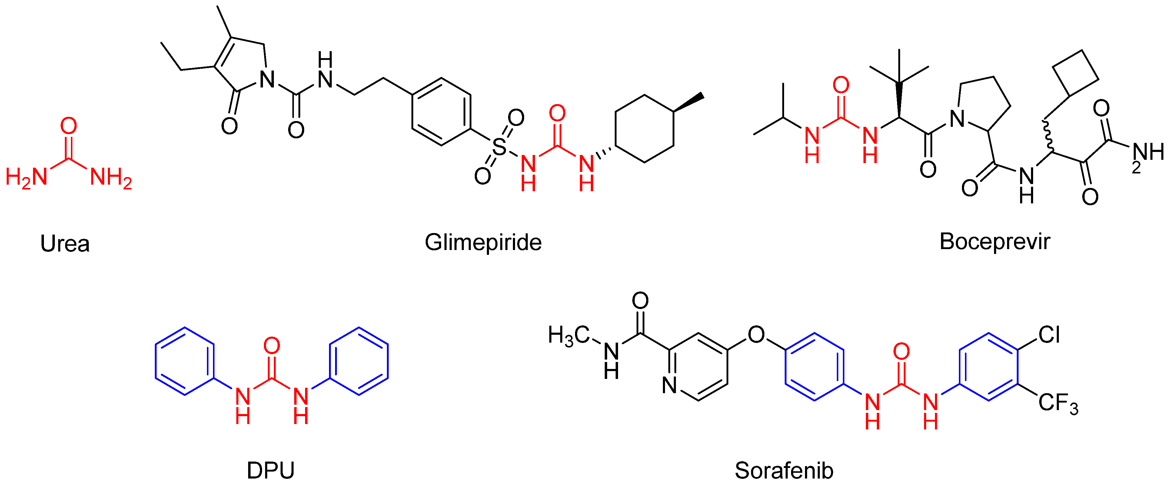
1. Introduction
2. Results and Discussion
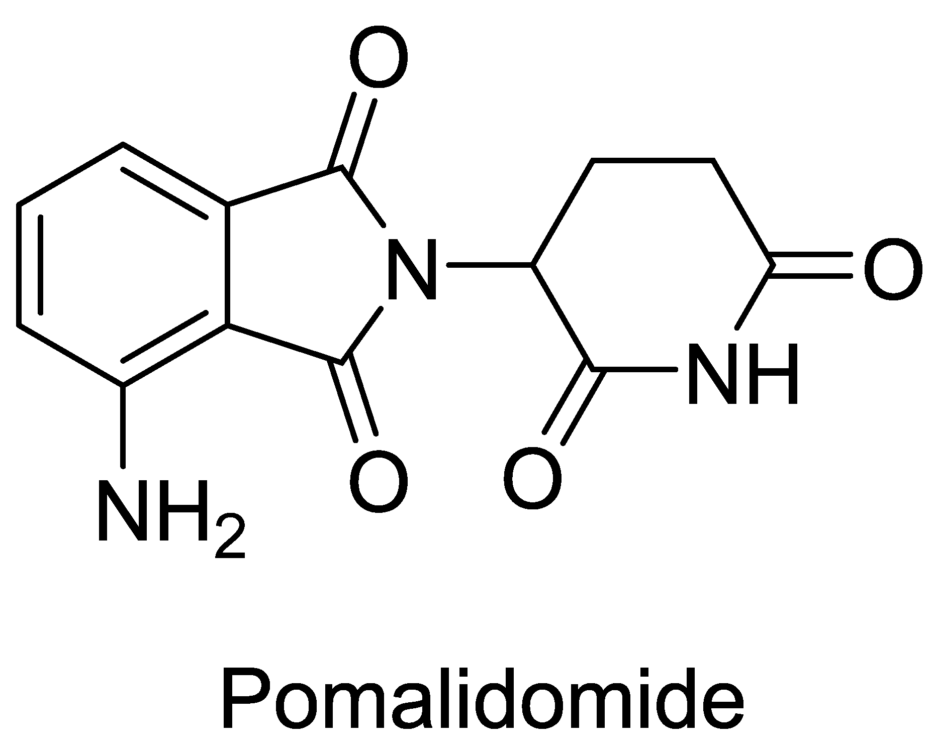
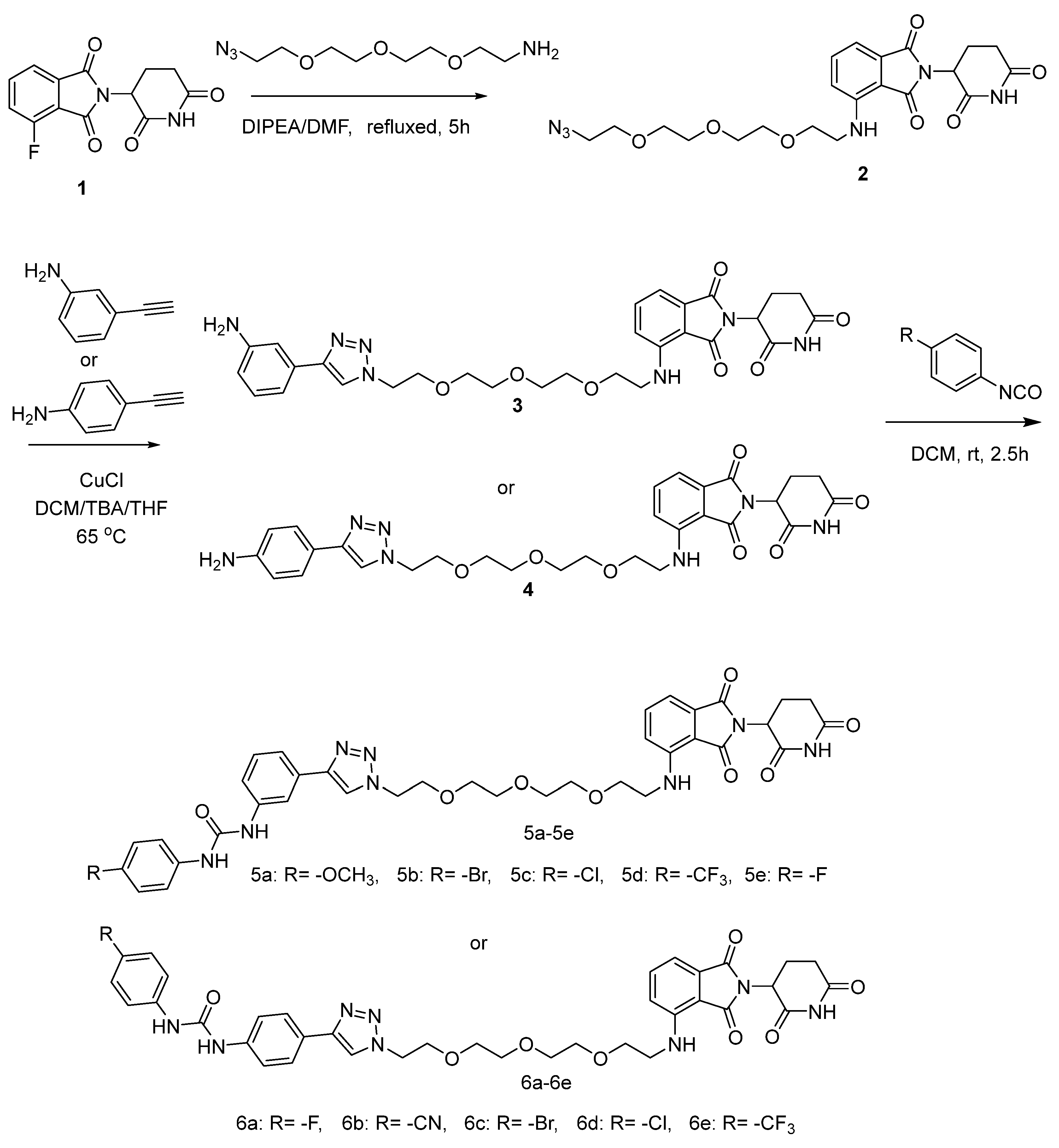
2.1. Chemistry
2.2. Biology
2.2.1. Effects of Novel Pomalidomide Derivatives Containing Urea Moieties on Cells Viability in Different Cancer Cell Lines
2.2.2. Novel Compound 5d Suppressed Cancer Cell Proliferation
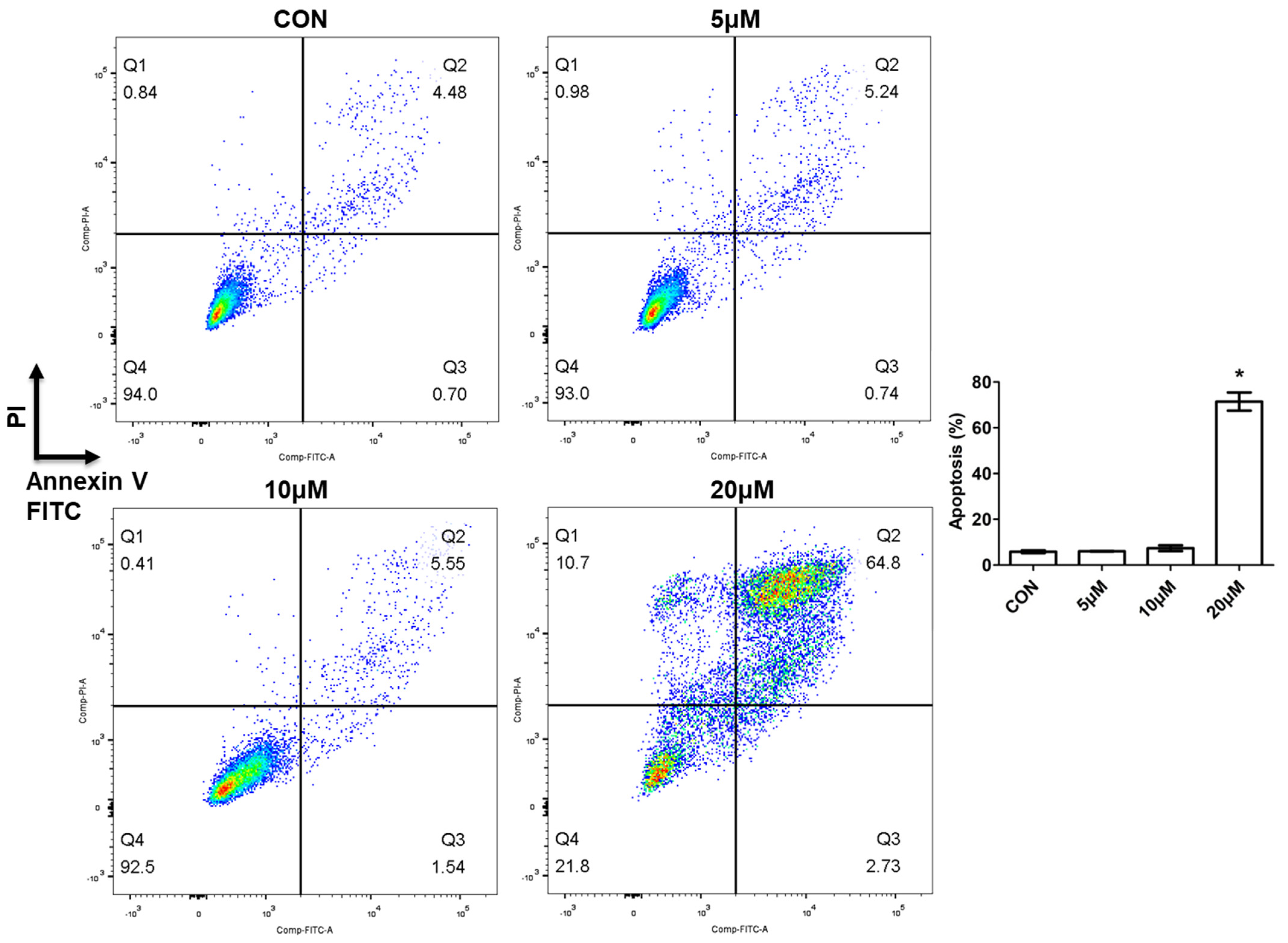
2.2.3. Novel Compound 5d Induced Cell Apoptosis
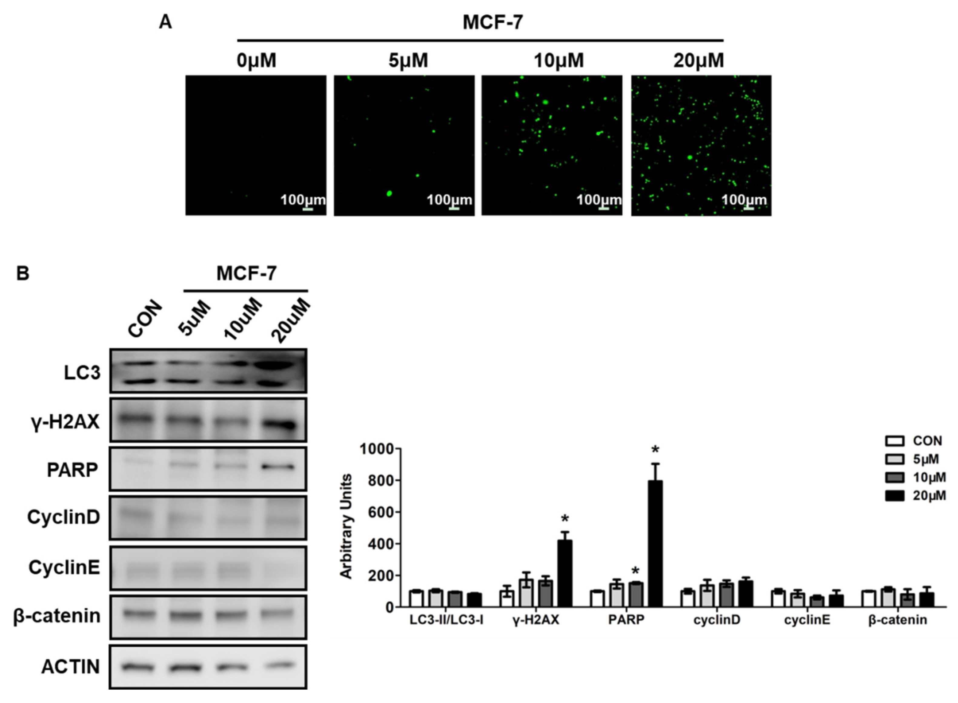
2.2.4. Novel Compound 5d Increased ROS Levels and Induced DNA Damage
3. Materials and Methods
3.1. Materials and Chemistry
3.2. General Procedure for Preparation of Compound 2
3.3. General Procedure for the Synthesis of Compound 3 (The Method Is Suitable for Compound 4)
3.4. General Procedure for the Preparation of Compound 5a (The Method Is Suitable for 5b~5e, 6a–6e)
3.5. Cell Culture
3.6. Cell Viability Assay
3.7. Live and Dead Cells Assay
3.8. Plate Clone Formation Assay
3.9. ROS Measurement
3.10. Apoptosis Measurement
3.11. Western Blot
3.12. Statistical Analyses
4. Conclusions
Supplementary Materials
Author Contributions
Funding
Institutional Review Board Statement
Informed Consent Statement
Data Availability Statement
Conflicts of Interest
References
- Boehncke, W.-H. Immunomodulatory drugs for psoriasis. BMJ 2003, 327, 634. [Google Scholar] [CrossRef]
- Dan, Z.; Corral, L.G.; Fleming, Y.W.; Stein, B. Immunomodulatory drugs Revlimid (lenalidomide) and CC-4047 induce apoptosis of both hematological and solid tumor cells through NK cell activation. Cancer Immunol. Immunother. 2008, 57, 1849–1859. [Google Scholar] [CrossRef]
- Lacy, M.Q.; Mccurdy, A.R. Pomalidomide. Blood 2013, 122, 2305–2309. [Google Scholar] [CrossRef] [PubMed]
- Richardson, P.G.; Siegel, D.S.; Vij, R.; Hofmeister, C.C.; Anderson, K.C. Pomalidomide alone or in combination with low-dose dexamethasone in relapsed and refractory multiple myeloma: A randomized phase 2 study. Blood 2014, 123, 1826–1832. [Google Scholar] [CrossRef]
- Escoubet-Lozach, L.; Lin, I.L.; Jensen-Pergakes, K.; Brady, H.A.; Gandhi, A.K.; Schafer, P.H.; Muller, G.W.; Worland, P.J.; Chan, K.W.H.; Verhelle, D. Pomalidomide and lenalidomide induce p21 WAF-1 expression in both lymphoma and multiple myeloma through a LSD1-mediated epigenetic mechanism. Cancer Res. 2009, 69, 7347–7356. [Google Scholar] [CrossRef]
- Chari, A.; Suvannasankha, A.; Fay, J.W.; Arnulf, B.; Kaufman, J.L.; Ifthikharuddin, J.J.; Weiss, B.M.; Krishnan, A.; Lentzsch, S.; Comenzo, R. Daratumumab plus pomalidomide and dexamethasone in relapsed and/or refractory multiple myeloma. Blood 2017, 130, 974–981. [Google Scholar] [CrossRef]
- Ruchelman, A.L.; Man, H.W.; Zhang, W.; Chen, R.; Capone, L.; Kang, J.; Parton, A.; Corral, L.; Schafer, P.H.; Babusis, D. Isosteric analogs of lenalidomide and pomalidomide: Synthesis and biological activity. Bioorg. Med. Chem. Lett. 2013, 23, 360–365. [Google Scholar] [CrossRef]
- Kelly, K.R.; Siegel, D.S.; Chanan-Khan, A.A.; Somlo, G.; Anderson, K.C. Indatuximab Ravtansine (BT062) in Combination with Low-Dose Dexamethasone and Lenalidomide or Pomalidomide: Clinical Activity in Patients with Relapsed/Refractory Multiple Myeloma. Blood 2016, 128, 4486. [Google Scholar] [CrossRef]
- Seki, J.; Sakurai, N.; Lam, W.; Reece, D. Pomalidomide desensitization in a patient hypersensitive to immunomodulating agents. Curr. Oncol. 2017, 24, 328–332. [Google Scholar] [CrossRef][Green Version]
- Owen, W.F., Jr.; Lew, N.L.; Liu, Y.; Lowrie, E.G.; Lazarus, J.M. The urea reduction ratio and serum albumin concentration as predictors of mortality in patients undergoing hemodialysis. N. Engl. J. Med. 1993, 329, 1001–1006. [Google Scholar] [CrossRef]
- Nakagawa, T.; Lomb, D.J.; Haigis, M.C.; Guarente, L.P. SIRT5 Deacetylates Carbamoyl Phosphate Synthetase 1 and Regulates the Urea Cycle. Cell 2009, 137, 560–570. [Google Scholar] [CrossRef]
- Fling, S.P.; Gregerson, D.S. Peptide and protein molecular weight determination by electrophoresis using a high-molarity tris buffer system without urea. Anal. Biochem. 1986, 155, 83–88. [Google Scholar] [CrossRef]
- Nauck, M.; Frid, A.; Hermansen, K.; Thomsen, A.B.; During, M.; Shah, N.; Tankova, T.; Mitha, I.; Matthews, D.R. Long-term efficacy and safety comparison of liraglutide, glimepiride and placebo, all in combination with metformin in type 2 diabetes: 2-year results from the LEAD-2 study. Diabetes Obes. Metab. 2013, 15, 204–212. [Google Scholar] [CrossRef]
- Kiser, J.J.; Burton, J.R.; Anderson, P.L.; Everson, G.T. Review and management of drug interactions with boceprevir and telaprevir. Hepatology 2012, 55, 1620–1628. [Google Scholar] [CrossRef]
- Escudier, B. Sorafenib in advanced clear-cell renal-cell carcinoma. N. Engl. J. Med. 2007, 356, 125–134. [Google Scholar] [CrossRef]
- Liu, L.; Cao, Y.; Chen, C.; Zhang, X.; Carter, C. Sorafenib Blocks the RAF/MEK/ERK Pathway, Inhibits Tumor Angiogenesis, and Induces Tumor Cell Apoptosis in Hepatocellular Carcinoma Model PLC/PRF/5. Cancer Res. 2007, 66, 11851–11858. [Google Scholar] [CrossRef]
- Raza, A.; Singh, A.; Amin, S.; Spallholz, J.E.; Sharma, A.K. Identification and biotin receptor-mediated activity of a novel seleno-biotin compound that inhibits viability of and induces apoptosis in ovarian cancer cells. Chem.-Biol. Interact. 2022, 365, 110071. [Google Scholar] [CrossRef]
- D’Autreaux, B.; Toledano, M.B. ROS as signalling molecules: Mechanisms that generate specificity in ROS homeostasis. Nat. Rev. Mol. Cell Biol. 2007, 8, 813–824. [Google Scholar] [CrossRef]
- Moloney, J.N.; Cotter, T.G. ROS signalling in the biology of cancer. Semin. Cell Dev. Biol. 2018, 80, 50–64. [Google Scholar] [CrossRef]
- Hayes, J.D.; Dinkova-Kostova, A.T.; Tew, K.D. Oxidative Stress in Cancer. Cancer Cell 2020, 38, 167–197. [Google Scholar] [CrossRef]
- Roos, W.P.; Thomas, A.D.; Kaina, B. DNA damage and the balance between survival and death in cancer biology. Nat. Rev. Cancer 2016, 16, 20–33. [Google Scholar] [CrossRef]
- Yang, Y.; Gao, H.; Sun, X.; Sun, Y.; Qiu, Y.; Weng, Q.; Rao, Y. Global PROTAC Toolbox for Degrading BCR-ABL Overcomes Drug-Resistant Mutants and Adverse Effects. J. Med. Chem. 2020, 63, 8567–8583. [Google Scholar] [CrossRef]


| Compd No. | IC50 (μM) | |
|---|---|---|
| MCF-7 | Huh7 | |
| 5a | 86.57 ± 8.59 | >200 |
| 5b | 114.13 ± 6.91 | >200 |
| 5c | 26.93 ± 2.40 | 132.27 ± 2.18 |
| 5d | 20.2 ± 5.70 | >200 |
| 5e | 53.44 ± 0.49 | >200 |
| 6a | >200 | >200 |
| 6b | >200 | >200 |
| 6c | 150.73 ± 5.90 | >200 |
| 6d | 107.60 ± 13.17 | >200 |
| 6e | 99.14 ± 9.51 | >200 |
| con1 | >200 | >200 |
| con2 | >200 | >200 |
| con3 | >200 | >200 |
Publisher’s Note: MDPI stays neutral with regard to jurisdictional claims in published maps and institutional affiliations. |
© 2022 by the authors. Licensee MDPI, Basel, Switzerland. This article is an open access article distributed under the terms and conditions of the Creative Commons Attribution (CC BY) license (https://creativecommons.org/licenses/by/4.0/).
Share and Cite
Guo, Y.; Wang, X.; Wang, Z.; Mao, L.; Wang, J.; Peng, L.; Xu, G. Synthesis and Anti-Tumor Effects of Novel Pomalidomide Derivatives Containing Urea Moieties. Pharmaceuticals 2022, 15, 1479. https://doi.org/10.3390/ph15121479
Guo Y, Wang X, Wang Z, Mao L, Wang J, Peng L, Xu G. Synthesis and Anti-Tumor Effects of Novel Pomalidomide Derivatives Containing Urea Moieties. Pharmaceuticals. 2022; 15(12):1479. https://doi.org/10.3390/ph15121479
Chicago/Turabian StyleGuo, Yajie, Xi Wang, Zhenzhen Wang, Longfei Mao, Jiahao Wang, Lizeng Peng, and Guiqing Xu. 2022. "Synthesis and Anti-Tumor Effects of Novel Pomalidomide Derivatives Containing Urea Moieties" Pharmaceuticals 15, no. 12: 1479. https://doi.org/10.3390/ph15121479
APA StyleGuo, Y., Wang, X., Wang, Z., Mao, L., Wang, J., Peng, L., & Xu, G. (2022). Synthesis and Anti-Tumor Effects of Novel Pomalidomide Derivatives Containing Urea Moieties. Pharmaceuticals, 15(12), 1479. https://doi.org/10.3390/ph15121479