Article

Synthesis and Glycosidase Inhibition Properties of Calix[8]arene-Based Iminosugar Click Clusters

1
Laboratoire d’Innovation Moléculaire et Applications (LIMA), University of Strasbourg | University of Haute-Alsace | CNRS (UMR 7042), Equipe de Synthèse Organique et Molécules Bioactives (SYBIO), ECPM, 25 Rue Becquerel, 67000 Strasbourg, France
2
Laboratory of Supramolecular Chemistry (SupraLab@UniSa), Dipartimento di Chimica e Biologia “A. Zambelli”, Università di Salerno, Via Giovanni Paolo II 132, I-84084 Fisciano, Italy
3
Laboratoire Vigne Biotechnologies et Environnement EA-3991, University of Haute-Alsace, 33 rue de Herrlisheim, 68008 Colmar CEDEX, France
*
Authors to whom correspondence should be addressed.
Pharmaceuticals 2020, 13(11), 366; https://doi.org/10.3390/ph13110366
Submission received: 14 October 2020 / Revised: 28 October 2020 / Accepted: 2 November 2020 / Published: 5 November 2020
(This article belongs to the Special Issue Glycomimetics and Glycoconjugates in Drug Discovery)

Abstract

:
A set of 6- to 24-valent clusters was constructed with terminal deoxynojirimycin (DNJ) inhibitory heads through C6 or C9 linkers by way of Cu(I)-catalyzed azide-alkyne cycloaddition (CuAAC) reactions between mono- or trivalent azido-armed iminosugars and calix[8]arene scaffolds differing in their valency and their rigidity but not in their size. The power of multivalency to upgrade the inhibition potency of the weak DNJ inhibitor (monovalent DNJ Ki being at 322 and 188 µM for C6 or C9 linkers, respectively) was evaluated on the model glycosidase Jack Bean α-mannosidase (JBα-man). Although for the clusters with the shorter C6 linker the rigidity of the scaffold was essential, these parameters had no influence for clusters with C9 chains: all of them showed rather good relative affinity enhancements per inhibitory epitopes between 70 and 160 highlighting the sound combination of the calix[8]arene core and the long alkyl arms. Preliminary docking studies were performed to get insights into the preferred binding modes.

Graphical Abstract

1. Introduction

Over the last 30 years a growing interest has been directed towards the molecular recognition of druggable biomolecular targets with calixarene derivatives [1,2,3,4,5,6,7]. This is due to the synthetic and conformational versatility of calixarenes [8,9,10] coupled with their ability to establish multiple interactions with biomolecules [1,2,3,4,5,6,7], which make them a class of compounds suitable for potential applications in the field of biopharmaceutical sciences. Several systems in biology interact through multiple simultaneous molecular contacts, and this concept has been adopted as a new strategy for the design and development of drug candidates. Following this approach, Hamilton and co-workers introduced at the upper rim of a conformationally-blocked, cone-shaped calix[4]arene scaffold four peptide functions which were able to establish multiple interactions with a PDGF growth factor, thus giving rise to antiangiogenic activity in vivo [11,12,13]. In another work, docking studies suggested that multiple H-bond and hydrophobic interactions play a crucial role in the interaction between a p-acetamidocalix[4]arene derivative and the protein disulfide isomerase (PDI), which is highly expressed in cancer cell types, including lung, brain, ovarian, melanoma, and prostate [14]. In a further work, in vitro studies [15] showed that conformationally-blocked calix[4]arene derivatives bearing aromatic naphthyl, pyrenyl, and aryl groups at the upper rim were able to inhibit HDAC enzymes, while docking calculations suggested that multiple hydrophobic interactions between the aromatic arms and the hydrophobic pockets close to the active site of the enzymes were essential for the stabilization of the HDAC-calixarene complex. Sansone and Fieschi also used calix[4]arene and calix[6]arene grafted with mannose residues to prevent binding of high mannose structures to the DC-SIGN receptor and thus potentially prevent HIV entry into immature dendritic cells [16]. In another example, Geraci and Spadaro showed that a calix[8]arene carrying eight antigens induced an improved antibody production in mice beyond the additive affect compared to its calix[4]arene counterpart [17]. Recently, glycosidase inhibitors have gained considerable clinical relevance [18,19,20,21] and several glycomimetics such as zanamivir®, miglustat®, migalastat®, voglibose®, and acarbose® reached the market. The selective hydrolysis of glycosidic bonds in carbohydrates and glycoconjugates is indeed involved in a plethora of key cellular processes in biological systems [22,23,24,25], and thus targeting glycosidases holds great potential for the treatment of many diseases such as type 2 diabetes [25,26,27], viral infection [21,27,28,29,30,31], tumor metastasis [21,27,32,33], or medical diagnosis [34].
Ten years ago, a breakthrough in the field of glycosidase inhibition was provided by the discovery of the large inhibitory multivalent effect for iminosugar-based multivalent clusters [35,36,37,38,39,40,41,42,43]. The simultaneous non-covalent interactions between several inhibitory epitopes (inhitopes) and one or several glycosidases provides higher specificity, thermodynamic, and kinetic stability compared to one single interaction and can be responsible for considerable affinity enhancements of up to 4700 per inhitope for the best multivalent effect obtained to date with compound 1e [44].
The rapid development of this field after the discovery of the first strong multivalent effect [35], was mainly based on structure–activity relationships established from the screening of libraries of multivalent glycomimetics [37,38,39,40,41,42]. Different physical techniques allowed for some rationalization of the observed effects [44,45,46,47,48,49,50,51]. More recently, the acquisition and solving of X-ray structures of two complexes between enzymes and a multimeric inhibitor allowed to draw explanations for the strongest inhibitory multivalent effect obtained to date with cluster 1e [50], or for an exceptional selectivity [49], and opened the way for a new area where rational design of multivalent inhibitors will be possible. Moreover, multivalent inhibitors have demonstrated their therapeutic potential and have proven to be able to cross membranes by in vitro [52,53] and in vivo [54] experiments. In this project, we wanted to build new multivalent constructs by associating the advantages of calixarene scaffolds with those of 1-deoxynojirimycin (DNJ), the inhitope that has led to the best inhibitory multivalent effect reported so far [44]. DNJ is indeed a moderate and non-selective glycosidase inhibitor that has a high potential for affinity improvement through multivalency [55]. The nature of the scaffold as well as the spatial distribution of the ligands is crucial for the modulation of the inhibitory activity and selectivity. At this regard, calix[n]arenes are particularly suitable as scaffolds for the synthesis of multivalent ligands, thanks to their variable number of reactive positions for inhitope attaching and thanks to the possibility of modulating their spatial orientation. Relatively few examples of calixarene-based iminosugar clusters have been synthesized so far. Tetravalent calix[4]arenes, either fixed in the 1,3-alternate [56] or in the cone structure, bearing four DNJ units at the upper [57] or lower [56,57] rim have been reported independently by Gouin and Marra, but showed modest multivalent effects for the inhibition of JBα-man except for rigid cluster 2 having an affinity enhancement per iminosugar of 70 and inducing aggregation of the enzyme. In all those clusters, the linkers between the inhitope and the scaffold were short— three to five methylene groups in total on both sides of the triazole—and the valency was only four. Previous studies indeed showed that longer linkers had a positive impact on the inhibitory multivalent effect [37,42]. In parallel, clusters 1a,b [58] of relatively low valency (6 to 8) and based on flexible cyclopeptoid scaffolds also lead to weak multivalent effects. Tripling the initial valency of these scaffolds by the grafting of trivalent azido-armed dendrons dramatically increased the multivalent effect by one to two orders of magnitude (see compounds 1a–e in Figure 1) [44].
Prompted by these considerations, we decided to synthesize a small library of DNJ-clusters based on the larger calix[8]arene macrocycle as scaffold, which can ensure a higher valency, with up to eight alkynes on the lower rim. A second more rigid 1,5-xylylene bridged [59] calix[8]arene scaffold was also used to probe valency and flexibility modulation. In order to evaluate further the influence of the chain length, but also valency and cluster global size, we grafted C6- and C9- alkylated DNJ heads and also a trivalent DNJ-dendron (Figure 2). The inhibition power of the different clusters has been determined on Jack Bean α-mannosidase (JBα-man) as a model enzyme which is the most sensitive to multivalent presentation known so far. JBα-man is indeed a commercially available source of GH 38 mannosidase and shares 29% of identity with a significant E-value of 2.10−95 with human Golgi α-mannosidase II as found by a Blast search with full sequence of JBα-man and lumenal domain of homo sapiens Golgi α-mannosidase II. The latter enzyme is a key member of the early steps of N-glycans trimming and has not been crystallized so far. Aberrant N-glycans at the tumor surface are associated with cancer growth and metastasis [60]. The inhibitor of lysosomal and Golgi α-mannosidase II (-)-swainsonine reached phase II in a clinical trial, highlighting the potential of α-mannosidase inhibitors [32].

2. Results and Discussion
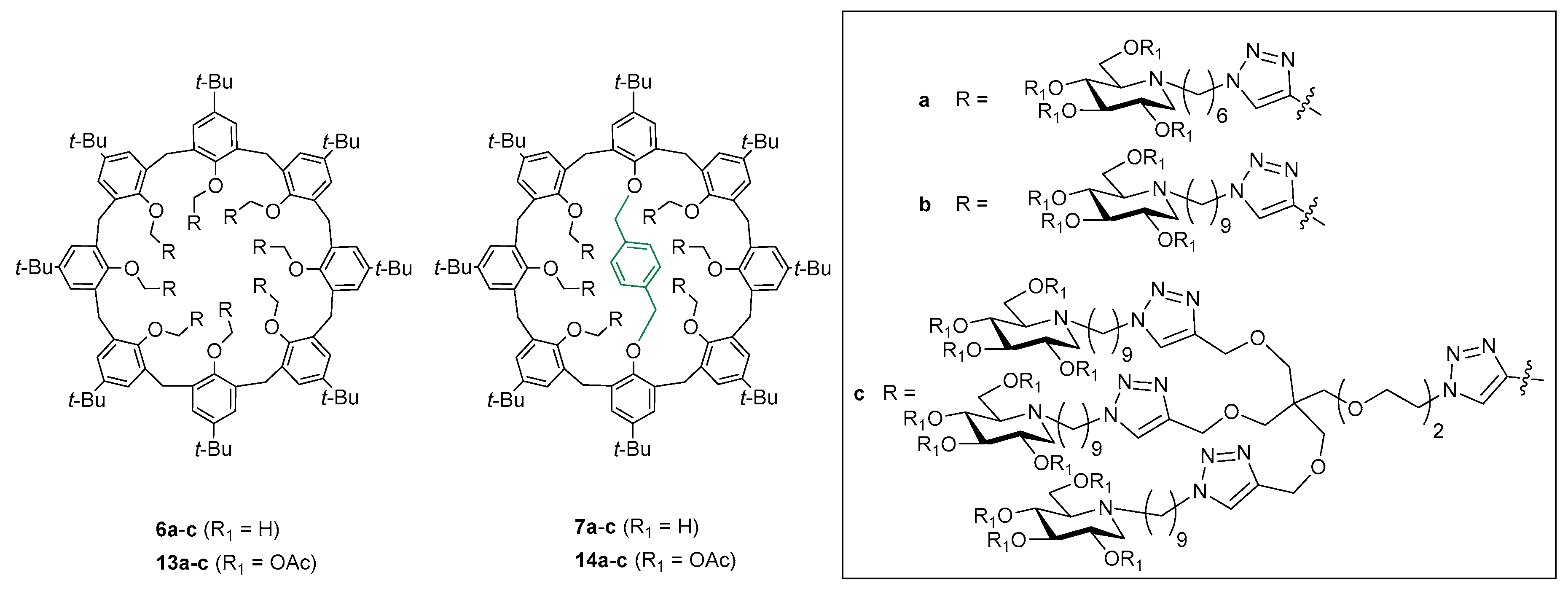
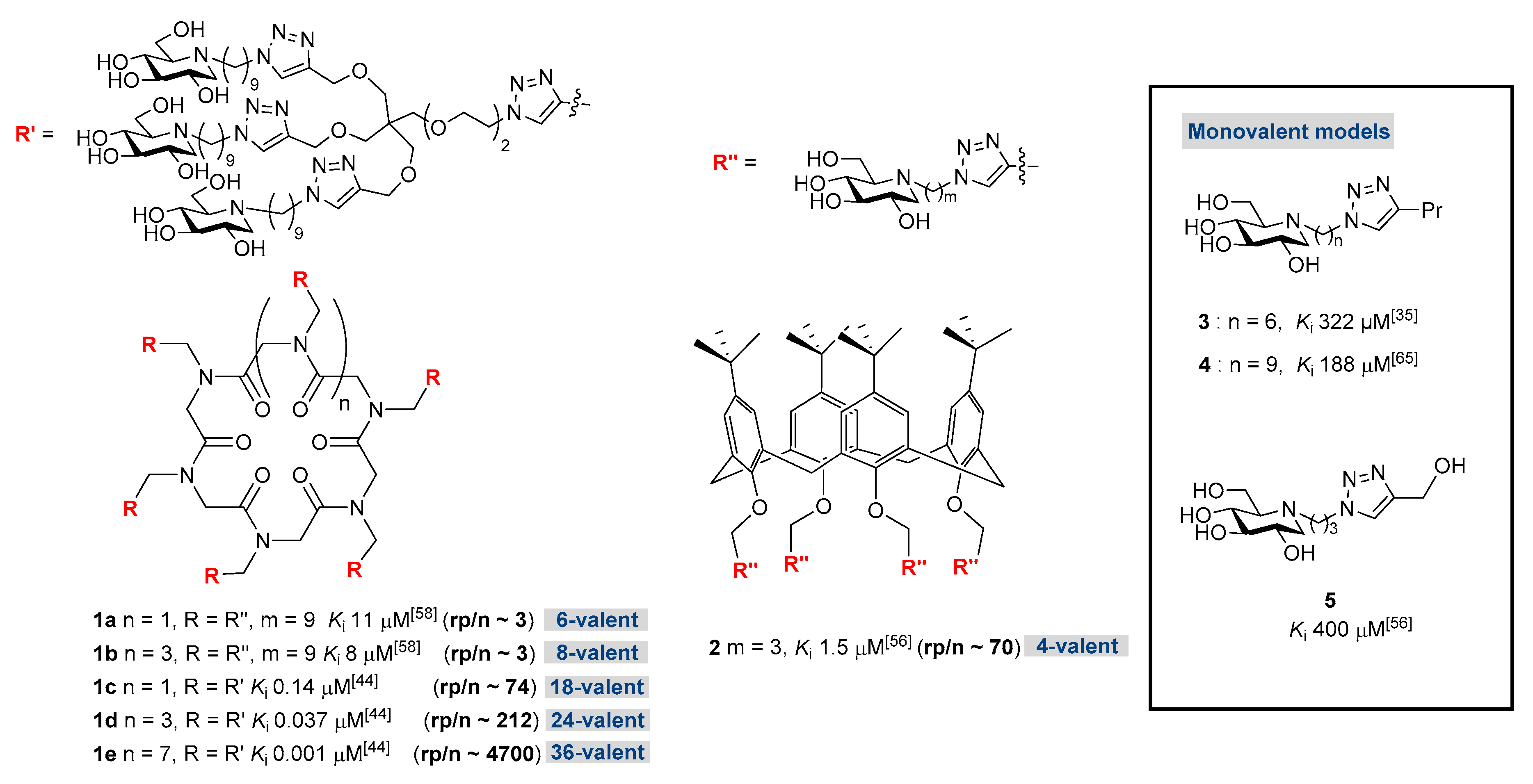
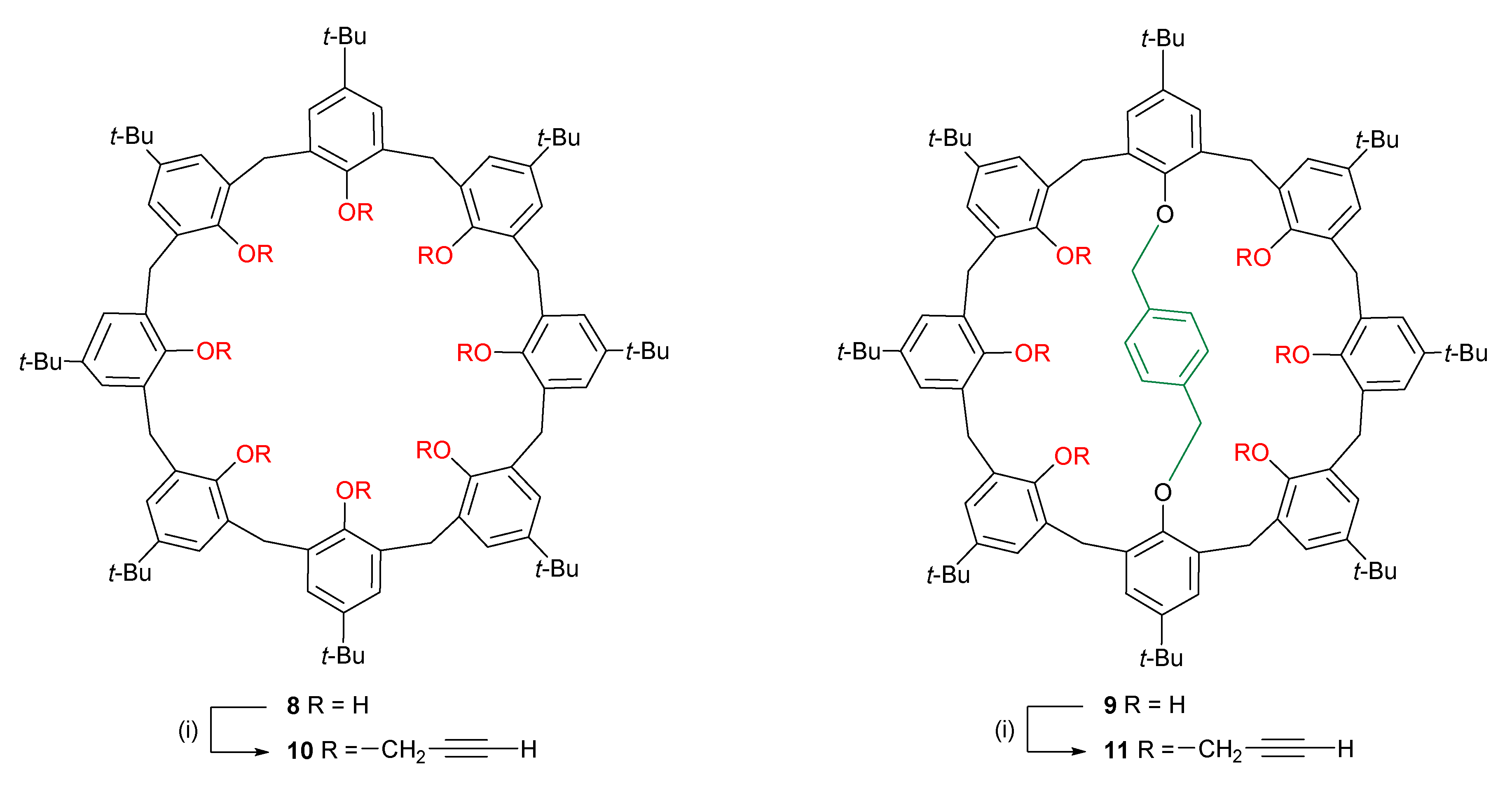
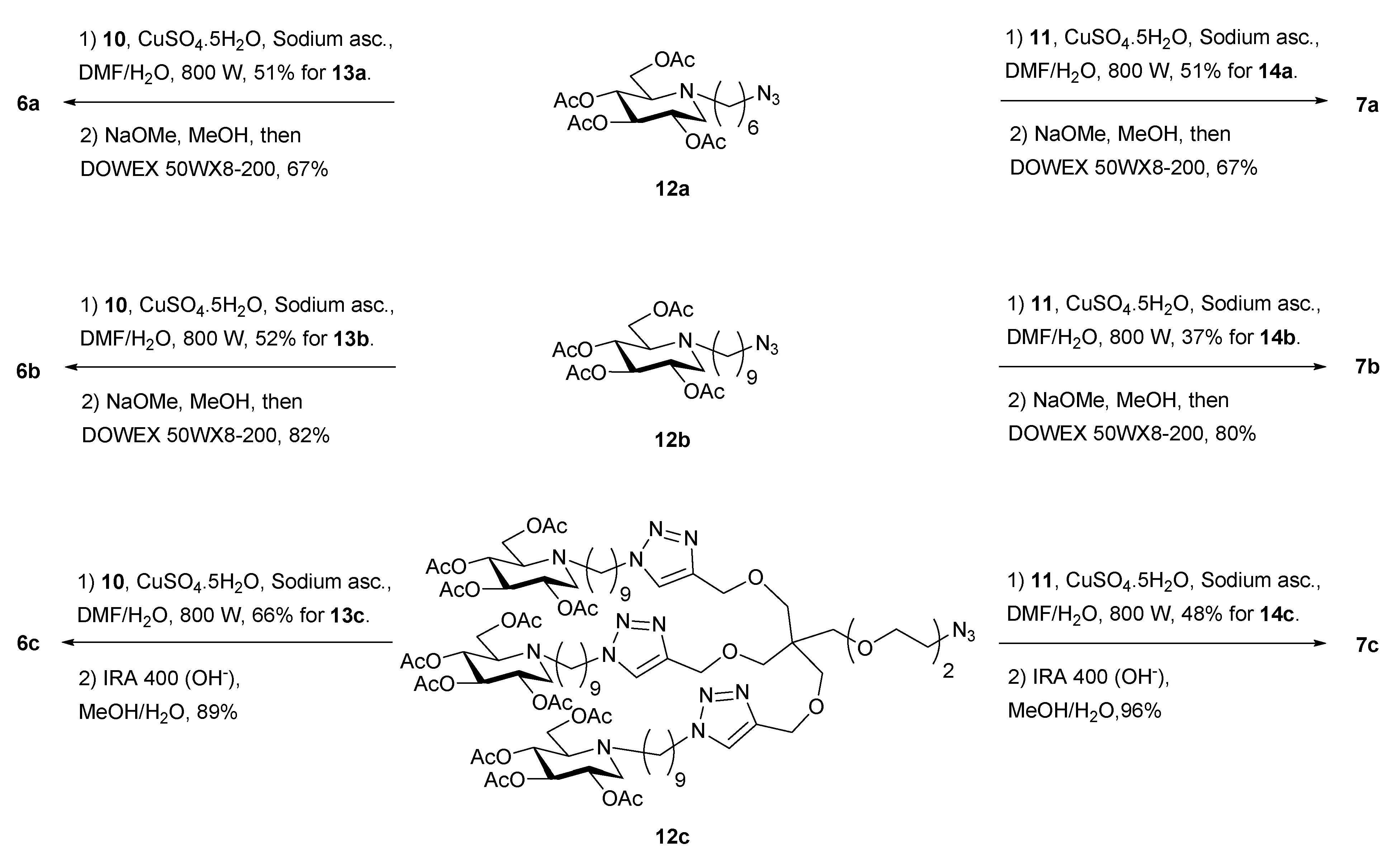
2.1. Chemistry

For the sake of efficiency, the synthesis of multivalent clusters usually relies on click reactions as defined by Sharpless [61]. The coupling of calix[4]arenes to get different neoglycoconjugates by amide coupling [62], nitrone cycloaddition [63], or photoinduced radical thiol-ene coupling [64] has already been used besides Cu(I)-catalyzed azide-alkyne cycloaddition (CuAAC) [56,57]. In our case, the general strategy towards targets 6 and 7 (Figure 2) differing by the nature of their central calixarene core and their DNJ coating, was to graft azide-armed DNJ ligands [65] or an azido-trivalent dendron [66] on calix[8]arene scaffolds by Cu(I)-catalyzed azide-alkyne cycloaddition (CuAAC).
For this purpose, full propargylation of p-tert-butylcalix[8]arene 8 [67] and the 1,5-p-xylylene-bridged tert-butylcalix[8]arene 9 [59] with propargyl bromide were performed under classical conditions [68] in the presence of Cs2CO3 as a base in acetone and conducted to propargylated calix[8]arenes 10 and 11 (Scheme 1). To evaluate the influence of the spacer length on the inhibitory activity, two different peracetylated azide-armed DNJ iminosugars 12a [45] and 12b [65] bearing, respectively, 6- or 9-carbon alkyl spacers have been grafted by CuAAC onto scaffolds 10 and 11 (Figure 2, Scheme 2). The click reactions were performed under classical conditions using copper sulfate and sodium ascorbate under microwave irradiation to give the corresponding peracetylated clusters 13a,b and 14a,b (Figure 2). Copper salts were removed by filtration over a small pad of silica using a solution of CH3CN/AcOEt/(30%) NH4OH before column chromatography, a procedure that previously showed its efficiency as proven by ICP-AES analysis of copper traces in final clusters [44]. Derivatives 13a,b and 14a,b were characterized by 1D and 2D NMR spectroscopy and ESI(+) MS (see SI).
The C8 symmetry of compound 13a is nicely shown in the 1H NMR spectrum (Figure S12) with only one singlet at δ = 0.89 ppm for all t-Bu groups, one singlet for the aromatic hydrogen atoms of the calix[8]arene skeleton at δ = 6.80 ppm and a singlet attributable to the triazole ring hydrogen atom at δ = 7.78 ppm. Similar spectra are found for the related 13b compound differing only from the linker length. The 13a,b spectra are averaged spectra of fast interconverting conformations in the NMR time scale. The interconversion is possible even with t-butyl groups because of the larger size of the calix[8]arene macrocycle compared to the calix[4]arene one [69].
Due to the presence of the 1,5-(p-xylylene) intramolecular bridge, derivative 14b has an overall C2 symmetry but shows characteristics of a C2v symmetry locally at the calix[8]arene core, with two perpendicular vertical symmetry planes and a C2 axis in their intersection, as reported previously with a derivative of 1,5-m-xylene-diyl bridged tert-butylcalix[8]arene (see SI Figure S38) [70]. Remarkably, its 1H NMR spectrum shows a thin singlet at δ = 3.34 ppm for the diethereal methylene of the intramolecular bridge showing nicely their equivalence due to the local C2v symmetry. The other methylene protons of the ring appear as two AX systems at 3.43/4.78 ppm (J = 14.2 Hz); and 3.51/4.39 ppm (J = 16.6 Hz), respectively, showing the rigidity and cone shape of the bridged calix[8]arene. Similarly, the t-butyl groups appear as three singlets at 0.41, 1.18, and 1.39 ppm. Two singlets attributed to triazole protons, one arising from the overlapped aromatics at 7.26 and the other one at 7.78 ppm are integrated in a 2:1 ratio. In addition, the presence of only five types of aromatic integrating for 4H as singlets at 5.84, 6.6, 6.96, 7.24, and 7.26 ppm further proves the symmetry. The DNJ heads which are further away from the core appear totally equivalent for all different arms with single signals for all the DNJ carbons. The 2D NMR spectra indeed allowed the full attribution of the 1H and 13C NMR spectra. Remarkably, the closely related compound 14a showed a loss of symmetry as seen by the non-equivalent aromatics of half the core and a more complex pattern of methylene protons of the rim. This may be due to the shorter linkers which bring closer the chiral DNJ heads, abolishing the apparent local C2v symmetry observed for 14b. The 1H NMR of cluster 14a thus exhibited exclusively a C2 symmetry showing two singlets attributable to triazole H-atoms at 7.22 and 7.77 ppm and 4 singlets (18 H each) attributable to the t-butyl groups at 0.40, 1.18, 1.19, and 1.42 ppm. In addition, two sets of signals for the hydrogen on the C1-position of the iminosugar units at 3.10 and 3.18 ppm integrate for four and two protons, respectively. These two different types of protons showed correlation with the corresponding geminal partners at δ = 2.26–2.30 (overlapped). The singlet at δ = 3.32 ppm was attributed to the aliphatic protons of the intramolecular bridge, while the corresponding aromatic protons were found at δ = 5.82 ppm. The signals of the aromatic protons of the calix[8]arene core were found at δ = 6.58, 6.61, 6.95, 6.98, and 7.22–7.28 ppm (overlapped), while the hydrogen atoms on the triazole rings were found at δ = 7.22–7.28 (overlapped with the aromatic signals) and 7.77 ppm.
Derivatives 13a,b and 14a,b have a valency of 8 and 6, respectively. In order to increase the valency of clusters by a factor of three, we adopted the strategy relying on the grafting of clickable trivalent dendron 12c [66], allowing the fast and efficient synthesis of iminosugar clusters with higher valencies. In this way, propargylated calix[8]arene 10 and 11 were reacted by CuAAc with dendron 12c to afford the 24-valent cluster 13c and the 18-valent cluster 14c in 66% and 48% yields, respectively. 1H NMR analysis of compound 13c shows again the C8 symmetry with well resolved signals of the iminosugar part, and single singlet signals for t-butyl groups, aromatic protons, and methylene groups of the core. Those singlets are, however, broader than for compounds 13a and 13b, suggesting a slower interconversion on the NMR time scale. 13C NMR also shows well resolved signals for the dendron part, but the calix[8]arene core is less resolved even at a higher number of scans due to the dilution of signals among the more intense ones of the dendritic part in addition to the slower interconversion. The t-butyl signal appears at 31.1 ppm as a broad signal and at 35.2 ppm for its quaternary center. The core methylene at 29.8 ppm, the aromatic C–H at 124.6 ppm, and the quaternary carbon at 144.2 ppm are also clearly visible as single signals. The ESI-MS of several ions of a multiply charged cluster further confirm the grafting of all positions of the scaffold.
Careful analysis of NMR spectra of compound 14c indicates the presence of mixture of atropoisomers. As for 14a,b, all iminosugars are equivalent in NMR and very well resolved as they are far from the core center. However, unlike the singlet observed for the bridge’s aromatic for compounds 14a and 14b at 5.8 pm, a broad peak at 5.78 ppm and a smaller one at 5.85 ppm integrate together for four protons. In a similar way, the t-butyl of the aromatic linked to the bridge appears as a major broad singlet at 0.37 and several other ones. Triazole protons of the dendrons appear as two singlets integrating for nine protons and the triazoles appear close to the calixarene core as several singlets. The characteristic methylenic protons of the core are unfortunately hidden by protons of the oligoethylene part of the dendron. The formation of atropoisomers for this compound can be explained by the fact that its precursor 11 undergoes conformational interconversion because the propargyl groups are not bulky enough to prevent it and are in accordance with the previously reported isolation of atropisomeric derivatives of 9 [71]. During the course of the CuAAC reaction, different blocked conformations arose by progressive grafting of the six large dendrons randomly. The result is a mixture of atropoisomers of the same valency that would be difficult to isolate. Full deprotection of clusters 13a,b and 14a,b into 6a,b and 7a,b, respectively, was achieved by treatment with NaOMe in dry methanol followed by protonation of the alcohols using Dowex 50WX8-200 (H+) ion-exchange resin, filtration and solvent and by-products evaporation. Multivalent clusters of higher valency 13c and 14c were deprotected with Amberlite IRA 400 (OH) resin allowing directly fully deprotected clean compounds 6c and 7c in quantitative yields (Scheme 2). Derivatives 6 and 7 were characterized by 1D and 2D NMR spectroscopy and ESI(+) MS. Clusters 6 are consistent with a C8 symmetry as their precursors, even if 6c has broader signals for the calix[8]arene core due to slower conformational inversion. Clusters 7a,b derived from the hexa-valent scaffold are nicely resolved with a C2 symmetry, but the dendritic 7c is obtained as an atropoisomer mixture as observed for their precursors. We decided to keep the mixture for final evaluation of inhibitory activity, being aware that only the valency effect and not the spatial arrangement would be taken into discussion.

2.2. Biological Assays

The inhibition potency of compounds 6 and 7 was then assayed against JBα-man. All clusters of small size 6a,b and 7a,b, directly derived from alkyl-DNJ were competitive inhibitors of this enzyme (Table 1, entry 3–6, Figures S83, S84 and S88, S89). As observed in a previous study with cyclodextrin-based clusters [65], the three methylene lengthening of the alkyl chain improves the affinity enhancement per inhibitory head (rp/n) of at least one order of magnitude (Table 1, entries 3 and 4, entries 5 and 6).
For the bridged calix[8]arene scaffold, the rp/n is improved by a factor of 10 (Table 1 entries 3 and 4), whereas a larger gain of 150 in rp/n is observed for the flexible calix[8]arene scaffold (Table 1 entries 5 and 6). Remarkably, the simple extension of the alkyl chain by three CH2 (from hexyl to nonyl) is thus sufficient to promote a 250-fold increase of inhibitory activity. This effect might be due to hydrophobic interactions between the chain and lipophilic glycine residues G788, G792, and G790 at the entry of the pocket as shown from the X-ray structure of compound 1e bound to JB α-man (Figure S7). The three supplementary methylene of the C9 chain indeed allow hydrophobic interaction with G788 which is probably missing with the shorter C6 chain. It is interesting to point out that for clusters with the smaller 6-carbon linker, there is a strong influence of the calixarene scaffold as shown by the one magnitude order difference in the Ki values and the absence of multivalent effect (rp/n < 1) for compound 6a (Table 1, entries 3 and 5). In this case, the more rigid scaffold, with the C2 symmetry, seems decisive. However, for the series with longer 9-carbon linkers, the scaffold rigidity has no impact on the inhibition constants and multivalent effects as shown by the comparison of entries 4 and 6 and 7 and 8 (Table 1). Interestingly, it seems that the higher degree of freedom obtained by the addition of three CH2 in the hexyl chain is sufficient to screen the mechanical strains due to some more rigid scaffold.
The jump in valency and cluster overall size by grafting trivalent dendron 12c has more impact on the inhibition and multivalent effect. Clusters 6c and 7c showed different inhibitory profiles. While cluster 7c has a mixed inhibition mode in the nanomolar range, 6c behavior was more difficult to analyze. Dixon plots showed with a good repeatability a strong nonlinearity (Figure S85). Moreover, double-reciprocal Lineweaver–Burk plots were suggesting a mixed inhibition; however, the respective secondary curves (slope or y-axis intercept as a function of inhibitor concentration) were non-linear (see Figure S86). The shape of those secondary curves did not correspond to hyperbolic partial mixed-type inhibition [72] and its assessment by the graphical method developed by Baici [73] rejected this inhibition modality. A possible slow-binding was searched by continuously following product formation over time in a longer time-scale with a decreased amount of enzyme and the perfect linearity observed showed the absence of a slow binding event. Finally, the competitive tight binding model was tested by fitting a Morrison equation [74,75] for fast tight binding competitive inhibition to the mean values of triplicate experiments. The good matching of the equation to our data gave us the inhibition constant of compound 6c (Ki = 50 ± 12 nM). The equations and protocol are described in our previous paper where we found a tight binding inhibition pattern for tri- tetra- and tetradecavalent clusters bearing inhibitors of higher affinity [55]. In this latter study, multimerisation of an enzyme-matching strong binding inhitope (mannoimidazole), whose monovalent inhibition was already in the nanomolar range (Ki 110 nM), led to a tight-binding inhibition with only three or four heads, whereas multimerization of a less good inhibitor (in the low micromolar range with Ki monovalent at 2.2 µM) could lead to tight binding only with a higher valency (14-valent). It is remarkable that a tight-binding behavior against the mannosidase is observed here for a cluster based on the low mismatching gluco inhibitor DNJ, which has a monovalent inhibition 100 times weaker, in the hundreds of the micromolar range. Presentation of 24 copies of DNJ allows us to reach an overall kinetic behavior that is attributed to high affinity inhibitors. The competitive tight binding behavior of cluster 6c is in agreement with the fixation of the DNJ heads into the active sites, as it has been observed in the X-ray structure of compound 1e with JB α-man [50]. Surprisingly, cluster 7c, is able to bind both the enzyme and the enzyme:substrate complex, but the latter is less favored with its higher uncompetitive component (Ki at 0.213 µM). This result, however, suggests that an access for the substrate remains when 7c is bound to the enzyme.
From a general point of view, the two larger clusters (in size and number of ligands) 6c and 7c show the best multivalent effects of the series, with an affinity increase per inhitope over a hundred, in the same range as those obtained with cyclopeptoid-based clusters of identical valencies, 24-valent DNJ 1d and 18-valent DNJ 1c, respectively (Figure 1) [44]. Due to conformational interconversion of the E/Z tertiary amide bonds, cyclopeptoids are known as a conformationally flexible class of scaffolds. The cyclopeptoid platforms are thus less rigid than the tert-butylcalix[8]arene scaffold, which itself is less rigid than its 1,5-xylylene-bridged analogue. The fact that close multivalent effects are found for dendron-derived clusters with three platforms of different rigidity further proves that the central core flexibility is not decisive for clusters constructed on trivalent dendron 12c. Those clusters are anyway very flexible due to the alkyl and oligoethylene chains and may allow dynamic reversible interactions with the enzymes, sometimes through a sandwich cross-linking complex as soon as their global size is large enough to link two JBα-man enzymes [44,50].
In contrast to cyclopeptoid-based clusters whose size is growing together with valency, the two calix[8]arene scaffolds of this study have the same size. Remarkably, all evaluated clusters with C9 alkyl chains show good multivalent effects (with rp/n values varying from 70 to 160) even for multimeric iminosugars of lower valencies (6-valent DNJ 7b and 8-valent DNJ 6b, Table 1). This is fairly good considering the fact that for the smaller cyclopeptoids 1a,b bearing also 6 and 8 DNJ units and the same C9 alkyl chains, the rp/n was of only 3 (Figure 1) [58]. The sole difference between pairs 1a/7b and 1b/6b being the scaffold, the gain in rp/n can be attributed to either the different ring size, different inhitopes local density or by a possible role of the aromatic core.

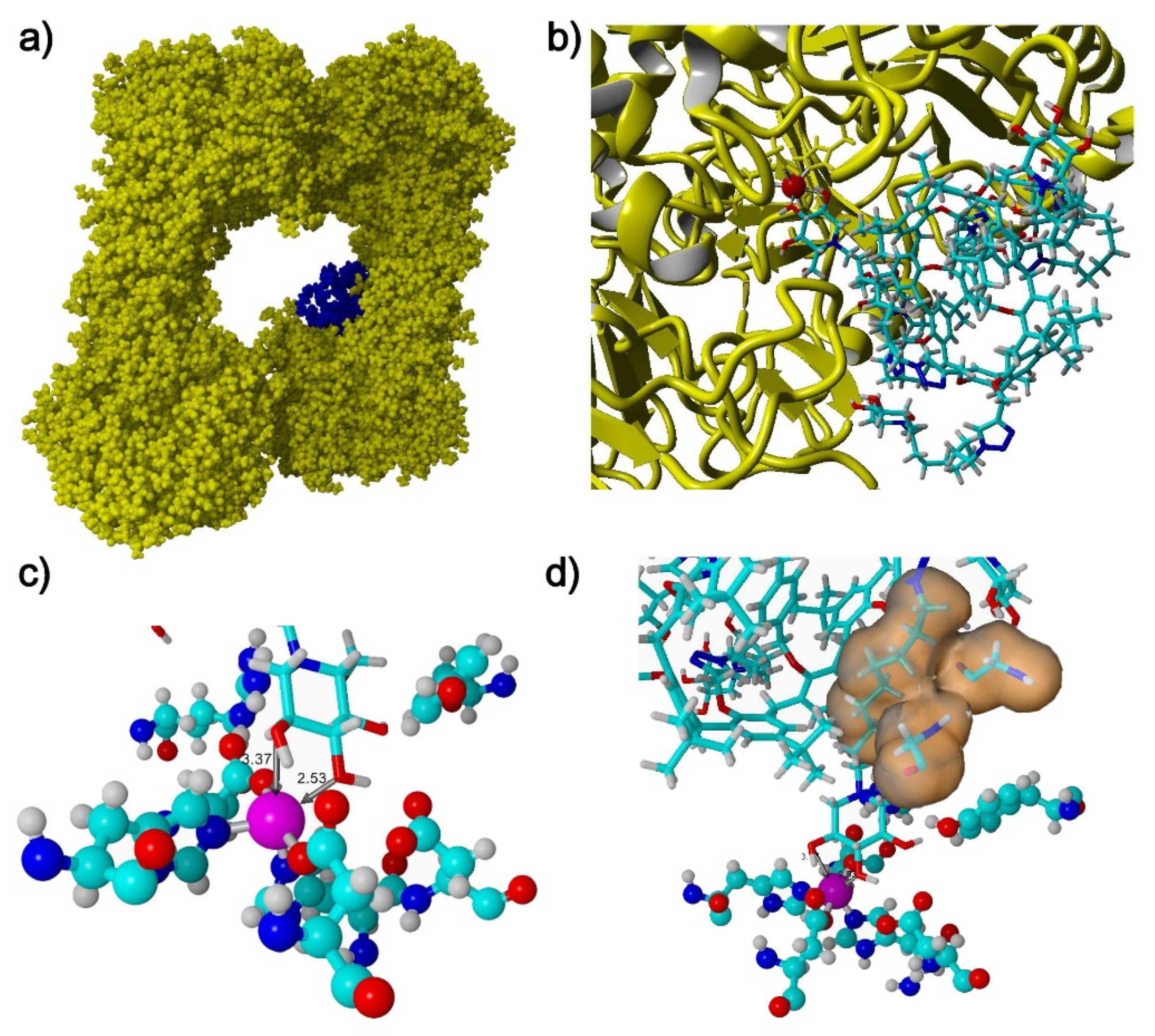
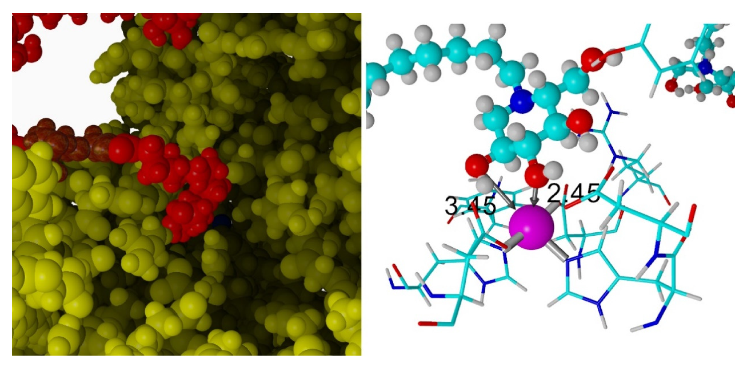
2.3. Docking Studies

In order to rationalize the biological data reported in Table 1 and to gain deeper insights into the possible interaction mode of calix[8]arene-based multivalent clusters with JBα-man, preliminary molecular docking studies were performed (Figure 3, Figure 4 and Figure 5 and Figures S91, S92). It is noteworthy that docking approaches have been recently reported for small calixarenes [15,76,77,78,79]. The multivalent cluster structures were optimized by molecular mechanics calculations and molecular dynamics simulations in a box of water molecules using the YASARA program [80] and AMBER force field [81,82,83,84]. Regarding the protein JBα-man, the previously reported X-ray structure [50] (Protein Data Bank, PDB entry 6B9O) was used as the starting structure for docking studies. As known, the crystallographic asymmetric unit contains one JBα-man protein composed of two LH heterodimers, each constituted by two distinct chains, L and H, to give a symmetrical (LH)2 complex (C2 axis). The tetrameric structure of the protein shows four open active sites with their zinc ion in the H-chain, turned toward a cavity located at the center of the 2x(LH)2 complex composed by the four LH heterodimers. Several complementary techniques showed that this large cavity accommodates large multivalent inhibitors (e.g., 1e) [44,50]. In solution, the apoprotein JBα-man is mainly found as (LH)2, but the 2x(LH)2 complex is also observed as a minor species as shown by analytical ultracentrifugation sedimentation velocity (AUC-SV) experiments [44]. While starting the docking studies with the 2x(LH)2 complex, the main objectives were to explore all possible binding sites and find the best one and see if some clusters are large enough to be able to bind several sites simultaneously.
The docking studies showed that 6-valent cluster 7b binds only one active site and one DNJ head occupies the active site pocket (Figure 3a,b), chelating the zinc atom as in the X-ray structure of compound 1e. Moreover, the hydrophobic chain of the Zn-coordinated DNJ of cluster 7b establishes hydrophobic interactions with amino acids G788 and G790 (Figure 3d), in accordance with electron density observed for the flexible chain near those two glycine residues in X-ray structure of 1e in complex with JBα-man [50].
Furthermore, the zinc atom adopts an octahedral coordination (Figure 3c) with oxygen atoms (O2 and O3) of the iminosugar head of 7b at 3.37 and 2.53 Å, but also two oxygen atoms of the side-chains of Asp145 and Asp25, and two histidine nitrogen atoms (His23 and His386).
Docking studies with derivative 6a (SI Figure S91), bearing a shorter spacer than 6b, indicated that hydrophobic interaction with G790 is missing compared to the longer C9 chain confirming the relevance of the spacer length.
For the huge dendritic cluster 6c, restrictions on the total atom number prevented the completion of the docking calculation. This problem was overcome by using a simplified cluster model bearing four DNJ heads. Interestingly, starting from an elongated geometry, we found that the global size of this simplified model of cluster 6c allows it to coordinate two zinc ions in distal binding sites of two enzymes (Figure 4). The cluster, located at the center of the enzyme dimer, adopts an octopus-like structure, cross-linking two distal binding sites. Analogous to what was observed for derivatives 6a and 7b, the zinc atom in the active site presents similar octahedral coordination with O2 and O3 of the iminosugar head of 6c at 2.45 and 3.45 Å (Figure 5), two oxygen atoms from Asp145 and Asp25, and two nitrogens from His23 and His386. Lipophilic interactions were also highlighted in this case, between the two residues of G788, G790, and the aliphatic chain of the Zn-coordinated DNJ of cluster 6c.
The overall results of the docking studies indicate that clusters 6a, 6b, and 7b bind only one active site at the same time. Their global size is indeed too short to cross-link two enzymes and the affinity enhancement observed may thus be only due to the bind-and-recapture effect. One supplementary lipophilic interaction between longer chains and glycine residues might be involved in the superiority of the C9 arms compared to the C6 ones. The cross linking of two enzymes (Zn-Zn distance ~60–70 A) observed with the longer dendron-elongated calix[8]arene cluster 6c shows that stabilization of the 2x(LH)2 complex is theoretically possible. This result could be extrapolated to cluster 7c which displays a similar overall size (same ring size and same flexible dendron length). Chelation of two more proximal sites (Zn-Zn distance ~40 A) of the same enzyme was not observed for 7c here but is not excluded with this cluster size. Even if those mechanisms are possible, they might not occur so frequently for 7c since the rp/n of compounds 6b, 7b and 6c, 7c are overall in the same range and one magnitude order below the one of the best multivalent inhibitor reported so far, 36-valent cluster 1e.

3. Materials and Methods

3.1. Chemistry

General Information: All chemicals were reagent grade and were used without further purification. Tetrahydrofuran was dried by heating under reflux over sodium wire in the presence of benzophenone as an indicator while dimethylformamide was dried by activated 3 Å molecular sieves. When necessary, the compounds were dried in vacuum over CaCl2. Reactions were monitored by Merck KGaA TLC silica gel plates (0.25 mm)( Kenilworth, NJ, USA) and visualized by 254 nm UV light, or by spraying with H2SO4-Ce(SO4)2. The derivatives 8 [67], 12a [45], 12b [65], and 12c [66] have been synthesized according to literature procedures. Proton 1H and carbon 13C nuclear magnetic resonance (NMR) experiments were recorded at 298K except when specified on Bruker Avance III HD 400 MHz, 500 MHz, or 600 MHz spectrometer (Billerica, MA, USA). Chemical shifts are reported relative to the residual solvent peak. A COSY spectrum was taken using a relaxation delay of 2 s with 30 scans and 170 increments of 2048 points each. HSQC spectra were performed with gradient selection, sensitivity enhancement, and phase sensitive mode using Echo/Antiecho-TPPI procedure. Optical rotations were measured at 589 nm (sodium lamp) and 20 °C on either a Perkin-Elmer 341 polarimeter (Waltham, MA, USA) or an Anton Paar MCP 200 polarimeter(Graz, Austria) with a path length of 1 dm. Infrared (IR) spectra were recorded neat on a Perkin–Elmer Spectrum Two FT-IR spectrometer. High-resolution (HRMS) electrospray ionization-time-of-flight (ESI-TOF) mass spectra were recorded on a Bruker micrOTOF® mass spectrometer.
Synthesis of derivatives 10 and 11: Cs2CO3 (1.25 equiv for each OH group) as added to a suspension of the appropriate calix[8]arene derivative 8 or 9 (1.0 g) in acetone (100 mL). The reaction mixture was stirred at reflux for 2 h. Then the system was cooled at room temperature and propargyl bromide (2.50 equiv for each OH group) was added. The reaction mixture was then stirred at reflux for 20 h, then it was cooled at room temperature and the solvent was removed under reduced pressure. An aqueous 1N solution of HCl (150 mL) was added to the reaction mixture and the suspension was stirred for 15 min. The aqueous phase was extracted and washed with CH2Cl2 (3 × 70 mL) and the organic phases were collected, dried over Na2SO4, filtered, and the solvent was removed under reduced pressure. The crude product was dissolved in a small amount of CH2Cl2 and then MeOH was added. The precipitated was filtered, affording the pure product as a white solid.
Compound 10: (76% yield); ESI(+) MS: m/z = 1603.1 [M + H]+, 1624.9 [M + Na]+; 1H NMR (400 MHz, CDCl3, 298 K): δ 1.11 (s, tBu, 72H), 2.30 (br t, 8H), 4.10 (s, 16H), 4.21 (d, J = 2.1 Hz, 16H), 6.95 (s, 16H) ppm; 13C NMR (100 MHz, CDCl3, 298K): δ 31.2, 31.5, 34.4, 60.9, 75.4, 79.7, 126.3, 133.4, 146.9, 153.1 ppm; Anal. calcd for C112H128O8: C, 83.96; H, 8.05; found C, 83.01; H, 8.16. Compound 11: (57% yield); ESI(+) MS: m/z = 1628.7 [M+H]+, 1647.9 [M+Na]+, 1667.9 [M+K]+; 1H NMR (600 MHz, CDCl3, 298K): δ 0.55 (s, 18H), 1.17 (s, 36H), 1.38 (s, 18H), 2.46 (br t, 4H), 2.56 (br t, 2H), 3.56 (s, 4H), 4.10 (s, 8H), 4.21 (s, 8H), 4.57 (s, 8H), 4.74 (s, 4H), 5.95 (s, 4H), 6.70 (s, 4H), 6.91 (s, 4H), 7.24 (br d, J = 2.2 Hz, 4H), 7.27 (s, 4H) ppm; 13C NMR (150 MHz, CDCl3, 298K): δ 31.0, 31.1, 31.6, 31.8, 34.0, 34.5, 60.6, 61.4, 75.2, 75.6, 80.0, 80.4, 124.6, 124.7, 126.3, 127.2, 128.3, 129.0, 132.4, 133.1, 133.6, 134.9, 136.43, 146.2, 146.4, 146.5, 151.5, 152.6, 154.4 ppm. Anal. calcd for C114H130O8: C, 84.09; H, 8.05; found C, 83.10; H, 8.14.
General procedure for the CuAAC with 12a and 12b: The calix[8]arene derivative 10 or 11 (10–15 mg) and the appropriate azide-armed DNJ iminosugar 12a or 12b (1.2 equiv for each alkyne unit) were dissolved in DMF (1.0 mL) in an ACE pressure tube. Then, a solution of Cu2SO4.5H2O (0.1 equiv. for each alkyne unit) and sodium ascorbate (0.2 equiv. for each alkyne unit) in water (0.25 mL) was added. The reaction mixture was heated under microwave irradiation for 20–30 min, using a CEM Corporation Discover LabMate system with the temperature controller. The mixture was concentrated, and traces of copper salts were removed by filtration through a short pad of silica gel eluting with CH3CN/AcOEt/NH4OH (9:1:1). The filtrate was concentrated and then purified by flash chromatography on silica gel.
Compound 13a: (AcOEt-AcOEt:MeOH 98:2, 51% yield; MS (ESI) m/z calcd for C272H384N32Na2O72 [M + 2Na]2+ 2648.3577; found 2648.3351; [α]D28 = +3.5 (c = 1, CHCl3); 1H NMR (300 MHz, CDCl3, 298K): δ 0.89 (s, 72H), 1.25 (br, 32H), 1.39 (br, 16H), 1.77 (br, 16H), 2.00–2.04 (m, 96H), 2.30 (t, J = 10.6 Hz, 8H), 2.53 (m, 8H), 2.61 (m, 8H), 2.70 (m, 8H), 3.16 (dd, J1 = 11.6 Hz, J2 = 4.9 Hz, 8H), 4.06 (s, 16H), 4.14 (br, 16H), 4.18 (overlapped, 16H), 4.79 (s, 16H), 4.92 (m, 8H), 5.03 (overlapped, 16H), 6.80 (s, 16H), 7.79 (s, 8H) ppm; 13C NMR (75 MHz, CDCl3, 298K): δ 20.9, 20.9, 21.1, 24.8, 26.6, 26.9, 30.2, 30.5, 31.4, 34.2, 50.2, 51.9, 53.1, 59.6, 61.6, 66.6, 69.6, 69.7, 74.9, 123.9, 126.1, 133.3, 144.1, 146.6, 152.7, 169.9, 170.2, 170.5, 171.0 ppm. Anal. calcd for C272H384N32O72: C, 62.18; H, 7.37; found C, 62.07; H, 7.48.
Compound 13b: (AcOEt:Petroleum Ether, 8:2, 52% yield); MS (ESI) m/z calcd for C296H432N32Na2O72 [M + 2Na]2+ 2816.5455; found 2816.5469; [α]D27 = +5.0 (c = 1, CHCl3); 1H NMR (600 MHz, CDCl3, 298K): δ 0.90 (s, 72H), 1.24 (overlapped, 80H), 1.39 (br, 16H), 1.76 (br, 16H), 2.00–2.06 (overlapped, 96H), 2.32 (t, J = 10.9 Hz, 8H), 2.55 (m, 8H), 2.62 (br d, J = 9.2 Hz, 8H), 2.70 (m, 8H), 3.17 (dd, J1 = 11.4 Hz, J2 = 5.1 Hz, 8H), 4.06 (s, 16H), 4.14 (br, 16H), 4.18 (br, 16H), 4.79 (s, 16H), 4.95 (m, 8H), 5.0–5.07 (overlapped, H3+H4, 16H), 6.80 (s, 16H), 7.78 (s, 8H) ppm; 13C NMR (150 MHz, CDCl3, 298 K): 20.9, 20.9, 21.1, 21.1, 24.8, 26.8, 27.5, 29.3, 29.7, 29.9, 30.6, 31.4, 34.2, 50.3, 52.1, 53.2, 59.7, 61.6, 66.6, 69.7, 69.7, 74.9, 123.9, 126.1, 133.3, 144.2, 146.6, 152.7, 170.0, 170.2, 170.6, 171.1 ppm. Anal. calcd for C296H432N32O72: C, 63.59; H, 7.79; found C, 63.50; H, 7.86.
Compound 14a: (AcOEt/Petroleum Ether 9:1-AcOEt, 51% yield); MS (ESI) m/z calcd for C234H322N24Na2O56 [M + 2Na]2+ 2205.1435; found 2205.1329; [α]D30 = +4.0 (c = 1, CHCl3); 1H NMR (600 MHz, CDCl3, 298K): δ 0.40 (s, 18H), 0.95–1.54 (overlapped, 44H), 1.17 (s, 36H), 1.41 (s, 18H), 1.86 (t, J = 7.2 Hz, 12H), 1.98–2.06 (overlapped, 72H), 2.25–2.30 (overlapped, 6H), 2.42–2.62 (overlapped, 14H), 2.69–2.74 (m, 4H), 3.10 (dd, J1 = 11.5 Hz, J2 = 5.2 Hz, 4H), 3.18 (dd, J1 = 11.5 Hz, J2 = 5.2 Hz, 2H), 3.32 (s, 4H), 3.44–3.48 (overlapped, 4H), 3.53 (m, 4H), 3.86–3.97 (overlapped, 8H), 4.07–4.17 (overlapped, 12H), 4.33 (t, J = 7.8 Hz, 4H), 4.36–4.40 (overlapped, 4H), 4.79 (d, J = 13.2 Hz, 4H), 4.89–4.96 (overlapped, 6H), 4.97–5.08 (overlapped, 12H), 5.82 (s, 4H), 6.58 (s, 2H), 6.61 (s, 2H), 6.95 (s, 2H), 6.98 (s, 2H), 7.22–7.28 (overlapped, 6H), 7.77 (s, 4H) ppm; 13C NMR (150 MHz, CDCl3, 298 K): δ 20.9, 21.0, 21.1, 24.4, 25.0, 26.5, 26.7, 26.8, 29.9, 30.3, 30.4, 30.6, 31.1, 31.6, 31.9, 33.9, 34.5, 34.5, 50.1, 50.4, 51.7, 51.9, 51.9, 53.1, 53.2, 59.4, 59.7, 61.5, 61.6, 61.9, 66.2, 67.0, 69.6, 69.7, 74.8, 75.0, 123.1, 123.3, 123.4, 124.0, 124.3, 127.3, 128.7, 129.2, 132.7, 133.4, 134.6, 134.7, 136.6, 144.0, 144.34, 146.5, 146.7, 152.4, 153.8, 169.9, 170.1, 170.3, 170.6, 171.1 ppm. Anal. calcd for C234H322N24O56: C, 64.36; H, 7.43; found C, 64.26; H, 7.51.
Compound 14b: (AcOEt/Petroleum Ether 72:28–AcOEt, 37% yield); MS (ESI) m/z calcd for C252H358N24Na2O56 [M + 2Na]2+ 2331.2844; found 2331.7650; [α]D31 = +7.5 (c = 1, CHCl3); 1H NMR (600 MHz, CDCl3, 298K): δ 0.41 (s, 18H), 1.05–1.30 (overlapped, 60H), 1.18 (s, 36H), 1.39 (s, 18H), 1.54 (br, 8H), 1.86 (br, 4H), 2.00–2.02 (overlapped, 72H), 2.29–2.34 (overlapped, 6H), 2.52–2.57 (overlapped, 6H), 2.61–2.64 (overlapped, 6H), 2.66–2.74 (overlapped, 6H), 3.14–3.20 (overlapped, 6H), 3.34 (s, H18, 4H), 3.43 (d, J = 14.4 Hz, 4H), 3.51 (d, J = 16.8 Hz, 4H), 3.89–3.99 (m, 8H), 4.10–4.17 (overlapped, 12H), 4.33 (t, J = 7.3 Hz, 4H), 4.39 (d, J = 16.6 Hz, 4H), 4.78 (d, J = 14.2 Hz, 4H), 4.92–4.97 (overlapped, 6H), 5.00–5.08 (overlapped, 24H), 5.84 (s, ArH, 4H), 6.60 (s, 4H), 6.96 (s, 4H), 7.24 (s, 4H), 7,26 (overlapped, 8H), 7.78 (s, 2H) ppm; 13C NMR (150 MHz, CDCl3, 298 K): δ 20.9, 21.0, 21.1, 21.2, 21.3, 22.9, 24.6, 24.9, 26.7, 26.7, 27.4, 27.5, 29.1, 29.2, 29.6, 29.7, 29.9, 30.1, 30.4, 30.6, 30.8, 31.1, 31.6, 31.8, 32.1, 33.9, 34.5, 50.3, 50.6, 52.0, 52.2, 53.2, 53.2, 59.7, 59.7, 60.6, 61.5, 61.7, 66.2, 67.1, 69.7, 69.7, 74.9, 75.0, 123.1,123.5, 124.0, 124.3, 127.3, 128.6, 129.2, 131.9, 132.8, 133.4, 134.7, 136.7, 144.2, 144.4, 145.9, 146.4, 146.5, 151.3, 152.4, 153.9, 169.9, 170.2, 170.2, 170.6, 171.1, 171.4 ppm. Anal. calcd for C252H358N24O56: C, 65.52; H, 7.81; found C, 65.43; H, 7.90.
General procedure for deacetylation of clusters 13a,b and 14a,b: The acetylated compound (10–15 mg) was dissolved in dry methanol (0.3–0.5 mL), and then NaOMe (0.45 equiv for each acetyl group) was added. The reaction mixture was stirred at room temperature for 6 h under nitrogen atmosphere. Methanol and water were added to dissolve the white precipitate. Dowex 50WX8-200 resin was added until pH=6–7 was reached. Then the solution was filtered and concentrated to give the pure compound.
Compound 6a: 67% yield; MS (ESI) m/z calcd for C208H320Na2O40 [M + 2Na]2+ 1676.1887; found 1976.1792; [α]D33= +13.0 (c = 0.067, MeOH); 1H NMR (600 MHz, MeOD, 298 K): δ 0.96 (s, 72H), 1.26–1.29 (overlapped, 32H), 1.50 (br, 16H), 1.73 (br, 16H), 2.31 (overlapped, 8H), 2.34 (overlapped, 8H), 2.68 (m, 8H), 2.88 (m, 8H), 3.08 (dd, J1 = 11.7 Hz, J2 = 5.04 Hz, 8H), 3.19 (t, J = 9 Hz, 8H), 3.40 (t, J = 9.2 Hz, 8H), 3.53 (m, 8H), 3.87 (br, 16H), 4.09 (s, 16H), 4.19 (br, 16H), 4.80 (br s, 16H), 6.89 (s, 16H), 7.96 (s, 8H), 8.51 (s, OH) ppm; 13C NMR (150 MHz, MeOD, 298 K): δ 25.1, 27.0, 27.5, 27.9, 28.2, 30.4, 30.5, 30.9, 31.3, 32.1, 33.2, 35.2, 36.6, 51.3, 53.9, 57.2, 58.7, 67.5, 70.2, 71.4, 80.1, 125.9, 131.0, 134.6, 145.2, 147.9, 154.1 ppm. Anal. calcd for C208H320N32O40: C, 63.91; H, 8.25; found C, 64.01; H, 8.16.
Compound 6b: 82% yield; MS (ESI) m/z calcd for C232H368N32Na2O40 [M + 2Na]2+ 2144.3765; found 2144.3665; [α]D27 = −4.0 (c = 0.24, MeOH); 1H NMR (600 MHz, MeOD, 313 K): δ 0.95 (s, 72H), 1.25 (overlapped, 80H), 1.57 (br, 16H), 1.76 (br, 16H), 2.46–2.49 (overlapped, 16H), 2.80 (m, 8H), 3.00 (m, 8H), 3.17 (dd, J1 = 11.3 Hz, J2 = 4.56 Hz, 8H), 3.23 (t, J = 9.1 Hz, 8H), 3.45 (t, J = 9.3 Hz, 8H), 3.57 (m, 8H), 3.87 (dd, J1 = 11.9 Hz, J2 = 2.5 Hz, 8H), 3.94 (dd, J1 = 12.1 Hz, J2 = 2.0 Hz, 8H), 4.07 (s, 16H), 4.21 (br, 16H), 4.81 (s, 16H), 6.88 (s, 16H), 7.96 (s, 8H), 8.43 (br s, 32H) ppm; 13C NMR (600 MHz, MeOD, 313 K): δ 25.0, 27.6, 28.2, 30.0, 30.3, 30.4, 30.5, 30.6, 30.7, 30.8, 31.2, 31.3, 31.9, 33.2, 35.1, 36.5, 49.8, 51.2, 53.9, 54.7, 56.6, 58.0, 67.4, 67.5, 69.6, 70.9, 79.6, 125.8, 127.2, 134.6, 145.2, 147.8, 154.0 ppm. Anal. calcd for C232H368N32O40: C, 65.63; H, 8.74; found C, 65.54; H, 8.84.
Compound 7a: 67% yield; MS (ESI) m/z calcd for C186H274N24Na2O32 [M + 2Na]2+ 1701.0168; found 1701.0117; [α]D29 = −2.0 (c = 1, MeOH); 1H NMR (600 MHz, MeOD, 298 K): δ 0.44 (s, 18H), 1.23 (s, 36H), 1.24–1.49 (overlapped, 44H), 1.48 (s, 18H), 1.81 (t, J = 7.2 Hz, 4H), 2.40–2.52 (overlapped, 12H), 2.68–2.75 (overlapped, 6H), 2.87–2.97 (overlapped, 6H), 3.11–3.16 (overlapped, 6H), 3.20–3.24 (overlapped, 6H), 3.36 (s, 4H), 3.40–3.46 (overlapped, 6H), 3.52–3.58 (overlapped, 6H), 3.60–3.65 (overlapped, 8H), 3.82–3.94 (overlapped, 20H), 4.37–4.42 (overlapped, 8H), 4.89–4.96 (overlapped, 8H), 5.15 (br s, 8H), 5.86 (s, 4H), 6.67 (s, 4H), 7.05 (d, J = 7.2 Hz, 4H), 7.36 (s, 4H), 7.39 (s, 4H), 7.42 (s, 4H), 8.28 (s, 2H), 8.44 (br s, OH) ppm; 13C NMR (150 MHz, MeOD, 298 K): δ 23.7, 24.5, 24.5, 24.7, 27.2, 27.27, 27.6, 30.3,30.5, 30.6, 30.7, 31.0, 31.1, 31.2, 31.9, 32.0, 32.2, 33.1, 34.9, 35.3, 35.6, 51.3, 51.3, 53.7, 53.7, 56.4, 56.5, 56.7, 57.5, 58.1, 66.7, 67.3, 67.4, 67.5, 69.4, 69.5, 69.7, 70.6, 71.0, 76.2, 79.5, 79.6, 79.7, 125.0, 126.0, 126.1, 128.7, 129.8, 130.3, 133.2, 133.2, 134.1, 134.9, 135.4, 137.7, 145.1, 145.1, 145.3, 147.1, 148.0, 148.1, 152.57, 153.5, 153.5, 154.8 ppm. Anal. calcd for C186H274N24O32: C, 66.52; H, 8.22; found C, 66.62; H, 8.12.
Compound 7b: 80% yield; MS (ESI) m/z calcd for C204H310N24Na2O32 [M + 2Na]2+ 1827.1576; found 1827.1540; [α]D29 = −4.0 (c = 1, MeOH); 1H NMR (600 MHz, MeOD, 298 K): δ 0.45 (s, 18H), 1.03–1.29 (overlapped, 60H), 1.22 (s, 36H), 1.45 (s, 18H), 1.48–1.56 (overlapped, 20H), 1.83 (m, 4H), 2.35–2.45 (overlapped, 12H), 2.71–2.78 (overlapped, 6H), 2.90–2.96 (overlapped, 6H), 3.09–3.14 (overlapped, 6H), 3.38 (s, 4H), 3.40–3.45 (overlapped, 6H), 3.50–3.56 (overlapped, 14H), 3.85–3.97 (overlapped, 20H), 4.36–4.42 (overlapped, 8H), 4.87–4.92 (overlapped, 8H), 5.11 (d, J = 12 Hz, 4H), 5.16 (s, 4H), 5.89 (s, 4H), 6.67 (s, 4H), 7.02 (s, 4H), 7.35 (s, 4H), 7.36 (s, 4H), 7.40 (s, 4H), 8.25 (s, 2H), 8.50 (br s, OH) ppm; 13C NMR (150 MHz, MeOD, 298 K): δ 23.7, 25.0, 25.2, 27.4, 27.5, 28.3, 29.8, 29.9, 30.2, 30.4, 30.7, 31.1, 31.3, 31.4, 31.7, 31.9, 32.0, 32.2, 34.8, 35.3, 35.3, 51.4, 53.9, 54.8, 56.9, 57.0, 58.3, 58.6, 66.8, 67.4, 67.5, 69.9, 70.0, 71.2, 71.3, 79.8, 79.9, 125.1, 125.3, 126.0, 128.7, 129.7, 130.3, 133.3, 134.1, 134.8, 135.6, 137.9, 145.2, 147.1, 147.8, 147.9, 152.5, 153.6, 154.9 ppm. Anal. calcd for C204H310N24O32: C, 67.86; H, 8.65; found C, 67.97; H, 8.56.
General Procedure for the CuAAC Reaction with trivalent dendron 12c: The calix[8]arene derivative 10 or 11 (10–15 mg) and the derivative 12c (1.2 equiv. for each alkyne unit) were dissolved in DMF (1.0 mL) in an ACE pressure tube. Then, a solution of Cu2SO4.5H2O (0.1 equiv. for each alkyne unit) and sodium ascorbate (0.2 equiv. for each alkyne unit) in water (0.25 mL) was added. The resulting suspension was heated under microwave irradiation at 80 °C for 30 min, then water (10 mL) was added, and the aqueous phase was extracted with EtOAc (3 × 12 mL). The organic phases were combined, dried with Na2SO4, and concentrated in vacuo. Traces of copper salts were removed by filtration through a short pad of silica gel eluting with CH3CN/H2O/NH4OH (15:0.5:0.5), and the residue was then purified by flash chromatography (SiO2; CH2Cl2/MeOH, 99:1 to 95:5) to give the desired iminosugar.
Compound 13c: 66% yield; [α]D20 = +3.5 (c = 1, MeOH); 1H NMR (CDCl3, 400 MHz): δ 7.85 (br s, 8H), 7.57 (s, 24H), 6.8 (br s, 16H), 5.10–4.99 (m, 48H), 4.98–4.91 (m, 24H), 4.81 (br s, 53H), 4.51 (s, 48H), 4.41 (br s, 10H), 4.33–4.27 (t, J = 7.4 Hz, 48H), 4.14 (s, 48H), 4.03 (br s, 92H), 3.52–3.36 (m, 96H), 3.21–3.15 (dd, J1 = 11.4, J2 = 5.0 Hz, 24H), 2.76–2.66 (m, 24H), 2.65–2.60 (m, 24H), 2.59–2.50 (m, 24H), 2.35–2.28 (dd, J1 = 10.9, J2=10.5 Hz, 24H), 2.06 (s, 72H), 2.02–1.99 (overlapped, 216H), 1.90–1.84 (m, 48H,), 1.46–1.16 (288H), 0.97–0.72 (m, 72H) ppm; 13C-NMR (CDCl3, 100 MHz): δ 171.0, 170.4, 170.1, 169.9, 145.3, 122.6, 74.9, 70.4–69.8, 69.7, 69.6, 69.4, 65.2, 61.6, 59.7, 53.1, 51.9, 50.4, 50.1, 45.5, 31.3, 30.5, 29.6, 29.2, 27.3, 26.7, 24.9, 21.0, 21.0, 20.9, 20.8 ppm; IR (neat) 1745 cm−1; MS (ESI, deconvoluted): m/z calcd for C808H1249N120O240 [M + 10H]10+ 1648.4740; found 1648.4710.
Compound 14c: 48% yield; [α]D20 = + 4.5 (c = 1, MeOH); 1H NMR (CDCl3, 400 MHz, 298K): δ 8.11–7.59 (m, 57H), 7.61–7.50 (m, 18H), 7.38–7.16 (m, 8H), 6.96–6.58 (m, 4H), 6.58 (s, 4H), 5.89–5.73 (m, 4H), 5.16–4.99 (m, 48H), 4.99–4.91 (m, 18H), 4.89–4.63 (m, 9H), 4.61–4.44 (m, 42H), 4.36–4.22 (m, 42H), 4.14 (s, 36H), 3.91–3.80 (m, 4H), 3.78–3.63 (m, 8H), 3.54–3.31 (m, 72H), 3.21–3.15 (dd, J1 = 11.5 and J2 =4.9 Hz, 18H), 2.76–2.66 (m, 18H), 265–2.60 (m, 18H), 2.59–2.49 (m, 18H), 2.35–2.28 (t, J = 10Hz, 18H), 2.09–2.04 (s, 54H), 2.03–2.00 (s, 108H), 2.01–1.98 (s, 54H), 1.93–1.79 (m, 36H), 1.49–1.10 (m, 270H), 0.37 (s, 18H) ppm; 13C NMR (CDCl3, 75 MHz, 298 K): δ 170.9, 170.4, 170.1, 169.8, 162.6, 145.42, 145.37, 144.2, 122.62, 122.53, 74.9, 69.8, 69.6, 69.5, 68.1, 65.2, 61.7, 59.8, 53.1, 52.0, 51.9, 50.4, 50.4, 45.6, 45.5, 36.6, 34.4, 34.4, 31.8, 31.6, 31.5, 31.1, 30.5, 29.6, 29.2, 27.4, 26.7, 25.8, 25.1, 25.0, 20.97, 20.94, 20.85, 20.80 ppm; MS (ESI, deconvoluted) m/z calcd for C636H978N90O182 [M + 14H]14+ 913.8864; found 913.9960.
General procedure for the deacetylation reaction of 13c and 14c: Amberlite resin IRA 400 (6n g/mmol of substrate; n = number of acetate groups) was added to a solution of acetylated iminosugar in a mixture of MeOH/H2O (1:1), and the resulting solution was stirred for 4 h by using a rotary evaporator at atmospheric pressure. The resin was then removed by filtration and washed with methanol and water. Solvents were evaporated under reduced pressure to give the desired deprotected iminosugar.
Compound 6c: 89% yield; [α]D20 =−2.5 (c = 1, MeOH); 1H NMR (MeOD, 400 MHz, 298K): δ 8.22–7.99 (br s, 8H), 7.97–7.82 (s, 24H), 7.19–6.59 (br s, 16H), 4.57–4.42 (m, 64H), 4.41–4.28 (m, 48H), 3.84 (s, 48H), 3.54–3.32 (overlapped, 64H), 3.20–3.07 (m, 24H,), 3.01–2.94 (dd, J1 = 11.9, J2 = 5.5 Hz, 24H), 2.83–2.71 (m, 24H), 2.62–2.50 (m, 24H), 2.20–2.12 (dd, J1 = 11.9, J2 = 11.5 Hz, 24H), 2.12–2.07 (m, 24H), 1.94–1.79 (m, 48H), 1.55–1.38 (m, 48H), 1.39–1.17 (m, 240H), 0.99–0.79 (m, 72H) ppm; 13C-NMR (MeOD, 100 MHz, 298K) δ 146.2, 124.9, 80.6, 72.1, 70.8, 67.4, 65.5, 59.6, 57.8, 53.8, 51.4, 32.1, 31.4, 30.6, 30.1, 28.6, 27.6, 25.3 ppm; IR (neat) 3356 (broad strong OH) cm−1; MS (ESI, deconvoluted) m/z calcd for C616H1058N120O144 [M + 10H]10+ 1244.9054; found 1244.9090.
Compound 7c: 96% yield; [α]D20 =−5.0 (c = 1, MeOH/H2O 1:1); 1H NMR (MeOD, 500 MHz, 298K): δ 8.33 (s, 2H), 7.99–7.85 (several s, 18H), 7.69 (several s, 4H), 7.41–7.20 (m, 8H), 6.96 (s, 4H), 6.80–6.55 (m, 4H), 5.95–5.81 (m, 4H), 5.21–4.98 (m, 12H), 4.64 (m, 4H), 4.54–4.25 (m, 88H), 3.84 (m, 40H), 3.73–3.58 (m, 8H), 3.54–3.22 (m, 120H), 3.13 (t, J = 9.0 Hz, 18H), 2.97 (dd, J = 11.2 and 5.1 Hz, 18H), 2.82–2.72 (m, 18H), 2.61–2.51 (m, 18H), 2.16 (dd, J = 11.1 and 10.0 Hz, 18H), 2.10 (m, 18H), 1.90–1.77 (m, 36H), 1.51–1.19 (m, 270H), 0.43 (s, 18H) ppm; 13C-NMR (MeOD, 125 MHz, 20480 scans) δ 155.0, 153.5, 152.4, 147.8, 147.5, 146.8, 146.1, 145.3, 145.1, 137.9, 135.8, 135.0, 134.2, 133.6, 133.3, 132.4, 130.4, 129.86, 129.8, 129.5, 128.8, 126.9, 126.8, 125.9, 125.2, 124.9, 80.6, 76.2, 72.0, 70.8, 71.3, 71.2, 70.3, 70.11, 69.99, 69.08, 67.3, 65.51, 65.46, 59.5, 57.8, 53.8, 51.3, 46.53, 46.50, 35.4, 35.3, 34.9, 32.3, 32.2, 32.0, 31.4, 30.6, 30.1, 28.6, 27.5, 25.2) ppm. IR (neat) 3338 (broad strong OH) cm−1. MS (ESI) m/z calcd for C492H830N90O110 [M + 10H]10+ 976.6409; found 976.6068.

3.2. Biological Assays

p-nitrophenyl-α-D-mannopyranoside and α-mannosidase (EC 3.2.1.24, from Jack Bean, Km = 2.0 mM pH 5.5) were purchased from Sigma Aldrich(St. Louis, MO, USA).
General procedure for Inhibition assay with basic quench: Inhibitory potencies were determined spectrophotometrically (Versamax 96-well plate spectrophotometer, Molecular Devices Corporation, Sunnyvale, CA, USA) measuring the residual hydrolytic activities of the mannosidase against p-nitrophenyl-α-D-mannopyranoside in the presence and absence of the inhibitor. All kinetics were performed at 25 °C and started by enzyme addition (0.015 U/mL) in a 100 µL assay medium (acetate buffer, 0.2 M, pH = 5) containing substrate (varying concentration from Km/8 to 2Km value) in presence or absence of various concentrations of inhibitor. After 40–50 min incubation, the reaction was quenched by addition of 1M Na2CO3 (100 µL). The absorbance of the resulting solution was determined at 405 nm. Ki values were determined in duplicate or triplicate, using the Dixon and Lineweaver–Burk graphical methods [72] with Microsoft Excel 97-03, or using non-linear regression with GraphPad Prism 7 Software [55]. As the inhibitors are only partially soluble in water, they were dissolved in DMSO for concentrated mother solutions and DMSO/buffer for diluted solutions with a final DMSO concentration under 4% in all wells. Previously, the stability of the enzymes in presence of various concentrations of DMSO was controlled and the enzyme activity was unaffected.
General procedure for inhibition assay measured continuously: The release of p-nitrophenol was measured continuously at 385 nm to determine initial velocities. All kinetics were performed at 35 °C and started by substrate addition in a 1 mL assay medium (acetate buffer, 0.2 M, pH = 5) containing α-mannosidase (0.025 U/mL), substrate (varying concentration from Km/4 to Km value) in presence or absence of various concentrations of inhibitor. Under these conditions, the p-nitrophenolate released led to optical densities linear with both reaction time and concentration of the enzyme. Ki values were determined in duplicate or quadruplate, using the Dixon graphical method [72]. As the inhibitors are only partially soluble in water, they were dissolved in DMSO for concentrated mother solutions and DMSO/buffer for diluted solutions with a final DMSO concentration under 5% in all vials. Previously, the stability of the enzymes in presence of various concentrations of DMSO was controlled and the enzyme activity was unaffected.

3.3. Docking Studies

Docking calculations were performed with the Autodock-VINA [85], program implemented in YASARA (software version 20.7.4) [86], using a general AMBER force field [81,82,83,84]. The X-ray structure of JBα-man (Protein Data Bank, PDB entry 6B9O] [50] was used. The protein and clusters were parameterized according to AutoSMILES, see also reference [80]. For all the docked structures, some bonds of the aliphatic chains were treated as active torsional bonds. The default VINA docking parameters in YASARA macro were used. Several different starting structures for each of the multivalent clusters were optimized by molecular dynamics simulation and molecular mechanics calculations in a box of water molecules, using YASARA software, and were then used in docking studies. Regarding dendron cluster 6c, the docking calculations were performed on a simplified cluster model bearing 4 DNJ heads in order to overcome computing power problems found during the docking studies with the complete dendron cluster 6c.

4. Conclusions

A series of multivalent clusters with deoxynojirimycin inhitopes was synthesized by CuAAC. Two alkyne-armed calix[8]arene scaffolds were used, a C8 symmetrical 8-valent calix[8]arene core and a C2v symmetrical 6-valent 1,5-xylylene bridged one, with the same size and a close valency but differing in their rigidity. With the shorter C6 linker, the flexibility of the scaffold plays a significant role since the C8 symmetrical cluster 6a did not show multivalent effect whereas the 1,5-xylylene bridged 7a had an affinity improvement per inhitope of 7. Clusters with a longer C9 linker, whatever their global size, in the rigidity and valency of the central core show good affinity enhancement per inhitope in the range around a hundred (70 to 160). The best results leading to Ki < 100 nM and rp/n > 2000 were achieved for larger clusters 6c and 7c, the ones with the higher valencies and larger size. These two multivalent inhibitors are the only ones in the series whose size allow for cross-linking two enzymes as shown by docking studies.

Supplementary Materials

The following are available online at https://www.mdpi.com/1424-8247/13/11/366/s1, Full structure of compounds 6a–c, 7a–c, 13a–c and 14a–c, Zoom of X-ray structure of JB -man in interaction with compound 1e for one active site (Figure S7), 1H, 13C NMR, HSQC and HBMC spectra of new compounds and kinetic plots for Ki determination. Details on molecular docking and coordinates of multivalent clusters and their complexes with JBα-man.

Author Contributions

Conceptualization, P.N. and P.C.; methodology, J.P.S., S.T., C.G., P.D.S. and A.B.; software, C.T. (Carmen Talotta), P.D.S.; validation, A.B., P.C., C.T. (Carmen Talotta), C.T. (Céline Tarnus), C.G. and P.N.; formal analysis, C.T. (Carmen Talotta), J.P.S. and A.B.; investigation, J.P.S., P.D.S., A.B. and S.T.; resources, C.T. (Carmen Talotta) and J.P.S.; data curation, S.T., C.T. (Carmen Talotta), P.D.S. and J.P.S.; writing—original draft preparation, A.B., S.T. and C.G.; writing—review and editing, A.B., P.N. and P.C.; visualization, Carmen Talotta, P.D.S. and A.B.; supervision, C.G., A.B. and P.C.; project administration, C.G. and P.C.; funding acquisition, P.C., P.N. and C.G. All authors have read and agreed to the published version of the manuscript.

Funding

This work was funded by the CNRS (UMR 7509), the University of Strasbourg, the Fondation pour la Recherche en Chimie (FRC Strasbourg) and the University of Salerno (FARB 2017 “Sintesi e studio delle proprietà di nuovi derivati cicloparafenilenici”).

Acknowledgments

The authors acknowledge the Regione Campania (POR CAMPANIA FESR 2007/2013 O.O.2.1, CUP B46D14002660009) for the FT-ICR mass spectrometer facilities, Farma-BioNet (CUP B25C13000230007), the Centro di Tecnologie Integrate per la Salute” (CITIS) (project PONa3_00138), for the 600 MHz NMR facilities.

Conflicts of Interest

The authors declare no conflict of interest.

References

  1. Peczuh, M.W.; Hamilton, A.D. Peptide and Protein Recognition by Designed Molecules. Chem. Rev. 2000, 100, 2479–2494. [Google Scholar] [CrossRef]
  2. Baldini, L.; Casnati, A.; Sansone, F.; Ungaro, R. Calixarene-based multivalent ligands. Chem. Soc. Rev. 2007, 36, 254–266. [Google Scholar] [CrossRef]
  3. Park, H.S.; Lin, Q.; Hamilton, A.D. Protein Surface Recognition by Synthetic Receptors:  A Route to Novel Submicromolar Inhibitors for α-Chymotrypsin. J. Am. Chem. Soc. 1999, 121, 8–13. [Google Scholar] [CrossRef]
  4. Sansone, F.; Casnati, A. Multivalent glycocalixarenes for recognition of biological macromolecules: Glycocalyx mimics capable of multitasking. Chem. Soc. Rev. 2013, 42, 4623. [Google Scholar] [CrossRef] [PubMed]
  5. Giuliani, M.; Morbioli, I.; Sansone, F.; Casnati, A. Moulding calixarenes for biomacromolecule targeting. Chem. Commun. 2015, 51, 14140–14159. [Google Scholar] [CrossRef]
  6. McGovern, R.E.; Fernandes, H.; Khan, A.R.; Power, N.P.; Crowley, P.B. Protein camouflage in cytochrome c-calixarene complexes. Nat. Chem. 2012, 4, 527–533. [Google Scholar] [CrossRef]
  7. Sansone, F.; Baldini, L.; Casnati, A.; Ungaro, R. Calixarenes: From biomimetic receptors to multivalent ligands for biomolecular recognition. New J. Chem. 2010, 34, 2715–2728. [Google Scholar] [CrossRef]
  8. Gutsche, C.D. Calixarenes: An Introduction, 2nd ed.; Royal Society of Chemistry: Cambridge, UK, 2008; ISBN 978-0-85404-258-6. [Google Scholar]
  9. Asfari, M.-Z.; Böhmer, V.; Harrowfield, J.; Vicens, J. Calixarenes 2001, 2001 ed.; Springer: Dordrecht, The Netherlands; Boston, MA, USA, 2001; ISBN 978-0-7923-6960-8. [Google Scholar]
  10. Neri, P.; Sessler, J.L.; Wang, M.-X. Calixarenes and Beyond; Springer International Publishing: Berlin/Heidelberg, Germany, 2016; ISBN 978-3-319-31865-3. [Google Scholar]
  11. Sebti, S.M.; Hamilton, A.D. Design of growth factor antagonists with antiangiogenic and antitumor properties. Oncogene 2000, 19, 6566–6573. [Google Scholar] [CrossRef] [PubMed] [Green Version]
  12. Blaskovich, M.A.; Lin, Q.; Delarue, F.L.; Sun, J.; Park, H.S.; Coppola, D.; Hamilton, A.D.; Sebti, S.M. Design of GFB-111, a platelet-derived growth factor binding molecule with antiangiogenic and anticancer activity against human tumors in mice. Nat. Biotechnol. 2000, 18, 1065–1070. [Google Scholar] [CrossRef]
  13. Zhou, H.; Wang, D.; Baldini, L.; Ennis, E.; Jain, R.; Carie, A.; Sebti, S.M.; Hamilton, A.D. Structure–activity studies on a library of potent calix[4]arene-based PDGF antagonists that inhibit PDGF-stimulated PDGFR tyrosine phosphorylation. Org. Biomol. Chem. 2006, 4, 2376–2386. [Google Scholar] [CrossRef]
  14. Tommasone, S.; Talotta, C.; Gaeta, C.; Margarucci, L.; Monti, M.C.; Casapullo, A.; Macchi, B.; Prete, S.P.; Ladeira De Araujo, A.; Neri, P. Biomolecular Fishing for Calixarene Partners by a Chemoproteomic Approach. Angew. Chem. Int. Ed. 2015, 54, 15405–15409. [Google Scholar] [CrossRef] [PubMed]
  15. Chini, M.G.; Terracciano, S.; Riccio, R.; Bifulco, G.; Ciao, R.; Gaeta, C.; Troisi, F.; Neri, P. Conformationally Locked Calixarene-Based Histone Deacetylase Inhibitors. Org. Lett. 2010, 12, 5382–5385. [Google Scholar] [CrossRef]
  16. Morbioli, I.; Porkolab, V.; Magini, A.; Casnati, A.; Fieschi, F.; Sansone, F. Mannosylcalix[n]arenes as multivalent ligands for DC-SIGN. Carbohydr. Res. 2017, 453–454, 36–43. [Google Scholar] [CrossRef]
  17. Geraci, C.; Consoli, G.M.L.; Granata, G.; Galante, E.; Palmigiano, A.; Pappalardo, M.; Di Puma, S.D.; Spadaro, A. First Self-Adjuvant Multicomponent Potential Vaccine Candidates by Tethering of Four or Eight MUC1 Antigenic Immunodominant PDTRP Units on a Calixarene Platform: Synthesis and Biological Evaluation. Bioconjugate Chem. 2013, 24, 1710–1720. [Google Scholar] [CrossRef]
  18. Hevey, R. Strategies for the Development of Glycomimetic Drug Candidates. Pharmaceuticals 2019, 12, 55. [Google Scholar] [CrossRef] [Green Version]
  19. Brás, N.F.; Cerqueira, N.M.; Ramos, M.J.; Fernandes, P.A. Glycosidase inhibitors: A patent review (2008–2013). Expert Opin. Ther. Patents 2014, 24, 857–874. [Google Scholar] [CrossRef]
  20. Wadood, A.; Ghufran, M.; Khan, A.; Azam, S.S.; Jelani, M.; Uddin, R. Selective glycosidase inhibitors: A patent review (2012–present). Int. J. Biol. Macromol. 2018, 111, 82–91. [Google Scholar] [CrossRef]
  21. Cipolla, L.; La Ferla, B.; Airoldi, C.; Zona, C.; Orsato, A.; Shaikh, N.; Russo, L.; Nicotra, F. Carbohydrate mimetics and scaffolds: Sweet spots in medicinal chemistry. Future Med. Chem. 2010, 2, 587–599. [Google Scholar] [CrossRef]
  22. Davies, G.J.; Gloster, T.M.; Henrissat, B. Recent structural insights into the expanding world of carbohydrate-active enzymes. Curr. Opin. Struct. Biol. 2005, 15, 637–645. [Google Scholar] [CrossRef]
  23. Rempel, B.P.; Withers, S.G. Covalent inhibitors of glycosidases and their applications in biochemistry and biology. Glycobiology 2008, 18, 570–586. [Google Scholar] [CrossRef] [Green Version]
  24. Herscovics, A. Importance of glycosidases in mammalian glycoprotein biosynthesis. Biochim. Biophys. Acta (BBA)—Gener. Subj. 1999, 1473, 96–107. [Google Scholar] [CrossRef]
  25. Asano, N. Glycosidase inhibitors: Update and perspectives on practical use. Glycobiology 2003, 13, 93R–104R. [Google Scholar] [CrossRef]
  26. Ghani, U. Re-exploring promising α-glucosidase inhibitors for potential development into oral anti-diabetic drugs: Finding needle in the haystack. Eur. J. Med. Chem. 2015, 103, 133–162. [Google Scholar] [CrossRef]
  27. Simone, M.I.; Mares, L.J.; Eveleens, C.A.; McCluskey, A.; Pappin, B.B.; Kiefel, M.J.; Houston, T.A. Back to (non-)Basics: An Update on Neutral and Charge-Balanced Glycosidase Inhibitors. MRMC 2018, 18, 812–827. [Google Scholar] [CrossRef] [PubMed]
  28. Williams, S.J.; Goddard-Borger, E.D. α-glucosidase inhibitors as host-directed antiviral agents with potential for the treatment of COVID-19. Biochem. Soc. Trans. 2020, BST20200505. [Google Scholar] [CrossRef]
  29. Pérez-García, L.A.; Martínez-Duncker, I.; Mora Montes, H.M. The Endoplasmic Reticulum Alpha-Glycosidases as Potential Targets for Virus Control. CPPS 2017, 18, 1090–1097. [Google Scholar] [CrossRef]
  30. Robina, I.; Moreno-Vargas, A.; Carmona, A.; Vogel, P. Glycosidase Inhibitors as Potential HIV Entry Inhibitors? CDM 2004, 5, 329–361. [Google Scholar] [CrossRef]
  31. Ikeda, K. Sialic Acid Derivative Synthesis and Inhibitory Activities against Human Parainfluenza Virus Type 1. Trends Glycosci. Glycotechnol. 2011, 23, 14–32. [Google Scholar] [CrossRef]
  32. Hossain, F.; Andreana, P.R. Developments in Carbohydrate-Based Cancer Therapeutics. Pharmaceuticals 2019, 12, 84. [Google Scholar] [CrossRef] [Green Version]
  33. Almeida, A.; Kolarich, D. The promise of protein glycosylation for personalised medicine. Biochim. Biophys. Acta (BBA)—Gener. Subj. 2016, 1860, 1583–1595. [Google Scholar] [CrossRef]
  34. Wu, L.; Armstrong, Z.; Schröder, S.P.; de Boer, C.; Artola, M.; Aerts, J.M.; Overkleeft, H.S.; Davies, G.J. An overview of activity-based probes for glycosidases. Curr. Opin. Chem. Biol. 2019, 53, 25–36. [Google Scholar] [CrossRef] [PubMed]
  35. Compain, P.; Decroocq, C.; Iehl, J.; Holler, M.; Hazelard, D.; Mena Barragán, T.; Ortiz Mellet, C.; Nierengarten, J.-F. Glycosidase Inhibition with Fullerene Iminosugar Balls: A Dramatic Multivalent Effect. Angew. Chem. Int. Ed. 2010, 49, 5753–5756. [Google Scholar] [CrossRef] [PubMed]
  36. Diot, J.; García-Moreno, M.I.; Gouin, S.G.; Ortiz Mellet, C.; Haupt, K.; Kovensky, J. Multivalent iminosugars to modulate affinity and selectivity for glycosidases. Org. Biomol. Chem. 2009, 7, 357. [Google Scholar] [CrossRef] [PubMed]
  37. Compain, P.; Bodlenner, A. The Multivalent Effect in Glycosidase Inhibition: A New, Rapidly Emerging Topic in Glycoscience. ChemBioChem 2014, 15, 1239–1251. [Google Scholar] [CrossRef]
  38. Gouin, S.G. Multivalent Inhibitors for Carbohydrate-Processing Enzymes: Beyond the “Lock-and-Key” Concept. Chem. Eur. J. 2014, 20, 11616–11628. [Google Scholar] [CrossRef]
  39. Zelli, R.; Longevial, J.-F.; Dumy, P.; Marra, A. Synthesis and biological properties of multivalent iminosugars. New J. Chem. 2015, 39, 5050–5074. [Google Scholar] [CrossRef]
  40. Matassini, C.; Parmeggiani, C.; Cardona, F.; Goti, A. Are enzymes sensitive to the multivalent effect? Emerging evidence with glycosidases. Tetrahedron Lett. 2016, 57, 5407–5415. [Google Scholar] [CrossRef]
  41. Mellet, C.O.; Nierengarten, J.-F.; Fernández, J.M.G. Multivalency as an action principle in multimodal lectin recognition and glycosidase inhibition: A paradigm shift driven by carbon-based glyconanomaterials. J. Mater. Chem. B 2017, 5, 6428–6436. [Google Scholar] [CrossRef]
  42. Compain, P. Multivalent Effect in Glycosidase Inhibition: The End of the Beginning. Chem. Rec. 2020, 20, 10–22. [Google Scholar] [CrossRef]
  43. González-Cuesta, M.; Ortiz Mellet, C.; García Fernández, J.M. Carbohydrate supramolecular chemistry: Beyond the multivalent effect. Chem. Commun. 2020, 56, 5207–5222. [Google Scholar] [CrossRef] [Green Version]
  44. Lepage, M.L.; Schneider, J.P.; Bodlenner, A.; Meli, A.; De Riccardis, F.; Schmitt, M.; Tarnus, C.; Nguyen-Huynh, N.-T.; Francois, Y.-N.; Leize-Wagner, E.; et al. Iminosugar-Cyclopeptoid Conjugates Raise Multivalent Effect in Glycosidase Inhibition at Unprecedented High Levels. Chem. A Eur. J. 2016, 22, 5151–5155. [Google Scholar] [CrossRef] [PubMed]
  45. Decroocq, C.; Joosten, A.; Sergent, R.; Mena Barragán, T.; Ortiz Mellet, C.; Compain, P. The Multivalent Effect in Glycosidase Inhibition: Probing the Influence of Valency, Peripheral Ligand Structure, and Topology with Cyclodextrin-Based Iminosugar Click Clusters. ChemBioChem 2013, 14, 2038–2049. [Google Scholar] [CrossRef]
  46. Rísquez-Cuadro, R.; García Fernández, J.M.; Nierengarten, J.-F.; Ortiz Mellet, C. Fullerene-sp2-Iminosugar Balls as Multimodal Ligands for Lectins and Glycosidases: A Mechanistic Hypothesis for the Inhibitory Multivalent Effect. Chem. Eur. J. 2013, 19, 16791–16803. [Google Scholar] [CrossRef] [Green Version]
  47. Mirabella, S.; D’Adamio, G.; Matassini, C.; Goti, A.; Delgado, S.; Gimeno, A.; Robina, I.; Moreno-Vargas, A.J.; Šesták, S.; Jiménez-Barbero, J.; et al. Mechanistic Insight into the Binding of Multivalent Pyrrolidines to α-Mannosidases. Chem. A Eur. J. 2017, 23, 14585–14596. [Google Scholar] [CrossRef] [PubMed]
  48. Moreno-Clavijo, E.; Carmona, A.T.; Moreno-Vargas, A.J.; Molina, L.; Wright, D.W.; Davies, G.J.; Robina, I. Exploring a Multivalent Approach to α-L-Fucosidase Inhibition. Eur. J. Organ. Chem. 2013, 2013, 7328–7336. [Google Scholar] [CrossRef]
  49. Martínez-Bailén, M.; Jiménez-Ortega, E.; Carmona, A.T.; Robina, I.; Sanz-Aparicio, J.; Talens-Perales, D.; Polaina, J.; Matassini, C.; Cardona, F.; Moreno-Vargas, A.J. Structural basis of the inhibition of GH1 β-glucosidases by multivalent pyrrolidine iminosugars. Bioorgan. Chem. 2019, 89, 103026. [Google Scholar] [CrossRef]
  50. Howard, E.; Cousido-Siah, A.; Lepage, M.L.; Schneider, J.P.; Bodlenner, A.; Mitschler, A.; Meli, A.; Izzo, I.; Alvarez, H.A.; Podjarny, A.; et al. Structural Basis of Outstanding Multivalent Effects in Jack Bean α-Mannosidase Inhibition. Angew. Chem. Int. Ed. 2018, 57, 8002–8006. [Google Scholar] [CrossRef] [Green Version]
  51. Hottin, A.; Wright, D.W.; Moreno-Clavijo, E.; Moreno-Vargas, A.J.; Davies, G.J.; Behr, J.-B. Exploring the divalent effect in fucosidase inhibition with stereoisomeric pyrrolidine dimers. Org. Biomol. Chem. 2016, 14, 4718–4727. [Google Scholar] [CrossRef]
  52. Joosten, A.; Decroocq, C.; de Sousa, J.; Schneider, J.P.; Etamé, E.; Bodlenner, A.; Butters, T.D.; Compain, P. A Systematic Investigation of Iminosugar Click Clusters as Pharmacological Chaperones for the Treatment of Gaucher Disease. ChemBioChem 2014, 15, 309–319. [Google Scholar] [CrossRef]
  53. Compain, P.; Decroocq, C.; Joosten, A.; de Sousa, J.; Rodríguez-Lucena, D.; Butters, T.D.; Bertrand, J.; Clément, R.; Boinot, C.; Becq, F.; et al. Rescue of Functional CFTR Channels in Cystic Fibrosis: A Dramatic Multivalent Effect Using Iminosugar Cluster-Based Correctors. ChemBioChem 2013, 14, 2050–2058. [Google Scholar] [CrossRef]
  54. Li, J.-J.; Wang, K.-R.; Li, R.-F.; Yang, J.-X.; Li, M.; Zhang, H.-X.; Cao, Z.-R.; Li, X.-L. Synthesis, self-assembly behaviours and multivalent glycosidase inhibition effects of a deoxynojirimycin modified perylene bisimide derivative. J. Mater. Chem. B 2019, 7, 1270–1275. [Google Scholar] [CrossRef]
  55. Pichon, M.M.; Stauffert, F.; Bodlenner, A.; Compain, P. Tight-binding inhibition of Jack bean α-mannosidase by glycoimidazole clusters. Org. Biomol. Chem. 2019, 17, 5801–5817. [Google Scholar] [CrossRef]
  56. Brissonnet, Y.; Ortiz Mellet, C.; Morandat, S.; Garcia Moreno, M.I.; Deniaud, D.; Matthews, S.E.; Vidal, S.; Šesták, S.; El Kirat, K.; Gouin, S.G. Topological Effects and Binding Modes Operating with Multivalent Iminosugar-Based Glycoclusters and Mannosidases. J. Am. Chem. Soc. 2013, 135, 18427–18435. [Google Scholar] [CrossRef]
  57. Marra, A.; Zelli, R.; D’Orazio, G.; La Ferla, B.; Dondoni, A. Synthesis and glycosidase inhibition properties of triazole-linked calixarene–iminosugar clusters. Tetrahedron 2014, 70, 9387–9393. [Google Scholar] [CrossRef]
  58. Lepage, M.L.; Meli, A.; Bodlenner, A.; Tarnus, C.; De Riccardis, F.; Izzo, I.; Compain, P. Synthesis of the first examples of iminosugar clusters based on cyclopeptoid cores. Beilstein J. Org. Chem. 2014, 10, 1406–1412. [Google Scholar] [CrossRef] [Green Version]
  59. Gaeta, C.; Gregoli, L.; Martino, M.; Neri, P. Convenient regioselective functionalization at the upper-rim of p-tert-butylcalix[8]arene through a protection–deprotection procedure. Tetrahedron Lett. 2002, 43, 8875–8878. [Google Scholar] [CrossRef]
  60. Ho, W.-L.; Hsu, W.-M.; Huang, M.-C.; Kadomatsu, K.; Nakagawara, A. Protein glycosylation in cancers and its potential therapeutic applications in neuroblastoma. J. Hematol. Oncol. 2016, 9, 100. [Google Scholar] [CrossRef] [Green Version]
  61. Kolb, H.C.; Finn, M.G.; Sharpless, K.B. Click Chemistry: Diverse Chemical Function from a Few Good Reactions. Angew. Chem. Int. Ed. 2001, 40, 2004–2021. [Google Scholar] [CrossRef]
  62. Marradi, M.; Cicchi, S.; Sansone, F.; Casnati, A.; Goti, A. Low-generation dendrimers with a calixarene core and based on a chiral C2 -symmetric pyrrolidine as iminosugar mimics. Beilstein J. Org. Chem. 2012, 8, 951–957. [Google Scholar] [CrossRef] [PubMed] [Green Version]
  63. Cardona, F.; Isoldi, G.; Sansone, F.; Casnati, A.; Goti, A. Building Multivalent Iminosugar-Based Ligands on Calixarene Cores via Nitrone Cycloadditions. J. Org. Chem. 2012, 77, 6980–6988. [Google Scholar] [CrossRef]
  64. Zelli, R.; Dumy, P.; Marra, A. Metal-free synthesis of imino-disaccharides and calix-iminosugars by photoinduced radical thiol–ene coupling (TEC). Org. Biomol. Chem. 2020, 18, 2392–2397. [Google Scholar] [CrossRef]
  65. Decroocq, C.; Rodríguez-Lucena, D.; Russo, V.; Mena Barragán, T.; Ortiz Mellet, C.; Compain, P. The Multivalent Effect in Glycosidase Inhibition: Probing the Influence of Architectural Parameters with Cyclodextrin-based Iminosugar Click Clusters. Chem. Eur. J. 2011, 17, 13825–13831. [Google Scholar] [CrossRef]
  66. Joosten, A.; Schneider, J.P.; Lepage, M.L.; Tarnus, C.; Bodlenner, A.; Compain, P. A Convergent Strategy for the Synthesis of Second-Generation Iminosugar Clusters Using “Clickable” Trivalent Dendrons: Synthesis of Second-Generation Iminosugar Clusters. Eur. J. Organ. Chem. 2014, 2014, 1866–1872. [Google Scholar] [CrossRef]
  67. Munch, J.H.; Gutsche, C.D. p-tert-Butylcalix[8]Arene. Org. Synth. 1990, 68, 243. [Google Scholar] [CrossRef]
  68. Cunsolo, F.; Consoli, G.M.L.; Piattelli, M.; Neri, P. Methylation of p-tert-Butylcalix[8]arene. Products Obtained in the Presence of Strong Bases. J. Org. Chem. 1998, 63, 6852–6858. [Google Scholar] [CrossRef]
  69. Consoli, G.M.L.; Cunsolo, F.; Piattelli, M.; Neri, P. Study on the Esterification of p-tert-Butylcalix[8]arene. J. Org. Chem. 1996, 61, 2195–2198. [Google Scholar] [CrossRef]
  70. Lapenta, R.; De Simone, N.A.; Buonerba, A.; Talotta, C.; Gaeta, C.; Neri, P.; Grassi, A.; Milione, S. Dinuclear zirconium complex bearing a 1,5-bridged-calix[8]arene ligand as an effective catalyst for the synthesis of macrolactones. Catal. Sci. Technol. 2018, 8, 2716–2727. [Google Scholar] [CrossRef]
  71. Consoli, G.M.L.; Cunsolo, F.; Geraci, C.; Gavuzzo, E.; Neri, P. Atropisomerism in 1,5-Bridged Calix[8]arenes. Org. Lett. 2002, 4, 2649–2652. [Google Scholar] [CrossRef]
  72. Segel, I.H. Enzyme Kinetics; Wiley: New York, NY, USA, 1975; p. 185. [Google Scholar]
  73. Baici, A. The Specific Velocity Plot. Eur. J. Biochem. 1981, 119, 9–14. [Google Scholar] [CrossRef]
  74. Morrison, J.F. Kinetics of the Reversible Inhibition of Enzyme-Catalyzed Reactions by Tight-Binding Inhibitors. Biochim. Biophys. Acta 1969, 185, 269–286. [Google Scholar] [CrossRef]
  75. Cha, S. Tight-binding inhibitors—I: Kinetic behavior. Biochem. Pharmacol. 1975, 24, 2177–2185. [Google Scholar] [CrossRef]
  76. Vovk, A.I.; Kalchenko, V.I.; Cherenok, S.A.; Kukhar, V.P.; Muzychka, V.M.; Lozynsky, M.O. Calix[4]arene methylenebisphosphonic acids as calf intestine alkaline phosphatase inhibitors. Org. Biomol. Chem. 2004, 2, 3162–3166. [Google Scholar] [CrossRef]
  77. Español, E.S.; Villamil, M.M. Calixarenes: Generalities and Their Role in Improving the Solubility, Biocompatibility, Stability, Bioavailability, Detection, and Transport of Biomolecules. Biomolecules 2019, 9, 90. [Google Scholar] [CrossRef] [Green Version]
  78. Khairutdinov, B.; Ermakova, E.; Sitnitsky, A.; Stoikov, I.; Zuev, Y. Supramolecular complex formed by DNA oligonucleotide and thiacalix[4]arene. NMR-spectroscopy and molecular docking. J. Mol. Struct. 2014, 1074, 126–133. [Google Scholar] [CrossRef]
  79. Jang, Y.M.; Yu, C.J.; Kim, J.S.; Kim, S.U. Ab initio design of drug carriers for zoledronate guest molecule using phosphonated and sulfonated calix[4]arene and calix[4]resorcinarene host molecules. J. Mater. Sci. 2018, 53, 5125–5139. [Google Scholar] [CrossRef] [Green Version]
  80. Ang, T.-F.; Salleh, A.B.; Normi, Y.M.; Leow, T.C. For a recent report regarding docking calculations using YASARA. In silico design of potentially functional artificial metallo-haloalkane dehalogenase containing catalytic zinc. Biotech 2018, 8, 314. [Google Scholar]
  81. Maier, J.; Martinez, C.; Kasavajhala, K.; Wickstrom, L.; Hauser, K.; Simmerling, C. ff14SB: Improving the accuracy of protein side chain and backbone parameters from ff99SB. J. Chem. Theory Comput. 2015, 11, 3696–3713. [Google Scholar] [CrossRef] [Green Version]
  82. Duan, Y.; Wu, C.; Chowdhury, S.; Lee, M.C.; Xiong, G.; Zhang, W.; Yang, R.; Cieplak, P.; Luo, R.; Lee, T.J. A point-charge force field for molecular mechanics simulations of proteins. Comput. Chem. 2003, 24, 1999–2012. [Google Scholar] [CrossRef]
  83. Jakalian, A.; Jack, D.B.; Bayly, C.I. Fast, efficient generation of high-quality atomic charge. AM1-BCC model: II. Parameterization and validation. J. Comp. Chem. 2002, 23, 1623–1641. [Google Scholar] [CrossRef]
  84. Wang, J.; Wolf, R.M.; Caldwell, J.W.; Koilman, P.A.; Case, D.A. Development and testing of a general amber force field. J. Comp. Chem. 2004, 25, 1157–1174. [Google Scholar] [CrossRef]
  85. Trott, O.; Olson, A.J. AutoDock VINA: Improving the speed and accuracy of docking with a new scoring function, efficient optimization and multithreading. J. Comput. Chem. 2010, 31, 455–461. [Google Scholar] [CrossRef] [Green Version]
  86. Krieger, E.; Vriend, G. YASARA View—Molecular graphics for all devices—From smartphones to workstations. Bioinformatics 2014, 30, 2981–2982. [Google Scholar] [CrossRef] [Green Version]
Figure 1. Structure and inhibition constant against Jack Bean α-mannosidase of multivalent clusters and their monovalent references.
Pharmaceuticals 13 00366 g001
Figure 2. Calix[8]arene based multivalent clusters 6ac and 7ac. For a full picture of those compounds see SI Figures S1–S6.
Pharmaceuticals 13 00366 g002
Scheme 1. Preparation of scaffolds 10 and 11. (i) Cs2CO3, acetone, reflux (10: 76%; 11: 57%).
Pharmaceuticals 13 00366 sch001
Scheme 2. Synthesis of multivalent clusters 6 and 7.
Pharmaceuticals 13 00366 sch002
Figure 3. Docking experiments. (a,b) The 3D model of the binding mode of 7b with JBα-man. In (a) the ligand 7b is given in blue while the protein is colored in yellow. (b) Magnification of the interaction with ligand 7b. (c) Detailed view of the octahedral coordination of Zn ion (in magenta) in enzyme pocket: the coordination of Zn ion with the oxygen atoms of the iminosugar is highlighted. (d) Detailed view of the hydrophobic interactions between the lipophilic alkyl chain of the Zn-coordinated DNJ of cluster 7b with G788 and G790.
Pharmaceuticals 13 00366 g003
Figure 4. Docking experiments. The 3D model of the octopus-like binding mode of dendritic cluster 6c to JBα-man dimer. The calculation was performed on a simplified cluster model bearing four DNJ heads in order to overcome computing limitations found during the docking calculation with the complete 24-valent cluster 6c.
Pharmaceuticals 13 00366 g004
Figure 5. Docking experiments. (Left) Detail of the threading of a DNJ head (in red) of 6c with JBα-man (in yellow). (Right) Octahedral coordination of Zn ion (in magenta) in enzyme pocket: the coordination of Zn ion with the oxygen atoms of the iminosugar is highlighted.
Pharmaceuticals 13 00366 g005
Table 1. Inhibition constants, relative inhibition potencies, and relative inhibition potencies per inhibitory unit of iminosugar clusters and monovalent references against JBα-man.
EntryCompoundAlkyl Chain LengthDNJ UnitKia (µM)rpbrp/nc
13 [35]C61322 [35]--
24 [65]C91188 [65]--
37aC667.7 ± 0.7427
47bC960.38 ± 0.0149582
56aC6880 ± 2440.5
66bC980.32 ± 0.0558873
77cC9180.092 ± 0.008 d
0.213 ± 0.042
2043113
86cC9240.050±0.012 e3760157
a Means ± SD obtained from experiments performed in triplicate; b Relative inhibition potency = Ki (monovalent reference 3 or 4)/Ki (cluster); c rp/n = rp/number of DNJ units; d mixed inhibition mode; e fast tight binding competitive inhibition mode.
Publisher’s Note: MDPI stays neutral with regard to jurisdictional claims in published maps and institutional affiliations.

