Integrated Love Wave Device Dedicated to Biomolecular Interactions Measurements in Aqueous Media

Abstract
:1. Introduction
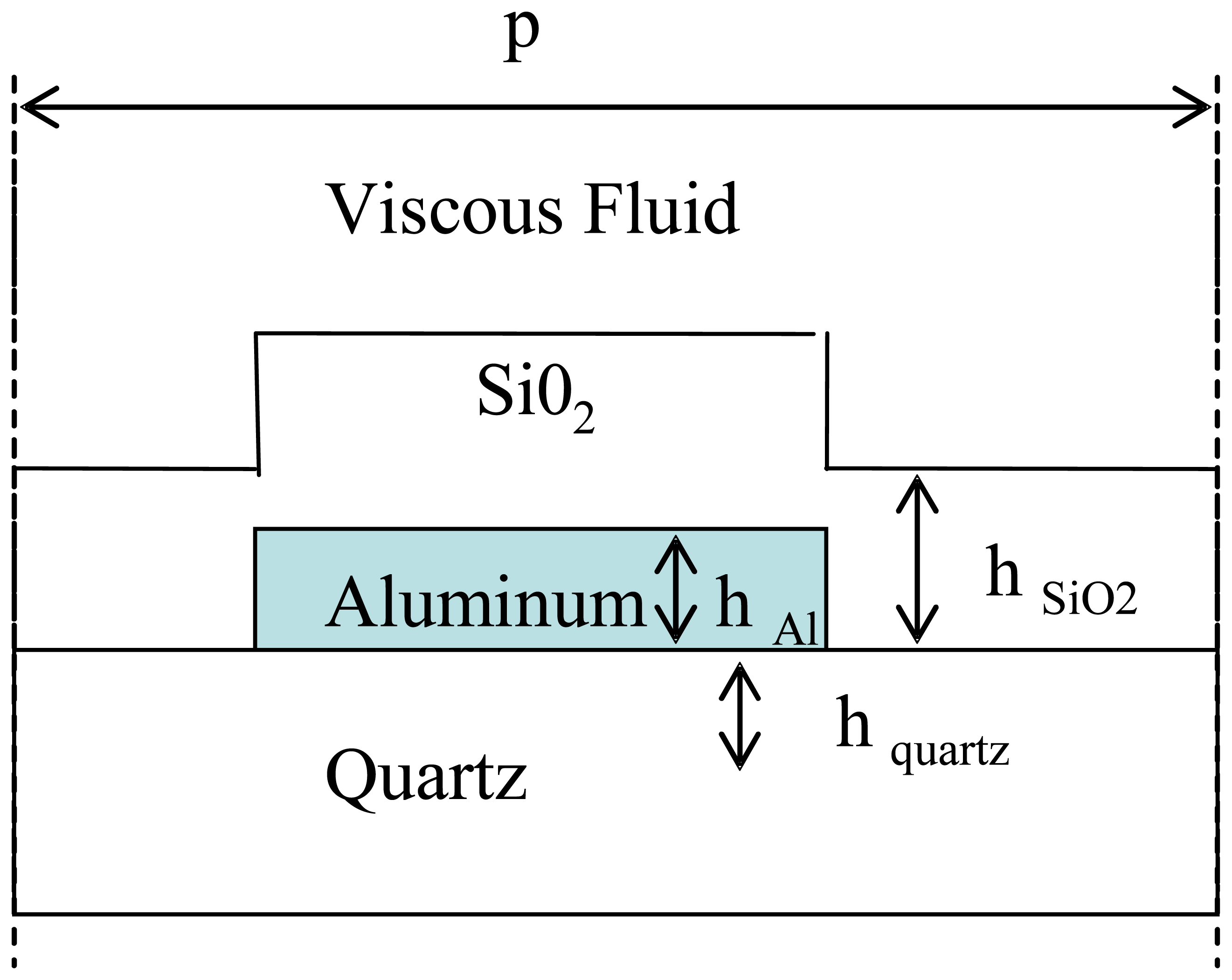
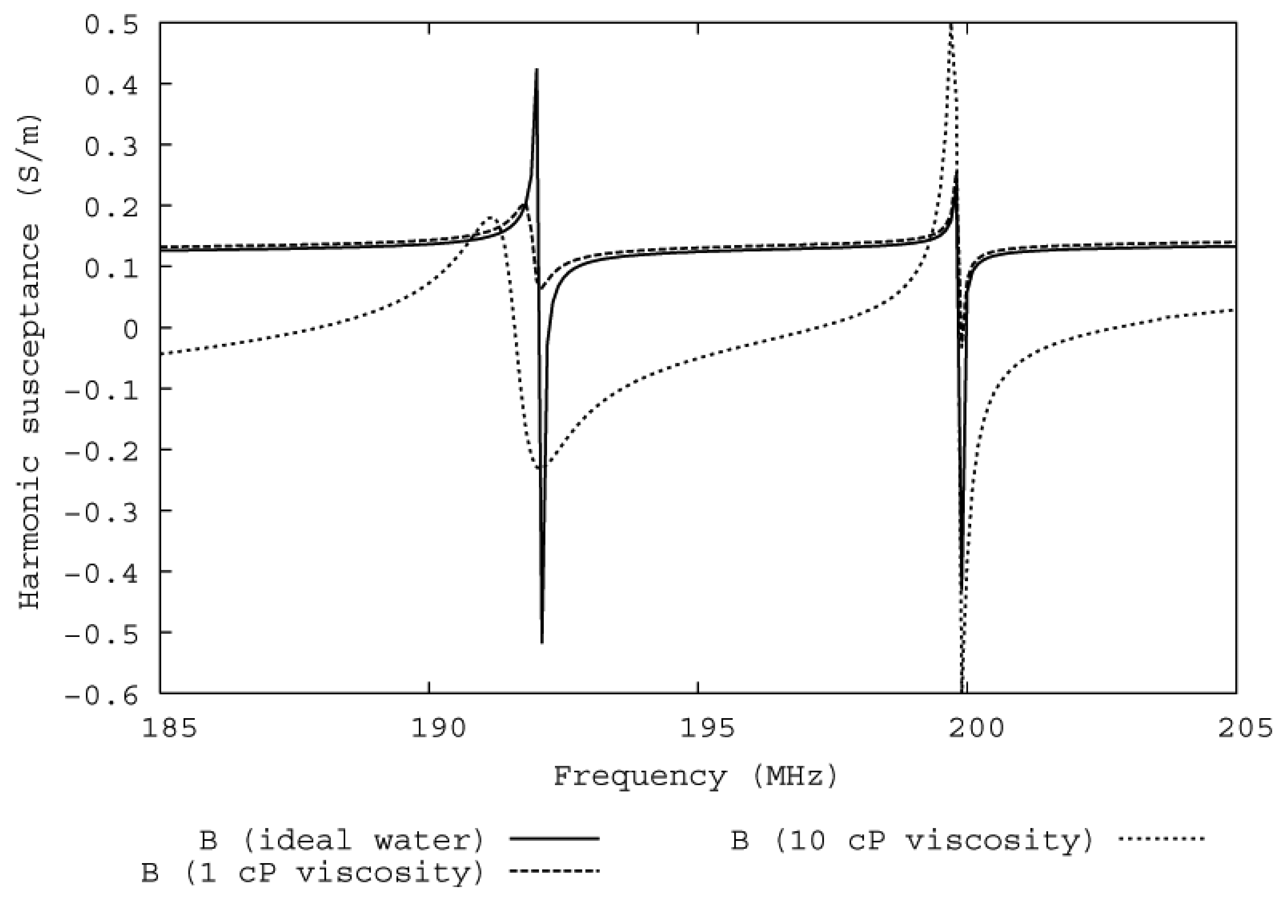
2. Theory and mass sensitivity measurements
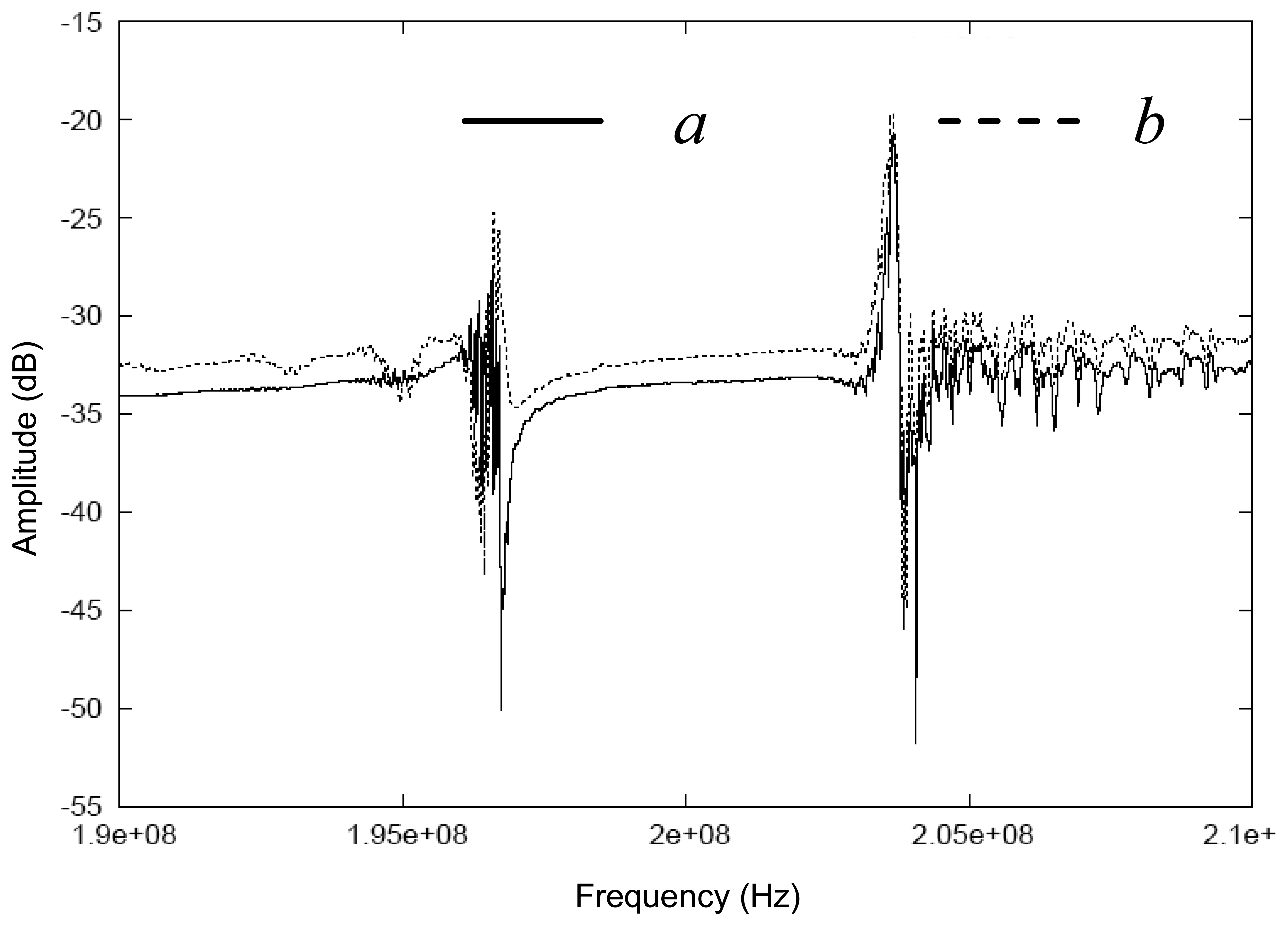
3. Results and discussion
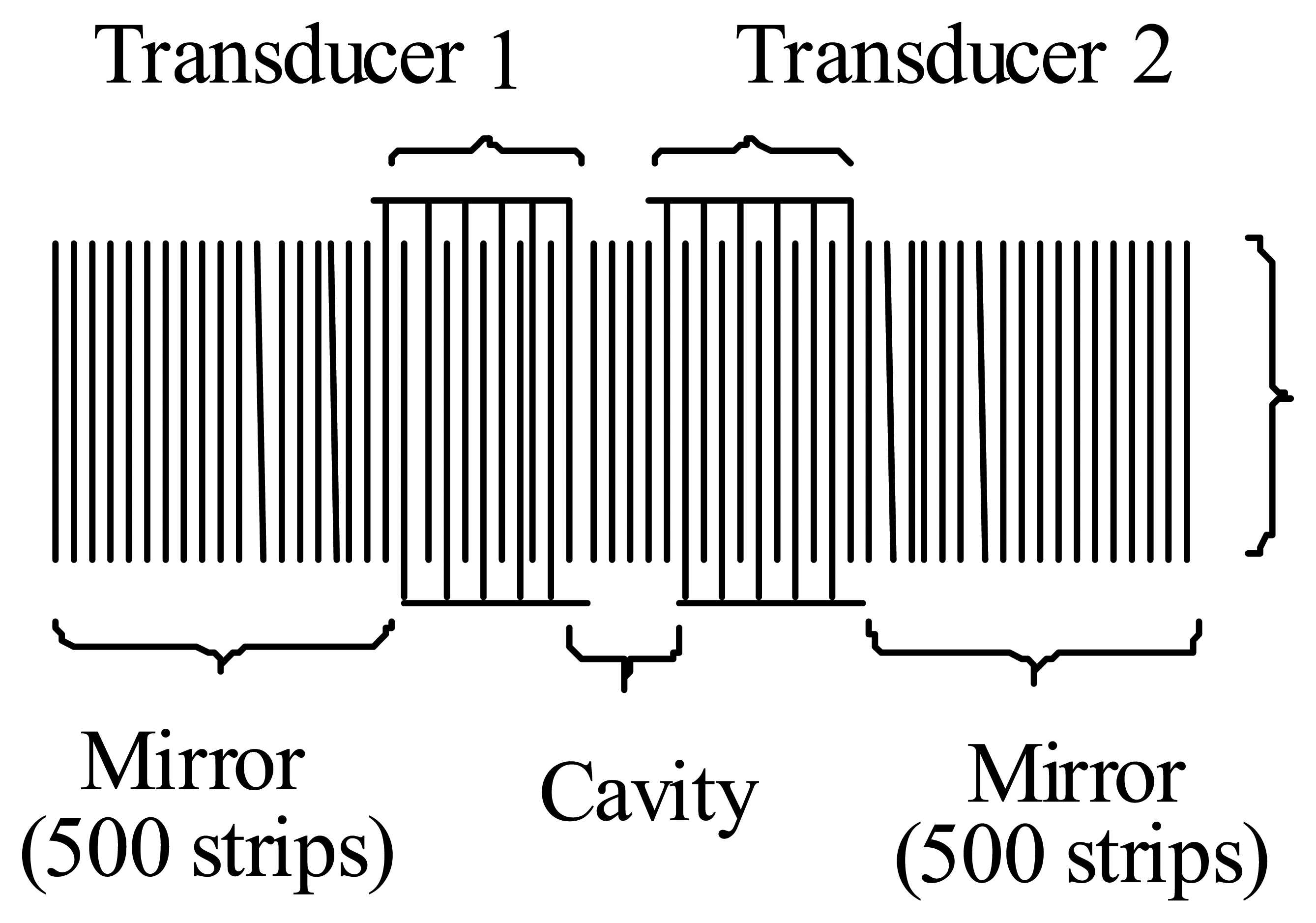
3.1 Design of the Love wave device
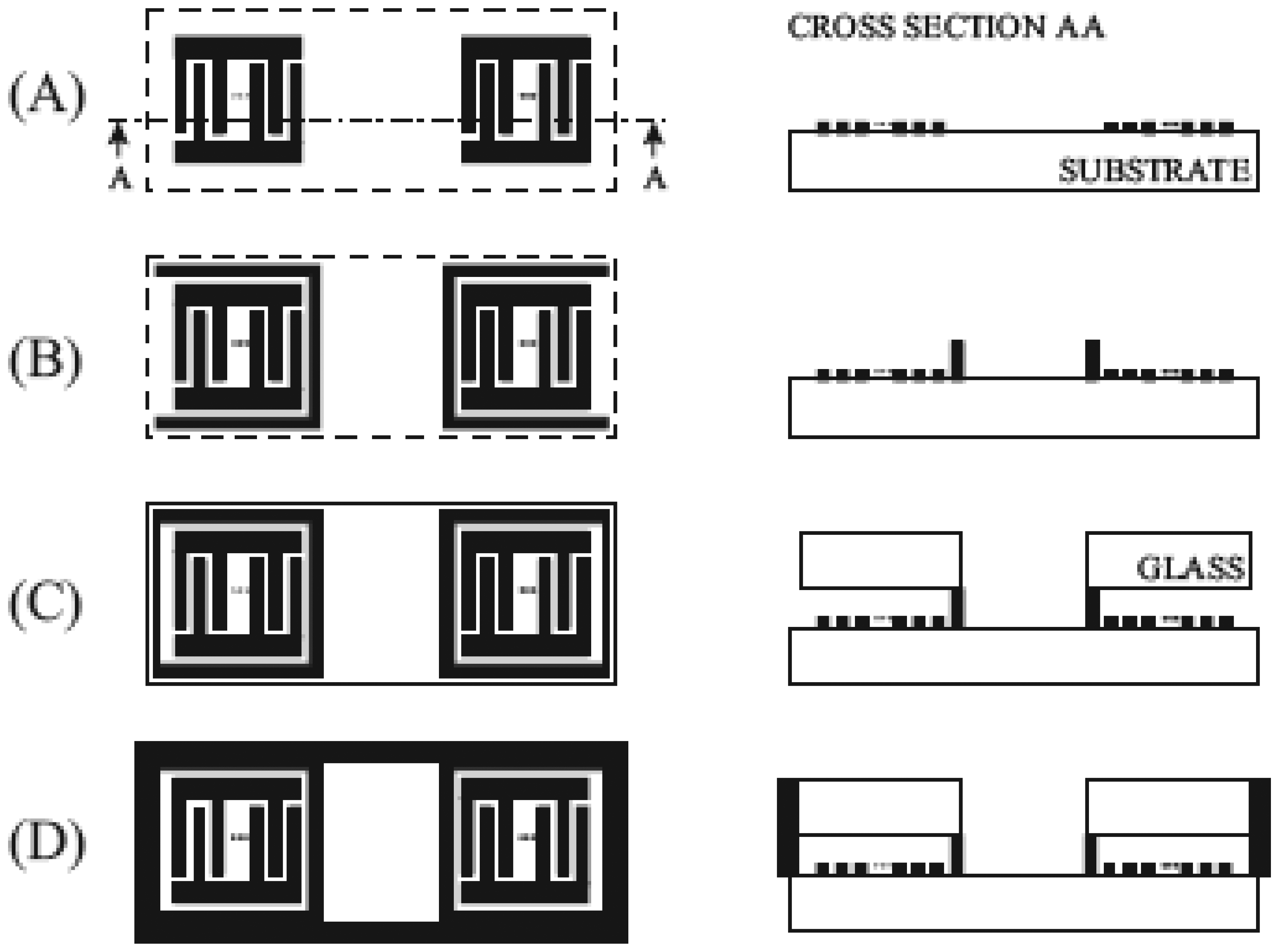
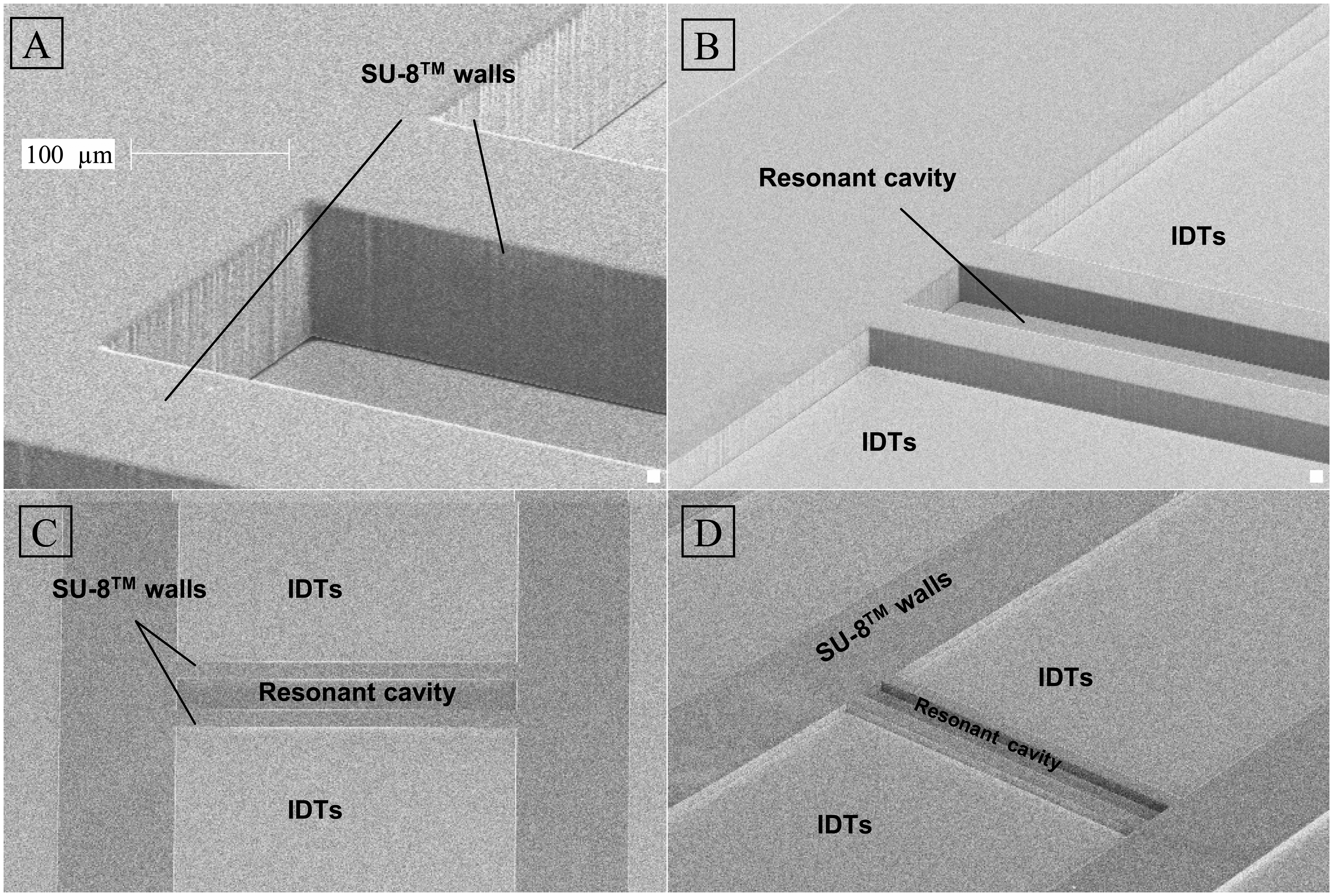
3.2 Fabrication of the Micro fluidic system
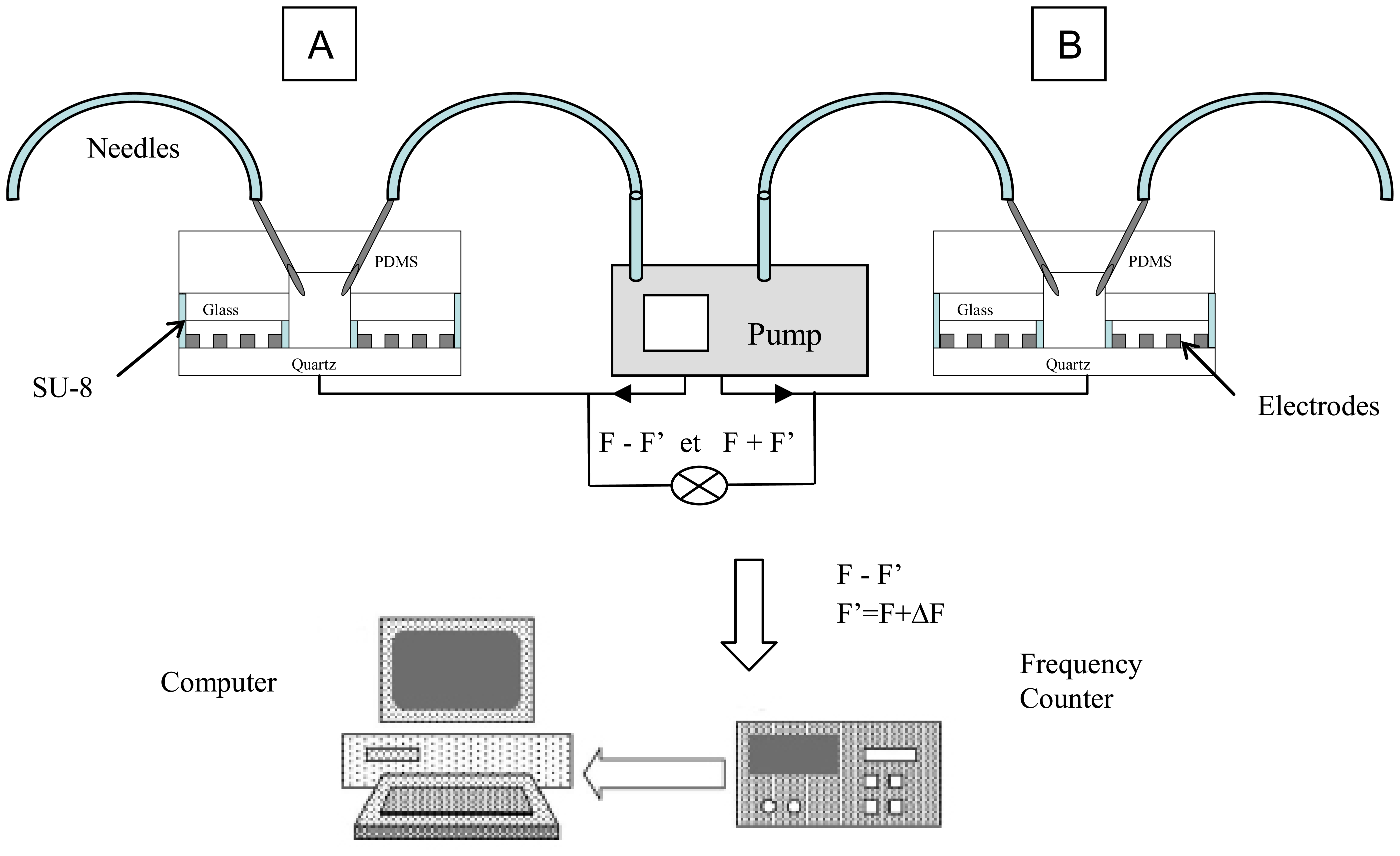
3.3 System set up
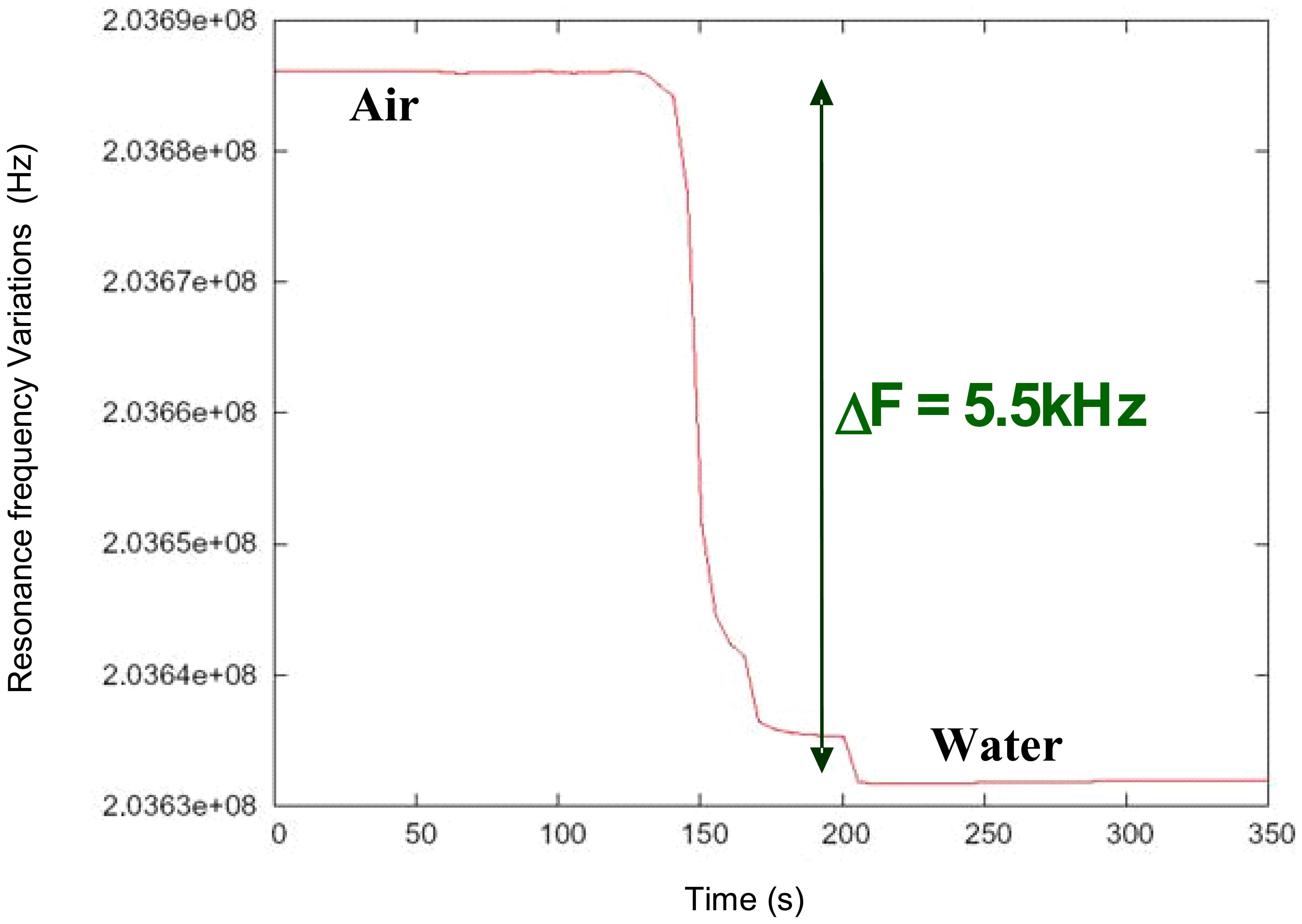
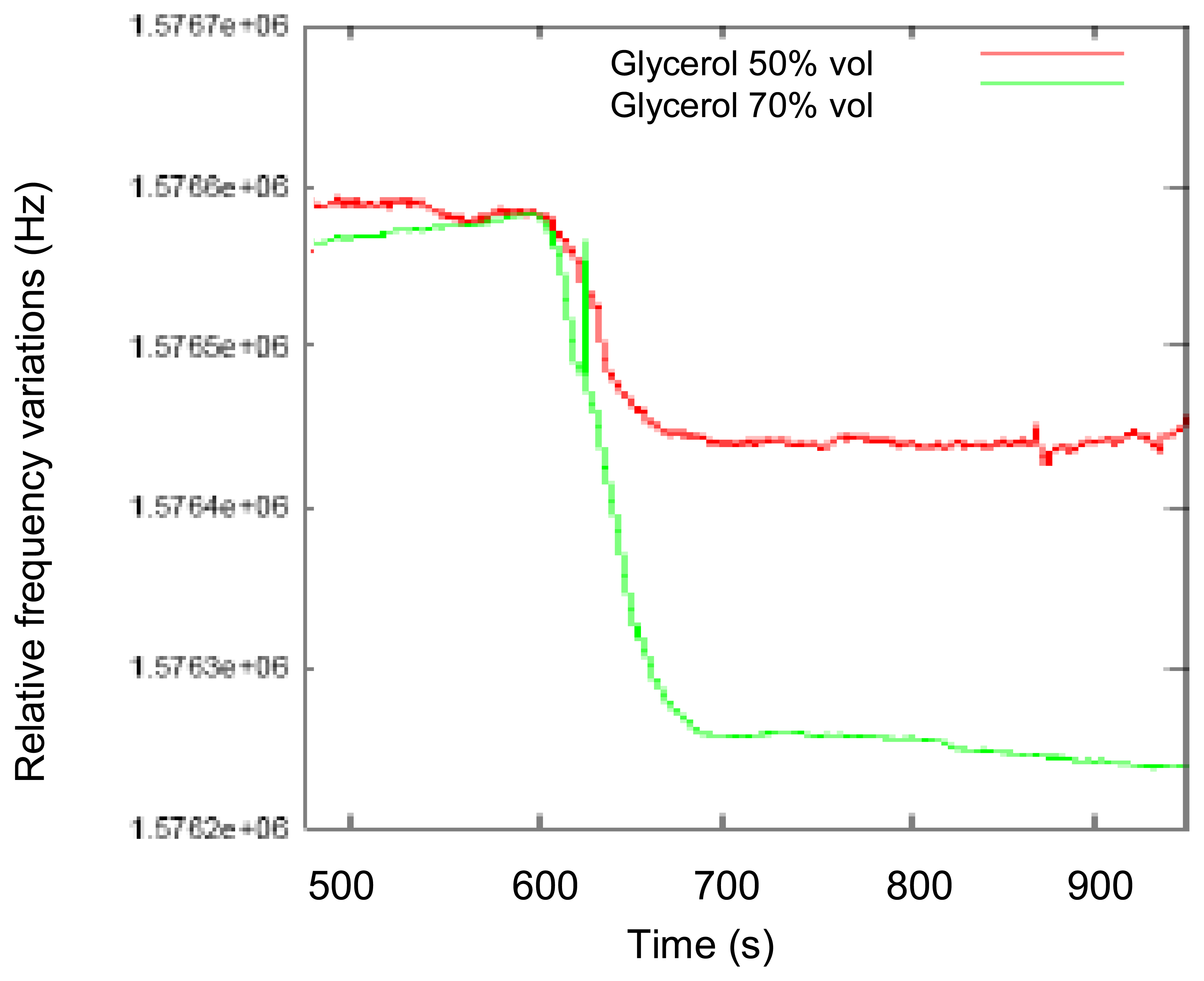
3.4 Measurements
Conclusion
References
- Rebeski, D.E.; Winger, E.M.; Shin, Y.K.; Lelenta, M.; Robinson, M.M.; Varecka, R.; Crowther, J.R. Identification of unacceptable background caused by non-specific protein adsorption to the plastic surface of 96-well immunoassay plates using a standardized enzyme-linked immunosorbent assay procedure. J. Immunol. Methods 1999, 226, 85–92. [Google Scholar]
- Rosengren, A.; Oscarsson, S.; Mazzocchi, M.; Krajewski, A.; Ravaglioli, A. Protein adsorption onto two bioactive glass-ceramics. Biomaterials 2003, 24, 147–55. [Google Scholar]
- Harding, G.L.; Du, J.; Dencher, P.R.; Barnett, D.; Howe, E. Sensors and Actuators A 1997, 61, 279–286.
- Jakoby, B.; Vellekoop, M.J. Viscosity sensing using a Love-wave device. Sensors and Actuators A: Physical 1998, 68, 275–281. [Google Scholar]
- Yantchev, V.; Strashilov, V. Reflectivity in surface transverse wave gratings. J. Appl. Phys. 2003, 94, 6212–6214. [Google Scholar]
- Nomura, T.; Saitoh, A.; Miyazaki, T. Liquid sensor probe using reflecting SH-SAW delay line. Sensors and Actuators B 2003, 91, 298–302. [Google Scholar]
- Hechner, J.; Soluch, W. Pseudo surface acoustic wave dual delay line on 41°YX LiNbO3for liquid sensors. Sensors and Actuators B 2005, 111-112, 436–440. [Google Scholar]
- Nomura, T.; Saitoh, A.; Horikoshu, Y. Measurement of acoustic properties of liquid using liquid flow SH-SAW sensor system. Sensors and Actuators B 2001, 68, 275–281. [Google Scholar]
- Jakoby, B.; Vellekoop, M. J. Properties of Loves waves: application in sensors. Smart Mater. Struct. 1997, 6, 668–679. [Google Scholar]
- Mchale, G.; Newton, M.I.; Martin, F.; Gizeli, E.; Melzak, K. A. Resonant conditions for Love wave guiding layer thickness. Appl. Phys. Lett. 2001, 79, 3542–3543. [Google Scholar]
- Blondeau-Patissier, V.; Ballandras, S.; Lengaigne, G.; Nadal, M.H.; Daniau, W.; Martin, G.; Hauden, D.; Blind, P. High sensitivity anhydride hexafluorhydric acid sensor. Sensors and Actuators B 2005, 111-112, 219–224. [Google Scholar]
- Raimbault, V.; Rebiere, D.; Dejous, C.; Guirardel, M.; Conedera, V.; Pistre, J. High viscosity sensing using a love wave acoustic platform combined with a PDMS microfluidic chip. ECS Transactions 2006, 4, 73–81. [Google Scholar]
- Ballandras, S.; Laude, V.; Pastureaud, Th.; Wilm, M.; Daniau, W.; Reinhardt, A.; Steichen, W.; Lardat, R. A FEA/BEM Approach to Simulate Complex Electrode Structures Devoted to Guided Elastic Wave Periodic Transducers. Proc. of the IEEE Ultrasonics Symposium; München, 2002; pp. 309–312. [Google Scholar]
- Ballandras, S.; Reinhardt, A.; Khelif, A.; Wilm, M.; Laude, V.; Daniau, W.; Blondeau-Patissier, V.; Boireau, W. Theoretical analysis of damping effects of SAW at solid/fluid interfaces. Proc. of the joint EFTF - IEEE Int. Freq. Cont. Symp. Tampa Bay, 2003; pp. 907–910. [Google Scholar]
- Ballandras, S.; Reinhardt, A.; Khelif, A.; Wilm, M.; Laude, V.; Daniau, W.; Blondeau-Patissier, V. Theoretical analysis of damping effects of guided elastic waves at solid/fluid interfaces. Journal of Applied Physicsm 2006, 99(5), 545–553. [Google Scholar]
- Reinhardt, A.; Khelif, A.; Wilm, M.; Laude, V.; Daniau, W.; Blondeau-Patissier, V.; Lengaigne, G.; Ballandras, S. Theoretical analysis of damping effects of acoustic waves at solid/fluid interfaces using a mixed periodic FEA/BEM approach. Proc. of the EFTF; Besançon, 2005. [Google Scholar]
- Royer; Dieulesaint, D.E. Elastics waves in solids 1, Ed. ed; Springer Verlag: Heidelberg, 2000. [Google Scholar]
- Grate, J.; Martin, S.; White, R. Acoustic wave micro sensors. Part. I. Anal. Chem. 1993, 65, 940–948. [Google Scholar]
- Kalantar-Zadeh, K.; Wlodarski, W.; Chen, Y. Y.; Fry, B. N.; Galastis, K. Novel Love mode surface acoustic wave based immunosensors. Sensors and Actuators B. 2003, 91, 143–147. [Google Scholar]
- Wang, Z.; Cheeke, J.D.N. Sensitivity analysis for Love mode acoustic gravimetric sensors. Appl. Phys. Lett. 1994, 64, 2940–2942. [Google Scholar]
- Francis, L.A.; Friedt, J.-M.; Bartic, C.; Campitelli, A. Proceedings of SPIE 2004, 5455, 353–363.
- Kim, J.-W.; Yamagat, Y.; Takasaki, M.; Lee, B.-H.; Ohmori, H.; Higuchi, T. A device for fabricating protein chips by using a surface acoustic wave atomizer and electrostatic deposition. Sensors and Actuators B 2005, 107, 535–545. [Google Scholar]
- McClellan, S.J.; Franses, E. I. Adsorption of bovine serum albumin at solid/aqueous interfaces Colloids and Surfaces A. Physicochem. Eng. Aspects 2005, 260, 265–275. [Google Scholar]


© 2007 by MDPI ( http://www.mdpi.org). Reproduction is permitted for noncommercial purposes.
Share and Cite
Blondeau-Patissier, V.; Boireau, W.; Cavallier, B.; Lengaigne, G.; Daniau, W.; Martin, G.; Ballandras, S. Integrated Love Wave Device Dedicated to Biomolecular Interactions Measurements in Aqueous Media. Sensors 2007, 7, 1992-2003. https://doi.org/10.3390/s7091992
Blondeau-Patissier V, Boireau W, Cavallier B, Lengaigne G, Daniau W, Martin G, Ballandras S. Integrated Love Wave Device Dedicated to Biomolecular Interactions Measurements in Aqueous Media. Sensors. 2007; 7(9):1992-2003. https://doi.org/10.3390/s7091992
Chicago/Turabian StyleBlondeau-Patissier, Virginie, Wilfrid Boireau, Bruno Cavallier, Gwladys Lengaigne, William Daniau, Gilles Martin, and Sylvain Ballandras. 2007. "Integrated Love Wave Device Dedicated to Biomolecular Interactions Measurements in Aqueous Media" Sensors 7, no. 9: 1992-2003. https://doi.org/10.3390/s7091992
APA StyleBlondeau-Patissier, V., Boireau, W., Cavallier, B., Lengaigne, G., Daniau, W., Martin, G., & Ballandras, S. (2007). Integrated Love Wave Device Dedicated to Biomolecular Interactions Measurements in Aqueous Media. Sensors, 7(9), 1992-2003. https://doi.org/10.3390/s7091992