

Varroa Mite Counting Based on Hyperspectral Imaging
Abstract
1. Introduction
2. Materials and Methods
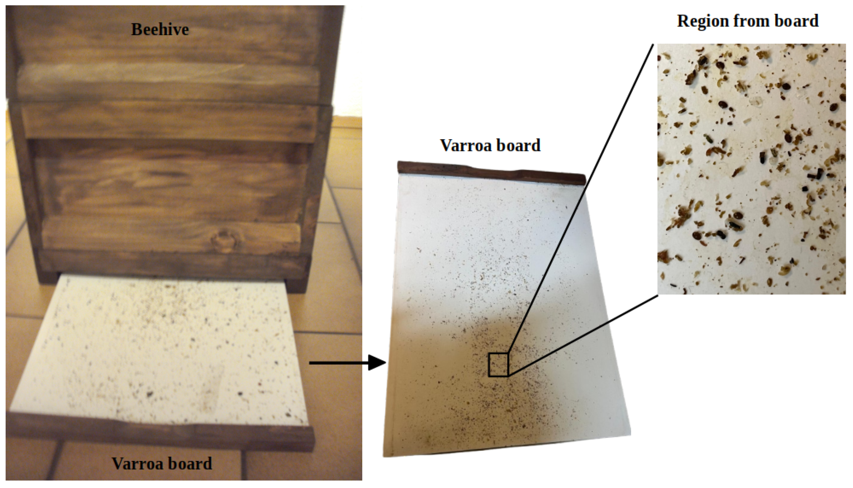
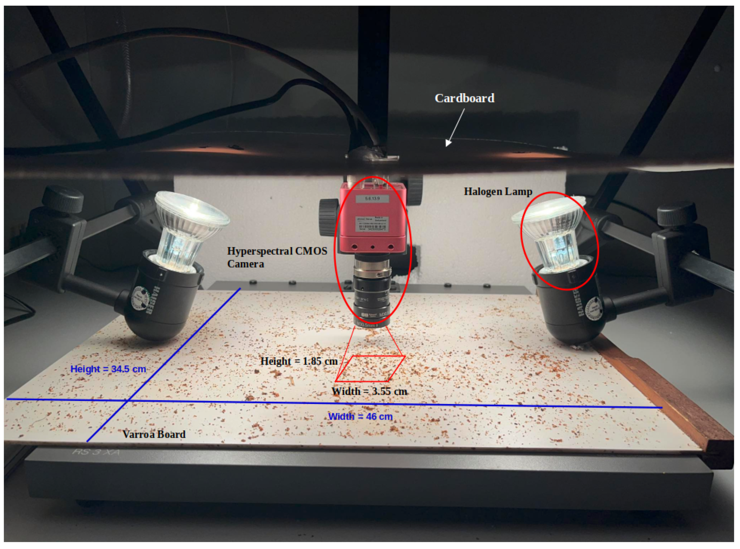
2.1. Image Acquisition Setup and Conditions
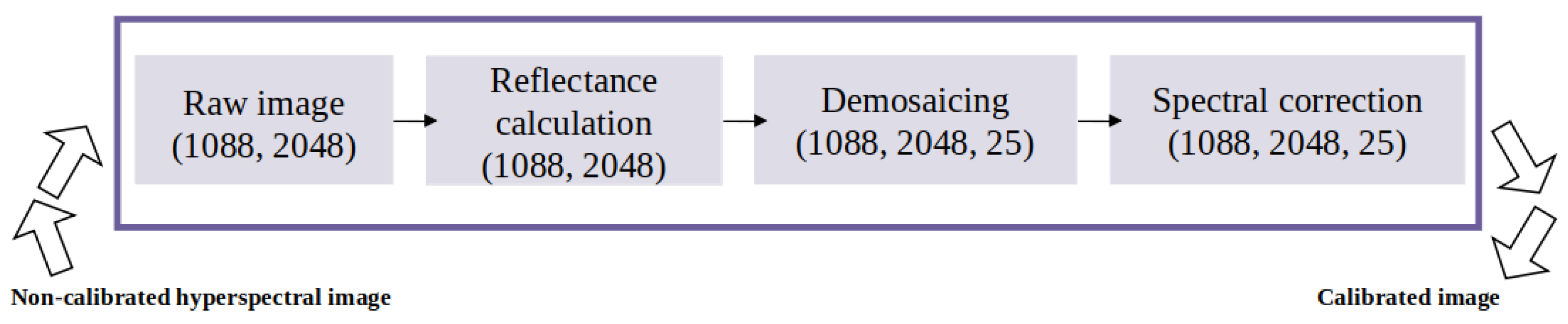
2.2. Recognition Pipeline and Calibration Process
2.3. Labeling and Data Generation
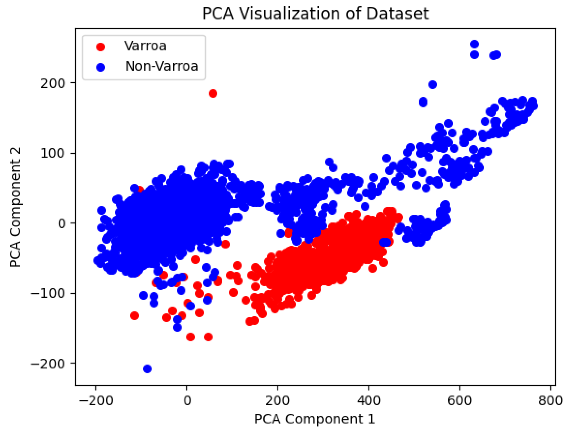
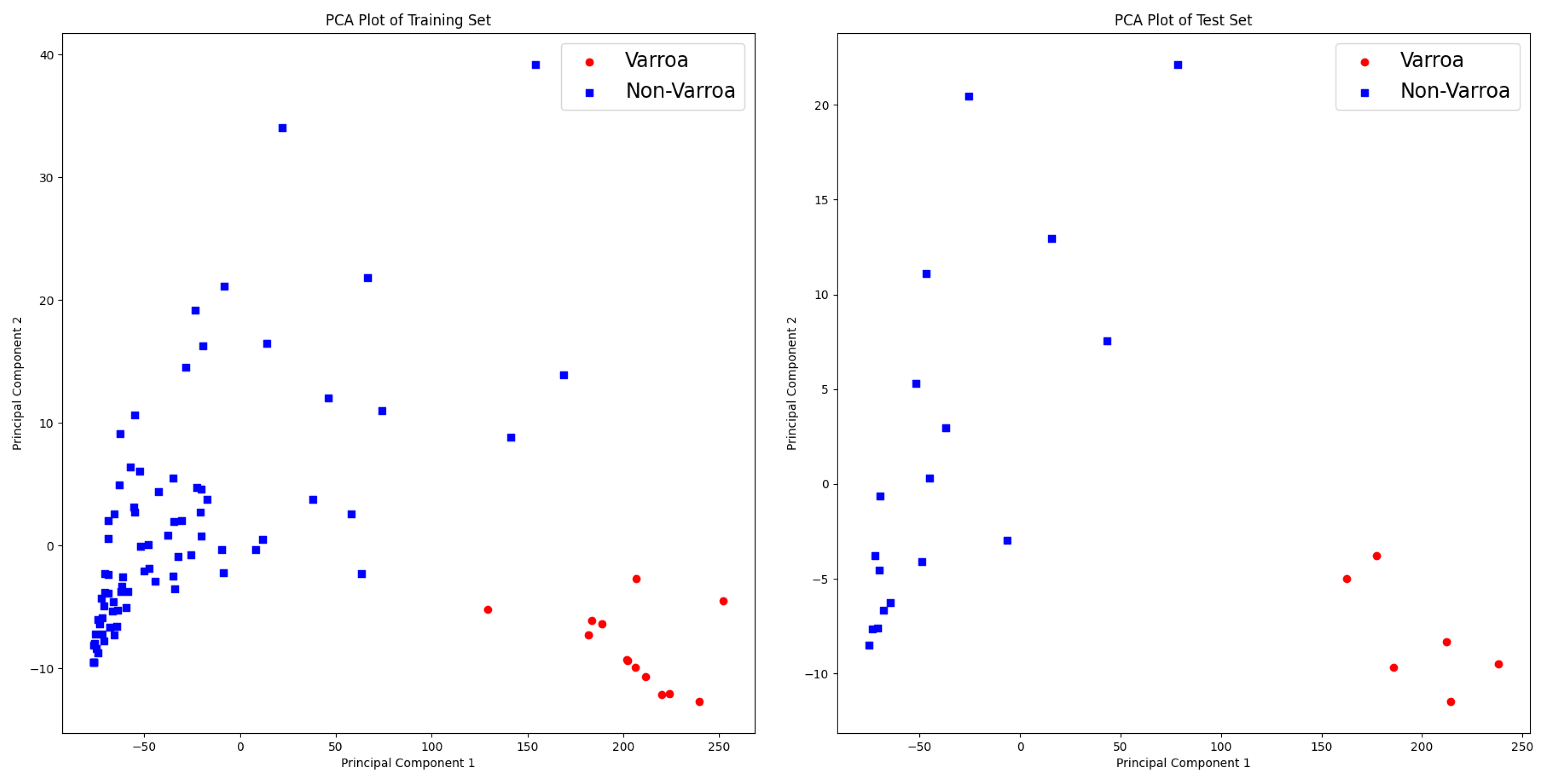
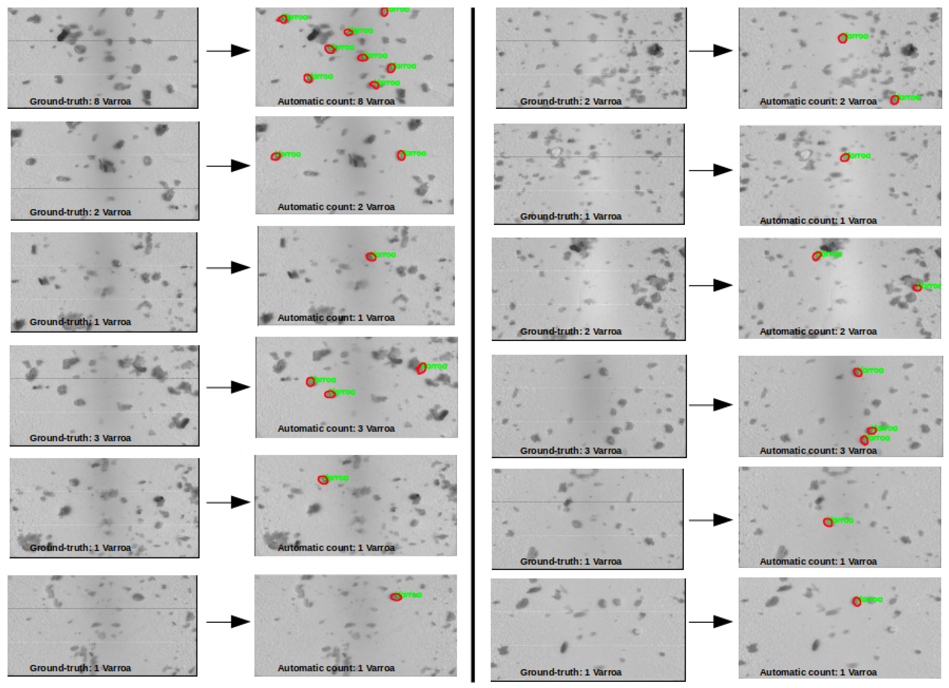
3. Results
- Training Set Confusion Matrix:
- Test Set Confusion Matrix:
4. Discussion
5. Conclusions
Author Contributions
Funding
Institutional Review Board Statement
Informed Consent Statement
Data Availability Statement
Acknowledgments
Conflicts of Interest
Abbreviations
| HS-Cam | Hyperspectral Camera |
| PCA | Principal Component Analysis |
| kNN | k-Nearest Neighbor |
| SVM | Support Vector Machine |
| NIR | Near Infrared |
| HSI | Hyperspectral Imaging |
| IR | Infrared |
| DL | Deep Learning |
References
- Decourtye, A.; Mader, E.; Desneux, N. Landscape enhancement of floral resources for honey bees in agro-ecosystems. Apidologie 2010, 41, 264–277. [Google Scholar] [CrossRef]
- Klein, A.M.; Vaissière, B.E.; Cane, J.H.; Steffan-Dewenter, I.; Cunningham, S.A.; Kremen, C.; Tscharntke, T. Importance of pollinators in changing landscapes for world crops. Proc. R. Soc. B Biol. Sci. 2007, 274, 303–313. [Google Scholar] [CrossRef] [PubMed]
- Rosenkranz, P.; Aumeier, P.; Ziegelmann, B. Biology and control of Varroa destructor. J. Invertebr. Pathol. 2010, 103, S96–S119. [Google Scholar] [CrossRef] [PubMed]
- Ostiguy, N.; Sammataro, D. A simplified technique for counting Varroa jacobsoni Oud. on sticky boards. Apidologie 2000, 31, 707–716. [Google Scholar] [CrossRef]
- Khan, A.; Vibhute, A.D.; Mali, S.; Patil, C. A systematic review on hyperspectral imaging technology with a machine and deep learning methodology for agricultural applications. Ecol. Inform. 2022, 69, 101678. [Google Scholar] [CrossRef]
- Li, T.; Wei, W.; Xing, S.; Min, W.; Zhang, C.; Jiang, S. Deep Learning-Based Near-Infrared Hyperspectral Imaging for Food Nutrition Estimation. Foods 2023, 12, 3145. [Google Scholar] [CrossRef] [PubMed]
- Elmasry, G.; Kamruzzaman, M.; Sun, D.W.; Allen, P. Principles and applications of hyperspectral imaging in quality evaluation of agro-food products: A review. Crit. Rev. Food Sci. Nutr. 2012, 52, 999–1023. [Google Scholar] [CrossRef] [PubMed]
- Nguyen, N.M.T.; Liou, N.S. Ripeness Evaluation of Achacha Fruit Using Hyperspectral Image Data. Agriculture 2022, 12, 2145. [Google Scholar] [CrossRef]
- Ye, W.; Xu, W.; Yan, T.; Yan, J.; Gao, P.; Zhang, C. Application of Near-Infrared Spectroscopy and Hyperspectral Imaging Combined with Machine Learning Algorithms for Quality Inspection of Grape: A Review. Foods 2022, 12, 132. [Google Scholar] [CrossRef] [PubMed]
- Martin, M.E.; Wabuyele, M.B.; Chen, K.; Kasili, P.; Panjehpour, M.; Phan, M.; Overholt, B.; Cunningham, G.; Wilson, D.; DeNovo, R.C.; et al. Development of an advanced hyperspectral imaging (HSI) system with applications for cancer detection. Ann. Biomed. Eng. 2006, 34, 1061–1068. [Google Scholar] [CrossRef] [PubMed]
- Cucci, C.; Delaney, J.K.; Picollo, M. Reflectance hyperspectral imaging for investigation of works of art: Old master paintings and illuminated manuscripts. Acc. Chem. Res. 2016, 49, 2070–2079. [Google Scholar] [CrossRef] [PubMed]
- Li, X.; Li, R.; Wang, M.; Liu, Y.; Zhang, B.; Zhou, J. Hyperspectral imaging and their applications in the nondestructive quality assessment of fruits and vegetables. In Hyperspectral Imaging in Agriculture, Food and Environment; IntechOpen: London, UK, 2018; pp. 27–63. [Google Scholar]
- Huang, H.; Liu, L.; Ngadi, M.O. Recent developments in hyperspectral imaging for assessment of food quality and safety. Sensors 2014, 14, 7248–7276. [Google Scholar] [CrossRef] [PubMed]
- ul Rehman, A.; Qureshi, S.A. A review of the medical hyperspectral imaging systems and unmixing algorithms’ in biological tissues. Photodiagn. Photodyn. Ther. 2021, 33, 102165. [Google Scholar] [CrossRef] [PubMed]
- Divasón, J.; Martinez-de Pison, F.J.; Romero, A.; Santolaria, P.; Yániz, J.L. Varroa Mite Detection Using Deep Learning Techniques. In Proceedings of the International Conference on Hybrid Artificial Intelligence Systems, Salamanca, Spain, 5–7 September 2023; Springer: Cham, Switzerland, 2023; pp. 326–337. [Google Scholar]
- Noriega-Escamilla, A.; Camacho-Bello, C.J.; Ortega-Mendoza, R.M.; Arroyo-Núñez, J.H.; Gutiérrez-Lazcano, L. Varroa Destructor Classification Using Legendre–Fourier Moments with Different Color Spaces. J. Imaging 2023, 9, 144. [Google Scholar] [CrossRef] [PubMed]
- Sevin, S.; Tutun, H.; Mutlu, S. Detection of varroa mites from honey bee hives by smart technology var-gor: A hivemonitoring and image processing device. Turk. J. Vet. Anim. Sci. 2021, 45, 487–491. [Google Scholar] [CrossRef]
- Koenig, A. VarroaCounter—Towards Automating the Varroa Screening for Alleviated Bee Hive Treatment. In Proceedings of the 5th International Conference on Sensors and Electronic Instrumentation Advances (SEIA 2019), Adeje, Spain, 25–27 September 2019. [Google Scholar]
- König, A. IndusBee 4.0–integrated intelligent sensory systems for advanced bee hive instrumentation and hive keepers’ assistance systems. Sens. Transducers 2019, 237, 109–121. [Google Scholar]
- König, A. BeE-Nose–An In-Hive Multi-Gas-Sensor Extension to the IndusBee4. 0 System for Hive Air Quality Monitoring and Varroa Infestation Level Estimation. Adv. Signal Process. Rev. 2021, 2, 443–463. [Google Scholar]
- König, A. An in-hive soft sensor based on phase space features for Varroa infestation level estimation and treatment need detection. J. Sens. Sens. Syst. 2022, 11, 29–40. [Google Scholar] [CrossRef]
- Photonfocus. Available online: https://www.photonfocus.com/de/support/software/ (accessed on 1 April 2024).
- Yu, K.Q.; Zhao, Y.R.; Zhu, F.L.; Li, X.L.; He, Y. Mapping of chlorophyll and SPAD distribution in pepper leaves during leaf senescence using visible and near-infrared hyperspectral imaging. Trans. ASABE 2016, 59, 13–24. [Google Scholar]
- Ramsey, S.D.; Ochoa, R.; Bauchan, G.; Gulbronson, C.; Mowery, J.D.; Cohen, A.; Lim, D.; Joklik, J.; Cicero, J.M.; Ellis, J.D.; et al. Varroa destructor feeds primarily on honey bee fat body tissue and not hemolymph. Proc. Natl. Acad. Sci. USA 2019, 116, 1792–1801. [Google Scholar] [CrossRef] [PubMed]
- IMEC. Calibration Files_Reference Manual, Internal PDF Document Provided by the Manufacturer upon Request. PDF Document Obtained from the Manufacturer; IMEC: Leuven, Belgium, 2021. [Google Scholar]


| Image Sensor Specifications | |
|---|---|
| Manufacturer/type | IMEC, CMV2K-SM5x5 |
| Technology | CMOS |
| Optical format | 2/3″ |
| Optical diagonal | 12.76 mm |
| Resolution | 2048 × 1088 |
| Pixel size | 5.5 μm × 5.5 μm |
| Active optical area | 11.26 mm × 5.98 mm |
| Dark current | 125e-/s |
| Read-out noise | 13e- |
| Full well capacity/SNR | 11ke- / 105:1 |
| Spectral range | Hyperspectral: 665 to 975 nm (25 pass bands) |
| Responsivity | Hyperspectral: 454 × 103 DN / (J/m2) @ 715 nm/8 bit |
| Quantum efficiency | Hyperspectral: <18% |
| Optical fill factor | 42% without microlenses |
| Dynamic range | 60 dB |
| Characteristic curve | Linear, piecewise linear |
| Shutter mode | Global shutter |
| Camera Specifications | |
| Interface | GigE |
| Frame rate | 42 fps |
| Pixel clock | 48 MHz |
| Camera taps | 2 |
| Grayscale resolution | 8 Bit/10 Bit |
| Fixed pattern noise (FPN) | <1DN RMS @ 8 Bit |
| Exposure time range | 13 μs–349 ms |
| Analog gain | yes |
| Digital gain | 0.1 to 15.99 (FineGain) |
| Trigger modes | Free running (non triggered), external trigger, SWTrigger |
| Features | Configurable region of interest (ROI), up to 8 regions of interest (MROIs), binning for data preprocessing, decimation in y-direction, 2 look-up tables (12-to-8 Bit) on user-defined image region (Region-LUT), constant frame rate independent of exposure time, crosshairs overlay on the image, temperature monitoring of camera, camera information readable over SDK, ultra low trigger delay and low trigger jitter, extended trigger input and strobe output functionality, status line in picture |
| Operation temperature/moisture | 0 °C … + 50 °C/20% … 80% |
| Storage temperature/moisture | −25 °C … 60 °C/20% … 95% |
| Power supply | +12 VDC (−10%) … +24 VDC (+10%) |
| Power consumption | <5.1 W |
| Lens mount | C-Mount (CS-Mount optional) |
| I/O inputs | 2× Opto-isolated 2× RS-422 Opto-isolated |
| I/O outputs | 2× Opto-isolated |
| Dimensions | 55 × 55 × 52 mm3 |
| Mass | 265 g |
| Connector I/O (power) | Hirose 12-pole (mating plug HR10A-10P-12S) |
| Connector interface | RJ-45 |
| Conformity | CE / RoHS / WEEE |
| IP code | IP40 |
| Classifiers | Training Set (Accuracy) | Test Set (Accuracy) | Hyperparameters | F1-Score |
|---|---|---|---|---|
| “Linear” SVM | 0.9932 | 0.9927 | “C”: 1 | 0.9581 |
| “Rbf” SVM | 0.912 | 0.915 | “C”: 0.1, “gamma”: 0.1 | 0.0 |
| kNN | 0.999 | 0.999 | “”: 1 | 0.9947 |
| ANN * | 0.991 | 0.992 | “”: 16, “epochs”: 10 | 0.995 |
| Classifiers | Test Set (Accuracy) | Cross-Validation (Accuracy) | Hyperparameters |
|---|---|---|---|
| “Linear” SVM | 1.0 | 1.0 | “C”: 1 |
| kNN | 1.0 | 0.9784 | “”: 3, “weights”: uniform |
| Random Forest | 1.0 | 0.9895 | “”: 10, “”: none |
| DecisionTree | 0.9583 | 0.9895 | “”: 5, “”: 2 |
| Method | Accuracy (%) | Recall (%) | Precision (%) | F1-Score (%) |
|---|---|---|---|---|
| HSI | 99.9 | 99.17 | 99.78 | 99.47 |
| HSI2RGB | 98.8 | 92.93 | 93.2 | 93.06 |
Disclaimer/Publisher’s Note: The statements, opinions and data contained in all publications are solely those of the individual author(s) and contributor(s) and not of MDPI and/or the editor(s). MDPI and/or the editor(s) disclaim responsibility for any injury to people or property resulting from any ideas, methods, instructions or products referred to in the content. |
© 2024 by the authors. Licensee MDPI, Basel, Switzerland. This article is an open access article distributed under the terms and conditions of the Creative Commons Attribution (CC BY) license (https://creativecommons.org/licenses/by/4.0/).
Share and Cite
Ghezal, A.; Peña, C.J.L.; König, A. Varroa Mite Counting Based on Hyperspectral Imaging. Sensors 2024, 24, 4437. https://doi.org/10.3390/s24144437
Ghezal A, Peña CJL, König A. Varroa Mite Counting Based on Hyperspectral Imaging. Sensors. 2024; 24(14):4437. https://doi.org/10.3390/s24144437
Chicago/Turabian StyleGhezal, Amira, Christian Jair Luis Peña, and Andreas König. 2024. "Varroa Mite Counting Based on Hyperspectral Imaging" Sensors 24, no. 14: 4437. https://doi.org/10.3390/s24144437
APA StyleGhezal, A., Peña, C. J. L., & König, A. (2024). Varroa Mite Counting Based on Hyperspectral Imaging. Sensors, 24(14), 4437. https://doi.org/10.3390/s24144437