
Image Noise Removal in Ultrasound Breast Images Based on Hybrid Deep Learning Technique

 , , , ,
  and
and
Abstract
:1. Introduction
2. Literature Review
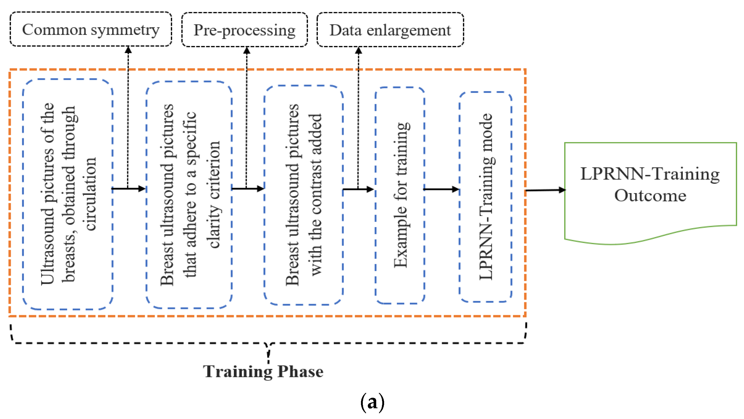
3. Proposed Framework Methodologies
3.1. Dataset Availability
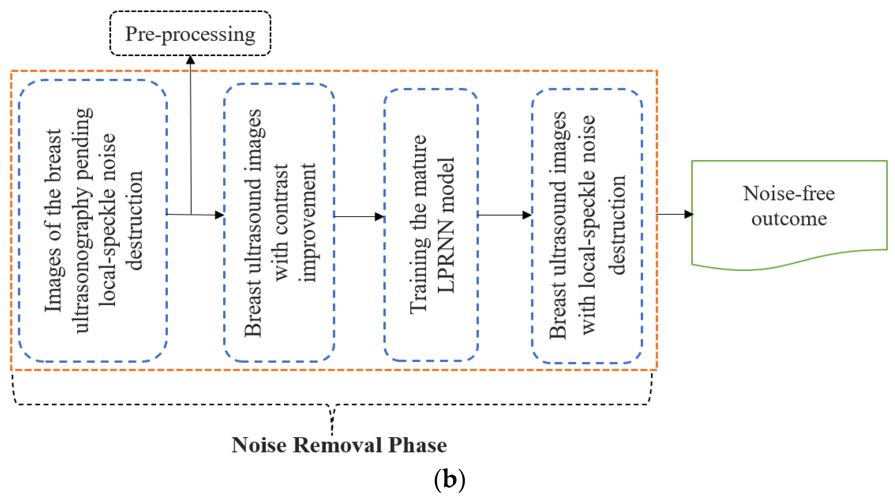
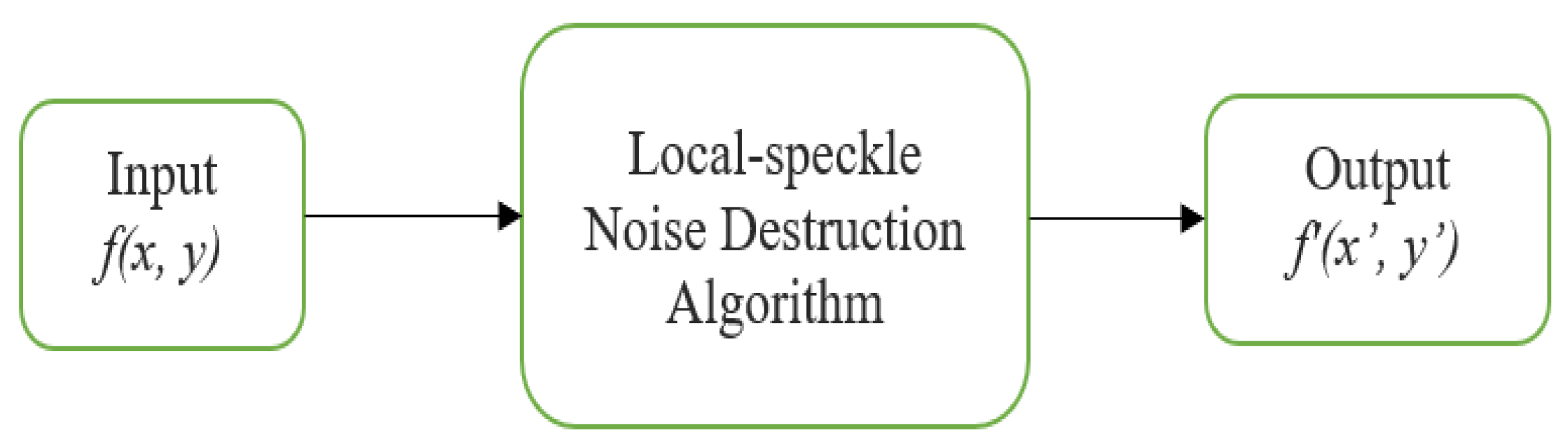
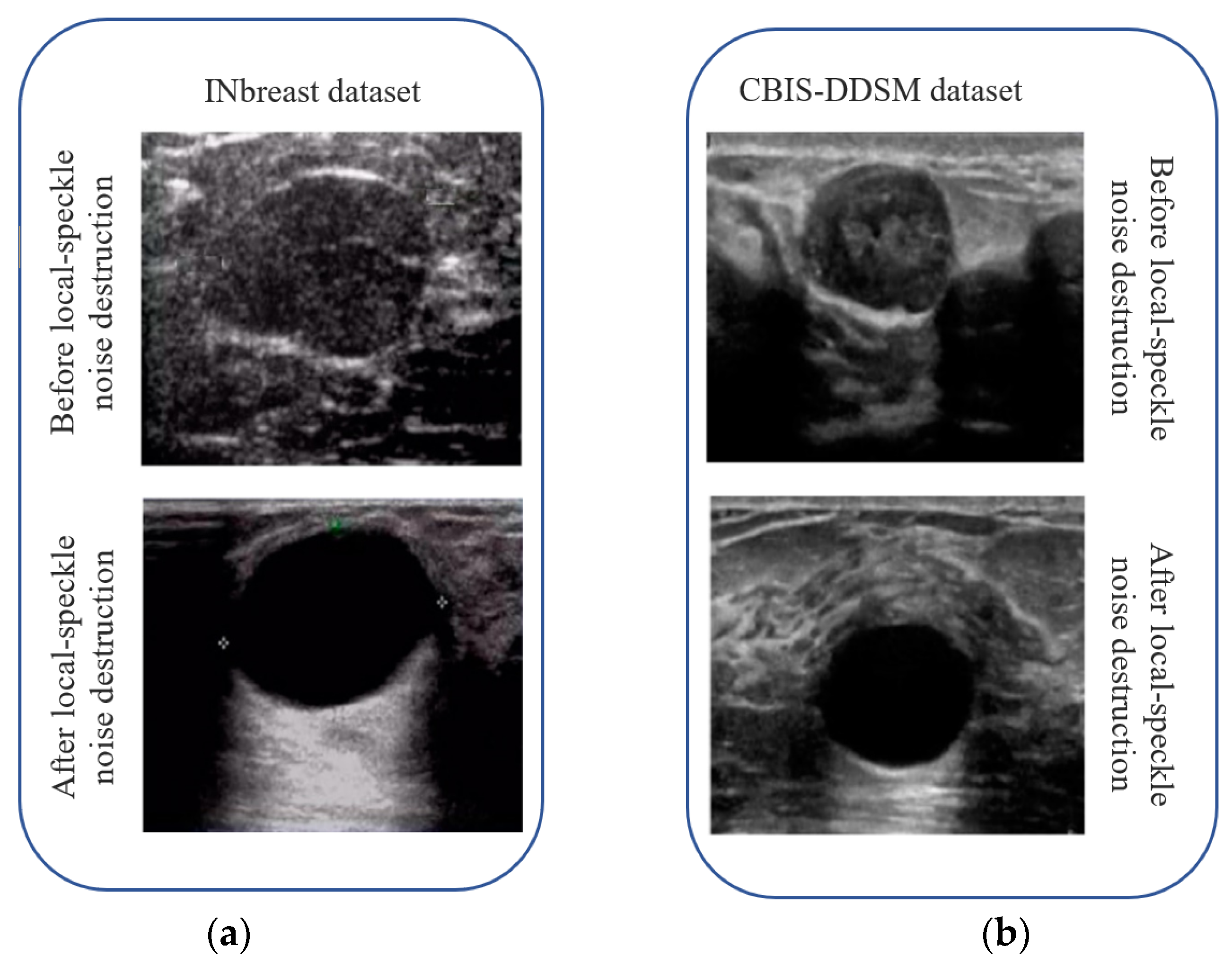
3.2. Effective Destruction of Local Speckle Noise in Breast Images
3.3. Breast Image Pre-Processing and Image Enhancement
3.4. High-Pass Ultrasound Image Spatial Filtering for Breast Images
- Step 1: To improve the process of acquiring the bootstrap image, we first determine a grayscale value of N, of the source ultrasound breast image f, and then use Equation (3) to identify the associated breast ultrasound image h that best fits the categorization.
- Step 2: Having completed Step 1, the ultrasound breast image is utilized as the reference image I’m of the guide-filtration algorithm.
- Step 3: I’’ is the ultrasound breast outcome, and it is achieved by improving the edge data of I’ along with a high-pass filter in order to enlarge the edge data retention.
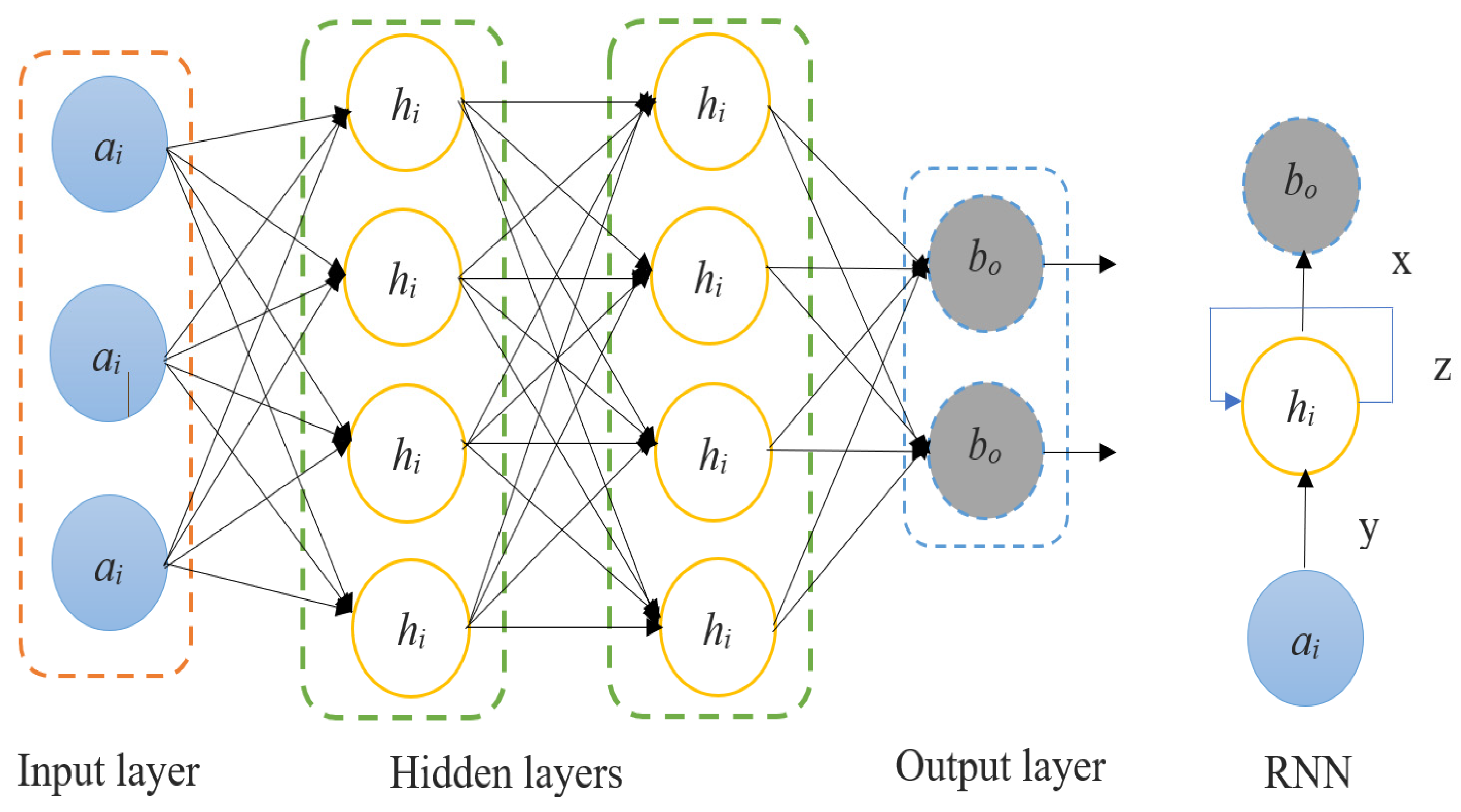
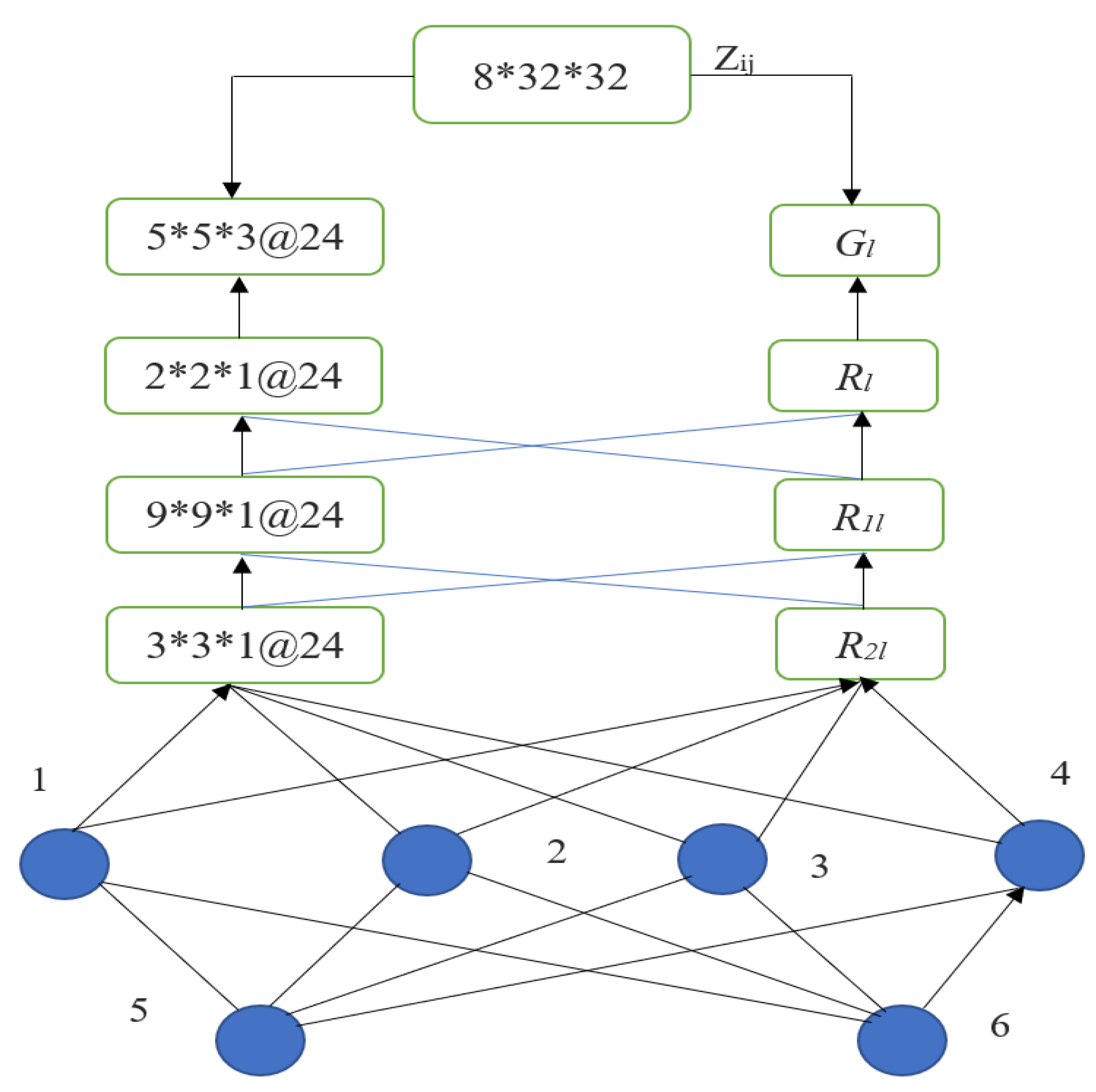
3.5. Logical-Pool Recurrent Neural Network—Local Speckle Noise Destruction
Local Speckle Noise Destruction Algorithm
| Algorithm 1. Noise removal of the local speckle noise. | |
| 1: | Begin |
| 2: | Logarithmic and computational transforms are used to improve the differentiation of the input ultrasound breast images; the algorithm (guided filter) is used to improve the details of the glandular ultrasound images; and the spatial high-pass filtering algorithm is used to denoise the over-sharpening of the ultrasound breast images, all based on their grayscale values |
| 3: | The pre-processed ultrasound breast images are fed into a local-speckle-noise destruction model of a logical-pool recurrent neural network |
| 4: | Ultrasound breast images are susceptible to losing image edge information during the local speckle noise reduction procedure. If we want to preserve the edge information after local speckle noise removal is applied, we will need to understand how that information is lost during processing. The meaning of “edge information loss”.
|
| 5: | In order to construct ultrasound image gradients, we first analyze the aforementioned stages and then use edge loss pairs to compare the edges of canonical clear images of ultrasound breast images. The unique anatomy of the breast emphasizes the significance of the gradients in the vertical plane. That is why we first use contrast in the vertical direction to depict breast ultrasound images. Integrating edge loss and L1 distance with a recurrent neural network yields the following objective function:
|
| 6: | Enhance the loss function to optimize the edge-specific improvement feature of the ultrasound images during training with the logical-pool recurrent neural network. The resulting model will be more responsive in edge local speckle noise destruction in ultrasound images, enhancing its effect on ultrasound breast images |
| 7: | While noise removal reduces the local speckle noise of ultrasound breast images, the edge information is preserved by the action of the advantage term in the logical-pool recurrent neural network as described above |
| 8: | End |
3.6. Performance Metric Evaluation Standards
4. Experimental Results
5. Discussion
6. Conclusions
Author Contributions
Funding
Institutional Review Board Statement
Informed Consent Statement
Data Availability Statement
Conflicts of Interest
Abbreviation
| Short Form | Abbreviation |
| LPRNN | Logical-Pool Recurrent Neural Network |
| SNR | Signal-to-Noise Ratio |
| PSNR | Peak Signal-to-Noise Ratio |
| DCNN | Deep Convolutional Neural Network |
| IR | InfraRed |
| 3D | Three Dimensional |
| KSVD | K-singular Value Decomposition |
| SARN | Spatial-wise Attention Residual network |
| CNN | Convolutional Neural Network |
| JPEG | Joint Photographic Experts Group |
| MRI | Magnetic Resonance Imaging |
| ISO | International Organization for Standardization |
| CBIS-DDSM | Curated Breast Imaging Subset-Digital Database for Screening Mammography |
| XML | Extensible Markup Language |
| SAR | Specific Absorption Rate |
| MSE | Mean Square Error |
| BPI | Border Protection Index |
References
- Tian, C.; Fei, L.; Zheng, W.; Xua, Y.; Zuo, W.; Lin, C.-W. Deep Learning on Image Denoising: An Overview. Neural Netw. 2020, 131, 251–275. [Google Scholar] [CrossRef] [PubMed]
- Zhou, H.; Mian, A.; Wei, L.; Creighton, D.; Hossny, M. Recent Advances on Single modal and Multimodal Face Recognition: A Survey. IEEE Trans. Hum.-Mach. Syst. 2014, 44, 701–716. [Google Scholar] [CrossRef]
- Jifara, W.; Jiang, F.; Rho, S.; Cheng, M.; Liu, S. Medical image denoising using convolutional neural network: A residual learning approach. J. Supercomput. 2019, 75, 704–718. [Google Scholar] [CrossRef]
- Xie, J.; Xu, L.; Chen, E. Image Denoising and Inpainting with Deep Neural Networks. Adv. Neural Inf. Process. Syst. 2012, 25, 1–9. [Google Scholar]
- Thayammal, S.; Sankaramalliga, G.; Priyadarsini, S.; Ramalakshmi, K. Performance Analysis of Image Denoising using Deep Convolutional Neural Network. IOP Conf. Ser. Mater. Sci. Eng. 2021, 1070, 012085. [Google Scholar] [CrossRef]
- Izadi, S.; Sutton, D.; Hamarneh, G. Image Denoising in the Deep Learning Era; Springer Nature: Dordrecht, The Netherlands, 2022; pp. 1–59. [Google Scholar]
- Gupta, K. Study of Deep Learning Techniques on Image Denoising. IOP Conf. Ser. Mater. Sci. Eng. 2021, 1022, 012007. [Google Scholar] [CrossRef]
- More, S.; Singla, J.; Song, O.-Y.; Tariq, U.; Malebary, S. Denoising Medical Images Using Deep Learning in IoT Environment. Comput. Mater. Contin. 2021, 69, 3127–3143. [Google Scholar] [CrossRef]
- Nirmal, A.; Raval, P.; Patel, S. Analysis of Image Denoising Techniques with CNN and Residual Networks in Deep Learning. J. Interdiscip. Cycle Res. 2020, 12, 222–246. [Google Scholar]
- López, M.M.; Frederick, J.M.; Ventura, J. Evaluation of MRI Denoising Methods Using Unsupervised Learning. Front. Artif. Intell. 2021, 4, 642731. [Google Scholar] [CrossRef] [PubMed]
- Trung, N.T.; Trinh, D.-H.; Trung, N.L.; Luong, M. Low Dose CT Image Denoising using Deep Convolutional Neural Networks with Extended Receptive Fields. Signal Image Video Process. 2022, 16, 1963–1971. [Google Scholar] [CrossRef]
- Wang, Y.; Huang, H.; Xu, Q.; Liu, J.; Liu, Y.; Wang, J. Practical Deep Raw Image Denoising on Mobile Devices. In European Conference on Computer Vision; Lecture Notes in Computer Science Book Series; Springer: Cham, Switzerland, 2020; Volume 12351, pp. 1–17. [Google Scholar]
- Lefkimmiatis, S. Non-local Color Image Denoising with Convolutional Neural Networks. Computer Vision and Pattern Recognition. In Proceedings of the IEEE Conference on Computer Vision and Pattern Recognition, Honolulu, HI, USA, 21–26 July 2017; pp. 1–15. [Google Scholar]
- Zheng, D.; Tan, S.H.; Zhang, X.; Shi, Z.; Ma, K.; Bao, C. An Unsupervised Deep Learning Approach for Real-World Image Denoising. In Proceedings of the International Conference on Learning Representations, Addis Ababa, Ethiopia, 26–30 April 2020; pp. 1–12. [Google Scholar]
- Latif, G.; Iskandar, D.A.; Alghazo, J.; Butt, M.; Khan, A.H. Deep CNN based MR image denoising for tumor segmentation using watershed transform. Int. J. Eng. Technol. 2018, 7, 37–42. [Google Scholar] [CrossRef]
- Saravanan, S.; Kumar, V.V.; Sarveshwaran, V.; Indirajithu, A.; Elangovan, D.; Allayear, S.M. Computational and Mathematical Methods in Medicine Glioma Brain Tumor Detection and Classification Using Convolutional Neural Network. Comput. Math. Methods Med. 2022, 2022, 4380901. [Google Scholar] [CrossRef] [PubMed]
- Aslam, M.A.; Aslam; Cui, D. Breast Cancer Classification using Deep Convolutional Neural Network. J. Phys. Conf. Ser. 2020, 1584, 012005. [Google Scholar] [CrossRef]
- Aljuaid, H.; Alturki, N.; Alsubaie, N.; Cavallaro, L.; Liotta, A. Computer-aided diagnosis for breast cancer classification using deep neural networks and transfer learning. Comput. Methods Programs Biomed. 2022, 223, 106951. [Google Scholar] [CrossRef] [PubMed]
- Zahoor, S.; Shoaib, U.; Lali, I.U. Breast Cancer Mammograms Classification Using Deep Neural Network and Entropy-Controlled Whale Optimization Algorithm. Diagnostics 2022, 12, 557. [Google Scholar] [CrossRef] [PubMed]
- Khikani, H.A.; Elazab, N.; Elgarayhi, A.; Elmogy, M.; Sallah, M. Breast Cancer Classification Based on Histopathological Images Using a Deep Learning Capsule Network. arXiv 2022, arXiv:2208.00594. [Google Scholar]


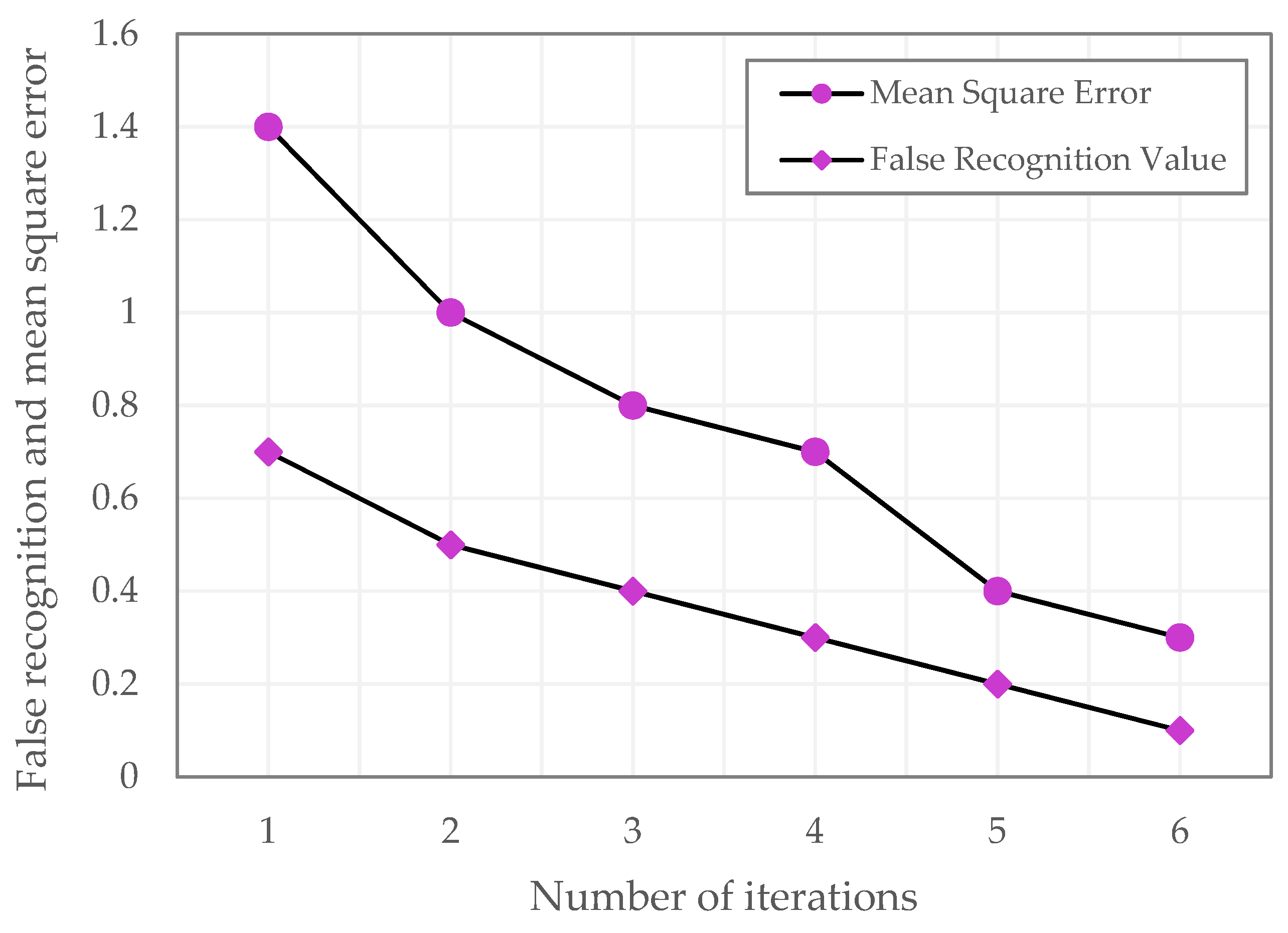
| No. of Iterations | Mean Square Error | False Recognition Value |
|---|---|---|
| 1 | 1.4 | 0.7 |
| 2 | 1 | 0.5 |
| 3 | 0.8 | 0.4 |
| 4 | 0.7 | 0.3 |
| 5 | 0.4 | 0.2 |
| 6 | 0.3 | 0.1 |
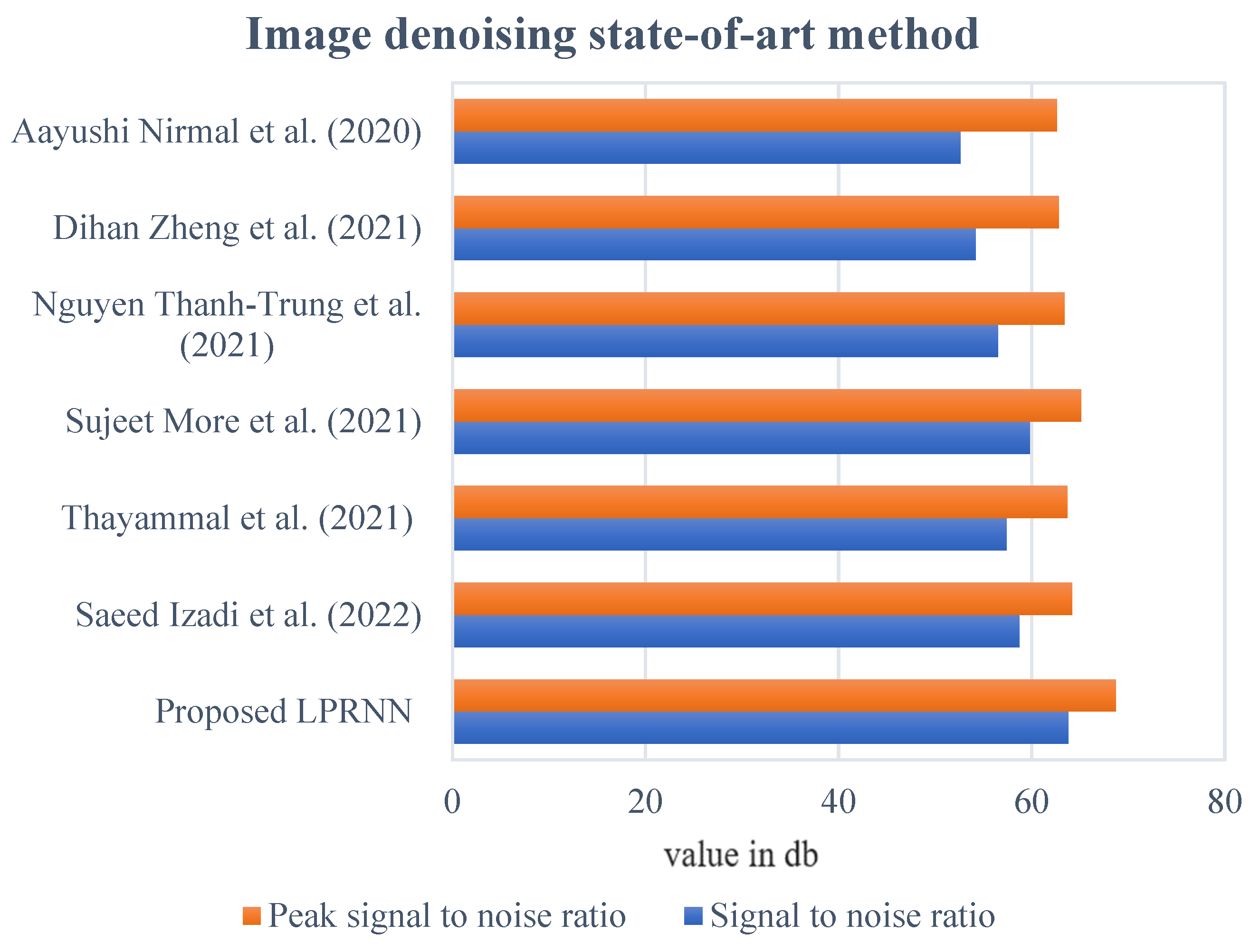
| Methods | Signal-to-Noise Ratio | Peak Signal-to-Noise Ratio |
|---|---|---|
| Value in dB | ||
| Proposed LPRNN | 63.8 | 68.7 |
| Saeed Izadi et al. (2022) [6] | 58.7 | 64.2 |
| Thayammal et al. (2021) [5] | 57.4 | 63.7 |
| Sujeet More et al. (2021) [8] | 59.8 | 65.1 |
| Nguyen Thanh-Trung et al. (2021) [11] | 56.5 | 63.4 |
| Dihan Zheng et al. (2021) [14] | 54.2 | 62.8 |
| Aayushi Nirmal et al. (2020) [9] | 52.6 | 62.6 |
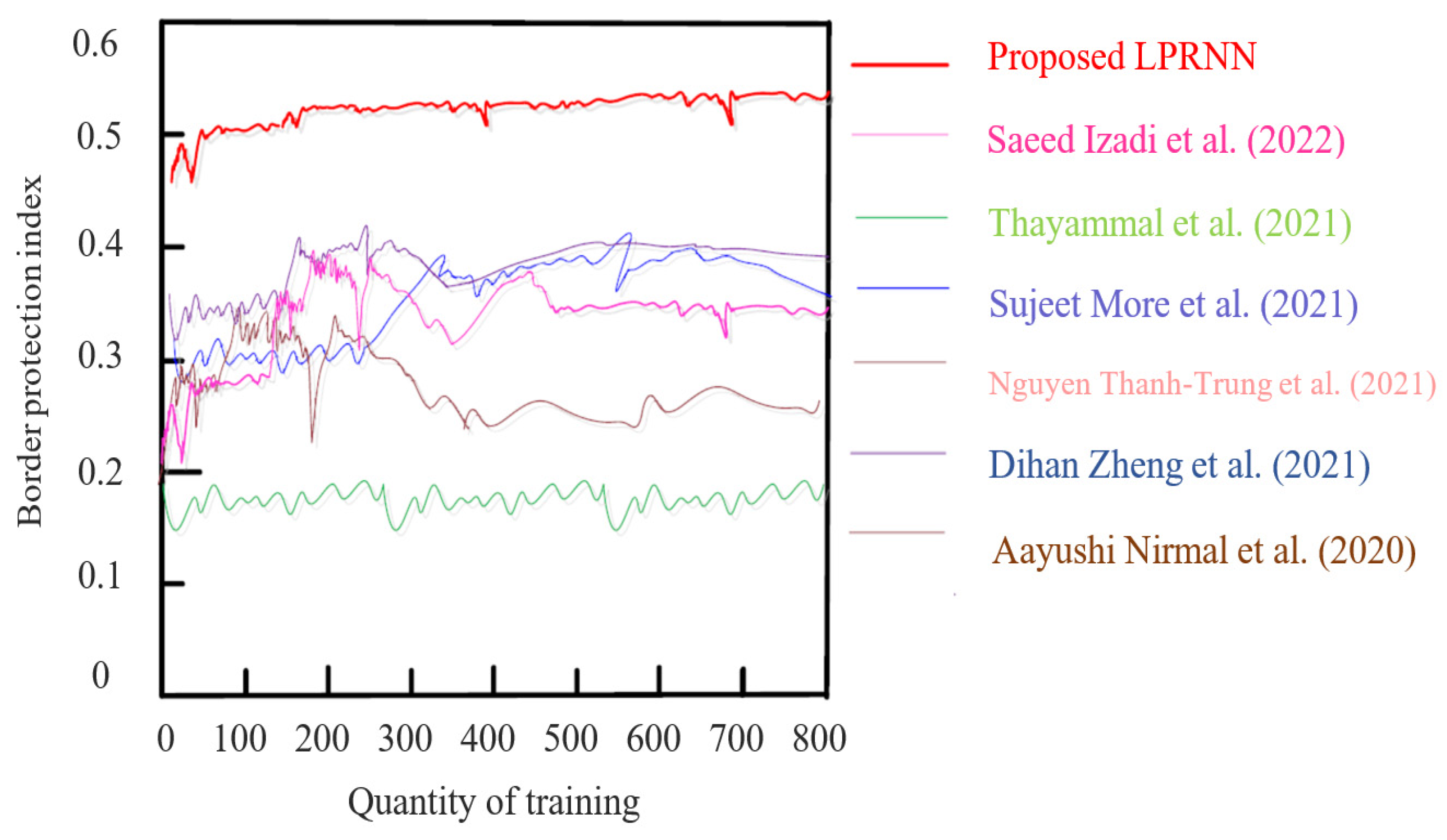
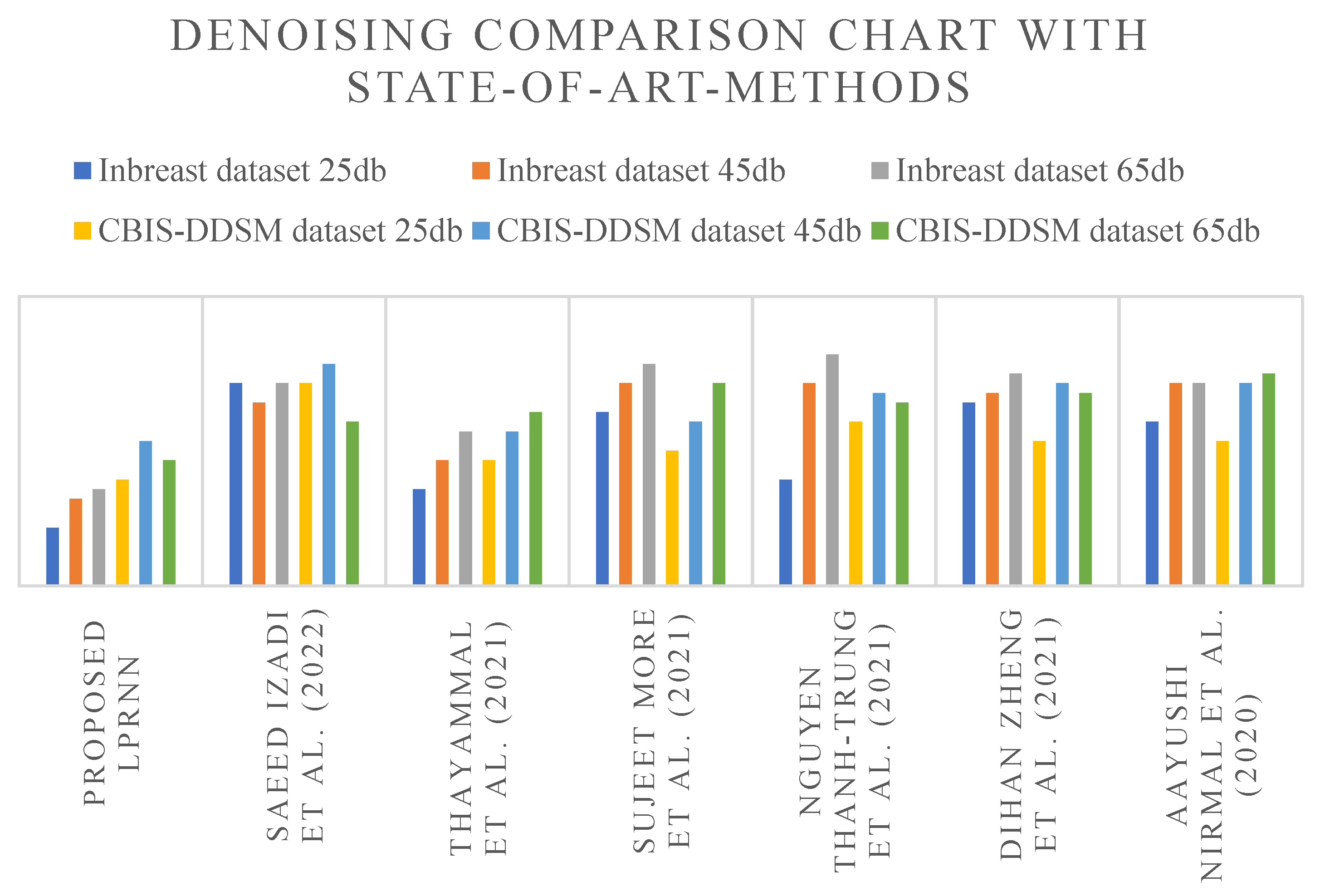
| Methods | INbreast Dataset | CBIS-DDSM Dataset | ||||
|---|---|---|---|---|---|---|
| 25 db | 45 db | 65 db | 25 db | 45 db | 65 db | |
| Proposed LPRNN | 6 | 9 | 10 | 11 | 15 | 13 |
| Saeed Izadi et al. (2022) [6] | 21 | 19 | 21 | 21 | 23 | 17 |
| Thayammal et al. (2021) [5] | 10 | 13 | 16 | 13 | 16 | 18 |
| Sujeet More et al. (2021) [8] | 18 | 21 | 23 | 14 | 17 | 21 |
| Nguyen Thanh-Trung et al. (2021) [11] | 11 | 21 | 24 | 17 | 20 | 19 |
| Dihan Zheng et al. (2021) [14] | 19 | 20 | 22 | 15 | 21 | 20 |
| Aayushi Nirmal et al. (2020) [9] | 17 | 21 | 21 | 15 | 21 | 22 |
Disclaimer/Publisher’s Note: The statements, opinions and data contained in all publications are solely those of the individual author(s) and contributor(s) and not of MDPI and/or the editor(s). MDPI and/or the editor(s) disclaim responsibility for any injury to people or property resulting from any ideas, methods, instructions or products referred to in the content. |
© 2023 by the authors. Licensee MDPI, Basel, Switzerland. This article is an open access article distributed under the terms and conditions of the Creative Commons Attribution (CC BY) license (https://creativecommons.org/licenses/by/4.0/).
Share and Cite
Vimala, B.B.; Srinivasan, S.; Mathivanan, S.K.; Muthukumaran, V.; Babu, J.C.; Herencsar, N.; Vilcekova, L. Image Noise Removal in Ultrasound Breast Images Based on Hybrid Deep Learning Technique. Sensors 2023, 23, 1167. https://doi.org/10.3390/s23031167
Vimala BB, Srinivasan S, Mathivanan SK, Muthukumaran V, Babu JC, Herencsar N, Vilcekova L. Image Noise Removal in Ultrasound Breast Images Based on Hybrid Deep Learning Technique. Sensors. 2023; 23(3):1167. https://doi.org/10.3390/s23031167
Chicago/Turabian StyleVimala, Baiju Babu, Saravanan Srinivasan, Sandeep Kumar Mathivanan, Venkatesan Muthukumaran, Jyothi Chinna Babu, Norbert Herencsar, and Lucia Vilcekova. 2023. "Image Noise Removal in Ultrasound Breast Images Based on Hybrid Deep Learning Technique" Sensors 23, no. 3: 1167. https://doi.org/10.3390/s23031167
APA StyleVimala, B. B., Srinivasan, S., Mathivanan, S. K., Muthukumaran, V., Babu, J. C., Herencsar, N., & Vilcekova, L. (2023). Image Noise Removal in Ultrasound Breast Images Based on Hybrid Deep Learning Technique. Sensors, 23(3), 1167. https://doi.org/10.3390/s23031167