
Analysis of the Active Measurement Systems of the Thoracic Range of Movements of the Spine: A Systematic Review and a Meta-Analysis


Abstract
1. Introduction
2. Methods
2.1. Eligibility Criteria
2.2. Information Sources and Search Strategy
2.3. Selection Process
2.4. Data Collection Process and Variables
2.5. Study Risk of Bias Assessment
2.6. Data Synthesis Methods and Meta-Analysis
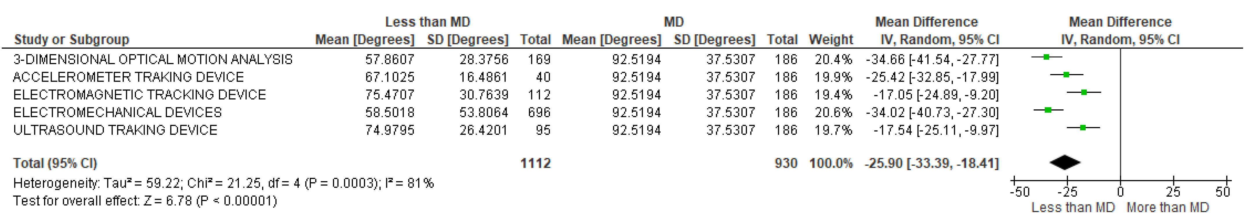
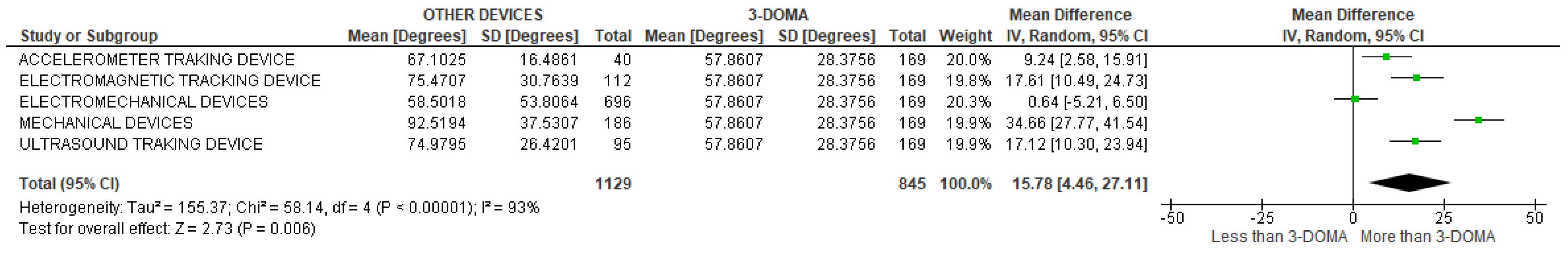
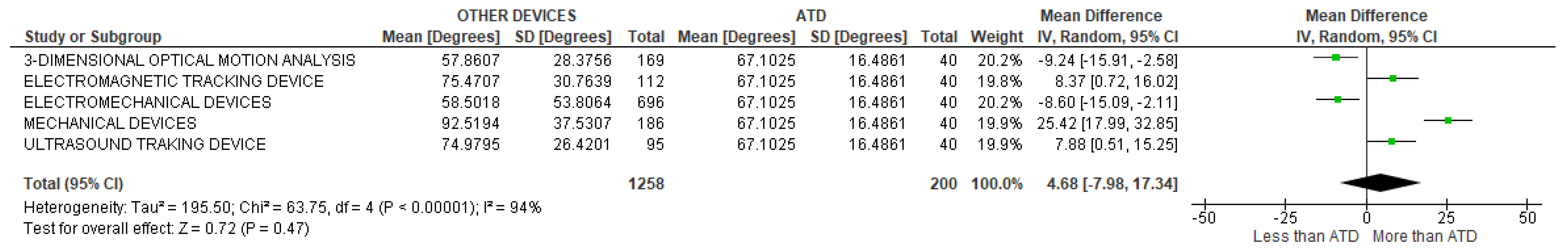
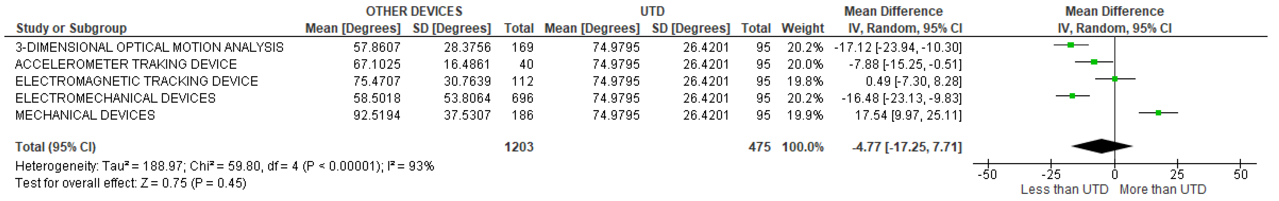
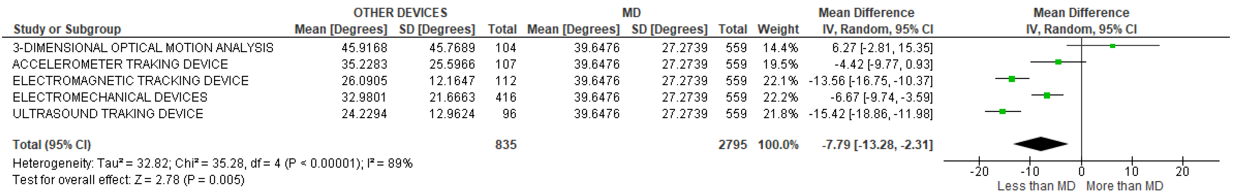
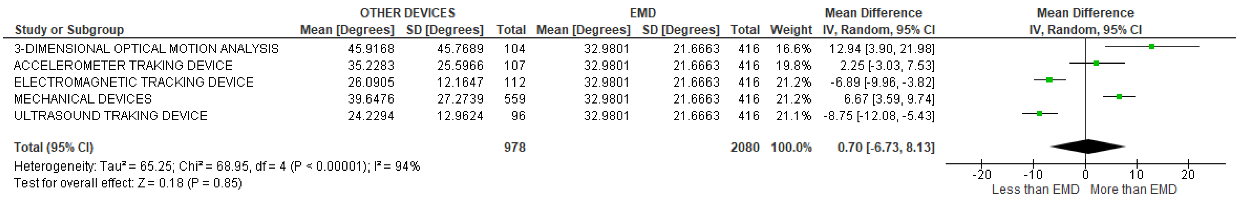
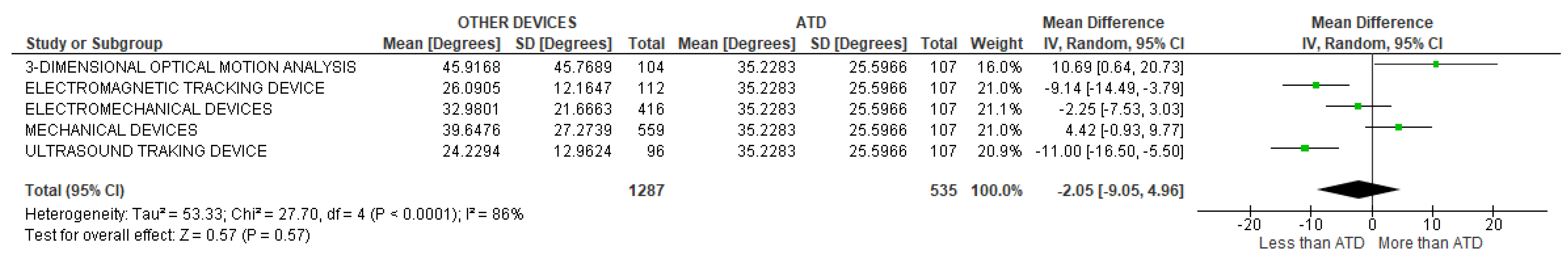
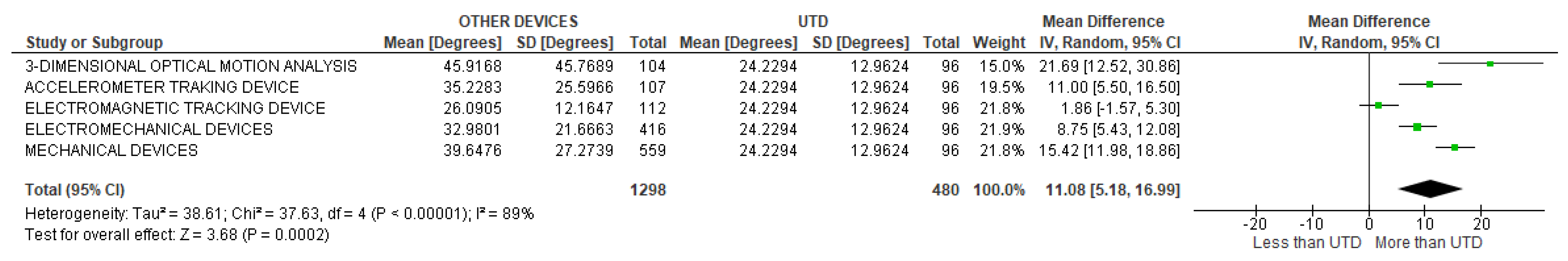
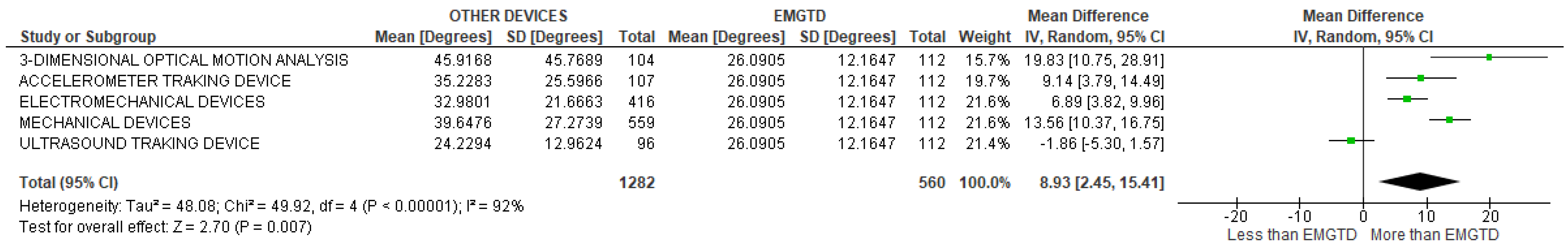
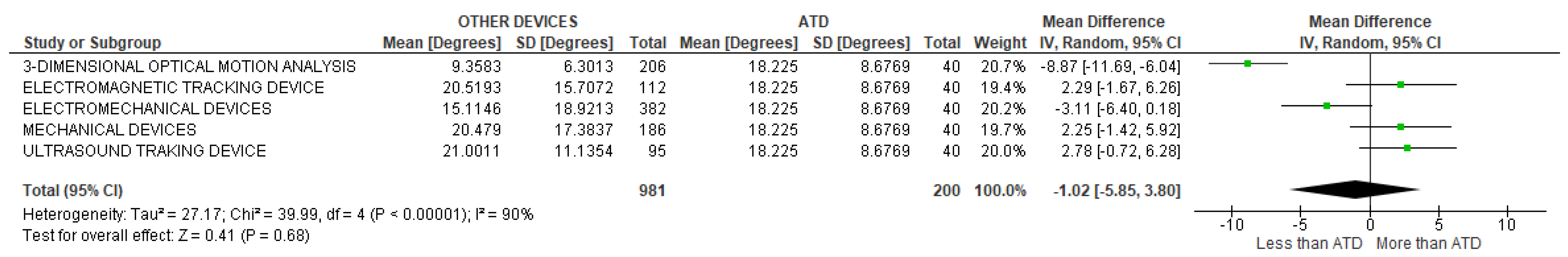
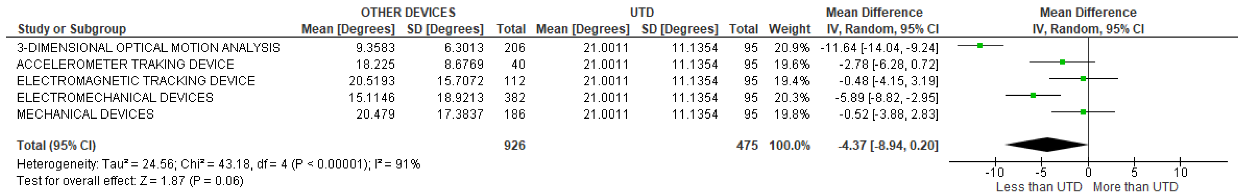
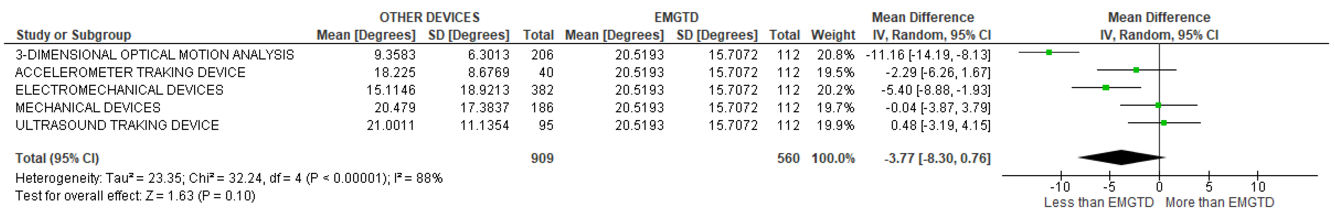
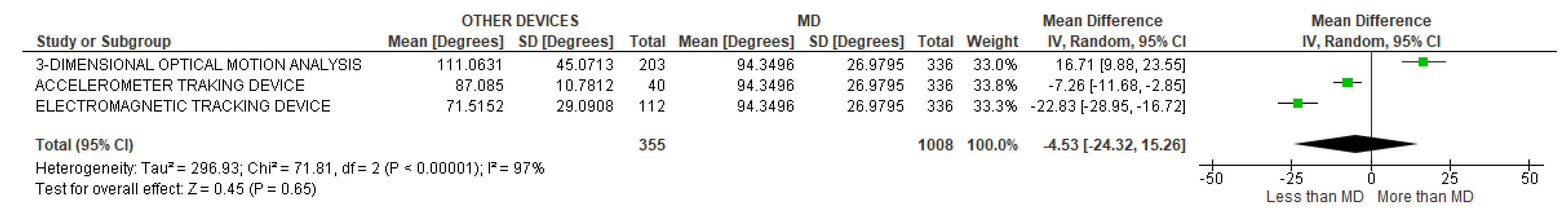
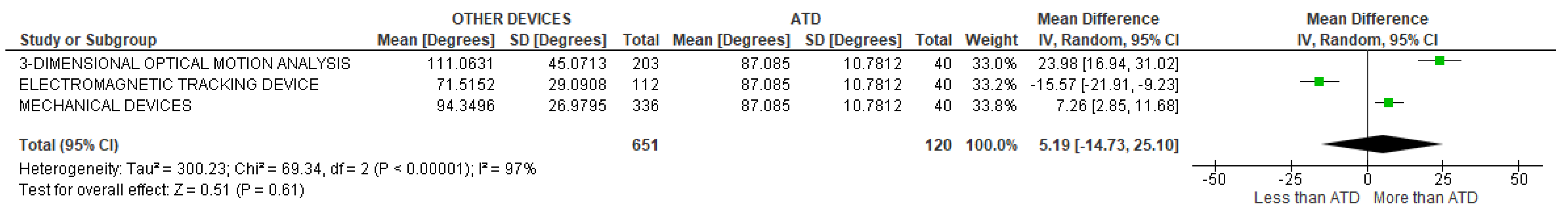
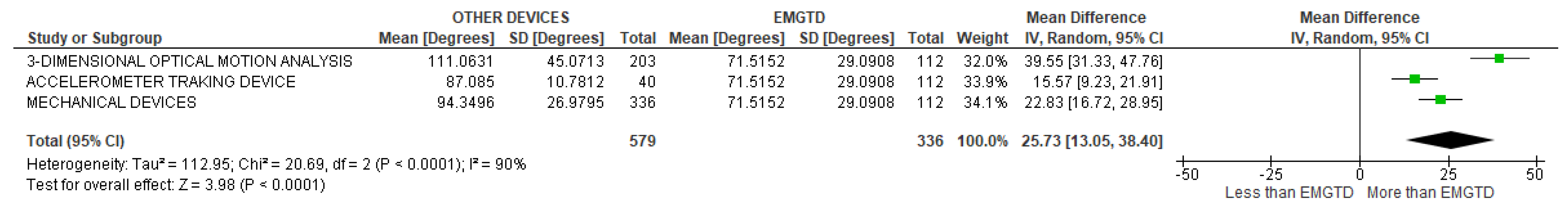
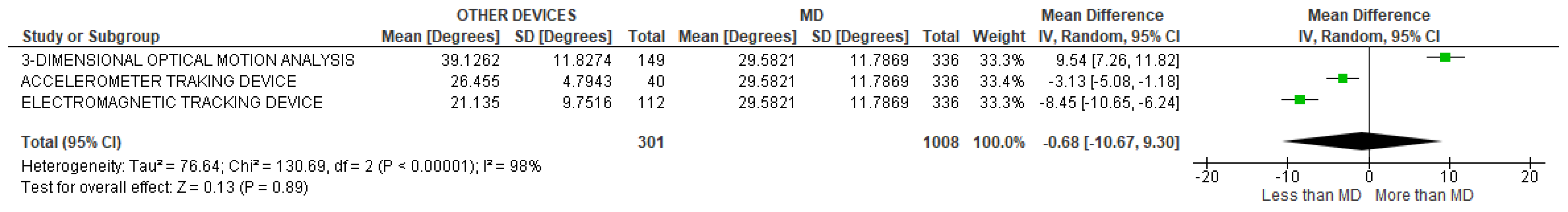
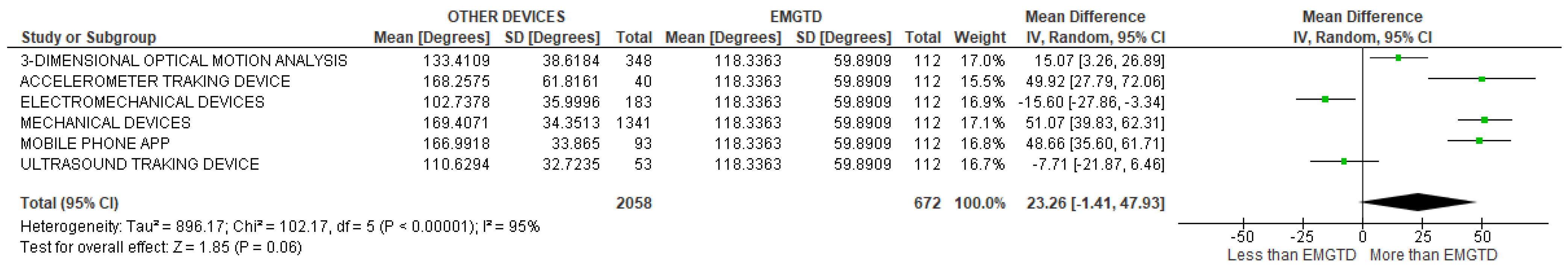
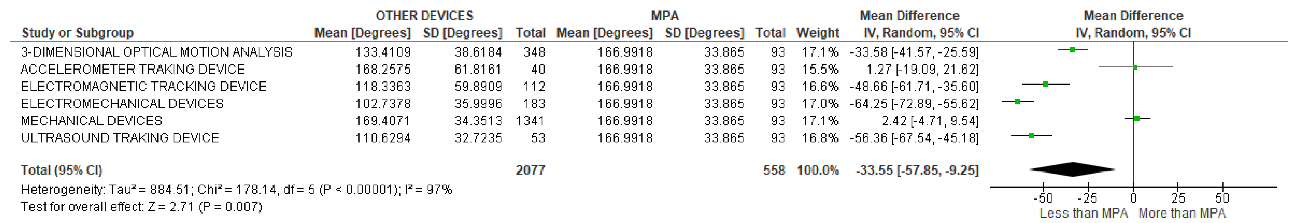
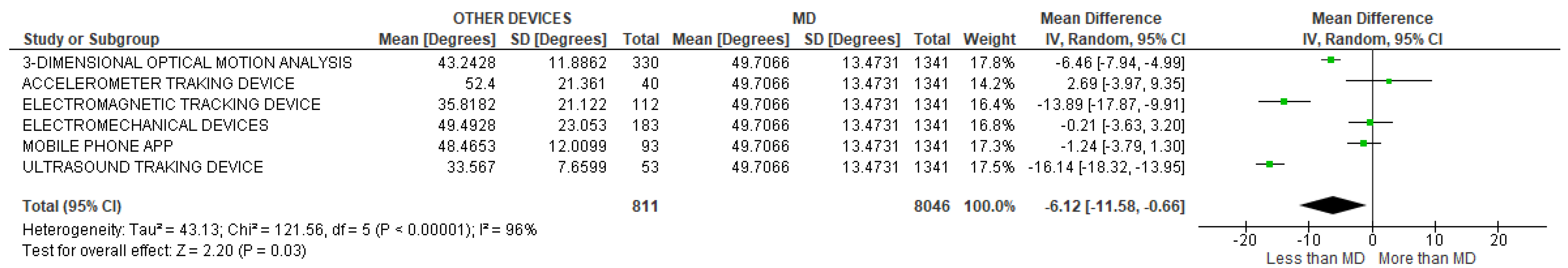
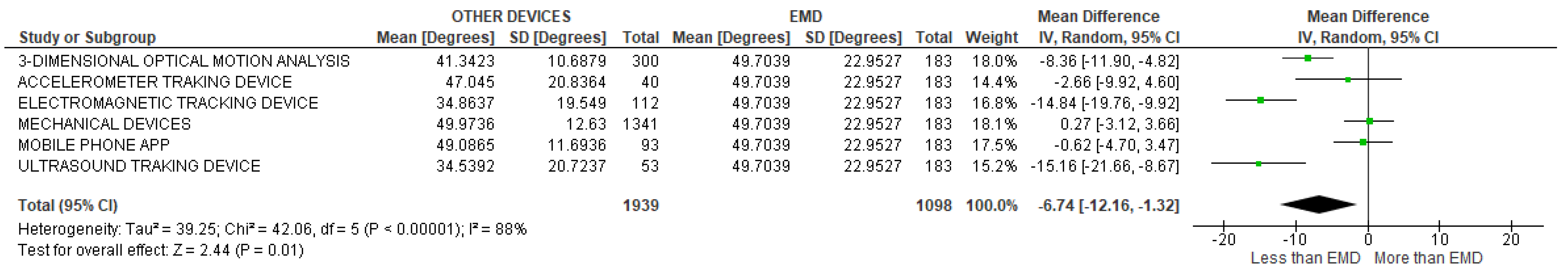
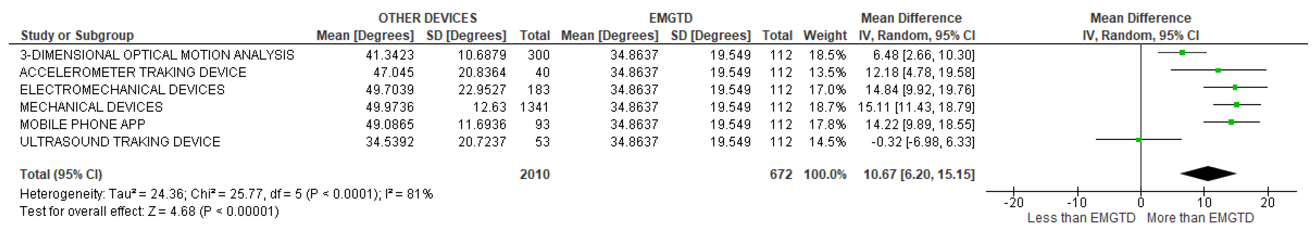
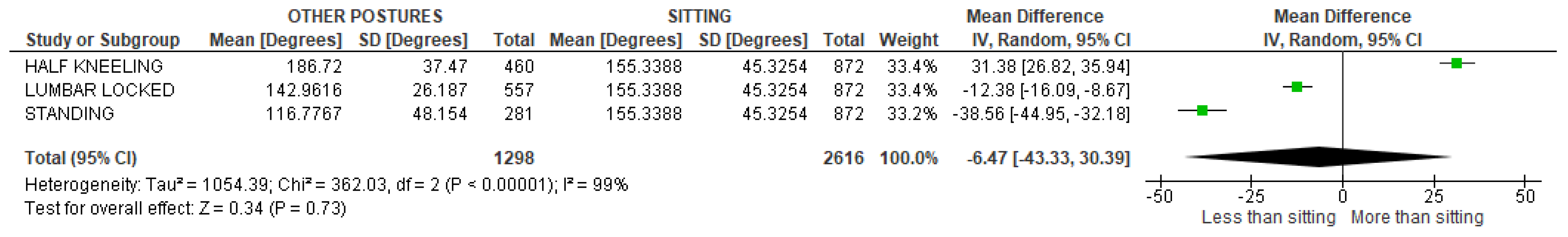
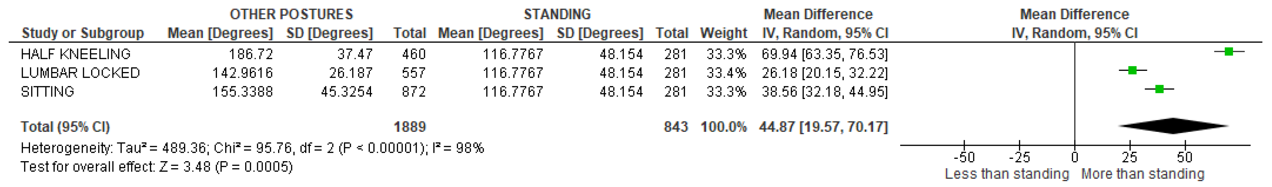
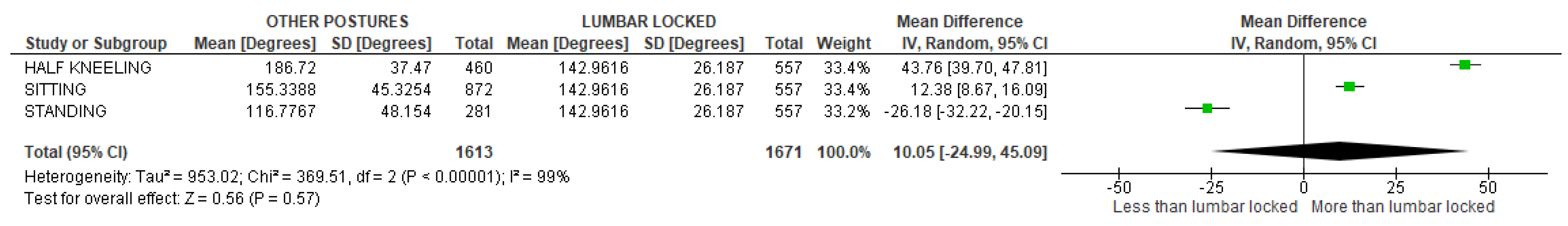
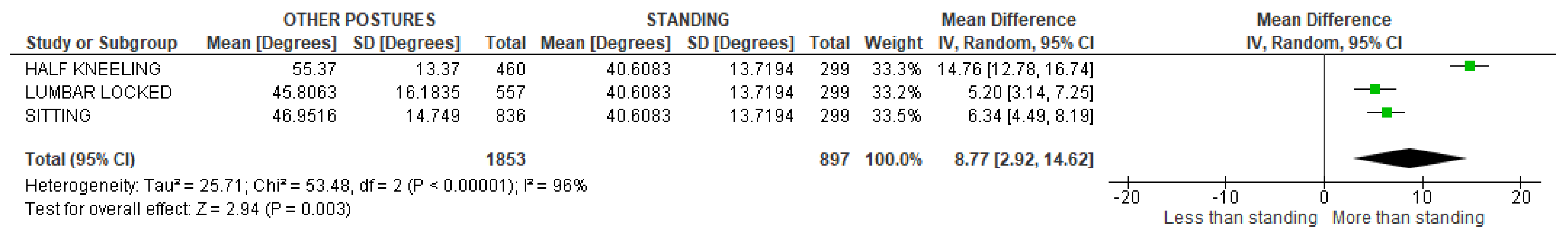
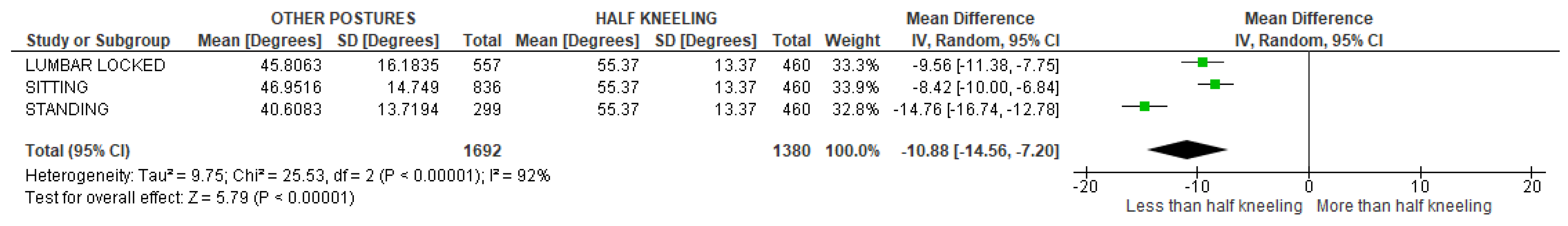
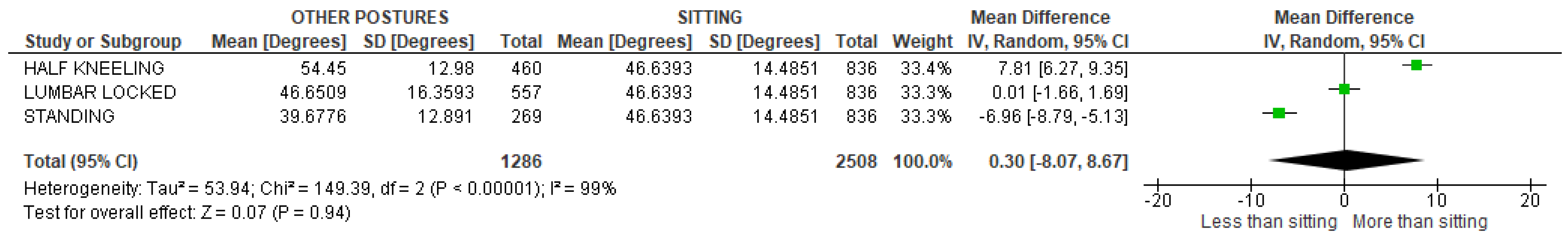
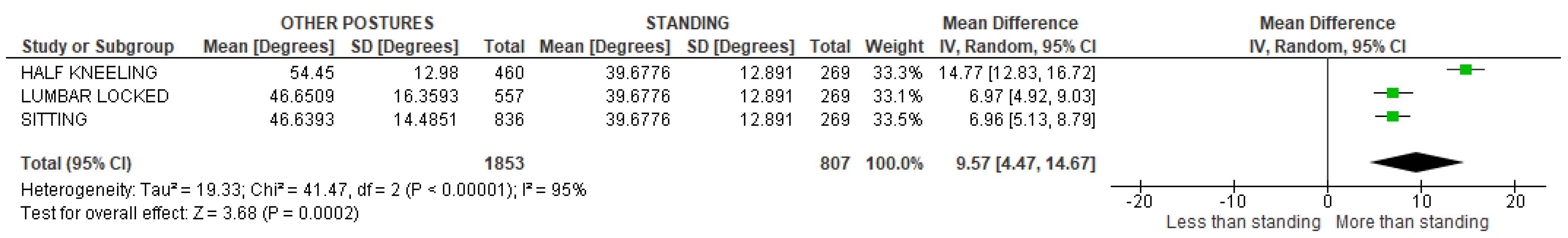
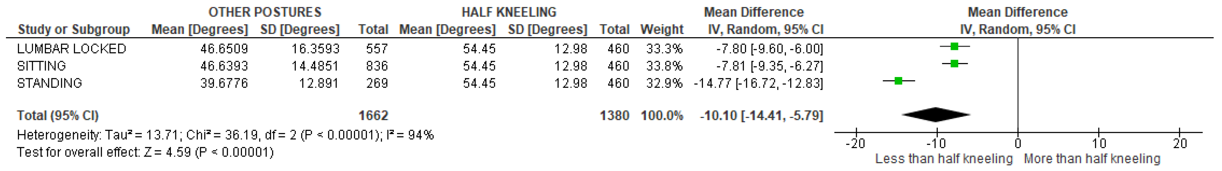
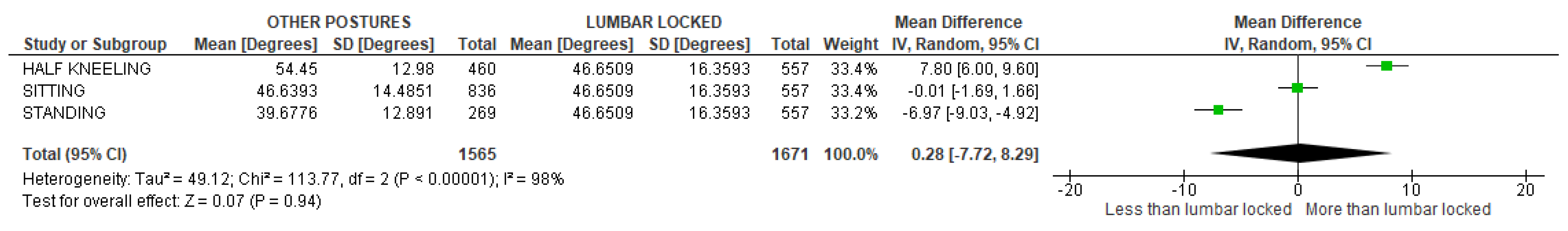
3. Results
3.1. Study Selection
3.2. Study Characteristics
4. Discussion
5. Conclusions
Author Contributions
Funding
Conflicts of Interest
Appendix A. Search String
Appendix B

Appendix C
| Study | Score | Methodological Quality | Number of Item of MINORS Scale | |||||||||||
|---|---|---|---|---|---|---|---|---|---|---|---|---|---|---|
| 1 | 2 | 3 | 4 | 5 | 6 | 7 | 8 | 9 | 10 | 11 | 12 | |||
| O’Gorman et al., 1987 [27] | 18/24 | Good | 2 | 2 | 2 | 2 | 2 | 2 | 2 | 0 | 1 | 1 | 1 | 1 |
| Mellin G. et al., 1991 [28] | 14/16 | Good | 2 | 2 | 2 | 2 | 2 | 2 | 2 | 0 | - | - | - | - |
| Crawford H.J. et al., 1993 [29] | 21/24 | Good | 2 | 2 | 2 | 2 | 2 | 2 | 2 | 0 | 1 | 2 | 2 | 2 |
| Willems J.M. et al., 1996 [30] | 15/16 | Excellent | 2 | 2 | 2 | 2 | 2 | 2 | 2 | 1 | - | - | - | - |
| Gilleard W. et al., 2002 [25] | 21/24 | Good | 2 | 2 | 2 | 2 | 2 | 2 | 2 | 1 | 2 | 2 | 1 | 1 |
| Mannion A.F. et al., 2004 [31] | 14/16 | Good | 2 | 2 | 2 | 2 | 2 | 2 | 2 | 0 | - | - | - | - |
| Post R.B. et al., 2004 [32] | 15/16 | Excellent | 2 | 2 | 2 | 2 | 2 | 2 | 2 | 1 | - | - | - | - |
| Edmondston S.J. et al., 2007 [34] | 16/16 | Excellent | 2 | 2 | 2 | 2 | 2 | 2 | 2 | 2 | - | - | - | - |
| Tedereko P. et al., 2007 [26] | 14/16 | Good | 2 | 2 | 2 | 2 | 2 | 2 | 2 | 0 | - | - | - | - |
| Hsu C.J. et al., 2008 [35] | 14/16 | Good | 2 | 2 | 2 | 2 | 2 | 2 | 2 | 0 | - | - | - | - |
| Mika A. et al., 2009 [36] | 23/24 | Excellent | 2 | 2 | 2 | 2 | 2 | 2 | 2 | 1 | 2 | 2 | 2 | 2 |
| Kasukawa Y. et al., 2010 [37] | 19/24 | Good | 2 | 2 | 2 | 2 | 2 | 2 | 2 | 0 | 2 | 1 | 1 | 1 |
| Theisen C. et al., 2010 [38] | 19/24 | Good | 2 | 2 | 2 | 2 | 2 | 2 | 2 | 0 | 2 | 1 | 1 | 1 |
| Heneghan N.R. et al., 2010 [39] | 14/16 | Good | 2 | 2 | 2 | 2 | 2 | 2 | 2 | 0 | - | - | - | - |
| Imagama S. et al., 2011 [40] | 14/16 | Good | 2 | 2 | 2 | 2 | 2 | 2 | 2 | 0 | - | - | - | - |
| Edmondston S.J. et al., 2011 [41] | 14/16 | Good | 2 | 2 | 2 | 2 | 2 | 2 | 2 | 0 | - | - | - | - |
| Edmondston S.J et al., 2012 [42] | 14/16 | Good | 2 | 2 | 2 | 2 | 2 | 2 | 2 | 0 | - | - | - | - |
| Fölsch C. et al., 2012 [43] | 14/16 | Good | 2 | 2 | 2 | 2 | 2 | 2 | 2 | 0 | - | - | - | - |
| Johnson K.D. et al., 2012 [44] | 14/16 | Good | 2 | 2 | 2 | 2 | 2 | 2 | 2 | 0 | - | - | - | - |
| Edmondston S.J. et al., 2012 [45] | 14/16 | Good | 2 | 2 | 2 | 2 | 2 | 2 | 2 | 0 | - | - | - | - |
| Wang H.J. et al., 2012 [46] | 19/24 | Good | 2 | 2 | 2 | 2 | 2 | 2 | 2 | 0 | 2 | 1 | 1 | 1 |
| Benjamin-Hidalgo P.E. et al., 2012 [47] | 20/24 | Good | 2 | 2 | 2 | 2 | 2 | 2 | 2 | 0 | 2 | 2 | 1 | 1 |
| Tsang S.M. et al., 2013 [48] | 14/16 | Good | 2 | 2 | 2 | 2 | 2 | 2 | 2 | 0 | - | - | - | - |
| Wirth B. et al., 2014 [50] | 22/24 | Excellent | 2 | 2 | 2 | 2 | 2 | 2 | 2 | 0 | 2 | 2 | 2 | 2 |
| Talukdar K. et al., 2015 [52] | 14/16 | Good | 2 | 2 | 2 | 2 | 2 | 2 | 2 | 0 | - | - | - | - |
| Alqhtani R.S. et al., 2015 [53] | 14/16 | Good | 2 | 2 | 2 | 2 | 2 | 2 | 2 | 0 | - | - | - | - |
| Hajibozorgi M. et al., 2016 [15] | 14/16 | Good | 2 | 2 | 2 | 2 | 2 | 2 | 2 | 0 | - | - | - | - |
| Schinkel-Ivy A. et al., 2016 [54] | 14/16 | Good | 2 | 2 | 2 | 2 | 2 | 2 | 2 | 0 | - | - | - | - |
| Furness J. et al., 2016 [55] | 20/24 | Good | 2 | 2 | 2 | 2 | 2 | 2 | 2 | 0 | 2 | 2 | 1 | 1 |
| Mazzone B. et al., 2016 [56] | 24/24 | Excellent | 2 | 2 | 2 | 2 | 2 | 2 | 2 | 2 | 2 | 2 | 2 | 2 |
| Zafereo J. et al., 2016 [57] | 16/16 | Excellent | 2 | 2 | 2 | 2 | 2 | 2 | 2 | 2 | - | - | - | - |
| Morais N. et al., 2016 [58] | 23/24 | Excellent | 2 | 2 | 2 | 2 | 2 | 2 | 2 | 2 | 2 | 2 | 1 | 2 |
| Rast F. et al., 2016 [59] | 21/24 | Good | 2 | 2 | 2 | 2 | 2 | 2 | 2 | 0 | 1 | 2 | 2 | 2 |
| Ishikawa Y. et al., 2017 [60] | 22/24 | Excellent | 2 | 2 | 2 | 2 | 2 | 2 | 2 | 2 | 2 | 2 | 1 | 1 |
| Bucke J. et al., 2017 [11] | 16/16 | Excellent | 2 | 2 | 2 | 2 | 2 | 2 | 2 | 2 | - | - | - | - |
| Roghani T. et al., 2017 [61] | 22/24 | Excellent | 2 | 2 | 2 | 2 | 2 | 2 | 2 | 2 | 2 | 2 | 1 | 1 |
| Hwang D. et al., 2017 [8] | 14/16 | Good | 2 | 2 | 2 | 2 | 2 | 2 | 2 | 0 | - | - | - | - |
| Narimani M. et al., 2018 [62] | 14/16 | Good | 2 | 2 | 2 | 2 | 2 | 2 | 2 | 0 | - | - | - | - |
| Heneghan N.R. et al., 2018 [63] | 22/24 | Excellent | 2 | 2 | 2 | 2 | 2 | 2 | 2 | 2 | 2 | 2 | 1 | 1 |
| Mousavi S.J. et al., 2018 [64] | 14/16 | Good | 2 | 2 | 2 | 2 | 2 | 2 | 2 | 0 | - | - | - | - |
| Furness J. et al., 2018 [65] | 22/24 | Excellent | 2 | 2 | 2 | 2 | 2 | 2 | 2 | 0 | 2 | 2 | 2 | 2 |
| Beaudette S.M. et al., 2019 [66] | 23/24 | Excellent | 2 | 2 | 2 | 2 | 2 | 2 | 2 | 1 | 2 | 2 | 2 | 2 |
| Schinkel-Ivy A. et al., 2019 [67] | 15/16 | Excellent | 2 | 2 | 2 | 2 | 2 | 2 | 2 | 1 | - | - | - | - |
| Welbeck A.N. Et al., 2019 [68] | 22/24 | Excellent | 2 | 2 | 2 | 2 | 2 | 2 | 2 | 0 | 2 | 2 | 2 | 2 |
| Hunter D.J. et al., 2020 [69] | 24/24 | Excellent | 2 | 2 | 2 | 2 | 2 | 2 | 2 | 2 | 2 | 2 | 2 | 2 |
References
- Kubas, C.; Chen, Y.-W.; Echeverri, S.; McCann, S.L.; Denhoed, M.J.; Walker, C.J.; Kennedy, C.N.; Reid, W.D. Reliability and Validity of Cervical Range of Motion and Muscle Strength Testing. J. Strength Cond. Res. 2017, 31, 1087–1096. [Google Scholar] [CrossRef] [PubMed]
- Shum, G.L.K.; Crosbie, J.; Lee, R.Y.W. Movement coordination of the lumbar spine and hip during a picking up activity in low back pain subjects. Eur. Spine J. 2006, 16, 749–758. [Google Scholar] [CrossRef] [PubMed]
- Ferrari, S.; Manni, T.; Bonetti, F.; Villafañe, J.H.; Vanti, C. A literature review of clinical tests for lumbar instability in low back pain: Validity and applicability in clinical practice. Chiropr. Man. Ther. 2015, 23, 14. [Google Scholar] [CrossRef] [PubMed]
- Bissolotti, L.; Gobbo, M.; Villafañe, J.H.; Negrini, S. Spinopelvic balance: New biomechanical insights with clinical implications for Parkinson’s disease. Eur. Spine J. 2014, 23, 576–583. [Google Scholar] [CrossRef]
- Negrini, S.; Imperio, G.; Villafañe, J.H.; Negrini, F.; Zaina, F. Systematic reviews of physical and rehabilitation medicine Cochrane contents. Part 1. Disabilities due to spinal disorders and pain syndromes in adults. Eur. J. Phys. Rehabil. Med. 2013, 49, 597–610. [Google Scholar]
- Tanaka, N.; An, H.; Lim, T.; Fujiwara, A.; Jeon, C.; Haughton, V. The relationship between disc degeneration and flexibility of the lumbar spine. Spine J. 2001, 1, 47–56. [Google Scholar] [CrossRef]
- Archer, I.A.; Moll, J.M.; Wright, V. Chest and spinal mobility in physiotherapists: An objective clinical study. Physiotherapy 1974, 60, 37–39. [Google Scholar]
- Hwang, D.; Lee, J.H.; Moon, S.; Park, S.W.; Woo, J.; Kim, C. The reliability of the nonradiologic measures of thoracic spine rotation in healthy adults. Phys. Ther. Rehabil. Sci. 2017, 6, 65–70. [Google Scholar] [CrossRef]
- Hyytiäinen, K.; Salminen, J.; Suvitie, T.; Wickström, G.; Pentti, J. Reproducibility of nine tests to measure spinal mobility and trunk muscle strength. Scand. J. Rehabil. Med. 1991, 23, 3–10. [Google Scholar]
- Johnson, K.D.; Grindstaff, T.L. Thoracic rotation measurement techniques: Clinical commentary. N. Am. J. Sports Phys. Ther. NAJSPT 2010, 5, 252–256. [Google Scholar]
- Bucke, J.; Spencer, S.; Fawcett, L.; Sonvico, L.; Rushton, A.; Heneghan, N.R. Validity of the Digital Inclinometer and iPhone When Measuring Thoracic Spine Rotation. J. Athl. Train. 2017, 52, 820–825. [Google Scholar] [CrossRef] [PubMed]
- Seichert, N.; Baumann, M.; Senn, E.; Zuckriegl, H. The “back mouse”—An analog-digital measuring device to record the sagittal outline of the back. Phys. Med. Rehabil. Kurortmed. 2008, 4, 35–43. [Google Scholar]
- Perry, M.; Smith, A.; Straker, L.; Coleman, J.; O’Sullivan, P. Reliability of sagittal photographic spinal posture assessment in adolescents. Adv. Physiother. 2009, 10, 66–75. [Google Scholar] [CrossRef]
- Troke, M.; Moore, A.; Cheek, E. Intra-operator and inter-operator reliability of the OSI CA 6000 Spine Motion Analyzer with a new skin fixation system. Man. Ther. 1996, 1, 92–98. [Google Scholar] [CrossRef]
- Hajibozorgi, M.; Arjmand, N. Sagittal range of motion of the thoracic spine using inertial tracking device and effect of measurement errors on model predictions. J. Biomech. 2016, 49, 913–918. [Google Scholar] [CrossRef] [PubMed]
- Heneghan, N.R.; Hall, A.; Hollands, M.; Balanos, G.M. Stability and intra-tester reliability of an in vivo measurement of thoracic axial rotation using an innovative methodology. Man. Ther. 2009, 14, 452–455. [Google Scholar] [CrossRef] [PubMed]
- Pearcy, M.; Hindle, R. New method for the non-invasive three-dimensional measurement of human back movement. Clin. Biomech. 1989, 4, 73–79. [Google Scholar] [CrossRef]
- Hutton, B.; Catalá-López, F.; Moher, D. La extensión de la declaración PRISMA para revisiones sistemáticas que incorporan metaanálisis en red: PRISMA-NMA. Med. Clin. 2016, 147, 262–266. [Google Scholar] [CrossRef]
- Page, M.J.; McKenzie, J.E.; Bossuyt, P.M.; Boutron, I.; Hoffmann, T.C.; Mulrow, C.D.; Shamseer, L.; Tetzlaff, J.M.; Akl, E.A.; Brennan, S.E.; et al. The PRISMA 2020 statement: An updated guideline for reporting systematic reviews. BMJ 2021, 372, 105906. [Google Scholar] [CrossRef]
- International Prospective Register of Systematic Reviews [Internet]. 2018. Available online: https://www.crd.york.ac.uk/prospero/ (accessed on 18 July 2018).
- Verhagen, A.P.; De Vet, H.C.; De Bie, R.A.; Kessels, A.G.; Boers, M.; Bouter, L.M.; Knipschild, P.G. The Delphi list: A criteria list for quality as-sessment of randomized clinical trials for conducting systematic reviews developed by Delphi consensus. J. Clin. Epidemiol. 1998, 51, 1235–1241. [Google Scholar] [CrossRef]
- Slim, K.; Nini, E.; Forestier, D.; Kwiatkowski, F.; Panis, Y.; Chipponi, J. Methodological index for non-randomized studies (MINORS ): Development and validation of a new instrument. ANZ J. Surg. 2003, 73, 712–716. [Google Scholar] [CrossRef] [PubMed]
- Villafañe, J.; Pedersini, P.; Bertozzi, L.; Drago, L.; Fernandez-Carnero, J.; Bishop, M.; Berjano, P. Exploring the relationship between chronic pain and cortisol levels in subjects with osteoarthritis: Results from a systematic review of the literature. Osteoarthr. Cartil. 2020, 28, 572–580. [Google Scholar] [CrossRef] [PubMed]
- Higgins, J.; Thomas, J. Cochrane Handbook for Systematic Reviews of Interventions, 2nd ed.; Chandler, J., Cumpston, M., Li, T., Page, M., Welch, V., Eds.; John Wiley & Sons: Chichester, UK, 2019. [Google Scholar]
- Gilleard, W.; Crosbie, J.; Smith, R. Effect of pregnancy on trunk range of motion when sitting and standing. Acta Obstet. Gynecol. Scand. 2002, 81, 1011–1020. [Google Scholar] [CrossRef]
- Tederko, P.; Krasuski, M.; Maciejasz, P. Restrainer of pelvis and lower limbs in thoracic and lumbar range of motion measurement. Ortop. Traumatol. Rehabil. 2007, 9, 156–167. [Google Scholar] [PubMed]
- O’Gorman, H.; Jull, G. Thoracic kyphosis and mobility: The effect of age. Physiother. Pract. 1987, 3, 154–162. [Google Scholar] [CrossRef]
- Mellin, G.; Kiiski, R.; Weckström, A. Effects of Subject Position on Measurements of Flexion, Extension, and Lateral Flexion of the Spine. Spine 1991, 16, 1108–1110. [Google Scholar] [CrossRef] [PubMed]
- Crawford, H.J.; Jull, G.A. The influence of thoracic posture and movement on range of arm elevation. Physiother. Theory Pract. 1993, 9, 143–148. [Google Scholar] [CrossRef]
- Willems, J.; Jull, G.; Ng, J.-F. An in vivo study of the primary and coupled rotations of the thoracic spine. Clin. Biomech. 1996, 11, 311–316. [Google Scholar] [CrossRef]
- Mannion, A.F.; Knecht, K.; Balaban, G.; Dvorak, J.; Grob, D. A new skin-surface device for measuring the curvature and global and segmental ranges of motion of the spine: Reliability of measurements and comparison with data reviewed from the literature. Eur. Spine J. 2004, 13, 122–136. [Google Scholar] [CrossRef]
- Post, R.B.; Leferink, V.J.M. Spinal mobility: Sagittal range of motion measured with the Spinal Mouse, a new non-invasive device. Arch. Orthop. Trauma Surg. 2004, 124, 187–192. [Google Scholar] [CrossRef]
- Holmström, E.; Ahlborg, B. Morning warming-up exercise—Effects on musculoskeletal fitness in construction workers. Appl. Ergon. 2005, 36, 513–519. [Google Scholar] [CrossRef] [PubMed]
- Edmondston, S.J.; Aggerholm, M.; Elfving, S.; Flores, N.; Ng, C.; Smith, R.; Netto, K. Influence of Posture on the Range of Axial Rotation and Coupled Lateral Flexion of the Thoracic Spine. J. Manip. Physiol. Ther. 2007, 30, 193–199. [Google Scholar] [CrossRef] [PubMed]
- Hsu, C.-J.; Chang, Y.-W.; Chou, W.-Y.; Chiou, C.-P.; Chang, W.-N.; Wong, C.-Y. Measurement of spinal range of motion in healthy individuals using an electromagnetic tracking device. J. Neurosurg. Spine 2008, 8, 135–142. [Google Scholar] [CrossRef] [PubMed]
- Mika, A.; Fernhall, B.; Mika, P. Association between moderate physical activity, spinal motion and back muscle strength in postmenopausal women with and without osteoporosis. Disabil. Rehabil. 2009, 31, 734–740. [Google Scholar] [CrossRef] [PubMed]
- Kasukawa, Y.; Miyakoshi, N.; Hongo, M.; Ishikawa, Y.; Noguchi, H.; Kamo, K.; Sasaki, H.; Murata, K.; Shimada, Y. Relationships between falls, spinal curvature, spinal mobility and back extensor strength in elderly people. J. Bone Miner. Metab. 2009, 28, 82–87. [Google Scholar] [CrossRef]
- Theisen, C.; van Wagensveld, A.; Timmesfeld, N.; Efe, T.; Heyse, T.J.; Fuchs-Winkelmann, S.; Schofer, M.D. Co-occurrence of outlet impingement syndrome of the shoulder and restricted range of motion in the thoracic spine—A prospective study with ultrasound-based motion analysis. BMC Musculoskelet. Disord. 2010, 11, 135. [Google Scholar] [CrossRef]
- Heneghan, N.R.; Balanos, G.M. Soft tissue artefact in the thoracic spine during axial rotation and arm elevation using ultrasound imaging: A descriptive study. Man. Ther. 2010, 15, 599–602. [Google Scholar] [CrossRef]
- Imagama, S.; Matsuyama, Y.; Hasegawa, Y.; Sakai, Y.; Ito, Z.; Ishiguro, N.; Hamajima, N. Back muscle strength and spinal mo-bility are predictors of quality of life in middle-aged and elderly males. Eur. Spine J. 2011, 20, 954–961. [Google Scholar] [CrossRef]
- Edmondston, S.; Waller, R.; Vallin, P.; Holthe, A.; Noebauer, A.; King, E. Thoracic Spine Extension Mobility in Young Adults: Influence of Subject Position and Spinal Curvature. J. Orthop. Sports Phys. Ther. 2011, 41, 266–273. [Google Scholar] [CrossRef]
- Edmondston, S.J.; Christensen, M.M.; Keller, S.; Steigen, L.B.; Barclay, L. Functional Radiographic Analysis of Thoracic Spine Extension Motion in Asymptomatic Men. J. Manip. Physiol. Ther. 2012, 35, 203–208. [Google Scholar] [CrossRef]
- Fölsch, C.; Schlögel, S.; Lakemeier, S.; Wolf, U.; Timmesfeld, N.; Skwara, A. Test-Retest Reliability of 3D Ultrasound Measurements of the Thoracic Spine. PM&R 2012, 4, 335–341. [Google Scholar] [CrossRef]
- Johnson, K.D.; Kim, K.-M.; Yu, B.-K.; Saliba, S.; Grindstaff, T.L. Reliability of Thoracic Spine Rotation Range-of-Motion Measurements in Healthy Adults. J. Athl. Train. 2012, 47, 52–60. [Google Scholar] [CrossRef] [PubMed]
- Edmondston, S.; Ferguson, A.; Ippersiel, P.; Ronningen, L.; Sodeland, S.; Barclay, L. Clinical and Radiological Investigation of Thoracic Spine Extension Motion During Bilateral Arm Elevation. J. Orthop. Sports Phys. Ther. 2012, 42, 861–869. [Google Scholar] [CrossRef] [PubMed]
- Wang, H.J.; Giambini, H.; Zhang, W.J.; Ye, G.H.; Zhao, C.; An, K.N.; Li, Y.K.; Lan, W.R.; Li, J.Y.; Jiang, X.S.; et al. A Modified Sagittal Spine Postural Classifi-cation and Its Relationship to Deformities and Spinal Mobility in a Chinese Osteoporotic Population. PLoS ONE 2012, 7, e38560. [Google Scholar]
- Hidalgo, B.; Gilliaux, M.; Poncin, W.; Detrembleur, C. Reliability and validity of a kinematic spine model during active trunk movement in healthy subjects and patients with chronic non-specific low back pain. J. Rehabil. Med. 2012, 44, 756–763. [Google Scholar] [CrossRef] [PubMed]
- Tsang, S.M.H.; Szeto, G.P.Y.; Lee, R.Y.W. Normal kinematics of the neck: The interplay between the cervical and thoracic spines. Man. Ther. 2013, 18, 431–437. [Google Scholar] [CrossRef]
- Battaglia, G.; Bellafiore, M.; Caramazza, G.; Paoli, A.; Bianco, A.; Palma, A. Changes in spinal range of motion after a flexibility training program in elderly women. Clin. Interv. Aging 2014, 9, 653–660. [Google Scholar] [CrossRef]
- Wirth, B.; Amstalden, M.; Perk, M.; Boutellier, U.; Humphreys, B. Respiratory dysfunction in patients with chronic neck pain—Influence of thoracic spine and chest mobility. Man. Ther. 2014, 19, 440–444. [Google Scholar] [CrossRef]
- Elenay, Ş.T.; Kaya, D.Ö.; Özüdogbreveru, A. Spinal postural training: Comparison of the postural and mobility effects of electrotherapy, exercise, biofeedback trainer in addition to postural education in university students. J. Back Musculoskelet. Rehabil. 2015, 28, 135–144. [Google Scholar]
- Talukdar, K.; Cronin, J.; Zois, J.; Sharp, A.P. The Role of Rotational Mobility and Power on Throwing Velocity. J. Strength Cond. Res. 2015, 29, 905–911. [Google Scholar] [CrossRef]
- Alqhtani, R.S.; Jones, M.D.; Theobald, P.S.; Williams, J.M. Reliability of an Accelerometer-Based System for Quantify-ing Multiregional Spinal Range of Motion. J. Manip. Physiol. Ther. 2015, 38, 275–281. [Google Scholar] [CrossRef] [PubMed]
- Schinkel-Ivy, A.; Drake, J.D. Breast size impacts spine motion and postural muscle activation. J. Back Musculoskelet. Rehabil. 2016, 29, 741–748. [Google Scholar] [CrossRef] [PubMed]
- Furness, J.; Climstein, M.; Sheppard, J.M.; Abbott, A.; Hing, W. Clinical methods to quantify trunk mobility in an elite male surfing population. Phys. Ther. Sport 2015, 19, 28–35. [Google Scholar] [CrossRef] [PubMed]
- Mazzone, B.; Wood, R.; Gombatto, S. Spine Kinematics During Prone Extension in People With and Without Low Back Pain and Among Classification-Specific Low Back Pain Subgroups. J. Orthop. Sports Phys. Ther. 2016, 46, 571–579. [Google Scholar] [CrossRef] [PubMed]
- Zafereo, J.; Wang-Price, S.; Brown, J.; Carson, E. Reliability and Comparison of Spinal End-Range Motion Assessment Using a Skin-Surface Device in Participants with and without Low Back Pain. J. Manip. Physiol. Ther. 2016, 39, 434–442. [Google Scholar] [CrossRef] [PubMed]
- Morais, N.; Cruz, J.; Marques, A. Posture and mobility of the upper body quadrant and pulmonary function in COPD: An exploratory study. Braz. J. Phys. Ther. 2016, 20, 345–354. [Google Scholar] [CrossRef]
- Rast, F.M.; Graf, E.; Meichtry, A.; Kool, J.; Bauer, C.M. Between-day reliability of three-dimensional motion analysis of the trunk: A comparison of marker based protocols. J. Biomech. 2016, 49, 807–811. [Google Scholar] [CrossRef]
- Ishikawa, Y.; Miyakoshi, N.; Hongo, M.; Kasukawa, Y.; Kudo, D.; Shimada, Y. Relationships among spinal mobility and sagittal alignment of spine and lower extremity to quality of life and risk of falls. Gait Posture 2017, 53, 98–103. [Google Scholar] [CrossRef]
- Roghani, T.; Zavieh, M.K.; Rahimi, A.; Talebian, S.; Manshadi, F.D.; Baghban, A.A.; King, N.; Katzman, W. The Reliability of Standing Sagittal Measurements of Spinal Curvature and Range of Motion in Older Women With and Without Hyperkyphosis Using a Skin-Surface Device. J. Manip. Physiol. Ther. 2017, 40, 685–691. [Google Scholar] [CrossRef]
- Narimani, M.; Arjmand, N. Three-dimensional primary and coupled range of motions and movement coordination of the pelvis, lumbar and thoracic spine in standing posture using inertial tracking device. J. Biomech. 2018, 69, 169–174. [Google Scholar] [CrossRef]
- Heneghan, N.R.; Baker, G.; Thomas, K.; Falla, D.; Rushton, A. What is the effect of prolonged sitting and physical activity on thoracic spine mobility? An observational study of young adults in a UK university setting. BMJ Open 2018, 8, e019371. [Google Scholar] [CrossRef] [PubMed]
- Mousavi, S.J.; Tromp, R.; Swann, M.C.; White, A.P.; Anderson, D.E. Between-session reliability of opto-electronic motion capture in measuring sagittal posture and 3-D ranges of motion of the thoracolumbar spine. J. Biomech. 2018, 79, 248–252. [Google Scholar] [CrossRef] [PubMed]
- Furness, J.; Schram, B.; Cox, A.J.; Anderson, S.L.; Keogh, J. Reliability and concurrent validity of the iPhone® Compass application to measure thoracic rotation range of motion (ROM) in healthy participants. PeerJ 2018, 6, e4431. [Google Scholar] [CrossRef] [PubMed]
- Beaudette, S.M.; Zwambag, D.P.; Graham, R.B.; Brown, S.H. Discriminating spatiotemporal movement strategies during spine flexion-extension in healthy individuals. Spine J. 2019, 19, 1264–1275. [Google Scholar] [CrossRef] [PubMed]
- Schinkel-Ivy, A.; Drake, J.D. Interaction Between Thoracic Movement and Lumbar Spine Muscle Activation Patterns in Young Adults Asymptomatic for Low Back Pain: A Cross-Sectional Study. J. Manip. Physiol. Ther. 2019, 42, 461–469. [Google Scholar] [CrossRef] [PubMed]
- Welbeck, A.N.; Amilo, N.R.; Le, D.T.; Killelea, C.M.; Kirsch, A.N.; Zarzour, R.H. Examining the link between thoracic rotation and scapular dyskinesis and shoulder pain amongst college swimmers. Phys. Ther. Sport 2019, 40, 78–84. [Google Scholar] [CrossRef]
- Boake, R.B.; Childs, K.T.; Soules, D.T.; Zervos, L.D.; Vincent, I.J.; MacDermid, C.J. Rasch analysis of The Shoulder Pain and Disability Index (SPADI) in a postrepair rotator cuff sample. J. Hand Ther. 2021, 34, 612–618. [Google Scholar] [CrossRef]
- Kasukawa, Y.; Miyakoshi, N.; Hongo, M.; Ishikawa, Y.; Kudo, D.; Suzuki, M.; Mizutani, T.; Kimura, R.; Ono, Y.; Shimada, Y. Age-related changes in muscle strength and spinal kyphosis angles in an elderly Japanese population. Clin. Interv. Aging 2017, 12, 413–420. [Google Scholar] [CrossRef]
- Pillastrini, P.; de Lima ESá Resende, F.; Banchelli, F.; Burioli, A.; Di Ciaccio, E.; Guccione, A.A.; Villafañe, J.H.; Vanti, C. Effectiveness of Global Postural Re-education in Patients with Chronic Nonspecific Neck Pain: Randomized Controlled Trial. Phys. Ther. 2016, 96, 1408–1416. [Google Scholar] [CrossRef]
- Asukai, M.; Fujita, T.; Suzuki, D.; Nishida, T.; Ohishi, T.; Matsuyama, Y. Sex-Related Differences in the Developmental Morphology of the Atlas: A Computed Tomography Study. Spine 2018, 43, 699–704. [Google Scholar] [CrossRef]
- Degulmadi, D.; Dave, B.R.; Krishnan, A. Age- and sex-related changes in facet orientation and tropism in lower lumbar spine: An MRI study of 600 patients. Eur. Spine J. 2019, 28, 961–966. [Google Scholar] [CrossRef] [PubMed]


| Type of Device | Device |
|---|---|
| Mechanical devices (MD) | INCLINOMETER, LIQUID GONIOMETER, GONIOMETER and BASELINE BUBBLE INCLINOMETER. |
| Electromechanical devices (EMD) | SPINAL MOUSE, VALEDOSHAPE, ACUMAR DI and ACCUMASTER. |
| Three-dimensional optical motion analysis (3-DOMA) | EXPERT VISION, 4-CAMERA AND SPHERICAL REFLECTIVE MARKERS, MET-SPOS, DIGITAL CAMERA AND SPHERICAL REFLECTIVE MARKERS, LATERAL DIGITAL PHOTOGRAPHS, OLYMPUS CAMERA AND PYRAMIDAL REFLECTIVE MARKERS, REFLECTIVE MARKERS AND CAMERA, VICON MX, POWERSHOT and OPTITRACK. |
| Accelerometer tracking device (ATD) | 3A SENSOR STRING, X-SENS MTX and HALO. |
| Ultrasound tracking device (UTD) | CMS 20 ZEBRIS and POLHEMUS SYSTEM. |
| Electromagnetic tracking device (EMTD) | FASTRAK and FLOCK OF BIRDS. |
| Mobile phone app (MPA) | CLINOMETTER APP and COMPASS APP. |
| Author and Year | Objective | Methodology | Results and Conclusion |
|---|---|---|---|
| O’Gorman et al., 1987 [27] | Establish methods suitable for external measurement of thoracic spine measurement and to document normative values of thoracic mobility as well as sagittal plane posture in an aging population. | 120 healthy adult female subjects were included in this study. Subjects were excluded from the study if they had thoracic spine pain, chronic neck pain, chronic low back pain, disease of the spine, or chest pathology. | This study has provided simple and repeatable methods of external measurement of thoracic kyphosis and movement suitable for a clinical setting. The changes of age were demonstrated. |
| Mellin G. et al., 1991 [28] | Compare reliability and range between spinal forward flexion in sitting and standing; extension in standing with and without the support and on an examination table; and lateral flexion in a free-standing position. | 27 healthy subjects (10 male) chosen by chance among staff members of the Rehabilitation Foundation were included in this study. Subjects were excluded from the study if they had low back pain or if they were obese. | They improve the average range and repeatability but should not affect a comparison between positions because the testing sequence of each position was dispersed equally between the subjects. |
| Crawford H.J. et al., 1993 [29] | Investigate if the angle of “normal” thoracic kyphosis was related to the range of available arm elevation, to document the range of thoracic extension used in this action and its percentage of total available extension range. | 60 healthy younger adult and healthy older female subjects were included in this study. Subjects were excluded from the study if they had thoracic spine pain, shoulder pain, scoliosis, chest conditions such as asthma, bronchitis, and emphysema, or conditions that may affect posture and movement. | Both normal young and older subjects use a high proportion of their thoracic extension range during bilateral arm elevation. Increased thoracic mobility in younger subjects is related to a large range of arm elevation, whereas an increased kyphosis in older adults is related to a reduced range of arm elevation. |
| Willems J.M. et al., 1996 [30] | Provide preliminary data on three-dimensional thoracic spine kinematics measured in vivo. | 60 healthy young adult subjects (30 male) were included in this study. Subjects were excluded from the study if they had a history of thoracic spine pain or injury, a history of thoracic surgery, or a history of scoliosis. | This study has provided some preliminary data of three-dimensional thoracic kinematics in vivo. Axial rotation is the dominant movement of the thoracic region followed by a sagittal and coronal plane motion. |
| Gilleard W. et al., 2002 [25] | Investigate the effects of pregnancy on the kinematics of the trunk segments during seated and standing forward flexion, side to side flexion, and seated axial rotation and compare it with control subjects. | 9 healthy maternal primiparous and multiparous subjects and 12 nulliparous subjects were included in this study. | The maternal subjects were similar to the control subjects in early pregnancy and at 8 weeks post-birth. In late pregnancy, the maternal subjects use strategies to minimize the effects of anatomical changes due to pregnancy. |
| Mannion A.F. et al., 2004 [31] | Assess the reliability of one of these types of devices, The Spinal Mouse. | 20 healthy volunteers Subjects (9 male) were included in this study. Subjects were excluded from the study if they had any low back pain at the time of testing or had experienced so within the preceding 2 weeks. | For global regions of the spine, the Spinal Mouse delivered consistently reliable results for standing curvatures and range of motion both within and between days also between investigators. |
| Post R.B. et al., 2004 [32] | Test the spinal Mouse inter-rater reliability as well as judge the device on its merits in clinical practice. | 111 subjects (75 male) were included in this study, 42 healthy subjects and 69 had sustained a spinal fracture. All spinal fracture subjects sustained their fracture at least 5 years previously and none of them had a neurological deficit. | The Spinal Mouse seems to be a good, reliable device for measuring sagittal spinal ROM, as tested inter-rater reliability. Measuring intersegmental RoM does not seem to be a reliable tool. |
| Holmström E. et al., 2005 [33] | Evaluate the effects on muscle stretchability, joint flexibility, muscle strength, and endurance in construction workers after a period of mourning warming-up exercise program of 3 months. | 57 male construction workers healthy subjects were included in this study. Subjects were excluded from the study if they did not work at construction during the last 12 months or had diseases or symptoms in the examinations. | Thoracic and lower back flexion mobility increased after a period of the morning warming-up program and differed significantly from the controls. The endurance decrease in the control group and muscular strength was not affected. |
| Edmondston S.J. et al., 2007 [34] | Use an optical motion analysis system to examine ranges of axial rotation and coupled axial rotation of the mid thorax in asymptomatic subjects and to determine whether these patterns of coupled movement are influenced by the posture. | 52 healthy subjects (25 male) were included in this study. Subjects were excluded from the study if they had conditions that may have affected the mobility of the thoracic spine such as trauma or surgery to the spine, spinal deformities, rheumatic disorders, or current thoracic pain. | In asymptomatic subjects, the rotational mobility of the thorax and the coupled lateral flexion are dependent on the posture from which the movement is initiated. |
| Tedereko P. et al., 2007 [26] | Present a prototypic station for active thoracic and lumbar ROM measurement with strict stabilization of the pelvis and lower limbs, analyze the repeatability, and analysis the neutral position reproducibility during the measurement. | 12 healthy subjects (4 male) were included in this study. Subjects were excluded from the study if they had a history of musculoskeletal disorder, postural abnormalities, and no pain in the examination. | Validated spinal measurements of active range of motion are useful in the monitoring of patients with musculoskeletal disorders. Determination of reference values of normal thoracic and lumbar range of motion is problematic because of discrepancies between measurement protocols. Immobilization of pelvis and lower limbs improves the repeatability of assessment of the thoracic and lumbar range of active motion and the reproducibility of the neutral position. |
| Hsu C.J. et al., 2008 [35] | Evaluate the 3D movement patterns of the spine and measure the ROM in healthy adults using an electromagnetic tracking device, and to analyze the relative contribution of the thoracic spine, the lumbar spine, and the hip to trunk movements. | 18 healthy male adult subjects were included in this study after signing informed consent. | With the electromagnetic tracking device, it is relatively simple and reliable to do a 3D dynamic measurement of the trunk movement objectively. |
| Mika A. et al., 2009 [36] | Determine whether the physical activity levels of postmenopausal women were associated with bone mineral density, the severity of thoracic kyphosis, and range of spinal motion. | 189 healthy female subjects were included in this study. Subjects were excluded from the study if they had a chronic disease or other conditions which may influence muscle strength such as spondylarthrosis, rheumatoid arthritis, or acute back pain at the time of the evaluation. | Moderately active women had a better range of spinal motion than sedentary women, but they did not differ significantly in the severity of kyphosis and bone mineral density. This study supports the importance of physical activity in postmenopausal women with bone loss. |
| Kasukawa Y. et al., 2010 [37] | Evaluate differences in spinal kyphotic angle, spinal mobility, muscle power, and postural imbalance in elderly people with or without a history or fear of falls. | 92 elderly subjects (23 male) who underwent a medical checkup for Musculoskeletal disorders from 2003 to 2007. Subjects were excluded from the study if they had neurological disorders. | The study reveals a relationship between spinal factors and falls. |
| Theisen C. et al., 2010 [38] | Compare the ROM of the thoracic spine in the sagittal plane in patients with outlet impingement syndrome and patients with no shoulder pathology. | 78 adult subjects (46 male) were included in this study, 39 (23 male) with shoulder impingement, and 39 (23 male) healthy. Subjects in the impingement group were excluded from the study if they had concomitant pathologic conditions of the shoulder. The healthy subject group was excluded if they had any problem, pathology, or pain. | The use of ultrasound topometry shows altered sagittal mobility of the thoracic spine in patients with an outlet impingement syndrome of the shoulder compared with patients who had no shoulder pathology. |
| Heneghan N.R. et al., 2010 [39] | Describe soft-tissue artifact as a first attempt in quantifying this unknown source of measurement error during functional movements in the thoracic spine and to evaluate whether there was any association between the ranges of thoracic motion and the amount of skin displacement. | 30 healthy subjects (14 male) were included in this study. Subjects were excluded from the study if they had previous neuromusculoskeletal spine conditions or who had scarring from abdominal surgeries. | This study describes soft-tissue artifacts during thoracic axial rotation and single-arm elevation using ultrasound imaging of bone and motion analysis to quantify the range of motion. The region of the greatest soft-tissue artifact was found in the mid-thoracic during axial rotation. |
| Imagama S. et al., 2011 [40] | Evaluate age-related changes in the lumbar spine, sagittal balance, spinal mobility, and back muscle strength in middle-aged and elderly males, and determine the relationship with quality of life | 100 male healthy subjects were included in this study. Subjects were excluded from the study if they had a history of spinal surgery, history of spinal compression fracture or if they did not agree to the study | Quality of life of middle-aged and elderly males is related to factors such as sagittal balance, lumbar lordosis angle, spinal ROM, and back muscle strength. |
| Edmondston S.J. et al., 2011 [41] | Examine the global and regional extension mobility of the thoracic spine in young, asymptomatic adults, using the habitual standing thoracic kyphosis as a reference from which to define the ROM and to evaluate the influence of the thoracic kyphosis on the thoracic extension in standing. | 40 healthy subjects (20 male) were included in this study. Subjects were excluded from the study if they had a history of spine pain in the previous 3 months, chronic respiratory disorders, and visually detected frontal plane deformities of the spine. | About the standing kyphosis, the sagittal mobility of the thoracic spine in young asymptomatic adults is relatively equal in flexion and extension. The magnitude of the thoracic kyphosis was associated with the end range extension position but not with the ROM toward an extension. |
| Edmondston S.J et al., 2012 [42] | Examine the range of thoracic spine extension motion in a group of young, asymptomatic subjects and compare the radiologically derived measurements with those obtained using photographic analysis and examine the relationship between the magnitude of neutral thoracic kyphosis and the range of thoracic spine extension motion. | 14 healthy male subjects were included in this study. All subjects were university staff or students who volunteered to participate and were recruited for 2 months. Subjects were excluded from the study if they had thoracolumbar scoliosis, chronic respiratory disorders, or spinal pain requiring treatment in the previous 3 months. | The method has been used to demonstrate considerable variability in thoracic spine extension in asymptomatic spine extension. Radiographic measurements were moderately correlated with angular photographic measurements. |
| Fölsch C. et al., 2012 [43] | Investigate the test–retest reliability of the CMS 20 ultrasound analysis system in the measurement of kyphosis angle, end-range flexion, and end-range extension of the thoracic spine. | 28 healthy subjects (14 male) were included in this study. Subjects were excluded from the study if they had pre-existing disease of the spine or pain within the previous year, pain during the examination, or failure to obey the instructions. | Ultrasound measurement analysis of static kyphosis angle of the thoracic spine in a sitting position provided good test–retest reliability. The ICC estimates were less for measurements of end-range flexion and even lower for the end-range extension. The high standard error of measurements and deviation differences seem to make this measurement unsuitable for motion analysis of the thoracic spine. |
| Johnson K.D. et al., 2012 [44] | Identify the most reliable techniques to measure thoracic spine rotation in healthy adults. | 46 healthy subjects (15 male) were included in this study. Subjects were excluded from the study if they had any pathologic condition of the spine, rib, shoulder, hip, or knee within the past 6 months, a history of scoliosis, a rheumatologic or respiratory condition, or any chance or pregnancy. | Our results indicate that the 5 techniques can be measured reliably by the same clinician within a day and between days. |
| Edmondston S.J. et al., 2012 [45] | Measure thoracic spine extension motion during bilateral arm elevation in asymptomatic male subjects using functional radiographic analysis and validity of photographic measurements of thoracic extension motion through comparison with the radiological measurements. | 21 healthy male subjects were included in this study. Subjects were excluded from the study if they had thoracolumbar scoliosis, Scheuermann disease, a history of spinal or shoulder pain in the previous 3 months, a body mass index greater than 25 kg/m2, radiographic or computerized tomography in the previous 12 months, or chronic respiratory disorders. | Functional radiographic analysis was used to measure the extension motion of the thoracic spine associated with bilateral arm elevation. When referenced to the thoracic kyphosis measured in the neutral standing position, the mean range of thoracic extension in the end range was 12.8 degrees with considerable variability among participants. |
| Wang H.J. Et al., 2012 [46] | Provide further evidence about the change of trunk mobility and the relationship between spinal curvature and balance and balance disorder, especially for the different types of global spine deformity in a Chinese population. | 476 elderly women subjects with and without osteoporosis were included in this study. Subjects were excluded from the study if they had a neurologic or musculoskeletal disease. | The present study classified and compared the mobility and the curvature in a Chinese population based on the entire spinal alignment. |
| Benjamin-Hidalgo P.E. et al., 2012 [47] | Evaluate the intra-examiner reliability of active trunk motion measurements in healthy subjects and those which chronic low back pain, to study the responsiveness of the model and to determine the sensitivity and specificity of ROM and speed measurements during active trunk movement. | 25 healthy subjects (10 male) and 25 subjects with low back pain (12 male) were included in this study. Healthy subjects were recruited voluntarily and had no incidence of low back pain in the 6 months before the experiment. | The quantitative analysis of kinematic motion patterns in subgroups of patients with chronic low back pain during trunk movements in different directions is of major importance because it can help clinicians to identify motion patterns that may contribute to chronic low back pain disorders and target interventions according to the quality of movement. The kinematic spine model and standardized protocol including 7 trunk motion tasks demonstrated good to excellent reliability. |
| Tsang S.M. et al., 2013 [48] | Examine the contribution and inter-regional coordination of the cervical and thoracic spine during active neck movements in a group of asymptomatic participants. This study will fill the knowledge gaps identified in this review, providing useful kinematic data of healthy participants which may help clinicians evaluate the neck mobility of patients with neck pain. | 34 healthy subjects (10 male) were included in this study. Subjects were excluded from the study if they had any limitation in performing pain-free neck movements actively, or had any orthopedic, neurological, or vestibular conditions. | The present study showed that the upper thoracic spine contributes significantly to overall neck mobility, although the extent depends on the direction of neck movement. The inter-regional coordination between the cervical and thoracic spine during active neck movements was found to be high. |
| Battaglia G. et al., 2014 [49] | Investigate the changes in spinal ROM after an 8-week flexibility training program in elderly women, modulating the volume (sets and repetitions) of workload training. | 37 healthy women subjects were included in this study. Subjects were included if they were over 60 years old, could provide informed consent, had a medical certificate attesting to their cognitive and physical suitability to participate in an experimental study, and were physically active. | In conclusion, our findings indicate that the flexibility training protocol performed for 8 weeks could improve spinal ROM in elderly women. These data might be suitable for increasing knowledge about the methodology of geriatric gymnastics. This study showed that a specific workload pattern (set, repetitions, type of exercise) could increase spinal ROM in elderly women. |
| Wirth B. et al., 2014 [50] | Investigate whether patients with chronic neck pain differ from healthy controls in terms of the thoracic spine and chest mobility and whether these parameters correlate positively with respiratory and neck function | 19 healthy subjects (7 male) and 10 subjects with chronic neck pain (7 male) were included in this study. Subjects were excluded from the study if they had spinal fracture or surgery or neurological or inflammatory pathology. | Thoracic spine and chest mobility were related only to MVV and not to the maximal respiratory pressures, the finding of the relationship to all cervical motions is of clinical importance. |
| Çelenay ŞT. et al., 2015 [51] | Investigate the effects of postural education on posture and mobility, and assess and compare the effects of electrotherapy, exercise, biofeedback trainer in addition to postural education in university students. | 96 healthy subjects (49 male) were included in this study. Subjects were excluded from the study if they had a systemic pathology including inflammatory disease; having a musculoskeletal injury, trauma, pathology, or structural deformity related to spine and extremities; or having active intervention including corticosteroid or any medication in the last 3 months. | Thoracic Spinal Stabilization Exercises were an effective and superior intervention on improving thoracic and lumbar spinal posture and mobility of university students. However, postural education itself was effective to change neither spinal posture nor mobility. |
| Talukdar K. et al., 2015 [52] | Investigate the role of upper-body rotational power and thoracic/hip mobility on cricket ball–throwing velocity. | 11 male professional cricket players and 10 under-19 club-level cricketers were included in this study. Subjects were excluded from the study if they had assisted physiotherapists in the 2 months before or had any major musculoskeletal injury. | Significant differences were observed between fast and slow throwers regarding the chop (work and force) but not for the lift. |
| Alqhtani R.S. et al., 2015 [53] | Investigate the reliability of a novel motion analysis device for measuring the regional breakdown of spinal motion and describing the relative motion of different segments of the thoracolumbar spine. | 18 healthy male subjects were included in this study. Subjects were excluded from the study if they had any spinal surgery, neurologic, or rheumatological disorders, or any disorder affecting the cervical, thoracic or lumbar region. | This multi-accelerometer system demonstrated excellent reliability and small errors to provide a viable and, largely practical, method of assessing multiregional clinical spinal motion. |
| Hajibozorgi M. et al., 2016 [15] | Measure total (T1–T12), lower (T5–T12) and upper (T1–T5) thoracic, lumbar (T12–S1), pelvis, and total trunk ROMs and their movement rhythms in the sagittal plane and | 40 young healthy male student subjects with no history of back surgery or recent back, hip, or knee complications were included in this study. | The thoracic spine ROM during forwarding trunk flexion could have implications in patient discrimination and biomechanical models. Inertial tracking devices allow for straightforward measurement of spinal ROMs. Thoracic sagittal ROM, mostly provided by movements from the lower (T5–T12) motion segments, was significantly smaller than that of the lumbar. |
| Schinkel-Ivy A. et al., 2016 [54] | Provide a preliminary indication of the relationships between breast size and spine motion and muscle activation variables in a sample of healthy young females. | 15 university-aged female subjects with all right dominant and without back pain were included in this study. | The results of the present study indicated that for a sample of young, healthy females across a range of breast sizes, increasing breast size was related to more extended Head and Trunk angles, as well as greater Thoracic flexion angles during flexion postures. |
| Furness J. et al., 2016 [55] | Develop a reliable method to quantify thoracic mobility in the sagittal plane; assess the reliability of an existing thoracic rotation method and quantify thoracic mobility in an elite male surfing population. | 57 healthy subjects (26 male) and 15 elite male surfers were included in this study. Subjects were excluded from the study if they had any cute or chronic spinal pathology in the past 3 months. | This study has illustrated reliable methods to assess the thoracic spine in the sagittal and horizontal planes. It has also quantified ROM in a surfing cohort; identifying thoracic rotation as a key movement. |
| Mazzone B. et al., 2016 [56] | Compare spine kinematics during prone extension in subjects with and without low back pain. Exploratory analyses were conducted to investigate differences among low back pain subgroups. | 17 healthy subjects (7 male) and 18 with low back pain (7 male) were included in this study. Subjects were excluded from the study if they were pregnant or had a history of serious spinal or other medical conditions except for low back pain for the study group. | There were no differences in overall trunk extension kinematics between subjects with and without low back pain. However, the distribution of movement differed between groups. Subjects with LBP displayed less low lumbar spine extension than subjects without low back pain. |
| Zafereo J. et al., 2016 [57] | Determine the reliability of using a skin-surface device to measure global and segmental thoracic and lumbar spine motion in participants with and without low back pain (LBP) and to compare global thoracic and lumbar motion between the 2 groups. | 20 healthy subjects (5 male) and 20 subjects with low back pain (5 male) were included in this study. Subjects were excluded from the study if they had the presence of red flag signs or symptoms such as tumor, infection or cauda equina syndrome, previous spinal surgery, presence of spinal fracture, pregnancy, unable to complete segmental mobility, or if they were older than 75 years old. | Global thoracic and lumbar end-range motion measurement using a skin-surface device has acceptable reliability for participants with LBP. Reliability for segmental end-range motion measurement was only acceptable for lumbar flexion in participants with LBP. |
| Morais N. et al., 2016 [58] | Explore whether postural alignment and mobility variables of the upper quadrant are associated with changes in pulmonary function and compare such variables between patients with chronic obstructive pulmonary disease (COPD) and healthy individuals. | 15 healthy subjects (7 male) and 15 subjects with COPD (7 male) were included in this study. COPD subjects were included if they were ≥18 years old, clinically stable over the past month, living in the community, able to walk, and able to follow instructions and were excluded if they had thoracic or abdominal surgery, recent musculoskeletal injury, or cardiovascular disorders | Patients with COPD presented impaired pulmonary function associated with pectoralis minor muscle length and mobility of the upper quadrant possibly as musculoskeletal adaptations to the chronic respiratory condition. |
| Rast F. et al., 2016 [59] | Quantify and compare the between-day reliability of trunk kinematics, when using an optoelectronic system and skin markers. | 20 healthy subjects (10 male) were included in this study. Subjects were excluded from the study if they were overweight (body mass index ≥25 kg/m2) | The additional markers and the point cloud algorithm used in this study did not improve the between-day reliability of trunk kinematics but resulted in different magnitudes of axial rotation angles. Furthermore, using a reference trial to define neutral position was found to be more reliable for analysis of frontal and transverse plane movements, whereas the definition by anatomical landmarks was more reliable for sagittal plane movements. |
| Ishikawa Y. et al., 2017 [60] | Investigate the relationships of total-body inclination, including the cranium, and sagittal alignment and mobility of the spine and lower extremities to quality of life (QOL) and falls, and to clarify which types of alignment and mobility of the spine and lower extremities correlate with QOL and falls among community-dwelling individuals. | 110 healthy subjects (41 male) were included in this study. All participants were able to walk and displayed no neurologic, scoliotic, or metabolic disorders related to spinal alignment. | Our results suggest that decreased extension range of motion of the lumbar spine is one of the most significant factors for falling. Screening those patients who demonstrate less ability to extend the spine and prescribing exercise therapy to regain extension mobility may reduce the incidence of falls. Forward-stooped posture and knee flexion deformities are associated with reduced QOL. |
| Bucke J. et al., 2017 [11] | Explore the criterion and concurrent validity of a digital inclinometer (DI) and iPhone Clinometer app for measuring thoracic spine rotation using the heel-sit position. | 23 healthy subjects (14 male) were included in this study. Subjects were excluded from the study if they had experienced a neuromusculoskeletal spine problem within the 12 months before the study, rheumatologic condition, current or chronic respiratory condition, were pregnant or were unable to adopt the heel-sit position. | The DI and iPhone provided valid measures of thoracic spine rotation in the heel-sit position. Both can be used in clinical practice to assess thoracic spine rotation, which may be valuable when evaluating thoracic dysfunction. |
| Roghani T. et al., 2017 [61] | The purpose of this study was to investigate the interrater reliability of a skin-surface instrument (Spinal Mouse, Idiag, Voletswil, Switzerland) in measuring standing sagittal curvature and global mobility of the spine in older women with and without hyperkyphosis. | 18 healthy women subjects and 20 women subjects with hyperkyphosis were included in this study. | Our study reports very high interrater reliability of the Spinal Mouse for the measurement of spinal curvature and mobility in older women with and without hyperkyphosis. Although the Spinal Mouse cannot replace the gold standard evaluation of spinal curvature with lateral spinal radiographs, our study suggests that this device can be used to reliably assess spinal curvature and mobility in older women with and without spinal deformities. |
| Hwang D. et al., 2017 [8] | Measure the accurate angle of thoracic rotation and determine which measurement device is the most reliable among the four commonly used by the therapist and suggest the most reliable and convenient way to measure thoracic ROM. | 40 healthy subjects (20 male) were included in this study. Subjects were excluded from the study if they had rheumatic disease, pain, congenital or acquired disease around the thoracic spine or if they were pregnant. | The use of the goniometer, bubble inclinometer, dual inclinometer, and smartphone clinometer for measurements in the lumbar locked posture are reliable and valid non-radiologic measures of thoracic rotational ROM in healthy adults. |
| Narimani M. et al., 2018 [62] | Measure T1, T5, T12, total (T1–T12) thoracic, lower (T5–T12) and upper (T1–T5) thoracic, lumbar (T12–S1), and pelvis primary and coupled ROM in all anatomical planes and directions (flexion, extension, left/right lateral bending, and left/right axial rotation) during unconstrained standing posture in healthy individuals. | 22 young healthy male subjects were included in this study. Subjects were excluded from the study if they had recent back, hip, or knee complications. | Pelvis, the lumbar, and thoracic spine had different/varying contributions/rhythms to generate total trunk (T1) movement, both within and between planes. The pattern of the coupled motions was inconsistent between subjects, but side bending was generally associated with twisting to the same side at the thoracic spine and the opposite side at the lumbar spine. |
| Heneghan N.R. et al., 2018 [63] | Investigate the influence of sedentary behavior on thoracic spine mobility. Investigate the influence of physical activity on thoracic spine mobility. To evaluate whether a relationship exists between duration of sitting and physical activity and thoracic mobility. | 96 healthy asymptomatic subjects (35 male) were included in this study. Subjects were included in the study if they were 18–30 years. They were excluded if they had current or previous neuromusculoskeletal spine condition, rheumatoid arthritis, current or chronic respiratory conditions, pregnancy, current or knee pathology, or were unable to adopt the heel-sit position. | This study provides evidence of reduced thoracic mobility in individuals who spend >7 h a day sitting and <150 min of physical activity a week. With observed associations between thoracic mobility and exercise and sitting duration, further research is now required to explore the possible causal relationship between physical activity behaviors on spinal musculoskeletal health and subsequently their relationship to spinal complaints. |
| Mousavi S.J. et al., 2018 [64] | Measure thoracic kyphosis (TK), lumbar lordosis (LL), and pelvic tilt (PT), as well as three-dimensional spine flexion, extension, lateral bending, and axial rotation ROMs, with three-dimensional marker clusters on the spine, and to determine the between-session reliability of these measurements. | 19 healthy subjects (11 male) were included in this study. Subjects were excluded from the study if they had recent back pain, history of spinal surgery, traumatic fracture, thoracic deformity, or conditions that affect balance, movement, or ability to stand. | this study demonstrates that optoelectronic motion capture measurements afford objective, quantitative and reliable data on a patient’s posture and kinematics. Importantly, we demonstrate that reliable data can be obtained with a reasonable number of trial repetitions for most outcomes. In addition, motion capture allows for three-dimensional and dynamic outcomes to be assessed, which would not be possible with standard diagnostic approaches such as radiographic studies. |
| Furness J. et al., 2018 [65] | Determine the reliability (intra-rater and inter-rater) and validity of the Compass app when assessing thoracic spine rotation ROM in healthy individuals. | 30 healthy subjects (10 male) were included in this study. Subjects were excluded from the study if they were currently experiencing back or trunk pain, had any back injury within 6 weeks before testing, had a history of spinal surgery, were younger than 18 years of age, or refuse to give informed consent. | This study reveals that a compass app is a reliable tool for measuring thoracic spine rotation which produces greater reproducibility of measurements both within and between raters than a universal goniometer (UG). As a significant positive correlation exists between the Compass app and UG, this supports the use of either device in clinical practice as a reliable and valid tool to measure thoracic rotation. |
| Beaudette S.M. et al., 2019 [66] | Identify if distinct spine spatiotemporal movement strategies are utilized in a homogenous sample of young healthy participants. | 51 healthy male subjects were included in this study. Subjects were excluded from the study if they had trunk or pelvic pain or any diagnosed allergies to adhesives. | Spatiotemporal spine flexion-extension patterns are not uniform across a population of young healthy individuals. |
| Schinkel-Ivy A. et al., 2019 [67] | Investigate the interaction between thoracic movement and lumbar muscle co-contraction when the lumbar spine was held in a relatively neutral posture. | 30 healthy subjects (15 male) were included in this study. All participants were right-hand dominant and were asymptomatic. | Tasks with thoracic movement and a neutral lumbar spine posture resulted in increases in co-contraction within the lumbar musculature compared with quiet standing and maximum trunk range-of-motion tasks. Findings indicated an interaction between the 2 spine regions, suggesting that thoracic posture should be accounted for during the investigation of lumbar spine mechanics. |
| Welbeck A.N. Et al., 2019 [68] | Examine the differences in thoracic spine rotation in swimmers with and without scapular dyskinesis and the relationship between thoracic spine rotation and shoulder pain/dysfunction according to the Kerlan-Jobe Orthopedic Clinic (KJOC) score. | 34 NCAA division 1 swimmer subjects (13 males) were included in this study. Subjects were included in the study if they were swimmers ranging in ages from 18 to 26 years old, currently on the roster of a varsity level college swimming team and cleared by medical personnel for full participation in training and competition. | In our cohort of NCAA Division 1 swimmers, no differences were found between swimmers with or without scapular dyskinesis and the extent of thoracic rotation. We found no correlation between thoracic rotation and the amount of self-reported pain and dysfunction experienced in the upper extremity. |
| Hunter D.J. et al., 2020 [69] | Investigate whether there is a relationship between Shoulder impingement syndrome and thoracic posture. | 39 healthy subjects (19 male) and 39 subjects with shoulder pain (20 male) were included in this study. Subjects were excluded from the study if they had any back injury within 6 weeks before testing, had a history of spinal surgery, were younger than 18 years of age, or refuse to give informed consent. | Individuals with SIS had a greater thoracic kyphosis and less extension ROM than age and gender-matched healthy controls. These results suggest that clinicians could consider addressing the thoracic spine in patients with SIS. |
| Authors and Year | Age Mean (SD) | Device | Type of Device | Posture | Gender Separated | RoM Plane |
|---|---|---|---|---|---|---|
| O’Gorman et al., 1987 [27] | 48.83 (17.02) | INCLINOMETER | Mechanical device | Sitting | Yes | Sagittal Coronal |
| Mellin G. et al., 1991 [28] | 30.6 (8.9) | INCLINOMETER | Mechanical device | Sitting and standing | No | Sagittal Coronal |
| Crawford H.J. et al., 1993 [29] | 43.25 (21.71) | INCLINOMETER | Mechanical device | Sitting | Yes | Sagittal |
| Willems J.M. et al., 1996 [30] | 21 (3) | FASTRAK | Electromagnetic tracking device | Sitting | No | Sagittal Coronal Transversal |
| Gilleard W. et al., 2002 [25] | 28 (7) | EXPERT VISION | Three-dimensional optical motion analysis | Sitting and standing | Yes | Sagittal Coronal Transversal |
| Mannion A.F. et al., 2004 [31] | 41.8 (7.75) | SPINAL MOUSE | Electromechanical device | Standing | No | Sagittal |
| Post R.B. et al., 2004 [32] | 39.2 (18) | SPINAL MOUSE | Electromechanical device | Standing | No | Sagittal |
| Holmström E. et al., 2005 [33] | 39.7 (13.6) | LIQUID GONIOMETER | Mechanical device | Standing | Yes | Sagittal |
| Edmondston S.J. et al., 2007 [34] | 23.2 (5.2) | 4-CAMERA AND SPHERICAL REFLECTIVE MARKERS | Three-dimensional optical motion analysis | Sitting | No | Transversal |
| Tedereko P. et al., 2007 [26] | 31.6 (13.6) | MET-SPOS | Three-dimensional optical motion analysis | Standing | No | Sagittal Coronal Transversal |
| Hsu C.J. et al., 2008 [35] | 31 (13) | FLOCK OF BIRDS ELECTROMAGNETIC TRACKING DEVICE | Electromagnetic tracking device | Standing | Yes | Sagittal Coronal Transversal |
| Mika A. et al., 2009 [36] | 64.7 (9) | GONIOMETER | Mechanical device | Standing | Yes | Sagittal Coronal |
| Kasukawa Y. et al., 2010 [37] | 72.9 (8.1) | SPINAL MOUSE | Electromechanical device | Standing | No | Sagittal |
| Theisen C. et al., 2010 [38] | 56.1 (19.5) | CMS 20 ZEBRIS | Ultrasound tracking device | Sitting | No | Sagittal |
| Heneghan N.R. et al., 2010 [39] | 23.83 (3.1) | POLHEMUS SYSTEM | Ultrasound tracking device | Sitting | No | Transversal |
| Imagama S. et al., 2011 [40] | 70.2 (7.1) | SPINAL MOUSE | Electromechanical device | Standing | Yes | Sagittal |
| Edmondston S.J. et al., 2011 [41] | 22.8 (3.2) | DIGITAL CAMERA AND SPHERICAL REFLECTIVE MARKERS | Three-dimensional optical motion analysis | Sitting and standing | Yes | Sagittal |
| Edmondston S.J et al., 2012 [42] | 30.2 (7) | LATERAL DIGITAL PHOTOGRAPHS | Three-dimensional optical motion analysis | Standing | Yes | Sagittal |
| Fölsch C. et al., 2012 [43] | 33 (14.8) | CMS 20 ZEBRIS | Ultrasound tracking device | Sitting | No | Sagittal |
| Johnson K.D. et al., 2012 [44] | 23.6 (4.3) | GONIOMETER AND INCLINOMETER | Mechanical device | Sitting, half kneeling, and lumbar locked rotation test | No | Transversal |
| Edmondston S.J. et al., 2012 [45] | 22.6 (3.2) | OLYMPUS CAMERA AND PYRAMIDAL REFLECTIVE MARKERS | Three-dimensional optical motion analysis | Standing | Yes | Sagittal |
| Wang H.J. et al., 2012 [46] | 73.34 (6.98) | SPINAL MOUSE | Electromechanical device | Standing | Yes | Sagittal |
| Benjamin-Hidalgo P.E. et al., 2012 [47] | 40 (11) | REFLECTIVE MARKERS AND CAMERA | Three-dimensional optical motion analysis | Sitting | No | Sagittal Transversal |
| Tsang S.M. et al., 2013 [48] | 34.5 (9.08) | FASTRAK | Electromagnetic tracking device | Sitting | No | Sagittal Coronal Transversal |
| Battaglia G. et al., 2014 [49] | 69.1 (7.14) | SPINAL MOUSE | Electromechanical device | Standing | Yes | Sagittal |
| Wirth B. et al., 2014 [50] | 56.5 (9.9) | SPINAL MOUSE | Electromechanical device | Standing | No | Sagittal |
| Çelenay ŞT. et al., 2015 [51] | 20.1 (1.1) | SPINAL MOUSE | Electromechanical device | Sitting and standing | No | Sagittal |
| Talukdar K. et al., 2015 [52] | 23.8 (2.27) | GONIOMETER | Mechanical device | Sitting | Yes | Transversal |
| Alqhtani R.S. et al., 2015 [53] | 30.6 (7.4) | 3A SENSOR STRING | Accelerometer tracking device | Standing | Yes | Sagittal Coronal Transversal |
| Hajibozorgi M. et al., 2016 [15] | 22.5 (1.8) | X-SENS MTX | Accelerometer tracking device | Standing | Yes | Sagittal |
| Schinkel-Ivy A. et al., 2016 [54] | 22.8 (2.7) | VICON MX | Three-dimensional optical motion analysis | Standing | Yes | Sagittal |
| Furness J. et al., 2016 [55] | 31.29 (11.2) | HALO AND INCLINOMETER | ACCELEROMETER TRACKING DEVICE and MECHANICAL DEVICE | Sitting and lumbar locked rotation test | No | Sagittal Transversal |
| Mazzone B. et al., 2016 [56] | 25.6 (8.7) | VICON MX | Three-dimensional optical motion analysis | Standing | No | Sagittal |
| Zafereo J. et al., 2016 [57] | 29.9 (10.18) | VALEDOSHAPE | Electromechanical device | Standing | No | Sagittal |
| Morais N. et al., 2016 [58] | 66.8 (7.47) | POWERSHOT | Three-dimensional optical motion analysis | Standing | No | Sagittal |
| Rast F. et al., 2016 [59] | 29.95 (8.5) | VICON MX | Three-dimensional optical motion analysis | Standing | No | Coronal Transversal |
| Ishikawa Y. et al., 2017 [60] | 72.9 (7.72) | SPINAL MOUSE | Electromechanical device | Standing | No | Sagittal |
| Bucke J. et al., 2017 [11] | 25.82 (4.28) | POLHEMUS SYSTEM, ACUMAR DI AND CLINOMETTER APP | Ultrasound tracking device, electromechanical device, and mobile phone app | Lumbar locked rotation test | No | Transversal |
| Roghani T. et al., 2017 [61] | 63 (6) | SPINAL MOUSE | Electromechanical device | Standing | Yes | Sagittal |
| Hwang D. et al., 2017 [8] | 22.5 (3.5) | GONIOMETER, BASELINE BUBBLE INCLINOMETER, INCLINOMETER, AND CLINOMETER APP | Mechanical devices and mobile phone app | Lumbar locked rotation test | No | Transversal |
| Narimani M. et al., 2018 [62] | 24.8 (1) | X-SENS MTX | Accelerometer tracking device | Standing | Yes | Sagittal Coronal Transversal |
| Heneghan N.R. et al., 2018 [63] | 21.2 (2.6) | ACUMAR DI (DIGITAL INCLINOMETER) | Electromechanical device | Standing | No | Transversal |
| Mousavi S.J. et al., 2018 [64] | 47 (17) | VICON MX | Three-dimensional optical motion analysis | Standing | No | Sagittal Coronal Transversal |
| Furness J. et al., 2018 [65] | 29.8 (8.9) | COMPASS APP AND GONIOMETER | Mobile phone app and mechanical device | Sitting | No | Transversal |
| Beaudette S.M. et al., 2019 [66] | 24 (3.3) | OPTITRACK | Three-dimensional optical motion analysis | Standing | Yes | Sagittal |
| Schinkel-Ivy A. et al., 2019 [67] | 23.9 (3.25) | VICON MX | Three-dimensional optical motion analysis | Standing | Yes | Sagittal Coronal Transversal |
| Welbeck A.N. et al., 2019 [70] | 19.6 (1.2) | ACCUMASTER (DIGITAL INCLINOMETER) | Electromechanical device | Lumbar locked rotation test | Yes | Transversal |
| Hunter D.J. et al., 2020 [68] | 55.7 (10.6) | INCLINOMETER | Mechanical device | Sitting | No | Sagittal |
| Measurements | n | Male | Female |
|---|---|---|---|
| rFE | 1092 | 528 | 564 |
| rF | 1292 | 464 | 828 |
| rE | 951 | 410 | 541 |
| rSSLF | 561 | 142 | 419 |
| rRLF | 569 | 152 | 417 |
| rLLF | 539 | 137 | 402 |
| rSSR | 858 | 419 | 439 |
| rRR | 876 | 434 | 442 |
| rLR | 846 | 419 | 427 |
| Total | 2365 | 1053 | 1312 |
| Plane Measurement | MD | EMD | 3-DOMA | ATD | UTD | EMGTD | MPA | Total Measures |
|---|---|---|---|---|---|---|---|---|
| rFE | 3 | 10 | 5 | 2 | 2 | 3 | 0 | 25 |
| rF | 6 | 8 | 6 | 4 | 2 | 3 | 0 | 29 |
| rE | 3 | 7 | 7 | 2 | 2 | 3 | 0 | 24 |
| rSSLF | 3 | 0 | 4 | 2 | 0 | 3 | 0 | 12 |
| rRLF | 3 | 0 | 4 | 2 | 0 | 3 | 0 | 12 |
| rLLF | 3 | 0 | 3 | 2 | 0 | 3 | 0 | 11 |
| rSSR | 7 | 3 | 6 | 2 | 2 | 3 | 3 | 26 |
| rRR | 7 | 3 | 6 | 2 | 2 | 3 | 3 | 26 |
| rLR | 7 | 3 | 5 | 2 | 2 | 3 | 3 | 25 |
| Total | 42 | 34 | 46 | 20 | 12 | 27 | 9 | 190 |
| Device | Sitting | Standing | Half Kneeling | Lumbar Locked Rotation Test |
|---|---|---|---|---|
| MD | 7 | 3 | 3 | 5 |
| EMD | 1 | 12 | 0 | 2 |
| 3-DOMA | 4 | 12 | 0 | 0 |
| ATD | 1 | 3 | 0 | 0 |
| UTD | 3 | 0 | 0 | 1 |
| EMGTD | 1 | 2 | 0 | 0 |
| MPA | 1 | 0 | 0 | 2 |
| Total | 18 | 32 | 3 | 10 |
| Posture Measure | n | Years (SD) | BMI (SD) |
|---|---|---|---|
| MD | 777 | 44.18 (19.58) | 26.27 (4.26) |
| EMD | 749 | 44.26 (23.87) | 23.78 (4.10) |
| 3-DOMA | 455 | 27.57 (11.26) | 23.80 (3.19) |
| ATD | 89 | 26.65 (7.69) | 24.03 (3.32) |
| UTD | 124 | 36.84 (18.74) | 22.87 (3.48) |
| EMGTD | 94 | 28.07 (7.47) | 23.79 (3.95) |
| MPA | 78 | 28.07 (7.47) | 23.75 (3.15) |
| Total | 2365 | 39.24 (20.64) | 24.44 (3.81) |
| Posture Measure | n | Years (SD) | BMI (SD) |
|---|---|---|---|
| SIT | 735 | 33.57 (17.03) | 23.46 (3.27) |
| ST | 1242 | 44.59 (22.52) | 24.60 (3.76) |
| HN | 77 | 23.5 (4.35) | 24.40 (3.30) |
| LL | 311 | 24.06 (6.36) | 24.04 (2.98) |
| Total | 2365 | 39.24 (20.64) | 24.44 (3.81) |
| Posture Measure | Standing | Sitting | Mean Difference | ||||
|---|---|---|---|---|---|---|---|
| Mean [Degrees] | SD [Degrees] | Total | Mean [Degrees] | SD [Degrees] | Total | IV, Fixed, 95% CI | |
| rFE | 62.8485 | 52.0603 | 818 | 71.0636 | 34.3889 | 480 | −8.22 [−12.93, −3.50] |
| rF | 35.3205 | 25.6757 | 906 | 36.2647 | 27.815 | 488 | −0.94 [−3.93, 2.04] |
| rE | 13.8573 | 16.1804 | 633 | 20.0032 | 12.4703 | 388 | −6.15 [−7.91, 4.38] |
| rssLF | 104.9546 | 36.1702 | 441 | 78.2215 | 27.7242 | 250 | 26.73 [21.92, 31.55] |
| rRLF | 33.2965 | 12.4733 | 423 | 23.88 | 10.2095 | 214 | 9.42 [7.69, 11.23] |
| rLLF | 30.4491 | 11.1596 | 393 | 23.5919 | 9.7917 | 214 | 6.86 [5.14, 8.57] |
Publisher’s Note: MDPI stays neutral with regard to jurisdictional claims in published maps and institutional affiliations. |
© 2022 by the authors. Licensee MDPI, Basel, Switzerland. This article is an open access article distributed under the terms and conditions of the Creative Commons Attribution (CC BY) license (https://creativecommons.org/licenses/by/4.0/).
Share and Cite
Esteban-González, P.; Sánchez-Romero, E.A.; Villafañe, J.H. Analysis of the Active Measurement Systems of the Thoracic Range of Movements of the Spine: A Systematic Review and a Meta-Analysis. Sensors 2022, 22, 3042. https://doi.org/10.3390/s22083042
Esteban-González P, Sánchez-Romero EA, Villafañe JH. Analysis of the Active Measurement Systems of the Thoracic Range of Movements of the Spine: A Systematic Review and a Meta-Analysis. Sensors. 2022; 22(8):3042. https://doi.org/10.3390/s22083042
Chicago/Turabian StyleEsteban-González, Pablo, Eleuterio A. Sánchez-Romero, and Jorge Hugo Villafañe. 2022. "Analysis of the Active Measurement Systems of the Thoracic Range of Movements of the Spine: A Systematic Review and a Meta-Analysis" Sensors 22, no. 8: 3042. https://doi.org/10.3390/s22083042
APA StyleEsteban-González, P., Sánchez-Romero, E. A., & Villafañe, J. H. (2022). Analysis of the Active Measurement Systems of the Thoracic Range of Movements of the Spine: A Systematic Review and a Meta-Analysis. Sensors, 22(8), 3042. https://doi.org/10.3390/s22083042