
Potentialities of High-Resolution 3-D CZT Drift Strip Detectors for Prompt Gamma-Ray Measurements in BNCT


 ,
, 
 ,
,  ,
,  , ,
, ,  , and
, and 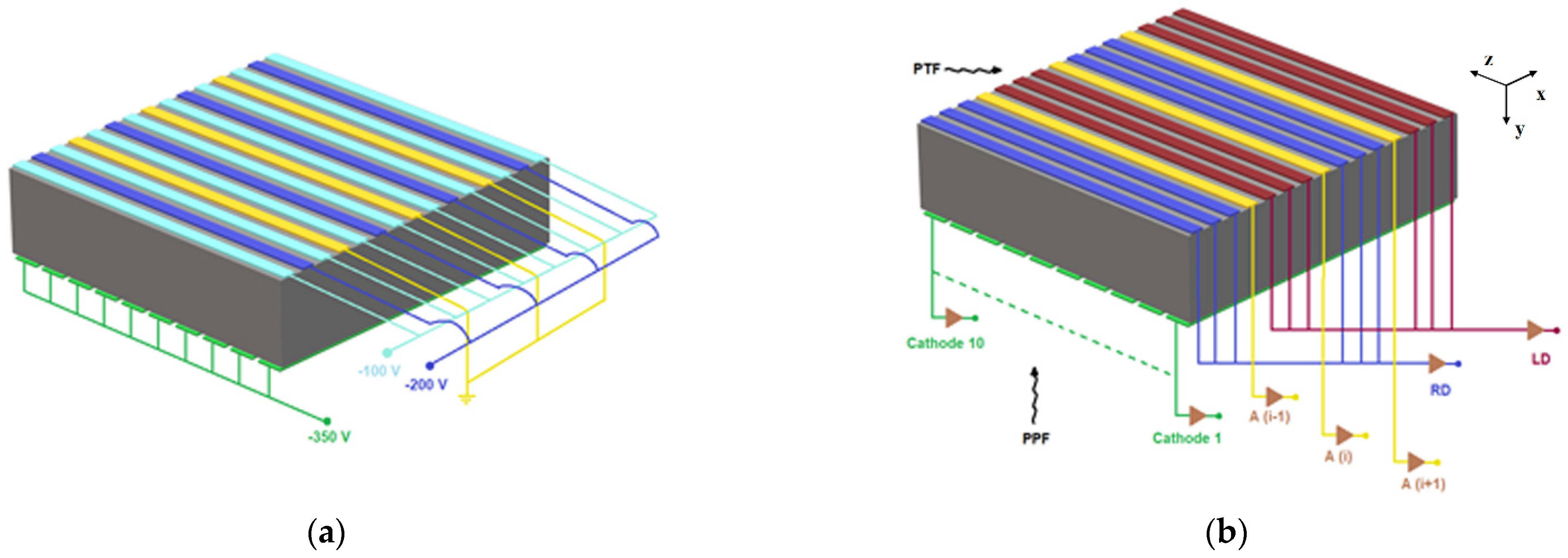
Abstract
:1. Introduction
2. The 3-D CZT Drift Strip Detectors
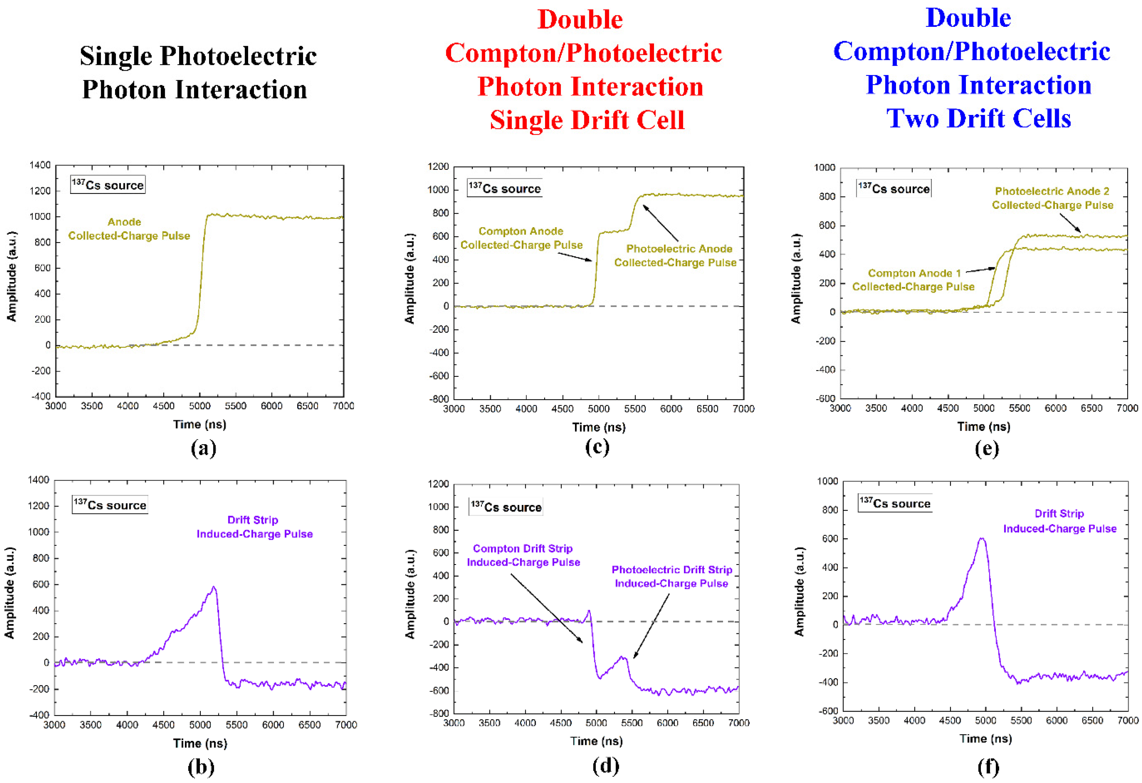
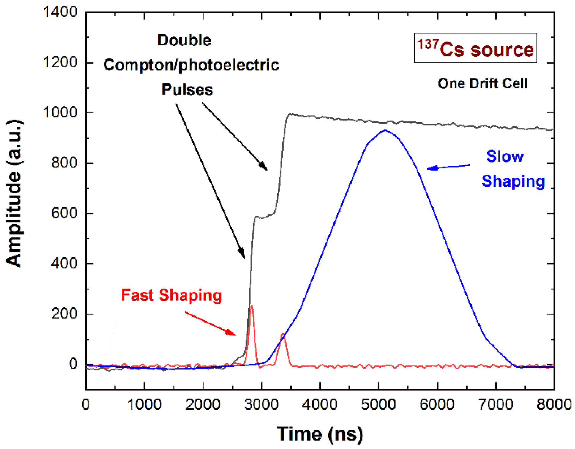
2.1. The Pulses from the Strips: Single and Double Photon Interactions
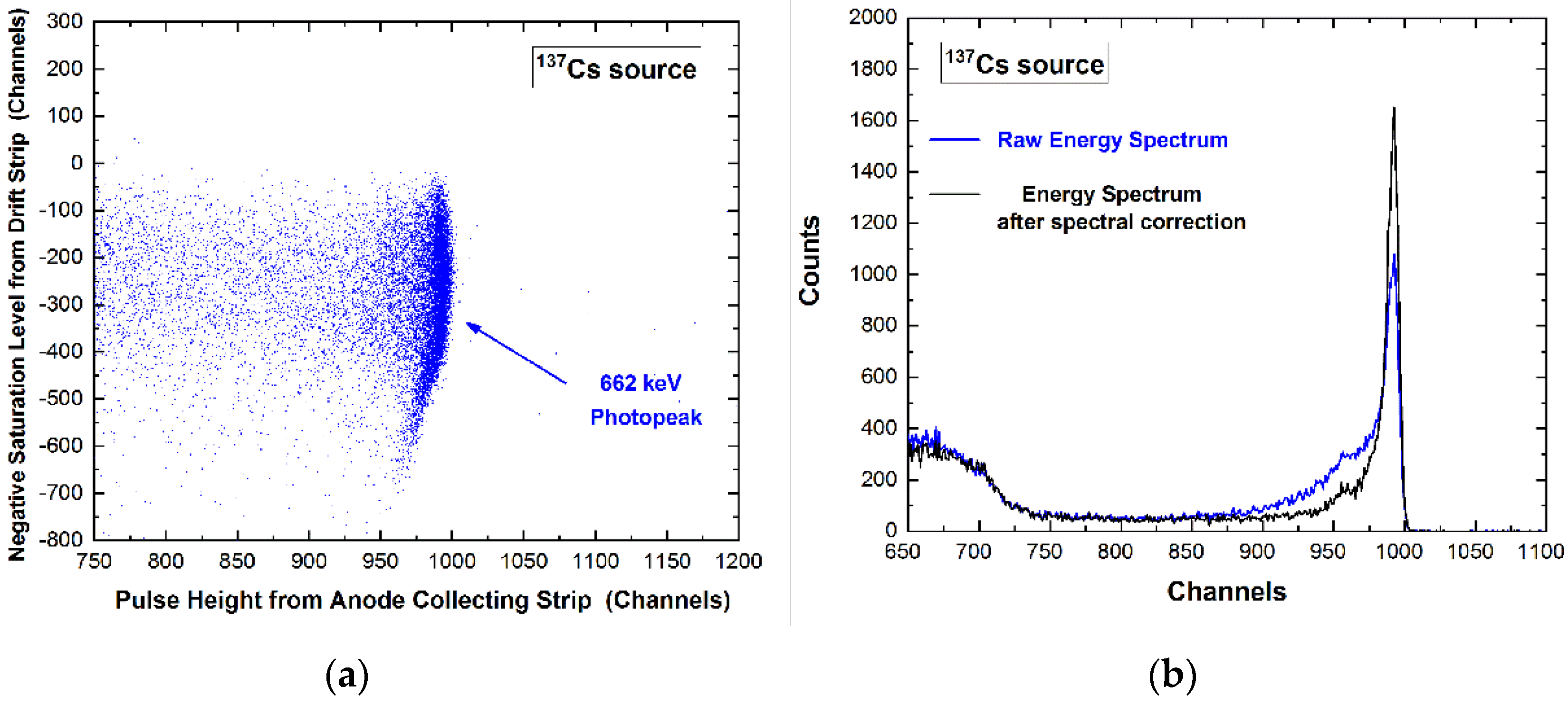
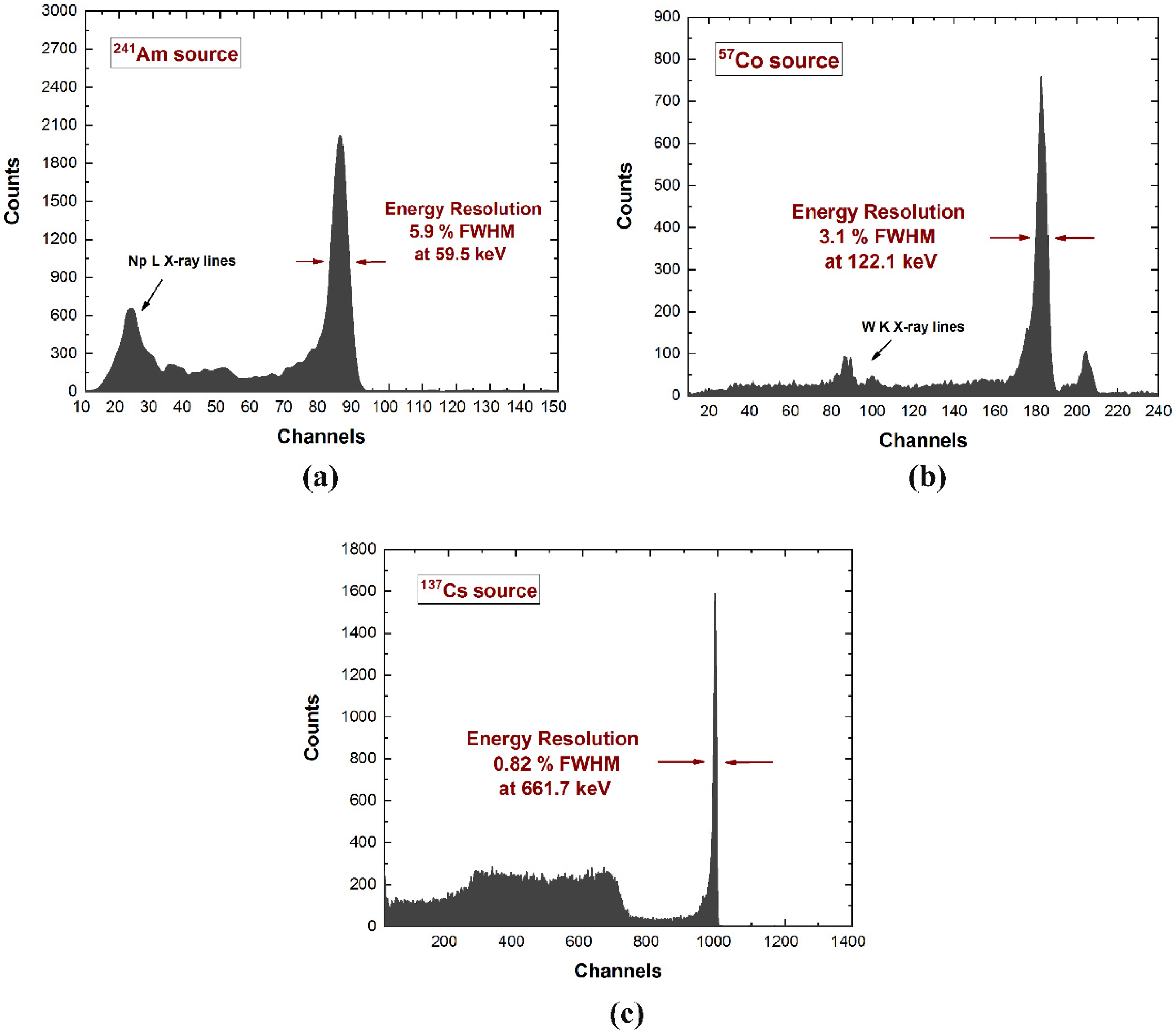
2.2. Spectroscopic Performance
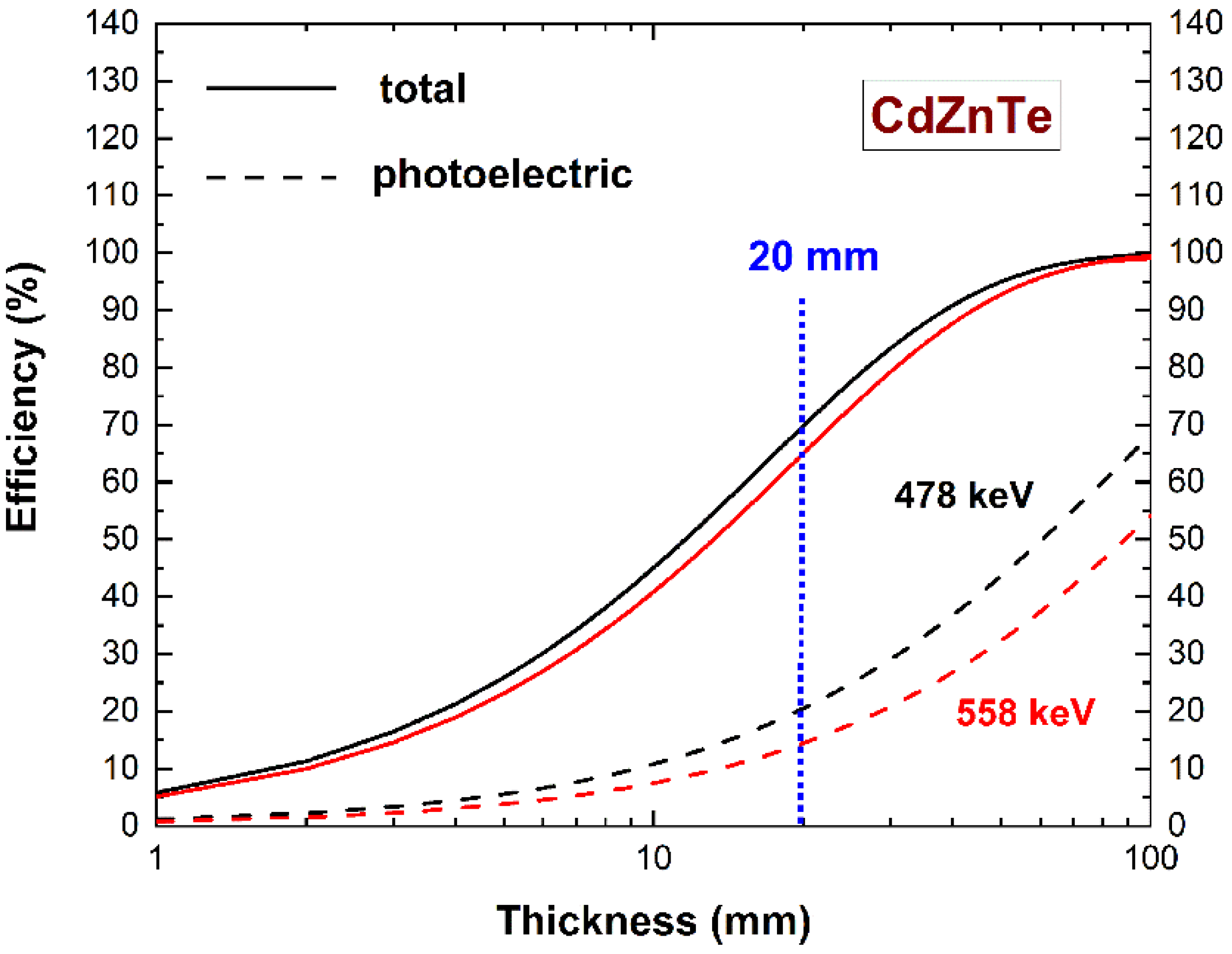
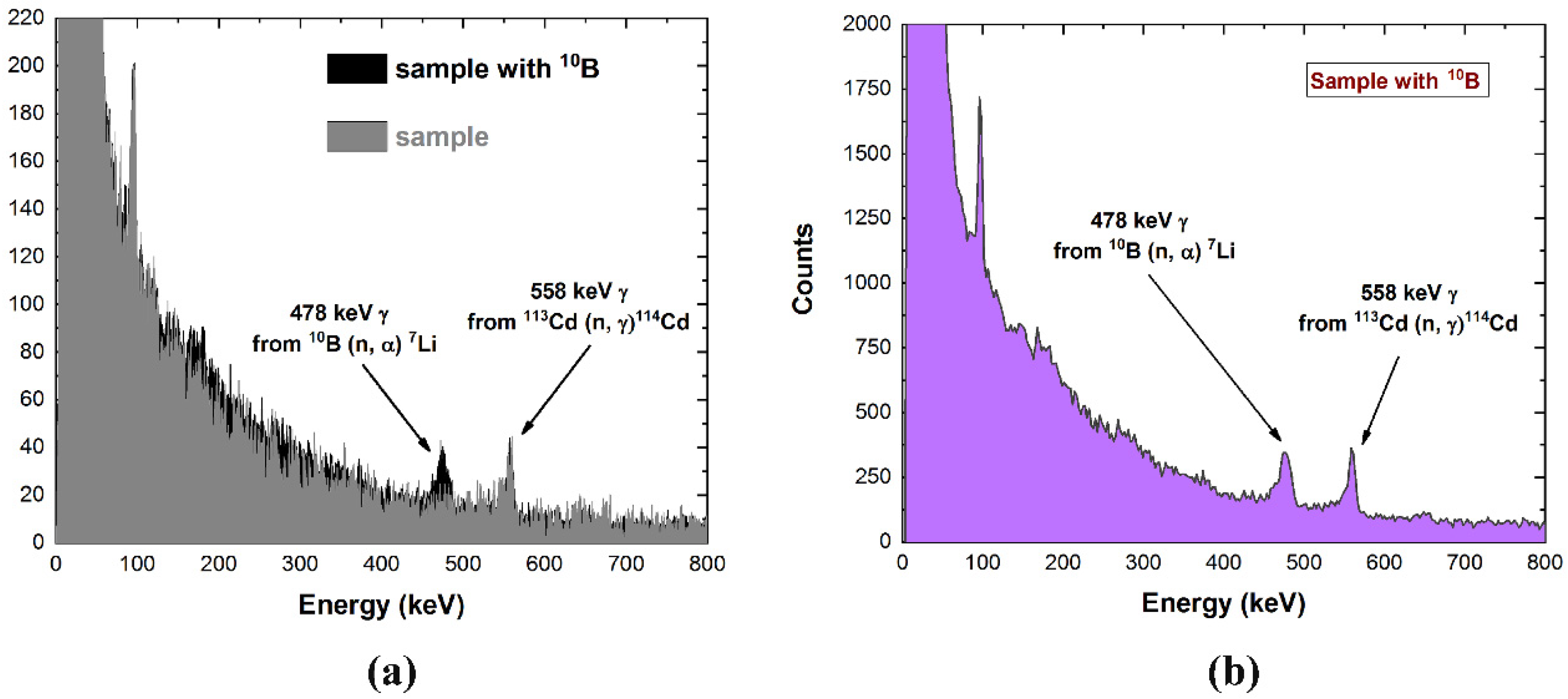
3. Prompt Gamma-Ray Measurements in BNCT Environment
4. Conclusions
Author Contributions
Funding
Acknowledgments
Conflicts of Interest
References
- Locher, G.L. Biological effects and therapeutic possibilities of neutrons. Am. J. Roentgenol. Radium Ther. 1936, 36, 1–13. [Google Scholar]
- Kruger, P.C. Some biological effects of nuclear disintegration products on neoplastic tissue. Proc. Natl. Acad. Sci. USA 1940, 26, 181–192. [Google Scholar] [CrossRef] [PubMed] [Green Version]
- Farr, L.E.; Sweet, W.H.; Locksley, H.B.; Robertson, J.S. Neutron capture therapy of gliomas using boron-10. Trans. Am. Neurol. Assoc. 1954, 79, 110–113. [Google Scholar]
- Hatanaka, T. Future possibility of neutron capture therapy of malignant tumors by use of low energy neutron from nuclear reactors and other sources. Gan No Rinsho 1969, 15, 367–369. [Google Scholar] [PubMed]
- González, S.J.; Bonomi, M.R.; Santa Cruz, G.A.S.; Blaumann, H.R.; Larrieu, O.A.C.; Menéndez, P.; Rebagliati, R.J.; Longhino, J.; Feld, D.B.; Dagrosa, M.A.; et al. First BNCT treatment of a skin melanoma in Argentina: Dosimetric analysis and clinical outcome. Appl. Radiat. Isot. 2004, 61, 1101–1105. [Google Scholar] [CrossRef]
- Joensuu, H.; Kankaanranta, L.; Seppälä, T.; Auterinen, I.; Kallio, M.; Kulvik, M.; Laakso, J.; Vähätalo, J.; Kortesniemi, M.; Kotiluoto, P.; et al. Boron neutron capture therapy of brain tumors: Clinical trials at the finnish facility using boronophenylalanine. J. Neurooncol. 2003, 62, 123–124. [Google Scholar] [CrossRef] [PubMed]
- Capala, J.; Stenstam, B.H.; Sköld, K.; Rosenschöld, P.M.; Giusti, V.; Persson, C.; Wallin, E.; Brun, A.; Franzen, L.; Carlsson, J.; et al. Boron neutron capture therapy for glioblastoma multiforme: Clinical studies in Sweden. J. Neurooncol. 2003, 62, 135–144. [Google Scholar] [CrossRef] [PubMed]
- Zonta, A.; Pinelli, T.; Prati, U.; Roveda, L.; Ferrari, C.; Clerici, A.M.; Zonta, C.; Mazzini, G.; Dionigi, P.; Altieri, S.; et al. Extra-corporeal liver BNCT for the treatment of diffuse metastases: What was learned and what is still to be learned. Appl. Rad. Isot. 2009, 67, S67–S75. [Google Scholar] [CrossRef]
- Fairchild, R.G.; Gabel, D.; Laster, B.H.; Greenberg, D.; Kiszenick, W.; Micca, P.L. Micro-analytical techniques for boron analysis using the 10B(n, alpha)7Li reaction. Med. Phys. 1986, 13, 50–56. [Google Scholar] [CrossRef]
- Matsumoto, T.; Aoki, M.; Aizawa, O. Phantom experiment and calculation for in vivo 10boron analysis by prompt gamma ray spectroscopy. Phys. Med. Biol. 1991, 36, 329–338. [Google Scholar] [CrossRef]
- Okazaki, K.; Tanaka, H.; Takata, T.; Kawabata, S.; Akabori, K.; Sakurai, Y. Evaluation of the energy resolution of a prompt gamma-ray imaging detector using LaBr3(Ce) scintillator and 8 × 8 array MPPC for an animal study of BNCT. Appl. Radiat. Isot. A 2020, 163, 109214. [Google Scholar] [CrossRef] [PubMed]
- Kobayashi, T.; Sakurai, Y.; Ishikawa, M. A noninvasive dose estimation system for clinical BNCT based on PG-SPECT—Conceptual study and fundamental experiments using HPGe and CdTe semiconductor detectors. Med. Phys. 2000, 27, 2124–2132. [Google Scholar] [CrossRef] [PubMed]
- Del Sordo, S.; Abbene, L.; Caroli, E.; Mancini, A.M.; Zappettini, A.; Ubertini, P. Progress in the development of CdTe and CdZnTe semiconductor radiation detectors for astrophysical and medical applications. Sensors 2009, 9, 3491–3526. [Google Scholar] [CrossRef] [PubMed]
- Murata, I.; Nakamura, S.; Manabe, M.; Miyamaru, H.; Kato, I. Characterization measurement of a thick CdTe detector for BNCT-SPECT—Detection efficiency and energy resolution. Appl. Radiat. Isot. A 2014, 88, 129–133. [Google Scholar] [CrossRef]
- Murata, I.; Mukai, T.; Nakamura, S.; Miyamaru, H.; Kato, I. Development of a thick CdTe detector for BNCT–SPECT. Appl. Radiat. Isot. 2011, 69, 1706–1709. [Google Scholar] [CrossRef]
- Winkler, A.; Koivunoro, H.; Reijonen, V.; Auterinen, I.; Savolainen, S. Prompt gamma and neutron detection in BNCT utilizing a CdTe detector. Appl. Radiat. Isot. 2015, 106, 139–144. [Google Scholar] [CrossRef]
- Hales, B.; Katabuchi, T.; Igashira, M.; Terada, K.; Hayashizaki, N.; Kobayashi, T. Predicted performance of a PG-SPECT system using CZT primary detectors and secondary Compton-suppression anti-coincidence detectors under near-clinical settings for boron neutron capture therapy. Nucl. Instr. Meth. A 2017, 875, 51–56. [Google Scholar] [CrossRef]
- Fatemi, S.; Gong, C.H.; Bortolussi, S.; Magni, C.; Postuma, I.; Bettelli, M.; Benassi, G.; Zambelli, N.; Zappettini, A.; Tang, X.B.; et al. Innovative 3D sensitive CdZnTe solid state detector for dose monitoring in Boron Neutron Capture Therapy (BNCT). Nucl. Instr. Meth. A 2019, 936, 50–51. [Google Scholar] [CrossRef]
- Fatemi, S.; Altieri, S.; Bortolussi, S.; Postuma, I.; Benassi, G.; Zambelli, N.; Bettelli, M.; Zanichelli, M.; Zappettini, A.; Protti, N. Preliminary characterization of a CdZnTe photon detector for BNCT-SPECT. Nucl. Instr. Meth. A 2018, 903, 134–139. [Google Scholar] [CrossRef]
- Abbene, L.; Gerardi, G.; Turturici, A.A.; Del Sordo, S.; Principato, F. Experimental results from Al/p-CdTe/Pt X-ray detectors. Nucl. Instr. Meth. A 2013, 730, 135–140. [Google Scholar] [CrossRef]
- Meuris, A.; Limousin, O.; Blondel, C. Characterization of polarization phenomenon in Al-Schottky CdTe detectors using a spectroscopic analysis method. Nucl. Instr. Meth. A 2011, 654, 293–299. [Google Scholar] [CrossRef]
- MacKin, D.; Polf, J.; Peterson, S.; Beddar, S. The effects of Doppler broadening and detector resolution on the performance of three stage Compton cameras. Med. Phys. 2013, 40, 012402. [Google Scholar] [CrossRef] [PubMed] [Green Version]
- Gong, C.-H.; Tang, X.-B.; Shu, D.-Y.; Yu, H.-Y.; Geng, C.-R. Optimization of the Compton camera for measuring prompt gamma rays in boron neutron capture therapy. Appl. Radiat. Isot. 2017, 124, 62–67. [Google Scholar] [CrossRef] [PubMed]
- Lee, T.; Lee, H.; Lee, W. Monitoring the distribution of prompt gamma rays in boron neutron capture therapy using a multiple-scattering Compton camera: A Monte Carlo simulation study. Nucl. Instr. Meth. A 2015, 798, 135–139. [Google Scholar] [CrossRef]
- Butler, J.F.; Lingren, C.L.; Doty, F.P. Cd1-xZnxTe gamma ray detectors. IEEE Trans. Nucl. Sci. 1992, 39, 605–609. [Google Scholar] [CrossRef]
- McGregor, D.S.; Lindsay, J.T.; Olsen, R.W. Thermal neutron detection with cadmium 1-x zinc x telluride semiconductor detectors. Nucl. Instr. Meth. A 1996, 381, 498–501. [Google Scholar] [CrossRef]
- Szeles, C.; Soldner, S.A.; Vydrin, S.; Graves, J.; Bale, D.S. CdZnTe Semiconductor Detectors for Spectroscopic X-ray Imaging. IEEE Trans. Nucl. Sci. 2008, 55, 572–582. [Google Scholar] [CrossRef]
- Iniewski, K. CZT detector technology for medical imaging. J. Inst. 2014, 9, C11001. [Google Scholar] [CrossRef]
- Iwanczyk, J.; Nygard, E.; Meirav, O.; Arenson, J.; Barber, W.C.; Hartsough, N.E.; Malakhov, N.; Wessel, J.C. Photon Counting Energy Dispersive Detector Arrays for X-ray Imaging. IEEE Trans. Nucl. Sci. 2009, 56, 535–542. [Google Scholar] [CrossRef] [Green Version]
- Seller, P.; Bell, S.; Cernik, R.J.; Christodoulou, C.; Egan, C.K.; Gaskin, J.A.; Jacques, S.; Pani, S.; Ramsey, B.D.; Reid, C.; et al. Pixellated Cd(Zn)Te high-energy X-ray instrument. J. Inst. 2011, 6, C12009. [Google Scholar] [CrossRef]
- Abbene, L.; Principato, F.; Gerardi, G.; Buttacavoli, A.; Cascio, D.; Bettelli, M.; Zappettini, A. Room-Temperature X-ray response of cadmium-zinc-Telluride pixel detectors grown by the vertical Bridgman technique. J. Synchrotron Rad. 2020, 27, 319–328. [Google Scholar] [CrossRef] [PubMed] [Green Version]
- Abbene, L.; Gerardi, G.; Raso, G.; Principato, F.; Zambelli, N.; Benassi, G.; Bettelli, M.; Zappettini, A. Development of new CdZnTe detectors for room-temperature high-flux radiation measurements. J. Synchrotron Rad. 2017, 24, 429–438. [Google Scholar] [CrossRef] [PubMed]
- Macri, J.R.; Apotovsky, B.A.; Butler, J.F.; Cherry, M.L.; Dann, B.K.; Drake, A.; Doty, F.P.; Guzik, T.G.; Larson, K.; Mayer, M.; et al. Development of an orthogonal-stripe CdZnTe gamma radiation imaging spectrometer. IEEE Trans. Nucl. Sci. 1996, 43, 1458–1462. [Google Scholar] [CrossRef]
- Kuvvetli, I.; Budtz-Jørgensen, C.; Caroli, E.; Auricchio, N. CZT drift strip detectors for high energy astrophysics. Nucl. Instr. Meth. A 2010, 624, 486–491. [Google Scholar] [CrossRef] [Green Version]
- Owe, S.H.; Kuvvetli, I.; Budtz-JØrgensen, C.; Zoglauer, A. Evaluation of a Compton camera concept using the 3D CdZnTe drift strip detectors. J. Inst. 2019, 14, C01020. [Google Scholar] [CrossRef]
- Abbene, L.; Gerardi, G.; Principato, F.; Buttacavoli, A.; Altieri, S.; Protti, N.; Tomarchio, E.; Del Sordo, S.; Auricchio, N.; Bettelli, M.; et al. Recent advances in the development of high-resolution 3D cadmium–zinc–telluride drift strip detectors. J. Synchrotron Rad. 2020, 27, 1564–1576. [Google Scholar] [CrossRef] [PubMed]
- Abbene, L.; Principato, F.; Gerardi, G.; Buttacavoli, A.; Altieri, S.; Gong, C.; Protti, N.; Bettelli, M.; Sarzi Amadè, N.; Zanettini, S.; et al. New results on high-resolution 3-D CZT drift strip detectors. In Proceedings of the 2020 IEEE Nuclear Science Symposium and Medical Imaging Conference (NSS/MIC), Boston, MA, USA, 31 October–7 November 2020. [Google Scholar] [CrossRef]
- Zanettini, S.; Pattini, F.; Amade, N.S.; Sidoli, M.; Zambelli, N.; Benassi, G.; Bettelli, M.; Rampino, S.; Gilioli, E.; Protti, N.; et al. Al2O3 Coating as Passivation Layer for CZT-based Detectors. In Proceedings of the 2018 IEEE Nuclear Science Symposium and Medical Imaging Conference Proceedings (NSS/MIC) 2018, Sydney, NSW, Australia, 10–7 November 2018; p. 8824623. [Google Scholar]
- Benassi, G.; Nasi, L.; Bettelli, M.; Zambelli, N.; Calestani, D.; Zappettini, A. Strong mechanical adhesion of gold electroless contacts on CdZnTe deposited by alcoholic solutions. J. Inst. 2017, 12, P02018. [Google Scholar]
- Abbene, L.; Principato, F.; Gerardi, G.; Bettelli, M.; Seller, P.; Veale, M.C.; Zambelli, N.; Benassi, G.; Zappettini, A. Digital fast pulse shape and height analysis on cadmium–zinc–telluride arrays for high-flux energy-resolved X-ray imaging. J. Synchrotron Rad. 2018, 25, 257–271. [Google Scholar] [CrossRef]
- Abbene, L.; Gerardi, G.; Raso, G.; Basile, S.; Brai, M.; Principato, F. Energy resolution and throughput of a new real time digital pulse processing system for x-ray and gamma ray semiconductor detectors. J. Instrum. 2013, 8, P07019. [Google Scholar] [CrossRef]


Publisher’s Note: MDPI stays neutral with regard to jurisdictional claims in published maps and institutional affiliations. |
© 2022 by the authors. Licensee MDPI, Basel, Switzerland. This article is an open access article distributed under the terms and conditions of the Creative Commons Attribution (CC BY) license (https://creativecommons.org/licenses/by/4.0/).
Share and Cite
Abbene, L.; Principato, F.; Buttacavoli, A.; Gerardi, G.; Bettelli, M.; Zappettini, A.; Altieri, S.; Auricchio, N.; Caroli, E.; Zanettini, S.; et al. Potentialities of High-Resolution 3-D CZT Drift Strip Detectors for Prompt Gamma-Ray Measurements in BNCT. Sensors 2022, 22, 1502. https://doi.org/10.3390/s22041502
Abbene L, Principato F, Buttacavoli A, Gerardi G, Bettelli M, Zappettini A, Altieri S, Auricchio N, Caroli E, Zanettini S, et al. Potentialities of High-Resolution 3-D CZT Drift Strip Detectors for Prompt Gamma-Ray Measurements in BNCT. Sensors. 2022; 22(4):1502. https://doi.org/10.3390/s22041502
Chicago/Turabian StyleAbbene, Leonardo, Fabio Principato, Antonino Buttacavoli, Gaetano Gerardi, Manuele Bettelli, Andrea Zappettini, Saverio Altieri, Natalia Auricchio, Ezio Caroli, Silvia Zanettini, and et al. 2022. "Potentialities of High-Resolution 3-D CZT Drift Strip Detectors for Prompt Gamma-Ray Measurements in BNCT" Sensors 22, no. 4: 1502. https://doi.org/10.3390/s22041502
APA StyleAbbene, L., Principato, F., Buttacavoli, A., Gerardi, G., Bettelli, M., Zappettini, A., Altieri, S., Auricchio, N., Caroli, E., Zanettini, S., & Protti, N. (2022). Potentialities of High-Resolution 3-D CZT Drift Strip Detectors for Prompt Gamma-Ray Measurements in BNCT. Sensors, 22(4), 1502. https://doi.org/10.3390/s22041502