Article

Hyalosira (Diatomeae: Grammatophoraceae) from Florida Keys, U.S.A., Including Two New Species with Consistent Ornamentation †

by
Christopher S. Lobban
Division of Natural Sciences, University of Guam, Mangilao, GU 96923, USA
ANSP-GC58474 and ANSP-GC58475.
Diversity 2025, 17(7), 448; https://doi.org/10.3390/d17070448
Submission received: 26 May 2025 / Revised: 20 June 2025 / Accepted: 20 June 2025 / Published: 24 June 2025

Abstract

Diatoms are the most numerous of algae and scanning electron microscopy continues to reveal ever-increasing diversity. Two new species of Hyalosira from Florida add new characters distinguishing species in this recently redefined genus: H. ornata sp. nov. from Florida Bay has consistent and elaborate ornamentation on both valves and undulate valve margins; H. sertifera sp. nov., from the Atlantic coast of Key Largo, has a thick, consistent, garland-like ring of cristae on only one valve; the former species has deep septa on all copulae except the valvocopula, the latter shallow septa or none on the copulae. Hyalosira hesperia was also found in the Atlantic sample; this is the first record outside the Mediterranean. The number of species known from SEM is now twelve, including four with septa, a diversity potentially useful in exploring outstanding questions of septum function; a key to these species is appended.

1. Introduction

Although diatoms are evolutionarily recent in geological time, the earliest clear fossils being about 75 million years ago [1], they have become extremely diverse, especially in comparison to other algae [2], so much so that Mann & Vanormelingen [3] paraphrased Haldane’s famous remark about God’s fondness for beetles to suggest that He also had an inordinate fondness for diatoms. As scanning electron microscopy (SEM) has become widespread, much finer distinctions between genera and between species have become necessary. Thus, in several monospecific or small genera more species were discovered in the limited regions studied, suggesting that there are likely to be many more [4,5,6,7,8].
A case in point is the genus Hyalosira Kützing 1844 [9]. Defined at a time when the details of the silica shells were not considered (the name means hyaline chains), this genus underwent several taxonomic revisions before the 21st Century, being pulled first into Striatella [10] (pl. 54, fig. 3) and then into Microtabella [11], before being put back into Hyalosira [12]. Moreover, the four taxa named by Kützing were revised as varieties within H. delicatula by Grunow in van Heurck (1881) [10] and then Hustedt (1931) [13] synonymized all Kützing’s species under Striatella delicatula, noting a distribution in Europe from Mediterranean to Arctic waters. Navarro & Williams [12] showed SEM of cells attributed to H. delicatula Kützing from the Baltic coast of Sweden but for two reasons it cannot be in this genus: (1) areolae of the valve are as small as the poroids of the apical pore field, and (2) there is only a single row of areolae on each copula. The ultrastructure of H. delicatula type materials is unknown, and this may be problematic because the one morphotype for which unmounted type material could be found (H. obtusangula), was taken as representative of the type species by [14]. Given the images in [12], we cannot assume that all Kützing’s species were in the same genus. Recently, Lobban et al. [14] contributed to a roundup of taxa observed in SEM in samples from various parts of the world [14] (table 2), adding five more species with striae ranging from uniseriate to triseriate and mixed, separating the true Hyalosira species in Grammatophoraceae from those in Rhabdonemataceae—renamed Placosira Ashworth & Majewska, and illustrating some unresolved diversity in both. Shortly after that paper was published, we found two more Hyalosira species [15], bringing the total to ten observed by SEM, but leaving a few known only from LM, some of which were shown to belong to other genera [14]. The new species showed that the number of rimoportulae per valve could range from 0 to 2 in different species, that there could be heterovalvy in the frustule, heteropolarity in the valve, and curvature along the apical axis. The most recent diagnosis of generic characters is, “Tabulate cells with apical pore fields; copulae numerous with two rows of pores; septa, if present, closed less than half the cell length; rimoportulae fan-shaped, adjacent to midline” [15]. We anticipated additional species as new regions were explored, and two new species were found in recent sampling in the Florida Keys. One is septate with two unexpected new characters: highly and consistently decorated on both valves, and undulate valve outline. The second has a consistent ring of cristae on only one valve. In addition, there was H. hesperia Álvarez-Blanco & S.Blanco emend Bizsel & S.Blanco, previously known only from the Mediterranean. A key to all species known from SEM is included.

2. Materials and Methods

Samples of seaweeds with epiphytic diatoms were hand collected from crab traps and their buoy lines in shallow water of Florida Bay, near Tavernier (25°02.456′ N, 80°30.766′ W), and from South Creek Village boat dock on the exposed side of Key Largo (25°07.7898′ N, 80°24.20004′ W) and preserved in 40% ethanol for transport to Guam. There, they were processed for microscopy following standard procedures [16], in short: boiling for 10 min in equal portions of water and concentrated nitric acid, remaining in the acid at room temperature for 24 h, and rinsed 10 times with distilled water. Drops of the cleaned shells were put on coverslips and cellulose nitrate filters, the former mounted on slides with Naphrax resin for light microscopy (LM) with a Nikon 80i (Tokyo, Japan) microscope with differential interference contrast illumination; the latter mounted on stubs and sputter-coated with gold for SEM with a Thermo-Fisher desktop Phenom XL G2 running (Waltham, MA, USA) at 10 kV and low emission settings.

3. Results

RHABDONEMATALES Round & Crawford
Grammatophoraceae Lobban & Ashworth
Hyalosira Kützing emend. Lobban, Ashworth and Majewska

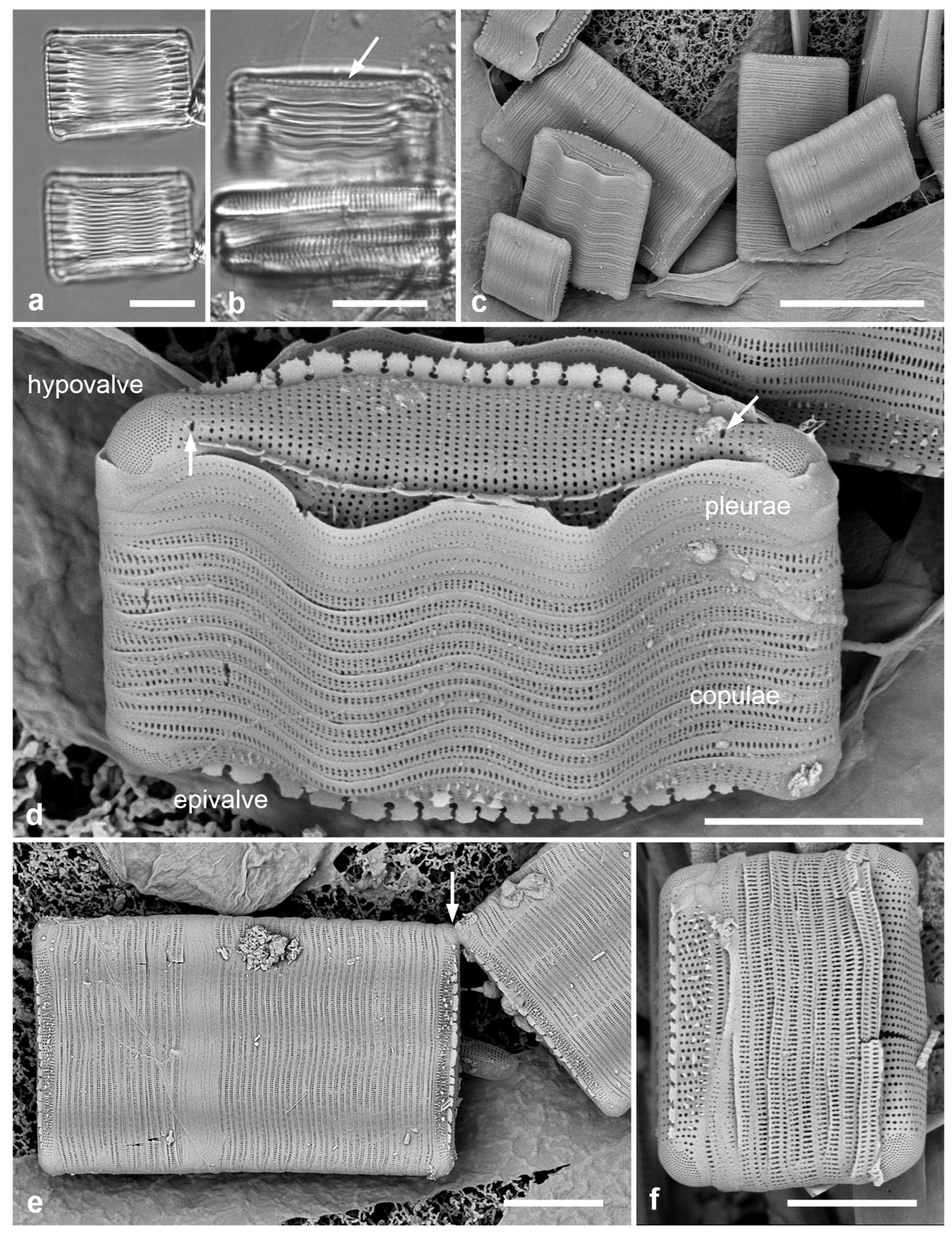
3.1. Hyalosira ornata sp. nov.—Figure 1 and Figure 2

Diagnosis: Valve elliptical in upper part but with an undulate mantle, two lines of sun-to-crown-shaped crista elements on the upper part; two rimoportulae per valve. Copulae with shallow septa but long midrib thickenings.
Holotype: Specimen at 19.7 mm E and 3.0 mm S of the mark on slide 4151, deposited in ANSP, accession number ANSP-GC58474. Figure 1a.
Type locality: Florida: Florida Bay (off Tavernier), 25°02.456′ N, 80°30.766′ W, sample FL-RH1645, on a crab trap buoy line, ca. 2 m deep, substrate mostly red filamentous seaweed, coll. C.S. Lobban, T.A. & D. Frankovich, 15 March 2025.
Figure 1. Hyalosira ornata. (a) Holotype at two focal planes in LM. (b) Frustule (partially obscured) in LM showing valve ornamentation (arrow). (cf) SEM from whole mount. (c) Several specimens at different angles on substrate, showing lack of chain formation. (d) Portrait of single complete frustule showing ornamentation; recently divided cell: new hypovalve at top showing two rimoportula openings (arrows) and the lines of crista elements (in end view on near side) (compare with those on epivalve), epivalve at bottom with epicingulum of copulae and pleurae. (e) Pair of frustules joined by mucilage at corner (arrow). (f) Small frustule with one undecorated valve, the decorated valve showing the shards and spines on the lower mantle; valve outline perhaps not undulating. Scale bars: (c) = 25 µm, (a,b,e) = 10 µm, (d,f) = 5 µm.
Diversity 17 00448 g001
Figure 2. Hyalosira ornata, SEM of acid cleaned material. (a). External view of valve surface showing the costae on the upper edge of the mantle and two rimoportula openings (arrows). (b) Portion of frustule in girdle view showing shards and spines on mantle. (c) Interior of valve with attached valvocopula, showing narrow shelf on copula extending nearly to open end, and the two rimoportulae (arrows). (d) Detail of isolated valve interior showing oblique view of rimoportula. (e) Broken frustule showing one valve at the bottom of the stack with open tips of valvocopula and polar aspect of copulae. Depth of septum indicated by double arrow. (f) Three sections of a cingulum, pleurae at far right, the copulae showing the extent of the shelves beyond the septum (arrow). Scale bars: (a,c,e,f) = 5 µm, (b,d) = 2 µm.
Diversity 17 00448 g002
Description: Cells usually single but occasionally joined corner to corner, tabulate in girdle view, recently divided cells rectangular in the apical axis, more mature cells becoming longer in the pervalvar axis as the hypocingulum grew (Figure 1a–f). Valves 13–27 µm long, greatest width 6–8 µm, slightly less across center; striae parallel, 34–36 in 10 µm (Table 1); elliptical valve face but with a broad mantle undulate in outline at margin (Figure 2a–d). Cristae with spiky, circular crista elements present near top of mantle on each side (Figure 1c–f and Figure 2a,d), detectable in LM as a series of dots (Figure 1b arrow); cristae on newly exposed valve face perhaps reflecting their original shape (Figure 1d), gradually eroding to irregular shapes. Additional shards and spines of silica present on the lower mantle (Figure 1e,f and Figure 2a,d). These ornaments usually occurred on both valves in a frustule; only occasionally one valve lacked them (Figure 1f). Two rimoportulae present on each valve, adjacent to the ends of an indistinct sternum (Figure 1d and Figure 2a,c, arrows). Copulae variable in number depending on hypocingulum development, but with up to 18 copulae in the mature epicingulum (Figure 1e), open bands arranged alternately, the closed ends deepened into ligules to fill the gap between open ends (Figure 1d–f and Figure 2d,e). Valvocopula with narrow shelf all around (Figure 2c,e,f), other copulae with septum extending from the closed end for about 14% of valve length but the midrib thickened almost to the open end; septa typically with some pores near the pole (Figure 2e,f). Areolae on the copulae, 55–58 in 10 µm, probably single on each side of the midrib but with delicate bar across the middle at the outer surface; areolae more numerous in the ligule. Pleurae distinct, with smaller pores and without septa (Figure 1d and Figure 2f).
Etymology: Ornata (L.), decorated, with reference to the diverse silica elements on the valve surface.
Phycobank registration: http://phycobank.org/105590.
Comments: Frustules in the sample were easy to distinguish in LM from various Microtabella spp. present by the relatively shallow septa and long thickenings, giving the appearance of alternating tuning forks (Figure 1a), whereas, septa in Microtabella reach the middle often giving the appearance of a central chain of rings.

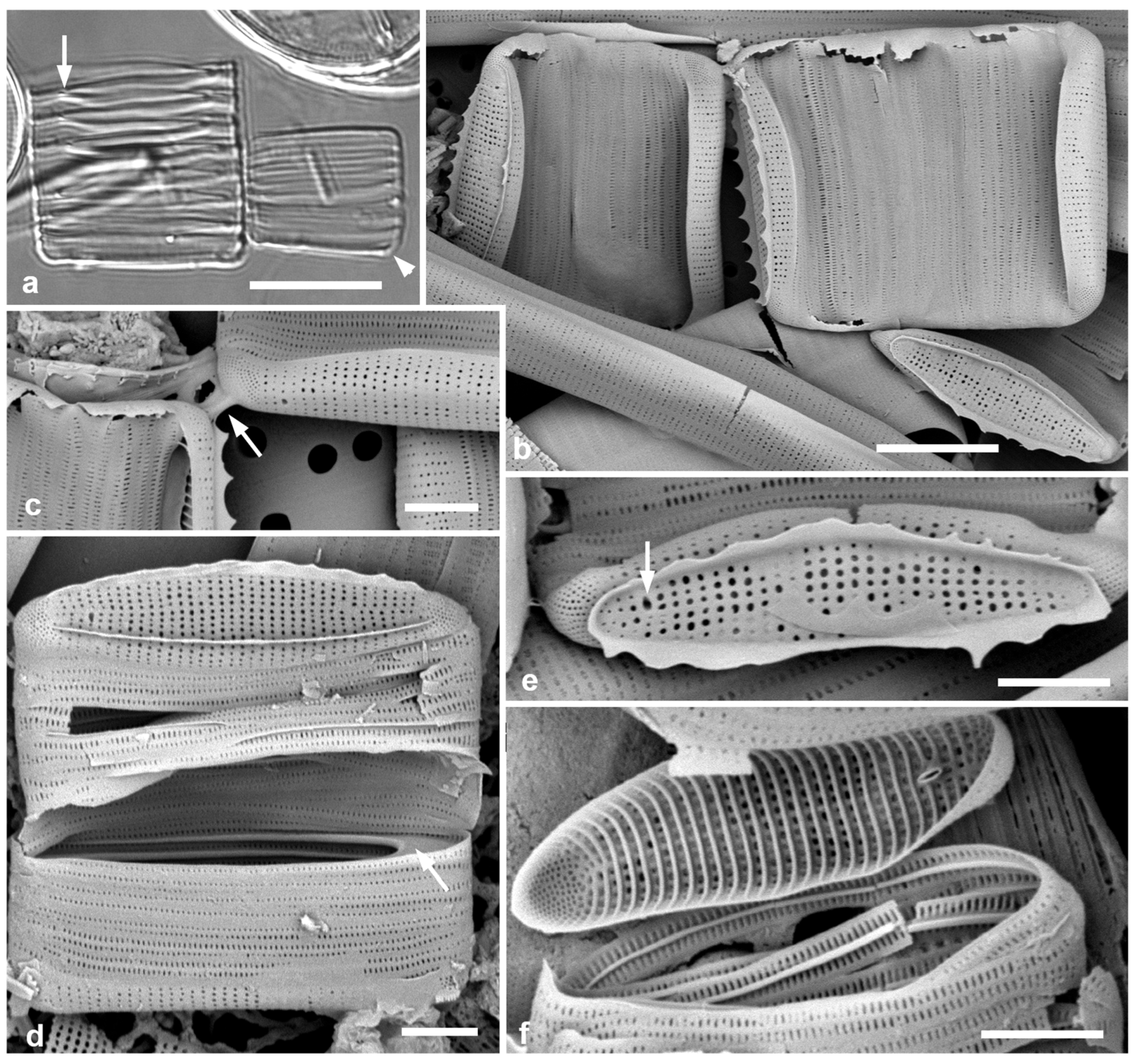
3.2. Hyalosira sertifera sp. nov.—Figure 3

Diagnosis: Distinguished from other species by the sturdy corona consistently present on one valve, and thus heterovalvar; distinguished from Hyalosira hesperia present in the same sample by shallow or absent septa and by stria densities of 38–40 in 10 µm on valve and 70–73 in 10 µm on copulae.
Holotype: Specimen at 17.3 mm E and 11.2 mm S of the mark on slide 4154, deposited in ANSP, accession number ANSP-GC58475. On right in Figure 3a.
Isotype: Frustule in SEM image Figure 3d.
Type locality: Florida: Key Largo, ocean side, South Creek Village dock, 25°07.7898′ N, 80°24.20004′ W, epiphytic on Heterosiphonia crispella var. laxa from rocks at waterline, sample FL-SCV-1, coll. C.S. Lobban & T.A. Frankovich, 17 March 2025.
Description: Cells apparently single as a rule but occasionally joined corner to corner (Figure 3b,c). Valves lanceolate-elliptical, 9–16 µm long, 3–4 µm wide; striae parallel, uniseriate, 38–40 in 10 µm. Sturdy, jagged cristae forming a continuous “garland” around the edge on the valve face-mantle boundary, on one valve only (Figure 3b,d,e). Single rimoportula per valve (Figure 3e,f). At least some copulae with shallow septa (<10% of valve length (Figure 3d,f, Table 1) but midribs thickened well past the middle of the band; the distribution of septa was not clear from the fragments observed. Areolae on the copulae 70–73 in 10 µm. Pleurae indistinct, (Figure 3b,d).
Etymology: From (L.) sertum (garland) + ferre (to bear): garland-bearer.
Phycobank registration: http://phycobank.org/105591.

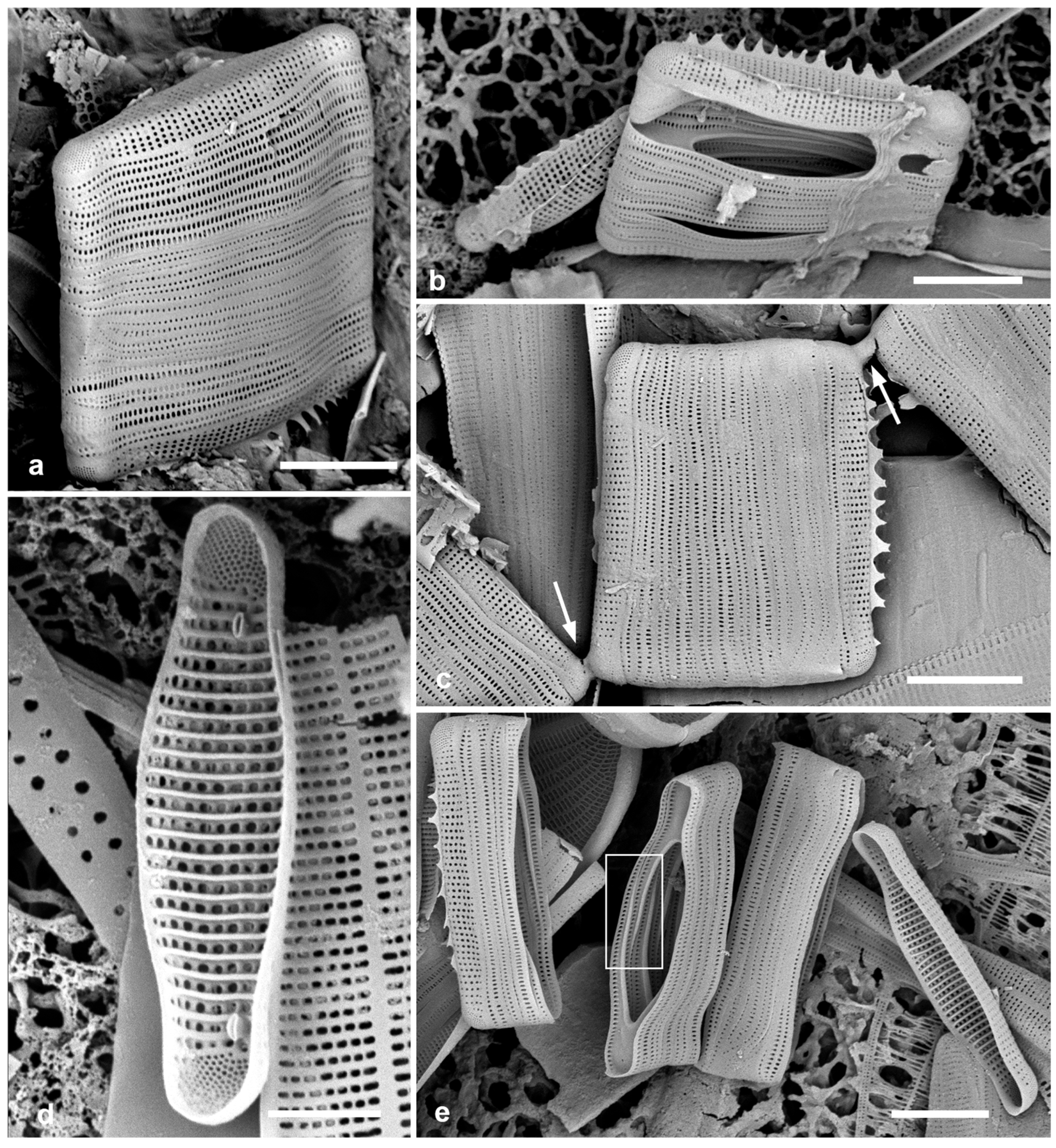
3.3. Hyalosira hesperia Álvarez-Blanco & Blanco Emend Bizsel & S.Blanco.—Figure 3a and Figure 4

Description: This species is recognized by the lanceolate, rostrate valves with uniseriate striae and two rimoportulae per valve, and presence of septa on the copulae. Length 7–20 µm, width ca. 3 µm; stria densities 28–32 in 10 µm on valve and 42–50 in 10 µm on copulae; septa tapering out about 65% of length of the band (Figure 4e) and visible in LM (on left in Figure 3a); rimoportula at each pole (Figure 4d). Cristae with irregular flaps sometimes well developed on one valve (Figure 4a–c); cells occasionally joined in chains of 2–4 (Figure 4c).
Comment: Distinguished from H. sertifera in the same sample by presence of septa and much coarser striae on the copulae; and from the septate H. pacifica Lobban by slightly higher stria densities on both valves and copulae (Table 1). The species was previously reported from the Mediterranean coasts of Spain and Turkey [15].
Figure 3. Hyalosira sertifera sp. nov. (a) LM showing holotype specimen complete frustule on right (no apparent septa); the characteristic “garland” not clearly distinguishable in LM, but possibly the apparently thickened bottom valve edge (arrowhead). Partial frustule of septate H. hesperia to the left, arrow indicating the septa present in the latter. (bf) SEM, from whole mount except (d,f) acid cleaned. (b) Two cells joined by the top corner and a cell in valve view. (c) Detail of a mucilage connection (arrow). (d) Designated isotype frustule with decorated valve in near-valve view showing “garland” and single rimoportula, along with a shallow septum on the copula adjacent to the pleurae (arrow). (e) Collapsed frustule with valve in valve view showing rimoportula (arrow) and ring of cristae. (f) Frustule showing valve interior with single rimoportula and thickenings on copulae, no septa evident. Scale bars: (a) = 10 µm, (b) = 5 µm, (cf) = 2 µm.
Diversity 17 00448 g003
Figure 4. Hyalosira hesperia SEM whole mounts (a,c) and acid cleaned (b,d,e). (a) Oblique view of whole frustule. (b) Broken frustule showing septa on copulae below collapsed pleurae, with isolated valve adjacent. (c) Chain of three cells showing mucilage pads between apical pore fields (arrows). (d) Valve interior view showing two rimoportulae. (e) Broken frustule showing overlapping ends of septa (rectangle). Scale bars: (ac,e) = 5 µm, (d) = 2 µm.
Diversity 17 00448 g004
Table 1. Character states of all Hyalosira taxa.
CharactersH. ornata, sp. nov.H. hesperia MediterraneanH. hesperia FloridaH. pacificaH. sertifera sp. nov.H. tropicalisH. septataH. fabaH. navarroanaH. mixtaH. triseriataH. obtusata [delicatula] ****H. parietina
Substratumepiphyticepilithic, epiphyticepiphyticepiphyticepiphyticepiphyticepizoicepizoicepiphyticepiphyticepiphyticepiphyticunknown
Shape in valve viewundulatelinear to weakly inflated, rostrate to weakly capitatelinear to weakly inflated, rostrate to weakly capitatelanceolate, rostrate to capitateelliptical, weakly rostrateoval to lanceolate, inflated, rostrate to capitatelinear to weakly inflated, rostrate to weakly capitatelanceolatelanceolate, weakly rostratelanceolate, apices rounded or subcapitatelanceolate, weakly rostratelinear to linear-lanceolateoval
Length × width, µm16–24 × 6–814–19 × 3–47–20 × 39–23 × 3–59–16 × 3–414–16 × 6–810–23 × 2.5–3.54.5–16 × 2.2–3.09–47 × 3–510–31 (4.5–15) × 3–4 (2.2) ***7–24 × 3.3–4.26–12 × 2.1–3.8 [3–20 × 1.5–3.5]6–9 × 2.5–3.5
Stria density/10 µm34–3627–2928–3222–2438–4020–2236–3827–3224–2725–27 (24–30)22–23[22–30]33
Stria characteruniseriateuniseriateuniseriateuniseriateuniseriateuniseriate, areolae various diameters, generally largebiseriate throughoutBiseriate except for row of reniform areolae along sternumbiseriate on mantle, uniseriate on valve facebiseriate except triseriate at distal endtriseriate throughoutuniseriate throughoutunknown
Rimoportulae per valve222211111111unknown
Cristaeline of spiky, circular elements on marginal costa plus shards and spines lower on mantlevariable up to extensive flaps on only one valveapparently inconsistent flaps from continuous base on only one valveprominent flaps from continuous basesturdy “garland” from continuous base on only one valveoften prominent when present (one valve)elaborate and extensive (one valve)prominent when present, continuous almost to apicesrow of mostly separate flakes along edge of valve face and onto pore fieldspoorly developedcontinuous line of silica with triangular ridges, both valvesprominent when presentunknown
Copula striae/10 µm *55–5840–4442–5031–3470–73approx. 45approx. 65approx. 50approx. 50approx. 42 (60)approx. 65approx. 46unknown
Septa relative dimensions **
   Approx. depth (see Figure 2e)14%25%27%25%10% when presentvery shallowto 30%very shallowvery shallowabsentunknownshallow or absentunknown
   Thickened midrib>95%65%65%65%90% 90%
Referencethis paper[15]this paper[15]this paper [14][14][14][14][14][13,14][17]
* estimated from published images for species described in [14,15]. ** Described here more precisely than in [14] to distinguish them. *** Dimensions for South African population (in parentheses) notable smaller than in Guam. **** data in brackets are for Striatella delicatula as defined by Hustedt [13], other data from [14].

4. Discussion

In species of Hyalosira described to date, external ornamentation has consisted of irregularly developed silica flaps along the edge of the valve face, either arising individually or along a longitudinal line of silica, and called cristae, with reference to a cockscomb; sometimes the longitudinal line occurs alone, effectively a marginal costa [14,15]. The most noteworthy thing about these cristae so far has been that they tend to occur on only one of the two valves of a frustule; this was seen in SEM of both generitype material of H. obtusangula and in modern gatherings, as is seen here in H. hesperia (Figure 4c,e). Because of the variability, it has not yet been possible to include them as a taxonomic character even at the level of presence/absence [14]. Thus, the external ornamentation of H. ornata is remarkable for three reasons. First, the size and shape of the elements of the cristae were consistent, both along the valve and between specimens; second, H. ornata valves without them were very rare and on small cells (Figure 1f). Similar elements were recorded occasionally in H. septata [14] (fig. 16H). Third, the additional shards and spines on the mantle of H. ornata have not been seen in other species. I, thus, consider the ornamentation of this species to be a strong taxonomic character, but even without that H. ornata is distinguished by the undulate valve margins, the first time this has been observed in the genus. Hyalosira sertifera also has strong, consistent cristae that often form a complete ring, but only on one valve.
Another useful character of H. ornata is in the septa, where the pattern is different from that typical of other septate Hyalosira spp. and other genera. Whereas septa, developed centripetally from ingrowth of the copula midrib [18], usually have a short arc from the center of the septum to the end of the thickening, thus little overlap of the thickening in adjacent (oppositely oriented) copulae (Figure 3b and Figure 4e), in H. ornata the thickening continues as a shelf almost to the open tip of the band (Figure 2f). Possible additional states of this character, not yet seen, are (a) depth of the septum (so far not greatly different among species that have them), and whether the valvocopula stands alone in having little or no septum in contrast to all other copulae, versus a gradual change in depth over several copulae. In my cleaning technique, cingula tend to break into random clusters rather than completely dissociating (Figure 2f and Figure 4e). The pattern of septa could not be determined for H. sertifera because girdle bands generally collapsed despite the short septa (Figure 3b), unlike the cingula of H. ornata (Figure 2f) and H. hesperia (Figure 4e).
Finally, the stria density on the copulae is useful for taxonomy. The striae usually consist of a single areola on each side of the midrib, but H. septata has 3–4 areolae on each side. Most species have a stria density of 40–50 in 10 µm, but H. sertifera is notably finer with 70–73 in 10 µm, while H. septata and H. triseriata (estimated from published images, Table 1) have ca. 65 in 10 µm, whereas, H. pacifica is notably coarser at 30–32 in 10 µm.
Taxon sampling for genetic data for this genus, as redefined in [14], is very limited; data were useful in separating Placosira from Hyalosira but most of the new species have not been cultured and sequenced. One might hope for resolution of morphologically similar taxa with DNA sequencing, but experience with another Grammatophoracean genus, Microtabella (formerly congeneric with Hyalosira) shows that this will not necessarily be the case (Lobban, Ashworth & Frankovich, in prep.).
In cells with many copulae, such as those of Striatella, Hyalosira, Microtabella, and Hanicella, there is commonly ingrowth of the copula midline, ranging from narrow shelves (Striatella) to septa infilling the closed ends of the bands to some extent—almost to the transapical axis in Microtabella and Hanicella [18], 25% or less of cell length in the known species of Hyalosira. Such septa hypothetically reinforce the bands in situations where physical stress might cause long series of bands to buckle. Cox [19] noted that in comparison to the valve structure, the cingulum and its copulae “…not only are critical to the integrity of the frustule, but the variation in their structure also both provides additional taxonomic characters and reveals the different ways in which diatoms potentially ‘solve’ engineering challenges.” In most of those genera, septa are present in all known species, but in Hyalosira, only a minority of known species have septa. Thus, in Hyalosira, comparisons might be made between those with and those without septa. Of the four septate Hyalosira spp. that have been described, Hyalosira hesperia was collected from coastal rocks and seaweeds [15] and herein; H. septata was on a sea turtle carapace, H. pacifica was found forming chains on seaweed in intertidal pools, and H. ornata (solitary, subtidal) was also on seaweeds. Yet, in terms of structural integrity, it seems counterintuitive to have reinforced copulae in Hyalosira, when the apparently more delicate pleurae, without septa, would seem to be a weaker area. There are many potential breakpoints in such frustules, plus the mucilage pads connecting them in chains when this occurs. We still know very little about the forces that could present ‘engineering challenges’ to benthic diatoms, but they are likely to include at least shear stress, especially in chain forming species that might extend beyond the hydrodynamic boundary layer; these stresses are likely to be different for epiphytes, especially on filamentous seaweeds, versus epizoic taxa on large marine animals moving at different speeds (turtles, dolphins, manatees) [7,20,21,22,23]. Can we even interpret the role of septa in present day habitats, e.g., the Key Largo sample where species with regular and apparently irregular septa were living side by side on intertidal seaweed, when the septum may have evolved in a different habitat but be of no disadvantage in another?
The Florida Hyalosira flora also has H. tropicalis Navarro [24], which has a widespread tropical distribution [17]; no doubt diversity in the flora is greater than known to date. Most of the species in the key to the genus (below) have been identified within the last 5 years, so that their biogeography beyond the type localities remains unknown. Some species, such as H. mixta Lobban & Majewska and H. navarroana Lobban & Majewska were described from both Guam (Western Pacific) and South Africa (Indian Ocean coast); the latter also found in SEM study of the Honduras (Gulf of Mexico) material described by Grunow in the 19th C. [14]. While some of the characters such as size, stria density, and presence of septa are visible in LM, there are, increasingly, taxa that differ in characters visible only in SEM, such as whether striae are uni-, bi- or multiseriate. Nor should one assume that an apparently distinctive shape, such as the arcuate frustule of H. flexa Lobban, will remain unique to that species. The term simulacrum was adopted by Sterrenburg [25] for species that could be distinguished with care on new characters recognized in LM but it also applies to characters that can only be seen with SEM. If one is interested in discovering diversity, one should start with the hypothesis that two species are different unless they can be shown to be the same; not seeing any differences in LM is not the same thing.

5. Key to Species of Hyalosira Known to Date from SEM

1. Frustules arcuate in girdle view, heterovalvar: different concave and convex valvesH. flexa
1. Frustules flat in girdle view axis, tabular2
   2. Valve outline undulate, both valves highly ornamented, copulae septateH. ornata
   2. Valve outline elliptical to linear; ornamentation consistently on only one valve, inconsistent, or absent; copulae ± septa3
3. Prominent septum filling closed end of ordinary copulae (VC may be different) (visible in LM)4
3. Septa shallow or absent6
   4. Striae bi- or multiseriate at least in part, one rimoportulaH. septata
   4. Striae uniseriate, two rimoportulae per valve5
5. Striae relatively coarse on both valve and copulae (22–24 and 31–34 in 10 µm, respectively)H. pacifica
5. Striae finer on valve and copulae (27–32 and 40–50 respectively)H. hesperia
   6. Striae bi- or multiseriate at least in part7
   6. Striae uniseriate throughout8
7. Biseriate on mantle, uniseriate on valve face and upper mantleH. navarroana
7. Biseriate except for row of bean-like areolae along sternumH. faba
7. Biseriate on mantle and valve face except triseriate at mantle edgeH. mixta
7. Triseriate throughoutH. triseriata
   8. Small cells (14–16 µm long) with coarse striae 20–25/10 µmH. tropicalis
   8. Very small cells with fine striae ≥ 30/10 µm9
9. Valve striae 30–32/10 µm, copula striae ca. 44/10 µm; irregular ornamentationH. obtusangula
9. Valve striae 38–40/10 µm, copula striae 70–73/10 µm; ornamentation a consistent “garland” on one valve; shallow septa on at least some copulaeH. sertifera

Funding

SEM and write-up were supported by the National Science Foundation under EPSCoR Grant Number OIA-1946352 for the University of Guam Ecosystems Collaboration for Corals and Oceans (GECCO) program, Biorepository component.

Institutional Review Board Statement

Not applicable.

Data Availability Statement

All images have been trimmed to fit into plates. The original image files and/or related images are available from the author on reasonable request.

Acknowledgments

Thanks to Tom and DeeDee Frankovich for field trips and hospitality during my visit to Florida and to Tom for review of a draft of the manuscript.

Conflicts of Interest

The author declares no conflicts of interest. The funders had no role in the design of the study; in the collection, analyses, or interpretation of data; in the writing of the manuscript; or in the decision to publish the results.

Abbreviations

The following abbreviations are used in this manuscript:
LMLight microscopy
SEMScanning electron microscopy

References

  1. Bryłka, K.; Alverson, A.; Pickering, R.A.; Richoz, S.; Conley, D.J. Uncertainties surrounding the oldest fossil records of diatoms. Sci. Rep. 2023, 13, 8047. [Google Scholar] [CrossRef] [PubMed]
  2. Guiry, M.D. How many species of algae are there? A reprise. Four kingdoms, 14 phyla, 63 classes and still growing. J. Phycol. 2024, 60, 214–228. [Google Scholar] [CrossRef] [PubMed]
  3. Mann, D.G.; Vanormelingen, P. An inordinate fondness? The number, distributions, and origins of diatom species. J. Euk. Microbiol. 2013, 60, 414–420. [Google Scholar] [CrossRef] [PubMed]
  4. MacGillivary, M.E.; Kaczmarska, I. Genetic differentiation within the Paralia longispina (Bacillariophyta) species complex. Botany 2012, 90, 205–222. [Google Scholar] [CrossRef]
  5. Gastineau, R.; Davidovich, N.; Hansen, G.; Rines, J.; Wulff, A.; Kaczmarska, I.; Ehrman, J.; Hermann, D.; Maumus, F.; Hardivillier, Y.; et al. Haslea ostrearia-like diatoms: Biodiversity out of the blue. In Advances in Botanical Research, Volume 71: Sea Plants; Bourgougnon, N., Ed.; Academic Press: Cambridge, MA, USA; Elsevier: New York, NY, USA, 2014; pp. 441–446. [Google Scholar]
  6. Sterrenburg, F.A.S.; Tiffany, M.A.; Hinz, F.; Herwig, W.E.; Hargraves, P.E. Seven new species expand the morphological spectrum of Haslea. A comparison with Gyrosigma and Pleurosigma (Bacillariophyta). Phytotaxa 2015, 207, 143–162. [Google Scholar] [CrossRef]
  7. Majewska, R.; Bosak, S.; Frankovich, T.A.; Ashworth, M.P.; Sullivan, M.J.; Robinson, N.J.; Lazo-Wasem, E.A.; Pinou, T.; Nel, R.; Manning, S.R.; et al. Six new epibiotic Proschkinia (Bacillariophyta) species and new insights into the genus phylogeny. Eur. J. Phycol. 2019, 54, 609–663. [Google Scholar] [CrossRef]
  8. Sims, P.A.; Ashworth, M.P.; Theriot, E.C.; Manning, S.R. Molecular and morphological analysis of Biddulphia sensu lato: A new diagnosis of Biddulphia, with a description of the new genera Biddulphiella and Neobrightwellia. Mar. Micropaleontol. 2023, 178, 102186. [Google Scholar] [CrossRef]
  9. Kützing, F.T. Die Kieselschaligen Bacillarien oder Diatomeen; W. Köhne: Nordhausen, Germany, 1844; pp. [i-vii], [1]–152, pls 1–30. [Google Scholar]
  10. Van Heurck, H. Synopsis des Diatomées de Belgique Atlas; Ducaju et Cie: Anvers, Belgium, 1881; pls XXXI-LXXVII [31-77]. [Google Scholar]
  11. Round, F.E.; Crawford, R.M.; Mann, D.G. The Diatoms: Biology and Morphology of the Genera; Cambridge University Press: Cambridge, UK, 1990; 747p. [Google Scholar]
  12. Navarro, J.N.; Williams, D.M. Description of Hyalosira tropicalis sp. nov. (Bacillariophyta) with notes on the status of Hyalosira Kützing and Microtabella Round. Diatom. Res. 1991, 6, 327–336. [Google Scholar] [CrossRef]
  13. Hustedt, F. Die Kieselalgen Deutschlands, Österreichs und der Schweiz. In Dr. L. Rabenhorst’s Kryptogamen-Flora, Band 7, Teil 2, Part 1; Akademischen Verlagsgesellschaft: Frankfurt am Main, Germany, 1931. [Google Scholar]
  14. Lobban, C.S.; Majewska, R.; Ashworth, M.; Bizel, N.; Bosak, S.; Kooistra, W.H.C.F.; Lam, D.W.; Navarro, J.N.; Pennesi, C.; Sato, S.; et al. Diatom genus Hyalosira (Rhabdonematales emend.) and resolution of its polyphyly in Grammatophoraceae and Rhabdonemataceae with a new genus, Placosira, and five new Hyalosira species. Protist 2021, 172, 125816. [Google Scholar] [CrossRef] [PubMed]
  15. Lobban, C.S.; Bizsel, N.; Blanco, S. Rimoportula distribution, heterovalvy and heteropolarity in Hyalosira (Bacillariophyta: Rhabdonematales), with revision of H. hesperia and two new species. Protist 2022, 173, 125869. [Google Scholar] [CrossRef] [PubMed]
  16. Lobban, C.S. Towards a biogeography of Mastogloia (Diatomeae: Bacillariophyceae): A preliminary checklist for Yap, Micronesia, with new ultrastructure images and a review of problems. Nova Hedwigia 2025, 120, 313–417. [Google Scholar] [CrossRef]
  17. Witkowski, A.; Lange-Bertalot, H.; Metzeltin, D. Diatom Flora of Marine Coasts I; Icon. Diatomol. 7; Gantner Verlag: Ruggell, Germany, 2000; 925p. [Google Scholar]
  18. Lobban, C.S.; Ashworth, M.P. Hanicella moenia gen. et sp. nov., a ribbon-forming diatom (Bacillariophyta) with complex girdle bands, compared to Microtabella interrupta and Rhabdonema cf. adriaticum: Implications for Striatellales, Rhabdonematales and Grammatophoraceae, fam. nov. J. Phycol. 2014, 50, 860–884. [Google Scholar] [CrossRef] [PubMed]
  19. Cox, E.J. Girdle band structure in the diatom genus Proschkinia (Bacillariophyta)—Another potential adaptation to stabilise a deep cingulum? Diversity 2023, 15, 734. [Google Scholar] [CrossRef]
  20. Vogel, S. Life in Moving Fluids: The Physical Biology of Flow, 2nd ed.; Princeton U. Press: Princeton, NJ, USA, 1994. [Google Scholar]
  21. Ashworth, M.P.; Majewska, R.; Frankovich, T.A.; Sullivan, M.; Bosak, S.; Filek, K.; Van de Vijver, B.; Arendt, M.; Schwenter, J.; Nel, R.; et al. Cultivating epizoic diatoms provides insights into the evolution and ecology of both epibionts and hosts. Sci. Rep. 2022, 12, 15116. [Google Scholar] [CrossRef] [PubMed]
  22. Totti, C.; Romagnoli, T.; De Stefano, M.; Camillo, D.C.G.; Bavestrello, G. The diversity of epizoic diatoms. In All Flesh Is Grass. Cellular Origin, Life in Extreme Habitats and Astrobiology; Dubinsky, Z., Seckbach, J., Eds.; Springer: Dordrecht, Germany, 2010; Volume 16. [Google Scholar] [CrossRef]
  23. Riaux-Gobin, K.; Ashworth, M.P.; Kociolek, J.P.; Chevallier, D.; Saenz-Agudelo, P.; Witkowski, A.; Daniszewska-Kowalczyk, G.; Gaspar, C.; Lagant, M.; Touron, M.; et al. Epizoic diatoms on sea turtles and their relationship to host species, behaviour and biogeography: A morphological approach. Eur. J. Phycol. 2021, 56, 359–372. [Google Scholar] [CrossRef]
  24. Frankovich, T.A.; Wachnicka, A. Epiphytic diatoms along phosphorus and salinity gradients in Florida Bay (Florida, USA), an illustrated guide and annotated checklist. In Microbiology of the Everglades Ecosystem; Entry, J.A., Gotlieb, A.D., Jayachandran, K., Ogram, A., Eds.; CRC Press: Boca Raton, FL, USA, 2015; pp. 239–286. [Google Scholar]
  25. Sterrenburg, F.A.S. Studies on the genera Gyrosigma and Pleurosigma (Bacillariophyceae) Gyrosigma balticum, (Ehrenberg) Rabenhorst, G. pensacolae sp. n. and simulacrum species. Bot. Mar. 1995, 38, 401–408. [Google Scholar] [CrossRef]
Disclaimer/Publisher’s Note: The statements, opinions and data contained in all publications are solely those of the individual author(s) and contributor(s) and not of MDPI and/or the editor(s). MDPI and/or the editor(s) disclaim responsibility for any injury to people or property resulting from any ideas, methods, instructions or products referred to in the content.

