Brief Report

Fungal Diversity in the Dry Forest and Salt Flat Ecosystems of Reserva Ecologica Arenillas, El Oro, Ecuador

1
Grupos de Investigación MS2E, GOBIO y BIETROP, Herbario HUTPL, Departamento de Ciencias Biológicas y Agropecuarias, Carrera de Biología, Universidad Técnica Particular de Loja, San Cayetano Alto s/n, C.P, Loja 110107, Ecuador
2
Ministerio del Ambiente, Agua y Transición Ecológica, Dirección de Áreas Protegidas y Otras Formas de Conservación, Reserva Ecológica Arenillas, Pintag Nuevo, Arenillas, C.P, El Oro 070403, Ecuador
3
Instituto Nacional de Biodiversidad, INABIO, Av. Río Coca E6-115 e Isla Fernandina, Jipijapa, C.P, Quito 170135, Ecuador
*
Authors to whom correspondence should be addressed.
Diversity 2025, 17(6), 422; https://doi.org/10.3390/d17060422
Submission received: 9 April 2025 / Revised: 8 June 2025 / Accepted: 12 June 2025 / Published: 15 June 2025
(This article belongs to the Section Biodiversity Conservation)

Abstract

:
Fungi are a diverse and essential group that play crucial ecological roles. However, they remain understudied in tropical countries like Ecuador in terms of their forest or protected areas, particularly across diverse ecosystem zones such as seasonal forests and salt flats. This study aimed to inventory fungal diversity in two specific zones: the dry forest (DF) and the salt flat (SF) within the Reserva Ecologica Arenillas (REAR), located in El Oro, Ecuador. The results recorded 162 specimens representing 47 species belonging to 34 genera, identified morphologically. Although statistically significant, the difference in species richness and abundance between the dry forest and the salt flat was minimal, with the dry forest showing slightly higher values. Nonetheless, certain species were prevalent in both ecosystems, such as Cerrena hydnoides, Pycnoporus sanguineus, Hexagonia tenuis, and Chondrostereum sp., alongside four species with resupinate habit, all of them growing on decayed wood. The Shannon and Simpson indices were calculated to assess alpha diversity, revealing higher diversity in the DF. To evaluate differences in community composition between habitats, non-metric multidimensional scaling (NMDS) and permutational analysis of variance (PERMANOVA) were applied, indicating greater species turnover and dominance of specific taxa in the DF compared to the SF. These findings highlight the importance of the fungal diversity found in the REAR while also pointing to the need for more exhaustive monitoring and comparative studies with other wild or protected areas to fully understand and conserve this biodiversity.

1. Introduction

The southern region of Ecuador, which includes the provinces of El Oro, Loja, and Zamora Chinchipe, stands out for its high levels of endemism. The provinces of El Oro and Loja, located to the west, belong to the Tumbesino Equatorial Dry Forest [1,2], an area recognized for its remarkable diversity of flora and fauna due to its location in the Tumbes Endemism Zone [1]. In addition, Ecuadorian dry forests are integrated into two important biodiversity hotspots: Tumbes–Chocó–Magdalena and the Tropical Andes [2].
However, these forests face a high rate of deforestation, especially in the south of the country, where the best-preserved areas are located. According to Rivas et al. [3], Ecuador’s seasonal dry forests experienced a net loss of 27% between 1990 and 2018, with an annual deforestation rate of −1.12%. Within the National System of Protected Areas, only the Machalilla National Park, on the central coast, and the REAR, in the province of El Oro, protect this kind of ecosystem.
The REAR spans over 13,527.49 hectares, distributed between the cantons of Arenillas and Huaquillas. This area encompasses extensive tropical dry forests characterized by a structured composition of dominant tree species, which reflects the ecological conditions and conservation status of the ecosystem. According to recent floristic and ecological assessments in the REAR [4], the most abundant and ecologically significant tree species, in descending order of relative abundance, are Bursera graveolens (Sapindales; Burseraceae), Ceiba trichistandra (Malvales; Malvaceae), Handroanthus chrysanthus subsp. chrysanthus (Lamiales; Bignoniaceae), Eriotheca ruizii (Malvales; Malvaceae), Cochlospermum vitifolium (Malvales; Bixaceae), Geoffroea spinosa (Fabales; Fabaceae), Capparicordis crotonoides (Brassicales; Capparaceae), Cynophalla flexuosa (Brassicales; Capparaceae), and Pithecellobium excelsum (Fabales; Fabaceae). These species dominate the canopy and subcanopy layers of the dry forest, indicating a relatively intact and resilient forest structure in certain zones of the REAR.
In addition, mangrove ecosystems are present in the lowland coastal areas of the reserve, primarily composed of Rhizophora × harrisonii and R. mangle (Malpighiales; Rhizophoraceae). Shrub species such as Malpighia emarginata (Malpighiales; Malpighiaceae) and members of the genus Croton (Malpighiales; Euphorbiaceae) are also distributed throughout the reserve. In contrast, the vegetation of the surrounding salt flats (locally known as “islas salinas”) is sparse and includes xerophytic and halophytic species such as Armatocereus cartwrightianus (Caryophyllales; Cactaceae), Cordia lutea (Boraginales; Cordiaceae), Laguncularia racemosa var. glabriflora (Myrtales; Combretaceae), Tillandsia usneoides (Poales; Bromeliaceae), and Malachra alceifolia (Malvales; Malvaceae) [4,5].
In the REAR, 381 [5] and 762 mammals have been recorded [6]. However, there are no studies that document the diversity of fungi in this territory or in the mangroves and dry forests of Ecuador. The only records available correspond to lignicolous species in coastal areas, with 26 species in their asexual form (anamorphs) reported by Tamayo [7] and 28 species of wood decomposing macrofungi documented in the Petrified Forest of Puyango [8].
Despite fulfilling essential ecological roles—such as decomposing organic matter, recycling nutrients, forming mycorrhizal symbioses that enhance nutrient uptake and water retention, and serving as a food source for various organisms—fungi in Ecuador remain significantly understudied [9]. In contrast, research carried out in other countries highlights the importance of fungi’s analysis due to their crucial role in ecological processes, as has been demonstrated in mangroves [10,11] and dry forests [12].
Given the limited research on fungal diversity in the REAR, it is necessary to conduct a detailed inventory to identify and characterize the fungal communities—funga—[13] present in both the DF and the SF. We hypothesize that the DF harbors greater fungal diversity compared to the SF near the mangrove. This expectation is based on two main factors: (1) the dry forest exhibits higher vascular plant richness and structural complexity, which provide a greater variety of ecological niches and substrates for fungal associations (e.g., mycorrhizal, endophytic, saprotrophic); (2) ecological studies suggest that each vascular plant species can host, on average, up to nine fungal species [14], implying that areas with greater plant diversity are likely to support proportionally richer fungal communities.
While most of the recorded taxa produce macroscopic fruiting bodies, the inventory also includes microscopic fungi such as species of Penicillium and resupinate forms, which are not considered mushrooms in the strict morphological sense. The funga of the REAR is under constant threat due to ecosystem fragmentation driven by agricultural expansion, shrimp farming, and livestock activities that illegally encroach upon the reserve, as well as the impacts of climate change [15]. The results of this study will provide essential baseline information for the documentation and conservation of fungal diversity in these fragile and understudied ecosystems.

2. Materials and Methods
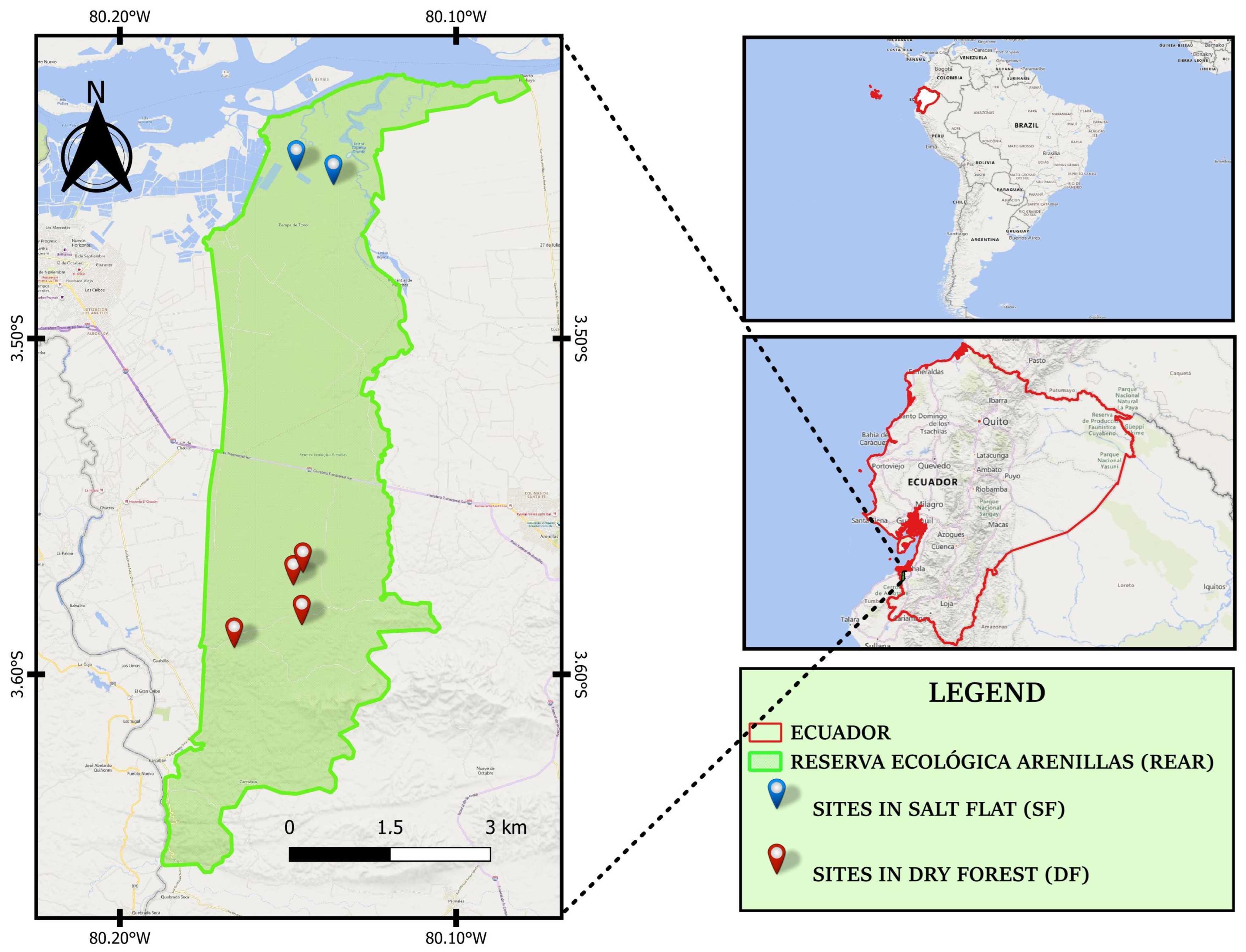
2.1. Sampling Area

Within the REAR, which is geographically bounded by the following coordinates: to the north at latitude −3.4316340 and longitude −80.1453494, to the south at latitude −3.6559638 and longitude −80.1795959, to the east at latitude −3.5436676 and longitude −80.1232910, and to the west at latitude −3.5436219 and longitude −80.1734161, six sampling sites were selected, and at each site, four 20 × 20 m (400 m2) plots were established, totaling 24 plots. Four of these sites were in the dry forest, while only two were established in the salt flat area (Figure 1), due to the limited extent of land with remaining vegetation, as shown in Figure S1A,B. The soil variability in the REAR is influenced by geomorphological processes such as alluvial and colluvial–alluvial deposition, as well as the presence of saline deposits. As a result, most soils are mineral in nature, except for an area in the northern part of the reserve that contains organic soil, as reported by Maza et al. [16].
The REAR has an elevation range of up to 300 m asl. The climate is classified as warm and dry, with an average temperature of 24 °C and varying levels of precipitation. There are three recognized climatic zones: warm–arid (less than 350 mm/year), warm–very dry (300–500 mm/year), and warm–dry (500–1000 mm/year) [17].
Four field trips were conducted in 2023, two in May (dry season) and two in July (rainy season). The sampling consisted of fungal search walks, covering 100% of each plot. Data on the fungi were recorded, including their shape, color, and growth substrate (such as soil, organic matter on the soil, fallen tree branches, logs, plant leaves, animal dung, among others). Photographs of the fungal fruiting bodies were also taken, capturing both the top of the cap (pileus) and the underside to identify gills, pores, and hymenophore characteristics. Samples were taken to the laboratory UTPL for further examination.

2.2. Specimen Identification

The identification of specimens was based on both macro- and microscopic characteristics, following the terminology of Largent [18] for fresh samples. Aspects such as the shape, texture, and color of different parts of the fruiting bodies, including ascomata, basidiomata, pileus, gills, pores, stipe, and other structures, were evaluated. Additionally, all specimens were examined microscopically, recording the forms of sexual and asexual spores, as well as reproductive structures such as asci, basidia, cystidia, and hyphae, according to the classification of Largent [18] and Hanlin [19]. The use of the nomenclature “cf.” indicates that the specimen shows morphological similarity to a known species, but its identification remains uncertain at the species level, particularly in the absence of phylogenetic confirmation. In contrast, the term “aff.” is used when a specimen closely resembles the holotype of a described species but likely represents a distinct, possibly undescribed, taxon.
The samples were observed using an Olympus BX51 microscope (Olympus Corporation, Tokyo, Japan) at 100× magnification. Microscopic photographs were taken using a ZEISS Axiocam 212 camera (Carl Zeiss, Oberkochen, Germany). Following microscopic analysis, most of the specimens collected during the sampling were deposited in the Fungarium (Table S1) of the Herbarium of the Universidad Técnica Particular de Loja (HUTPL).

2.3. Diversity Analysis

Alpha diversity, which reflects the diversity of species within a community, was evaluated by considering both species richness and abundance in the two ecosystems studied (DF and SF). For this purpose, the Simpson (D) [20] and Shannon (H’) [21] indices were used. Additionally, the non-parametric Mann–Whitney test was applied to compare species richness between the two forests type. Accumulation and rarefaction curves were generated using ACE and Chao2 [22,23] richness estimators to estimate sampling effort and the number of species recorded over time. To ensure a fair and comparable assessment of species diversity between habitats with different sampled areas, diversity calculations were based on standardized sampling effort, and rarefaction analysis was applied to account for differences in sampling size.
Differences in species composition between ecosystems were evaluated using Bray–Curtis dissimilarity and visualized through NMDS to explore compositional variations between the dry forest and salt flat. To assess the statistical significance of these differences, a PERMANOVA was conducted. All analyses were performed using R version 4.3.3 using “vegan” package [24].

3. Results and Discussion

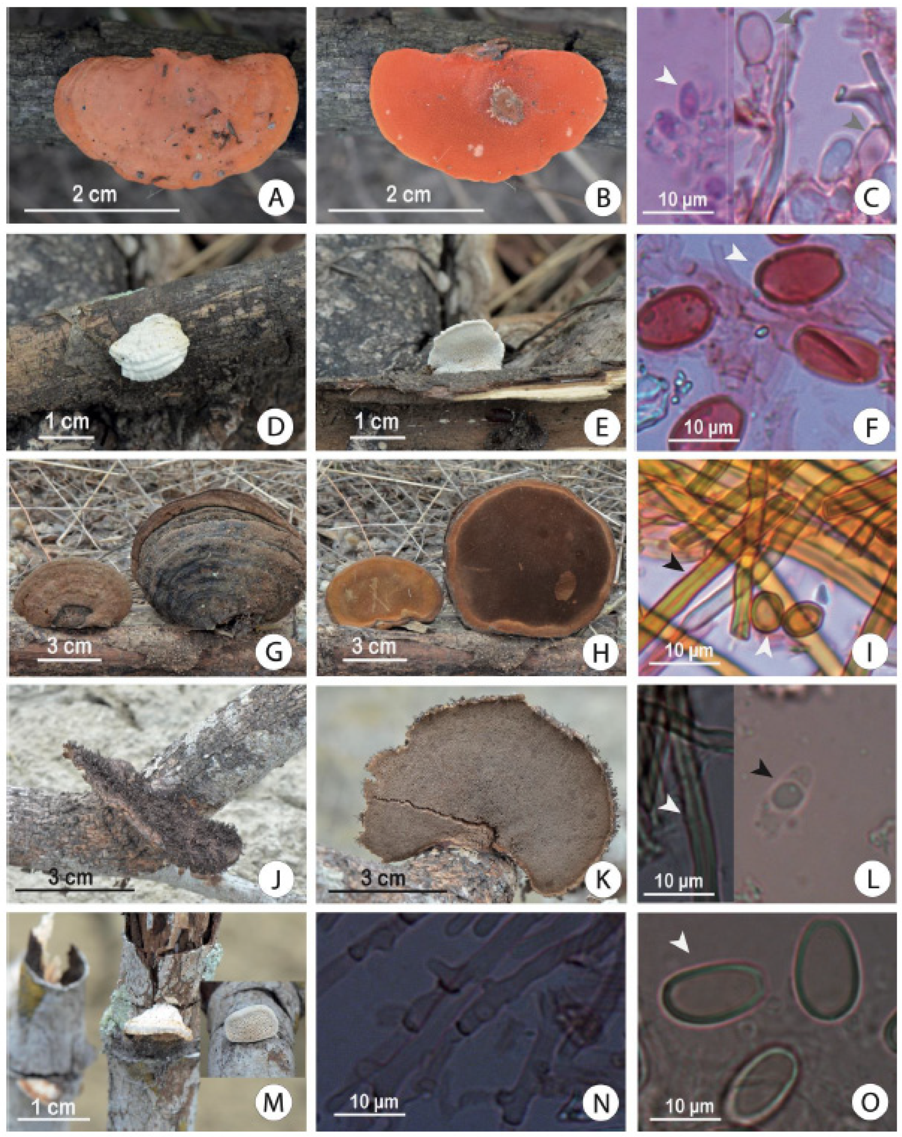
3.1. Morphological and Descriptive Diversity Recorded in the Dry Forest and Salt Flat

A total of 162 specimens corresponding to 47 species were recorded, mostly identified morphologically. These species belong to 34 genera. Additionally, nine specimens were grouped as unidentified into (Ascomycota incertae sedis, Dothidiomycetes incertae sedis, Polyporales incertae sedis, Basidiomycota incertae sedis with resupinate habit, and Ustilaginales incertae sedis.) due to insufficient morphological information for deeper taxonomic level assignment (Figure 2). The main limitation for identification was the absence of distinctive morphological features at certain developmental stages, combined with limited access to the molecular tools that could support morphological analysis. Additionally, the lack of reference data—such as taxonomic keys—particularly for understudied groups like resupinate Basidiomycetes in tropical regions, further hindered accurate classification (Figures S2 and S3, Table S1).
The DF exhibited the highest species diversity, with 37 species distributed in 28 genera (Figure 2). Basidiomycetes were the predominant group during the rainy season (Figure 3), which may be associated with the increased availability of woody substrates and decomposing organic matter resulting from higher moisture levels, conditions that favor their growth due to their lignocellulose-degrading capacity. In the SF ecosystem, 29 species belonging to 22 genera were recorded (Figure 2), with a greater representation of Ascomycetes during the dry season (Figure 3). This pattern may reflect seasonal differences in fruiting body production or environmental tolerance, though further studies would be needed to clarify the underlying mechanisms.
Studies in other tropical and subtropical ecosystems, such as India’s tropical dry evergreen biome, reveal notable differences in species richness and community composition, with a clear dominance of the phylum Basidiomycota (96%) over Ascomycota (4%). The most represented groups were the orders Agaricales (families Agaricaceae, Marasmiaceae) and Polyporales (Polyporaceae) [25], findings closely aligned with our own. In contrast, a study conducted in coastal mangrove ecosystems in Guyana reported low species richness (19 spp.) but also a high dominance of Basidiomycota (89%) [26], differing from our results in salt flats, where Ascomycota was slightly more dominant. Nevertheless, these environmentally extreme habitats consistently host wood-decaying fungi, particularly from the Polyporales and Agaricales orders. Macrofungal diversity and community composition vary significantly depending on forest or vegetation type, influenced by factors such as canopy structure, soil moisture, elevation [27], and habitat conservation status [25,26], reinforcing the importance of preserving mature vegetation and diverse woody substrates to sustain fungal ecological functions.
The species recorded in this study (Figure 2) were documented through photographs (Figure 4, Figure 5, Figures S2 and S3) and preserved in the HUTPL Herbarium (Table S1). Microscopic identification was possible for several species through the observation of reproductive structures such as ascospores and basidiospores (Figure 4 and Figure 5). However, a considerable number of specimens were either immature or degraded (Figures S2 and S3), which limited the ability to perform more detailed taxonomic analyses. Among the most representative species observed mainly on trunks in both forest types were Cerrena hydnoides, Chondrostereum sp., Hexagonia tenuis, and Pycnoporus sanguineus and several unidentified species, particularly Resupinate spp. (Figure 5, Figures S2 and S3), which lacked well-developed reproductive structures (e.g., spores).
Two noteworthy species, classified within the genus Truncospora, were designated as Truncospora aff. tropicalis and Truncospora cf. mexicana. The former was identified based on general morphological affinity, despite notable differences in spore size and the presence of a zonate, whitish pileus, contrasting with the pale ochre and non-zonate pileus typical of the type species [28]. The latter was assigned due to morphological similarities with T. mexicana, although it was found to be growing in apparent parasitic association with Laguncularia racemosa var. glabriflora, a relationship not previously reported for T. mexicana or T. tropicalis. Molecular data will be necessary to confirm the taxonomic placement of these species [29].
Although this study does not explore ecological responses, the persistence of these fungi in challenging or seasonal environments may be linked to their ability to form resistant fruiting bodies that can enter a dormant state through dehydration, as well as their efficiency in lignocellulose degradation [30]. In some cases, the strong melanization observed in Ascomycetes and Basidiomycetes might also represent an adaptive mechanism to cope with varying environmental conditions, as suggested by previous studies [31,32].
The diversity of plants, soils, logs, and leaf litter likely creates microhabitats that favor the growth and development of a greater variety of fungi, as evidenced in previous studies highlighting the influence of plant diversity on fungal species richness [14]. In contrast, lower diversity was observed in salt flats predominantly composed of Ascomycetes, which have been widely reported in halophilic areas of various regions, especially in areas of the Atlantic Ocean [17]. Additionally, the salt flats, with their nutrient-poor soils and high salinity, significantly limit fungal diversity [32].

3.2. Alpha Diversity

Analysis using the Mann–Whitney test reveals a significant difference in species richness between the two forest types, DF vs. SF, with a p-value of 0.0184, below the 0.05 threshold. This result suggests that the distribution of species richness varies between the forest types compared.
A total of 47 fungal species, primarily macrofungi, were recorded across the DF and SF ecosystems of the REAR. However, the DF exhibited a higher species richness, with 33 fungal species recorded (Figure 6). According to the ACE and Chao2 richness estimators, this number could increase to an estimated range of 47 to 52 species, taking into account the sampling effort. In contrast, the SF showed lower richness, with 23 species documented, and an estimated range of 24 to 25 species, based on the same estimators (Figure 6). Despite this, the documented diversity represents only a small fraction (1.37%) of the estimated potential fungal richness for the REAR, which is estimated at approximately 3429 species. This estimate is based on the ratio of nine fungi per plant species proposed by Hawksworth and Lücking [14], and the most recent record of 381 documented plant species in the REAR [4].
This species diversity is high according to the calculations using the Shannon (H’) and Simpson (D) indices for both forest types, DF and SF. The Shannon index shows values above H’ = 3, while the Simpson index shows values close to D’ = 1, confirming high diversity in both ecosystems (Table 1).

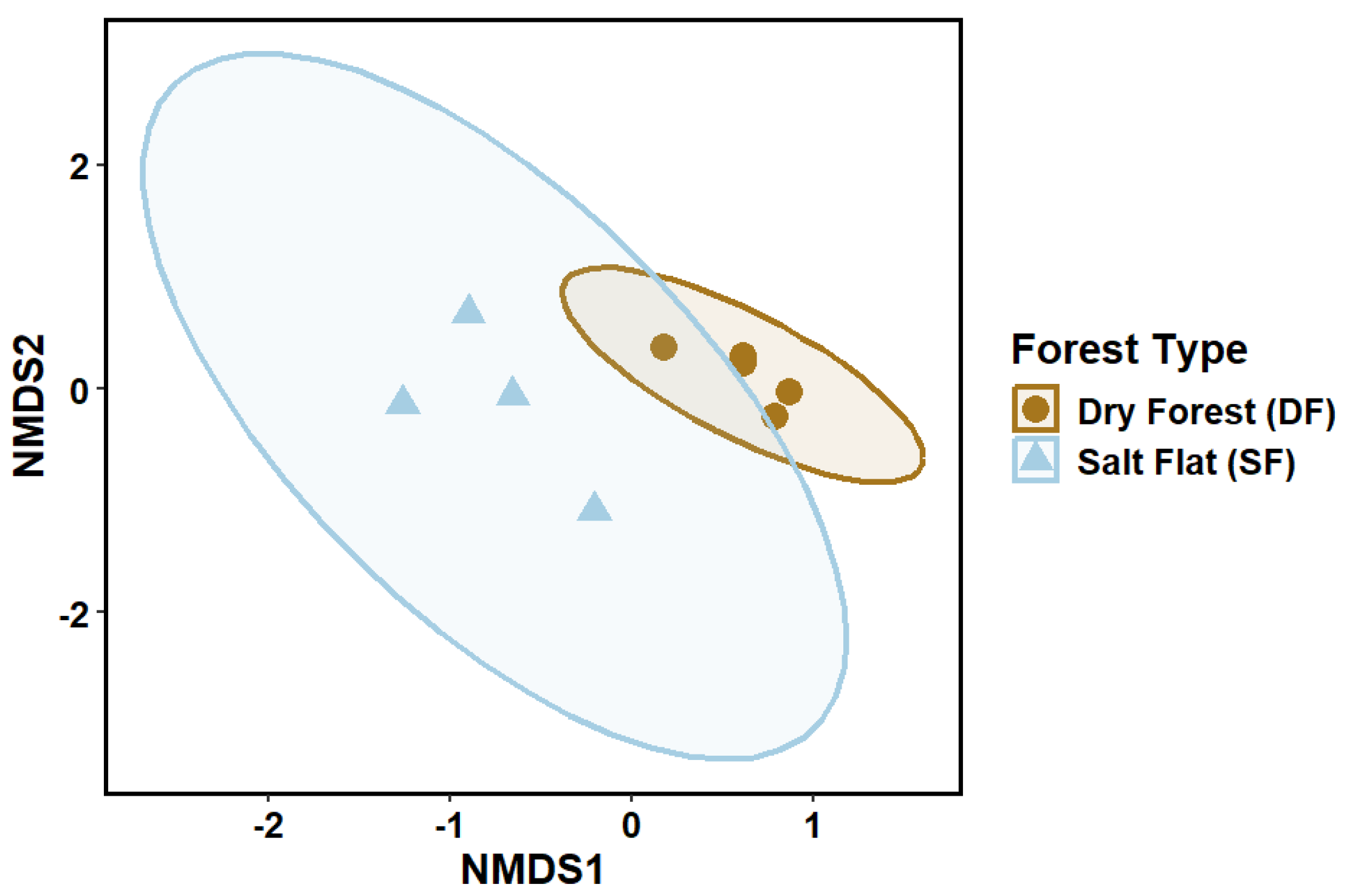
3.3. Ordination and Statistical Analysis

The NMDS based on Bray–Curtis distance revealed differences in species composition between the DF and SF ecosystems, with a stress value of 0.070 indicating a good two-dimensional representation. Ellipses also indicated that the SF exhibits greater variability in species composition, while the DF presents less variability and a more homogeneous community. In addition, the distribution of the points in the DF suggests a more uniform arrangement of species compared to that observed in the SF (Figure 7).
Statistically, the PERMANOVA analysis supports the results shown in the NMDS analysis, indicating that the DF and SF forest types have a significant difference in species composition in these ecosystems (Table 2).
The results of this study demonstrate that the REAR harbors a remarkable diversity of fungi in its ecosystems, especially in extreme areas like the salt flat, which, according to these data, act as an important biodiversity refuge. Given these findings, the ecosystem environments of the REAR must be conserved and protected from anthropogenic impacts, such as the expansion of agricultural areas and activities like shrimp farming [33], which are in high demand in this region.

4. Conclusions

Fungal diversity showed slight variation between ecosystems, with the dry forest exhibiting a minimally significant higher diversity, likely attributable to its greater structural complexity and substrate availability. Despite its harsher conditions, the salt flat supported resilient fungal taxa, particularly Ascomycetes. Both ecosystems contribute uniquely to the region’s overall fungal diversity and highlight the importance of targeted conservation efforts.

Supplementary Materials

The following supporting information can be downloaded at: https://www.mdpi.com/article/10.3390/d17060422/s1, Table S1. Checklist of fungi recorded in the Reserva Ecologica Arenillas (REAR) and corresponding voucher numbers in the Herbarium of the Universidad Técnica Particular de Loja (HUTPL); Figure S1. Forest types sampled in the REAR: (A,B) salt flat during the dry and rainy season; (C) dry forest during the dry season; (D) dry forest during the rainy season; Figure S2. Macroscopic visual guide for selected fungi recorded in the REAR: (A) Leucocoprinus birnbaumii; (B) Crinipellis trinitanis; (C) Chondrostereum sp.; (D) Humphreya sp.; (E) Schizophyllum commune; (F) Auricularia mesenterica; (G) Auricularia polytricha; (H) Xylaria sp.; (I) Cytospora rhizophorae; (J) Phlebopus cf. beniensis; (K) Phellinus sp.; Figure S3. Macroscopic visual guide for selected fungi recorded in the REAR: (A) Coriolopsis rigida; (B) Hexagonia tenuis; (C) cf. Lentinus; (D) Ascomycetes sp.; (E) Camillea sp.; (F) Xylaria sp.; (G) Ustilaginales sp.; (H) Resupinate sp. 3; (I) Rhytidhysteron sp.; (J) Resupinate sp. 1; (K) Resupinate sp. 2.

Author Contributions

D.C. conceived the research; D.C. and D.M. carried out the work of observation in the field; D.C. and D.M. wrote and critically reviewed the intellectual content of the manuscript; D.M. edited the tables and figures; D.C. took the photographs of all the figures; Á.B., F.L., and T.O. gave a strong critical revision of the manuscript. All authors contributed with their experience to improve the whole manuscript. All authors have read and agreed to the published version of the manuscript.

Funding

This research was funded by Universidad Técnica Particular de Loja with USD 2000 into the internal project PROY_INV_BA_2022_3623 named Diversidad de hongos y briofitos en bosques secos, manglares y salitrales del Sur de Ecuador.

Institutional Review Board Statement

Not applicable.

Informed Consent Statement

Not applicable.

Data Availability Statement

Data is contained within the article and Supplementary Materials.

Acknowledgments

We appreciate the availability of the Laboratory of Cultivation and Conservation of Microorganisms of the Universidad Técnica Particular de Loja.

Conflicts of Interest

The authors declare no conflicts of interest.

References

  1. Best, B.J.; Kessler, M. Biodiversity and Conservation in Tumbesian Ecuador and Peru; BirdLife International: Cambridge, UK, 1995. [Google Scholar]
  2. Mittermeier, R.A.; Turner, W.R.; Larsen, F.W.; Brooks, T.M.; Gascon, C. Global biodiversity conservation: The critical role of hotspots. In Biodiversity Hotspots: Distribution and Protection of Conservation Priority Areas; Springer: Berlin/Heidelberg, Germany, 2011; pp. 3–22. [Google Scholar]
  3. Rivas, C.A.; Guerrero-Casado, J.; Navarro-Cerillo, R.M. Deforestation and fragmentation trends of seasonal dry tropical forest in Ecuador: Impact on conservation. For. Ecosyst. 2021, 8, 46. [Google Scholar] [CrossRef]
  4. Luna-Florin, A.D.; Nole-Nole, D.A.; Rodríguez-Caballero, E.; Molina-Pardo, J.L.; Giménez-Luque, E. Ecological Characterization of the Flora in Reserva Ecológica Arenillas, Ecuador. Appl. Sci. 2022, 12, 8656. [Google Scholar] [CrossRef]
  5. Cerón, C.; Reyes, C. Características botánicas de la Reserva Militar y Ecológica Arenillas, El Oro-Ecuador. Cinchonia 2006, 7, 115–130. [Google Scholar]
  6. Espinosa, C.I.; Jara-Guerrero, A.; Cisneros, R.; Sotomayor, J.-D.; Escribano-Ávila, G. Reserva Ecológica Arenillas; ¿un refugio de diversidad biológica o una isla de extinción? Ecosistemas 2016, 25, 5–12. [Google Scholar] [CrossRef]
  7. Tamayo-Cevallos, R. Lignicolous marine fungi in mangrove ecosystem: State of knowledge and research perspective in Ecuador. Rev. Ecuat. De Med. Y Cienc. Biol. 2020, 41, 95–106. [Google Scholar]
  8. Cruz, D.; Masache, D. Listado y guía visual preliminar de macrohongos del Bosque Petrificado de Puyango-Ecuador. ACI Av. En Cienc. E Ing. 2023, 15, 2939. [Google Scholar] [CrossRef]
  9. Piepenbring, M.; López, F.; Cáceres, O. La importancia de los Hongos en los Ecosistemas. Puente Biológico 2016, 8, 57–91. [Google Scholar]
  10. Ghate, S.D.; Sridhar, K.R. Contribution to the knowledge on macrofungi in mangroves of the Southwest India. Plant Biosyst.-Int. J. Deal. All Asp. Plant Biol. 2016, 150, 977–986. [Google Scholar] [CrossRef]
  11. Dutta, A.K.; Pradhan, P.; Basu, S.K.; Acharya, K. Macrofungal diversity and ecology of the mangrove ecosystem in the Indian part of Sundarbans. Biodiversity 2013, 14, 196–206. [Google Scholar] [CrossRef]
  12. Luna-Fontalvo, J.A.; Abaunza, C.; Barrios, A.; Ramírez-Roncallo, K.; Guerrero, R.J.; Negritto, M.A. New records of agaricoid macrofungi (Agaricales, Basidiomycota) in an urban fragment of tropical dry forest from Colombian Caribbean Region. Check List 2023, 19, 371–379. [Google Scholar] [CrossRef]
  13. Rodríguez-Zúñiga, M.T.; Troche-Souza, C.; Vázquez-Lule, A.D.; Márquez-Mendoza, J.D.; Vázquez-Balderas, B.; Valderrama-Landeros, L.; Velázquez-Salazar, S.; Cruz-López, M.I.; Ressl, R.A.; Uribe-Martínez, A.; et al. Manglares de México/Extensión, Distribución y Monitoreo; Comisión Nacional para el Conocimiento y Uso de la Biodiversidad: México City, Mexico, 2013; p. 128. [Google Scholar]
  14. Hawksworth, D.L.; Lücking, R. Fungal Diversity Revisited: 2.2 to 3.8 Million Species. Microbiol. Spectr. 2017, 5, 1–17. [Google Scholar] [CrossRef] [PubMed]
  15. Moreira, N.M.; Chacón, N.V.; Flor, J.P.; Tamayo, J.L.; de María Valverde, F. Composición Florística y Nuevos Registros para la Reserva Ecológica Arenillas, El Oro-Ecuador. Investigatio 2016, 8, 111–132. [Google Scholar] [CrossRef]
  16. Maza Maza, J.E.; Sánchez Asanza, A.W.; Añazco Loaiza, H.E.; García Ochoa, J.A. Estudio de las carac-terísticas físico-químicas del suelo en áreas prioritarias de la reserva ecológica Arenillas. Rev. Científica Agro-Ecosistemas 2021, 9, 30–40. [Google Scholar]
  17. Ministerio del Ambiente. Plan de Manejo de la Reserva Ecológica Arenillas; Ministerio del Ambiente: Quito, Ecuador, 2015; 68p.
  18. Largent, D. How to Identify Mushrooms to Genus, I. Macroscopic Features; Mad River Press: Eureka, CA, USA, 1973; 125p. [Google Scholar]
  19. Hanlin, R.T. Illustrated Genera of Ascomycetes; APS Press: St Paul, MN, USA, 1990. [Google Scholar]
  20. Simpson, E.H. Measurement of diversity. Nature 1949, 168, 688. [Google Scholar] [CrossRef]
  21. Shannon, C.E.; Weaver, W. The Mathematical Theory of Communication; University of Illinois Press: Urbana, IL, USA, 1964; pp. 1–117. [Google Scholar]
  22. Chao, A. Nonparametric estimation of the number of classes in a population. Scand. Stat. Theory Appl. 1984, 11, 265–270. [Google Scholar]
  23. Colwell, R.K.; Coddington, J.A. Estimating terrestrial biodiversity through extrapolation. Philos. Trans. R. Soc. Lond. B Biol. Sci. 1994, 345, 101–118. [Google Scholar] [CrossRef]
  24. Oksanen, J.; Blanchet, F.G.; Kindt, R.; Legendre, P.; Minchin, P.R.; O’Hara, R.B.; Simpson, G.L.; Solymos, P.; Stevens, M.H.H.; Wagner, H.H. vegan: Community Ecology Package (R package version 2.5-5). 2019. Available online: https://CRAN.R-project.org/package=vegan (accessed on 10 February 2025).
  25. Priyamvada, H.; Akila, M.; Singh, R.K.; Ravikrishna, R.; Verma, R.S.; Philip, L.; Marathe, R.R.; Sahu, L.K.; Sudheer, K.P.; Gunthe, S.S. Terrestrial Macrofungal Diversity from the Tropical Dry Evergreen Biome of Southern India and Its Potential Role in Aerobiology. PLoS ONE 2017, 12, e0169333. [Google Scholar] [CrossRef]
  26. Sharif, W.R.; Da Silva, P.N.B. Macrofungal Diversity in Selected Coastal Mangrove Ecosystems in Guyana, South America. Eur. Mod. Stud. J. 2023, 7, 38. [Google Scholar] [CrossRef]
  27. Huang, J.-M.; Xu, T.-M.; Zhao, W.; Mumin, R.; Zeng, L.; Sun, Y.-F.; Cui, B.-K. Species Diversity and Community Composition of Macrofungi in the Dongling Mountains, Western Beijing, China. J. Fungi 2025, 11, 155. [Google Scholar] [CrossRef]
  28. Schoch, C.L.; Sung, G.H.; López-Giráldez, F.; Townsend, J.P.; Miadlikowska, J.; Hofstetter, V.; Robbertse, B.; Matheny, P.B.; Kauff, F.; Wang, Z.; et al. The Ascomycota tree of life: A phylum-wide phylogeny clarifies the origin and evolution of fundamental reproductive and ecological traits. Syst. Biol. 2009, 58, 224–239. [Google Scholar] [CrossRef]
  29. Oberwinkler, F. Evolutionary trends in Basidiomycota. Stapfia 2012, 96, 45–104. Available online: https://www.zobodat.at/pdf/STAPFIA_0096_0045-0104.pdf (accessed on 10 February 2025).
  30. Shen, C.; Wang, J.; He, J.Z.; Yu, F.H.; Ge, Y. Plant Diversity Enhances Soil Fungal Diversity and Microbial Resistance to Plant Invasion. Appl. Environ. Microbiol. 2021, 87, e00251-21. [Google Scholar] [CrossRef] [PubMed]
  31. Calabon, M.S.; Jones, E.B.G.; Promputtha, I.; Hyde, K.D. Fungal Biodiversity in Salt Marsh Ecosystems. J. Fungi 2021, 7, 648. [Google Scholar] [CrossRef] [PubMed]
  32. Bai, X.; Zhang, E.; Wu, J.; Ma, D.; Zhang, C.; Zhang, B.; Liu, Y.; Zhang, Z.; Tian, F.; Zhao, H.; et al. Soil fungal community is more sensitive than bacterial community to modified materials application in saline-alkali land of Hetao Plain. Front. Microbiol. 2024, 15, 1255536. [Google Scholar] [CrossRef]
  33. Biológica, S.D. La Biodiversidad y la Agricultura: Salvaguardando la Biodiversidad Y Asegurando Alimentación Para El Mundo; Secretaría del Convenio Sobre Diversidad Biológica: Montreal, QC, Canada, 2008. [Google Scholar]
Figure 1. Geographic location map of the Reserva Ecologica Arenillas and sampling site’s locations.
Diversity 17 00422 g001
Figure 2. Frequency of identified and unidentified fungal species by season and forest type in the REAR. Capital letters preceding the taxon names indicate higher taxonomic ranks assigned to unidentified specimens: C. = clase, P. = phylum, O. = order. Specimens labeled as Basidiomycota Incertae sedis represent distinct morphotypes with a resupinate growth habit.
Diversity 17 00422 g002
Figure 3. Distribution of the total frequency of fungi by phyla according to the type of forest and season.
Diversity 17 00422 g003
Figure 4. Macroscopic and microscopic visual guide for selected fungi recorded in the REAR: (AC) Pycnoporus sanguineus. (A,B) Pileus and hymenophore. (C) Ellipsoidal basidiospores (white arrowhead) and immature basidia (gray arrowhead). (DF) Truncospora aff. tropicalis. (D,E) Pileus and hymenophore. (F) Ellipsoidal and truncated basidiospores (white arrowhead). (GI) Phellinus sp. (G,H) Pileus and hymenophore. (I) Subglobose basidiospores (white arrowhead) alongside skeletal hyphae (black arrowhead). (JL) Cerrena hydnoides. (J,K) Pubescent pileus and hymenophore. (L) Ellipsoidal basidiospore (white arrowhead) and skeletal hyphae (black arrowhead). (MO) Truncospora cf. mexicana. (M) Pileus and hymenophore growing on Laguncularia racemosa var. glabriflora. (N) Branched hyphae. (O) Ellipsoidal and truncated basidiospores (white arrowhead).
Diversity 17 00422 g004
Figure 5. Macroscopic and microscopic visual guide for selected fungi recorded in the REAR: (AC) Hymenochaete rubiginosa. (A) Hairy, effuse-flattened basidioma. (B) Ellipsoidal to subcylindrical basidiospore (arrowhead). (C) Cystidium (white arrowhead) and skeletal hyphae (black arrowhead). (D,E) Calvatia cf. cyathiformis. (D) Basidioma with violet gleba. (E) Round, ornamented spores (white arrowhead) and capillitium (black arrowhead). (F,G) Rhytidhysteron cf. rufulum. (F) Apothecial ascomata. (G) Dark ascospores. (H,I) Pleospora herbarum. (H) Ascomata. (I) Mature muriform ascospore (black arrowhead). (JL) Platystomum sp. (J) Perithecium. (K) Ascospores (black arrowhead). (L) Ascus containing eight ascospores. (MO) Hypoxylon sp. (M) Ascocarp. (N) Dark fusoid ascospores with a central guttule. (O) Eight-spored asci (white arrowhead).
Diversity 17 00422 g005
Figure 6. Species accumulation and rarefaction curves for the dry forest (DF) and salt flat (SF), including richness estimates based on ACE (purple lines and points), Chao2 (gray lines and points), and observed fungal species richness (green points).
Diversity 17 00422 g006
Figure 7. Non-metric multidimensional scaling of fungal community composition by sampling plot in dry forest (circles) and salt flat (triangles). The stress value (0.070) indicates a reliable two-dimensional representation of the data.
Diversity 17 00422 g007
Table 1. Diversity indices for different forest types.
Forest TypeShannon (H’)Simpson (D)
DF3.240.95
SF2.970.94
Table 2. PERMANOVA results. Df = degrees of freedom; SS = sum of squares; R2 = coefficient of variation; F = F-statistics.
DfSSR2Fp-Value
Season10.425650.187781.95200.93
Forest type10.605640.267182.77750.012 *
Season: Forest Type10.145190.064050.66580.786
Residual51.090270.48099
Total82.266741.00000
* = Value showing significant difference.
Disclaimer/Publisher’s Note: The statements, opinions and data contained in all publications are solely those of the individual author(s) and contributor(s) and not of MDPI and/or the editor(s). MDPI and/or the editor(s) disclaim responsibility for any injury to people or property resulting from any ideas, methods, instructions or products referred to in the content.

