Article

Fungal Associates of the Moss Leucobryum candidum (Brid. ex P. Beauv.) Wilson in Southeast Queensland, Australia

by
Lana Valeska Misic
1,
Alison Shapcott
1,
Andrew J. Franks
2 and
D. İpek Kurtböke
1,*
1
School of Science, Technology and Education, University of the Sunshine Coast, Maroochydore BC, QLD 4558, Australia
2
GHD Pty Ltd., 145 Ann Street, Meanjin, Brisbane, QLD 4000, Australia
*
Author to whom correspondence should be addressed.
Diversity 2025, 17(6), 370; https://doi.org/10.3390/d17060370
Submission received: 11 March 2025 / Revised: 15 April 2025 / Accepted: 24 April 2025 / Published: 22 May 2025
(This article belongs to the Special Issue Women’s Special Issue Series: Diversity)

Abstract

:
The suite of fungi that associate with mosses, bryophilous fungi, can be explored further to provide insights into the symbiotic functionality of mosses as well as the ecosystems in which they reside. So far, in-depth studies on the taxonomic diversity, ecology, and physiological functions of bryophilous fungi associated with the Australasian moss species Leucobryum candidum are lacking. To generate information on the physiology, biology, and ecology of these organisms and their interactions with the moss host, the combined use of selective isolation and molecular characterisation of the fungal associates was carried out. Once the pure cultures of the fungal associates were obtained, a bioactivity assay was used to investigate the effect of fungal metabolites on moss growth in vitro. Overall, L. candidum species collected from six different locations within Southeast Queensland exposed to different environmental parameters were found to have a highly diverse community of fungal species from 10 orders and 17 families. A total of 25 of the 33 isolates that were identified using molecular sequencing techniques were unique species, confirming high beta diversity of the fungal associates of L. candidum collected from coastal, forest, and urban environments in Southeast Queensland. The highest numbers of culturable isolates came from coastal and forest sites. Urban sites accounted for the lowest numbers of culturable isolates. The taxonomic matches for these associates were known to have diverse endophytic, saprophytic, and parasitic roles within vascular plants. Selected moss samples were inoculated with fermentation extracts of fungal isolates (USC-F426 and USC-F427) and their effects on the moss samples were observed for any change in heights, weights, diameters, and morphological characteristics. A significant (p ≤ 0.05) difference in the heights of the in vitro-assessed L. candidum between treatments was observed. No significant differences, however, were seen between the weights and diameters and no discernible host symptoms were detected, other than a few morphological change observations.

1. Introduction

Fungi form important mutualistic or parasitic symbiotic relationships with vascular and non-vascular plants [1,2,3,4,5,6]. Such symbiotic interactions between plants and fungi are thought to have enhanced the nutrient uptake strategies of charophytes (green algae) and contributed to the divergence of bryophytes (non-vascular land plants, e.g., mosses) from their green algal ancestors [7,8], thus aiding the evolutionary advances that led to the colonisation of land by plants [7,8]. Bryophytes and the fungi in association with them perform various ecosystem services that are integral for the healthy functioning of ecosystems [1,9,10,11,12,13,14,15,16,17,18]. Fungi alone provide ecosystem services that are vital for the functioning of natural and non-natural ecosystems and form important symbiotic associations with bryophytes [18,19]. These interactions and services are so numerous that it would be difficult to estimate their economic and ecological value [20,21,22].
Bryophilous symbiosis with fungi can maintain plant nutrition, health, and quality, but also enhances micro fungi-driven carbon sequestration, ecosystem functioning, and nutrient cycling [23,24]. Despite these important roles in sustaining environmental health and their biopotential across multiple disciplines, limited scientific research has focused on bryophytes, particularly mosses, and their reservoir of undescribed fungal species [15]. Research so far has mostly focused on fungal associates of liverworts or mosses within certain ecosystems such as peat bogs [25] or the Antarctic tundra [26].
Advances in microbiology, such as culture-based and non-culture-based molecular approaches, currently provide the most accurate methods of assigning taxonomy to fungi isolated from plant material and categorising fungal species into phylogenies [27,28]. Whilst an association of a diversity of fungi, particularly ascomycetes and agaricomycetes, with mosses has been extensively identified [3,13,16], currently advancing molecular methods are yet to reveal many more fungal species associated with mosses such as the Australasian moss Leucobryum candidum (Brid. ex P. Beauv.) Wilson.
Mosses are distributed across all terrestrial biomes and certain species are common throughout Australasian regions such as L. candidum [29] which is a species of both scientific and historical interest generally found in moist, forested environments [29]. However, very few if any Australian bryomycological studies focus on this species. A study by Michel et al. (2011) [30] analysed the species–environment relationships of several New Zealand mosses and revealed that L. candidum was unique in positively responding to distribution models with increasing temperatures and slope, signifying a potential preference of warmer, well-drained forested environments. In contrast, a study by Downing and Marner (1998) [29] suggested Leucobryaceous taxa prefer moist, forested environments, often growing on the substrate of rotting logs, sometimes forming large mats or cushions in unison with Pyrrhobryum species. The dissimilarity between recorded habitat preference may suggest that the species can thrive in a variety of widespread environments that may thus exhibit differing fungal associates.
In the light of the above presented information, fungal associates of L. candidum within Southeast Queensland were investigated across a variety of environments. This study aimed to determine whether the composition and diversity of fungal symbionts vary across different external environments and to evaluate whether some of these fungi have a beneficial effect on the growth of L. candidum through the metabolites they produce.

2. Materials and Methods

2.1. Background: Subject Moss

Leucobryum candidum is a species of moss belonging to the Leucobryaceae family, within the order Dicranales [29]. Members of Leucobryum have a Leucobryaceous leaf anatomy that consists of a broad costa occupying almost the entire leaf width. In cross section, the costa consists of a single layer of chlorophyllose cells (chlorocysts) sandwiched between layers of empty hyaline cells (leucocysts). Leucocysts are large, empty, thin walled, often colourless water storage cells that are dead at maturity. Five Leucobryum species are recorded from Australia with L. candidum being the most widespread.
Recorded mainly along the east coast of Australia and Tasmania, L. candidum forms mats on rotting logs, soft bark, and occasionally tree fern trunks. It has been recorded from rainforests and wet forests but also extends into drier eucalypt forests [29]. The anatomy, distribution, and ecology of L. candidum make it an ideal subject species to investigate its fungal associates.

2.2. Study Sites and Sampling Methods

L. candidum specimens were sampled from the uniform substrate of rotting logs from six sites in Southeast Queensland (SEQ) from each of the following three landscape types: urban, coastal, and forested to capture the diversity across different environments (Table 1). Two samples were taken from each location with two specimen collections occurring from each. This resulted in a total of 12 specimens with four replicate samples in each of the environment types. The three landscape or environment types all supported eucalypt open forest but were categorised differently: large, unfragmented forests (forested), urban forest within 30 m of a housing development (urban), and coastal eucalypt forests (coastal). To avoid contamination, gloves were worn to collect 5 × 5 cm specimens with a sterilised fork before transferring them into bags. Samples were placed in ice for preservation and transportation back to the University of the Sunshine Coast (UniSC) laboratory. Voucher specimens were lodged (for all samples except 6A and 6B) in the Queensland Herbarium (BRI) under the following BRI AQ accession numbers: 1002521, 1002522, 1002523, 1002525, 1002526.

2.3. Fungal Isolation and Preservation

Each sample of L. candidum was surface-sterilised using the method described by El-Tarabily et al. (1997) [31] and four small sections of the sterile specimen were transferred onto Potato Dextrose Agar (PDA) (OXOID, Brisbane, Australia) plates supplemented with streptomycin sulphate (70 ppm) to prevent bacterial growth (Bakerspigel and Miller 1953) [32]. After 14 days of incubation at 22 °C, plugs were cut from emerging fungal hyphae from the moss specimens and were transferred to a new set of PDA plates with sterile loops. Isolates were separated by site and categorised into morphotypes based on their gross morphology. Purified fungal isolates were subsequently grown in PDA slants in McCartney bottles and once fully grown, they were covered with paraffin oil (Astral Scientific Pty Ltd., Taren Point, NSW, Australia) and stored at room temperature (25 to 30 °C).

2.4. Molecular-Level Characterisation of Fungal Isolates

2.4.1. DNA Extraction and PCR

Fungal thallus (50–100 mg) from each isolate was placed in rounded Eppendorf tubes (1.2 mm) with small quantities of 2.3 mm and 0.1 mm zirconia silica grinding beads (Biospec, DainTree Scientific, St Helens, TAS, Australia) for the optimal isolation of nucleic acids from fungal isolates [33]. Samples were frozen in liquid nitrogen then placed into a Qiagen TissueLyser Adapter (Qiagen, Clayton, VIC, Australia; Thermo Scientific, Scoresby, VIC, Australia) set (2 × 5) on a program of 23,000 RPM for 40 s. The positions of the rotors were alternated in direction six times between liquid nitrogen freezing to ensure efficient grinding. Isolates were extracted using a Qiagen DNeasy Plant Kit (Qiagen, Australia) following the manufacturer’s instructions. DNA extractions were tested for their quality and quantity of DNA (ng) via the NanoDropTM (Thermo Fisher Scientific, Scoresby, VIC, Australia).
Polymerase chain reaction (PCR) was used to amplify the ITS region of the highly conserved 18S rRNA fungal DNA as the (ITS) region has the highest probability of successful identification of the most inclusive range of fungal species [33]. The standard forward ITS1 (5′-TCCGTAGGTGAACCTGCGG-3′) and reverse ITS4 (5′-TCCTCCGCTTATTGATATGC-′3) primers [34] were used. The following equation was used to calculate an average of the minimum in silico annealing temperatures of the primers (58 °C): 4 × (number of G + number of C) + 2 × (number of A + number of T).
After trialling various quantities and durations of the reactions, a standard PCR method was used with the addition of Trehaleose to optimise PCR amplification [34]. The following quantities were pipetted into sterile PCR tubes to make a total reaction volume of 40 μL: 20 μL of Bioline PCR ready mix, 2 μL of ITS1 (Forward Primer), 2 μL of ITS4 of ITS1 (Forward Primer), 2 μL of ITS4 (Reverse Primer), 10 μL DNA template, and 6 μL of Trehelose. Thermal cycling (Bio-Rad T100 Thermocycler, McHugh Electronics, Caboolture, QLD, Australia) was programmed to run for preheat activation (95 °C for 3 min) followed by 35 cycles of denaturation (95 °C for 1 min), annealing (58 °C for 1 min), extension (72 °C for 12 min), a post-cycle extension (72 °C for 10 min), and held infinitely at 12 °C upon completion.
The results were visualised by loading 5 μL of PCR amplicon and 2 μL of loading dye in a 1% agarose gel stained with Ethidium Bromide at 110 V for 25 min. Images of gels were taken with the BIORAD ChemiDocTM XRS+ System and Image Lab Software (Version 3). A 100 bp DNA ladder was used to quantify the approximate sizing of double-stranded DNA fragments with positive results being considered as the bands that fluoresced at the estimated fragment size of 600 bp. To ensure the quality of the amplicons, negative controls containing only the master mix or H2O were included within each PCR cycle and gel electrophoresis procedure.

2.4.2. Sequencing Amplicons

A minimum of 30 μL of each PCR product for 37 samples was pipetted into a 96-well plate (Axygen, Fisher Biotec, Perth, WA, Australia) and submitted to Macrogen, South Korea, (http://dna.macrogen.com/eng/) (accessed on April 2019) for purification and Sanger sequencing [35]. The outputs were imported into the software program Geneious (Version 8) (https://www.geneious.com) (accessed on May, 2019) so that they could be visualised, edited, and trimmed before using the de novo assemble tool to create contigs of the forward and reverse sequences. The final consensus sequences were edited and trimmed in Geneious to enhance their quality before performing Basic Local Alignment Search Tool (BLAST, Version 3). The BLAST search was conducted within the National Centre for Biotechnology Information (NCBI) database to compare nucleotide sequences to group databases and compute the measurable statistical significance of matches as well as using the MycoBank database (http://www.mycobank.org) (accessed on June 2019) to surmise useful and evolutionary connections between fungal successions and compare sequence matches. The combination of DNA-based molecular methods and use of multiple databases allowed for a more accurate categorisation and differentiation between the species, sub-species, and genera [34]. Sequences with high similarity (>97%) to GenBank–EMBL accessions were considered nonchimeric [36] and thus 3% variance was within species level. A phylogenetic tree (Supplementary Figure S3) was constructed using the Geneious program Version 8.0 (https://www.geneious.com/). The alignment was achieved using the Multiple Sequence Comparison by Log-Expectation (MUSCLE) (https://www.ebi.ac.uk/jdispatcher/msa) alignment tool within Geneious using the standard MUSCLE align settings [37,38]. The alignment was trimmed at both ends to ensure only the highest quality of each sequence remained (approx. 800 bp). For data exploration, a preliminary neighbour-joining tree and an Unweighted Pair Group Method with Arithmetic Averaging (UPGMA) tree were created. UPGMA adequately represented the data and showed bootstrap values for each branch. The consensus UPGMA tree settings were as follows: Tamura–Nei distance metric with bootstrap resampling of 100 replications. The output was selected to be visualised as a cladogram tree within the Geneious program Version 8.0 (Biomatters) and later exported as an image file. The phylogenetic tree was reconstructed (Figure 1) using the maximum likelihood (ML) method by PhyML program (via SeaView program [39]. The sequences of USC-F425, USC-F432, USC-F404, USC-F413, USC-F400, USC-F434, USC-F411, USC-F427, USC-F436, USC-F423, USC-F408, and USC-F410 were aligned using MUSCLE program version 3.8.31 [38] via SeaView program version 4.6.3 [39,40]. The analysis performed with the “GTR+Gamma” nucleotide substitution model and bootstrap analysis of the tree was performed by 1000 replications of pseudodata.

2.5. In Vitro Culture Assay
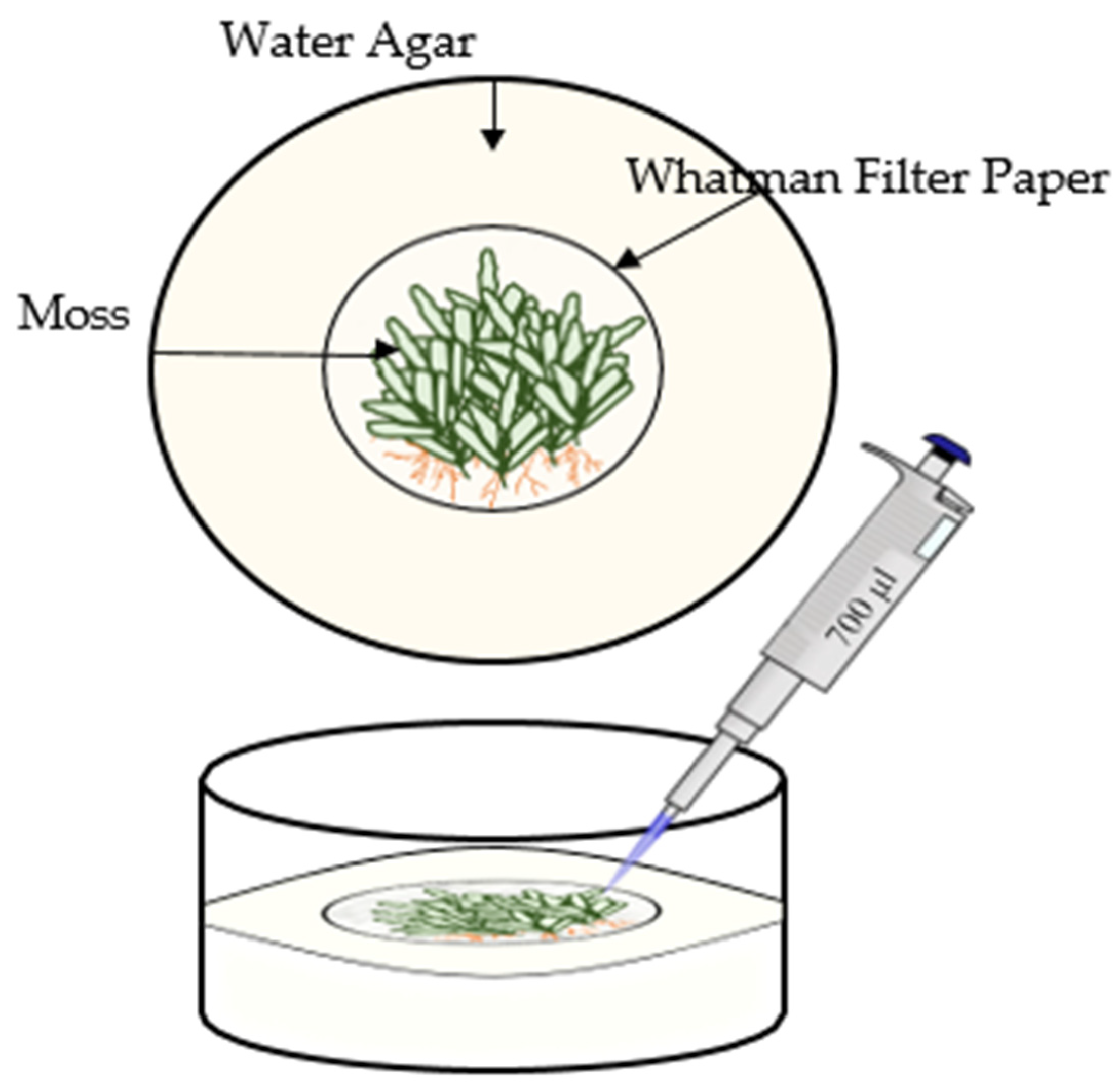
For the in vitro culture assays, Potato Dextrose Broth (PDB) (Oxoid, Australia) and a moss-based broth (MB) was inoculated with fungal isolates and fermented to test a medium close to the environment (Table 2). To prepare the MB, Sphagnum moss (Brunnings, Australia) was used due to the anatomical similarities it shares with L. candidum and its availability as a sterile, dried material (Bunnings, Australia). In a 2 L beaker, 250 mg of dried sphagnum moss was suspended in 500 mL of Milli Q H2O. The suspension was brought to boiling point in a microwave then put in a 99.9 °C water bath for 30 min. The lysate was separated, decanted, and filter sterilised. The moss broth medium was then prepared with 5.31 g glucose, 2.81 g yeast, and 375 mL of H2O and added to the 275 mL of moss extract and was autoclaved for 40 min at 121 °C. Both media were inoculated with five 5 × 5 mm plugs of Isolate USC-F426. This was replicated with isolate USC-F427 (2). The second culture flasks of each broth were inoculated with three 5 × 5 mm plugs of both isolates and placed in the orbital shaker/incubator at 25 °C 90 RPM for 7 days. The liquid was transferred to 50 mL Eppendorf tubes and put in an ultra-centrifuge (Thermo ScientificTM Sorvall RC6t centrifuge, Thermo Scientific, Scoresby, VIC, Australia) at 10,000 RPM and spun for 10 min. The extract was then filter-sterilised and stored at −4 °C immediately and −20 °C for longer-term storage.

2.6. Bioassay

Moss samples used in the assay were prepared by removing underlying debris and soil via rinsing in sterile Milli Q water three times and then cutting sections 9 cm in diameter by scalpel. Each moss sample was then placed in a Petri dish and soaked in an antibiotic solution containing Nystatin, Cycloheximide, Nalidixic acid, and Streptomycin in equal concentrations (50 ppm) to remove surface microorganisms. This was repeated using a new sterile Petri dish twice more with soaking durations of 1 min. The moss samples were removed and blotted dry with sterile Whatman filter paper (grade 1) then rinsed thoroughly with sterile Milli Q water. Sections of each specimen (3 cm in diameter) were cut by scalpel and placed into 70 × 40 mm glass crystallising dishes with 1.5 cm of water agar (15 g of agar suspended in 1 L of Milli Q and autoclaved at 121 °C for 40 min). A layer of Whatman filter paper was pre-soaked in 700 μL of the treatment and placed between the agar medium and the moss sample (Figure 2). Three replicates of each of the nine treatments equated to a total of twenty-seven experiments within the assay. Glass lids were applied onto the deep Petri dishes used during the experiment followed by parafilm sealing to prevent airborne contamination in the laboratory and placed in an incubator (Sanyo Incubator MIR-253, Australia) operating at 28 °C with light cycles of 16 h light/8 h dark. Fermentation broth samples (700 μL) were added to all settings every three days except controls, which had sterile water of equal amount.
At the beginning and the completion of the assay (42 days of incubation), the heights, weights, and diameters of each moss culture were recorded. Height was recorded as the total height (cm) of the gametophyte from the agar surface. Total weight of the culture assay was measured to the nearest gram before and after incubation. The difference was calculated and added extract (0.7 g), and H2O (0.7 g) weights were subtracted. Diameter (cm) was measured as the area of gametophyte growth on the agar surface. A statistical two-way ANOVA test was performed within the IBM SPSS Statistics (Version 25.0) to assess significant changes in height, weight, and diameter before and after the incubation period as well as between treatments. The means and standard deviations of the combined result from each replicate and treatment were also calculated. To repeat and refine the experiment, single shoots of sterile L. candidum were placed within the agar and were directly inoculated with the treatment. Protocols outlined in the above section were followed except for the following: zirconia silica beads were soaked in the treatment and imbedded into the agar underneath the placement of the gametophyte shoot (five per crystallising dish).

3. Results

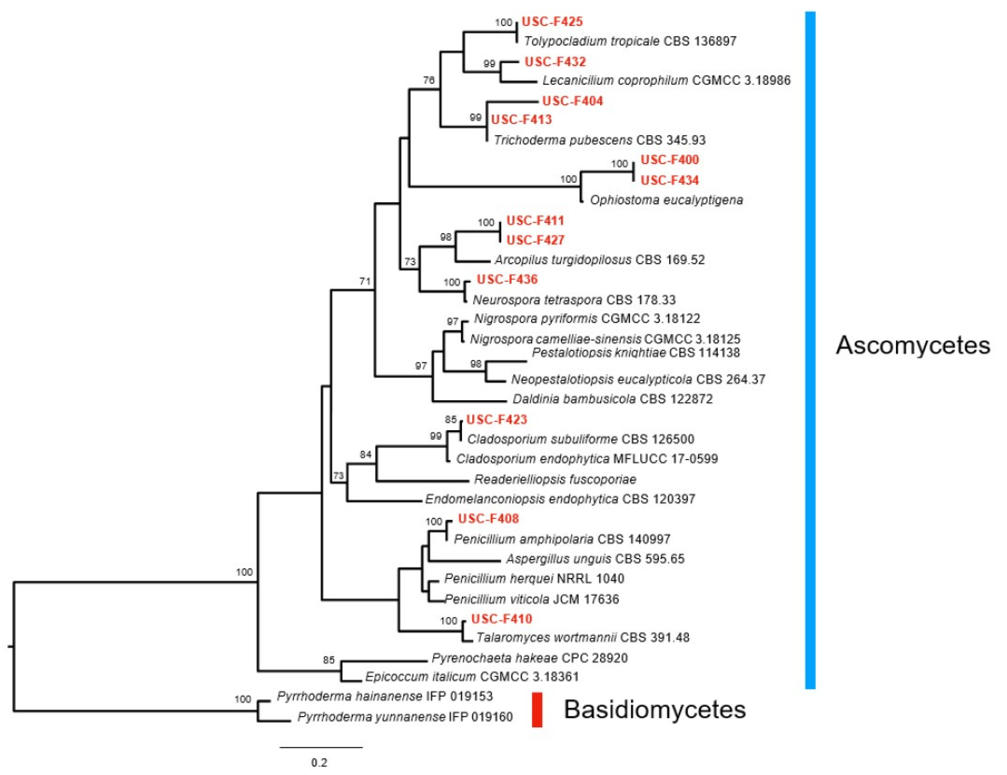
3.1. Fungal Isolates Cultured from Leucobryum candidum and Their Molecular Characterisation

A total of 70 fungal isolates were successfully cultured from the L. candidum samples. There was considerable morphological variation amongst isolates, suggesting a diverse array of taxa were cultured. Thirty-three morphologically different isolates were selected for further analysis. They were organised into morpho groups unique to each site (Table 3). Seven were unique to D’Aguilar NP (A); four morphotypes were only isolated from Bunyaville CP (B) (Supplementary Figure S1). Three morphotypes were unique to Mt. Mee and five were only isolated from Noosa NP. Six were uniquely isolated from Maroochy River CP (Supplementary Figure S2). Overall, each site appeared to have a unique set of fungal morphotypes isolated from L. candidum moss, with few cross-over species, suggesting high beta diversity.
The number of culturable isolates varied across the different environments/sites (Table 3). Coastal isolates accounted for 42% and forest collected samples accounted for 39%. Urban sites contributed the fewest number of culturable isolates with 18%, respectively. DNA sequence data were obtained for the 33 fungal isolates, which were found to closely affiliate with common cosmopolitan and saprobic taxa as well as known endophytes or pathogens of vascular plants (Figure 3, Supplementary Table S1).
Saprotrophic fungi comprised 48.48% of isolates. Endophytic ascomycetes formed 33.3% of the total isolates and parasitic fungi accounted for 18.19%. Categories were assigned based on the host plant and habitat of each isolate’s closest BLAST match to type specimens. Analysis of all sequenced ITS regions using BLAST searches within the NCBI GenBank database returned archived sequences with moderate to high (80–100%) identity to each fungal isolate. A single isolate returned a lower (75.2%) sequence identity to an archived ITS region (isolate USC-F413 from Noosa NP), and this was likely due to DNA contamination of the isolate that could not be removed through sub-culturing. Summary data for the BLAST results from isolates are presented in Supplementary Tables S2 and S3.
The BLAST search with altered criterion to include environmental samples (Supplementary Table S3) resulted in new matches scoring > 98% except for four isolates, which identified closest with the orders Ophiostomatales, Hypocreales, and one undescribed order (Fungal endophyte isolate 517). Based on the rule-of-thumb < 3% species delineation for fungal ITS sequences [41], isolates USC-F434, USC-F400, USC-F404, and USC-F433 (sequence BLAST matches < 97%.) are species without reliable sequence matches.
Three isolates scoring > 98% identity and 99% query cover affiliated most closely to Capnodiales, an order known for both latent saprophytic and parasitic activity. Eurotiales, known for cosmopolitan saprophytes, was the most common order with representation by four isolates. The order Hypocreales (with two isolates) and the Class Sordariomycetes (with one isolate) include members that are known to function as latent saprophytes as well as plant pathogens. Sordariales members are described as a mostly saprobic order and accounted for three isolates. Trichosphaeriales can be saprotrophic or parasitic and accounted for one isolate. Xylariales also accounted for a single order and is usually saprotrophic. The putative taxon match for isolate USC-F436 was Neurospora tetraspora (97.715%) when the BLAST search was limited to return TYPE specimen matches. This match was revised to Mycosphaerella sp. (99.469%) when BLAST was performed to include unverified environmental samples. Six other revisions of putative taxa differentiated between BLAST searches and are as follows: isolates USC-F405, USC-F424, and USC-F431 from Penicillium viticola to Penicillium cainii; isolate USC-F402 from Readerielliopsis fuscoporiae to Readeriella guyanensis; and isolate USC-F413 from Trichoderma pubescens to Trichoderma sp. Three isolate matches demonstrated taxonomic or phylogenetic affinities to orders, Amphisphaeriales, Botryosphaeriales, and Trichosphaeriales, that have not previously been reported from moss substrates.
The phylogenetic tree (Figure 1) indicated that 12 isolates showed close affiliation with the reference species used to construct the phylogram. Two isolates showed phylogenetic affinities with high (100%) bootstrap confidence to members of the Ophiostomatales order. Another two isolates showed 100% confidence grouping with the Eurotiales order, one from the Penicillium genus, the other from the Talaromyces genus. Isolate USC-F408 displayed phylogenetic affinities (100%) with the Capnodiales whilst another three isolates showed 100% bootstrapping confidence to members of Sordariales. The remaining four isolates grouped with the Hypocreales order with high bootstrap support (>99%).

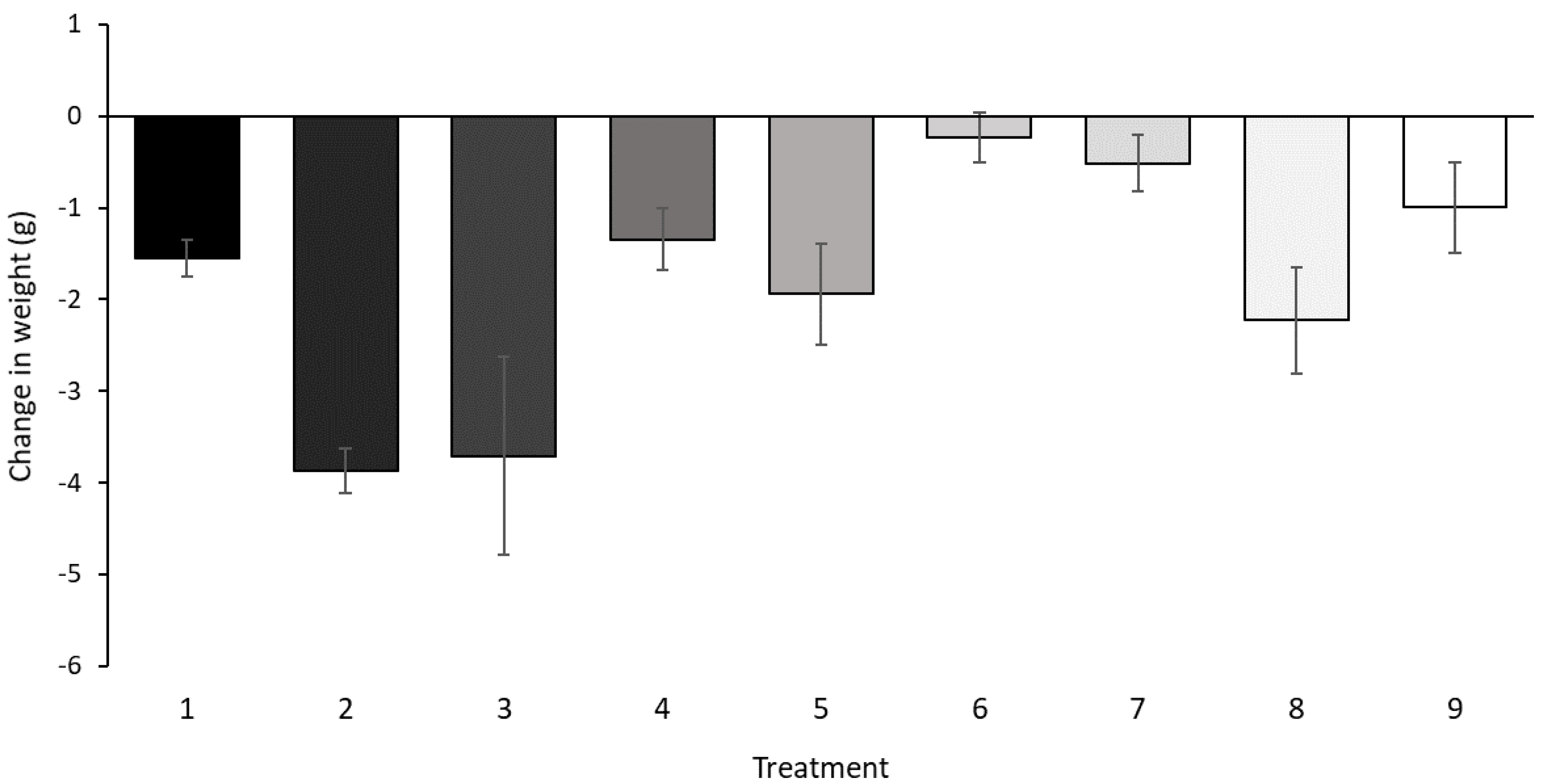
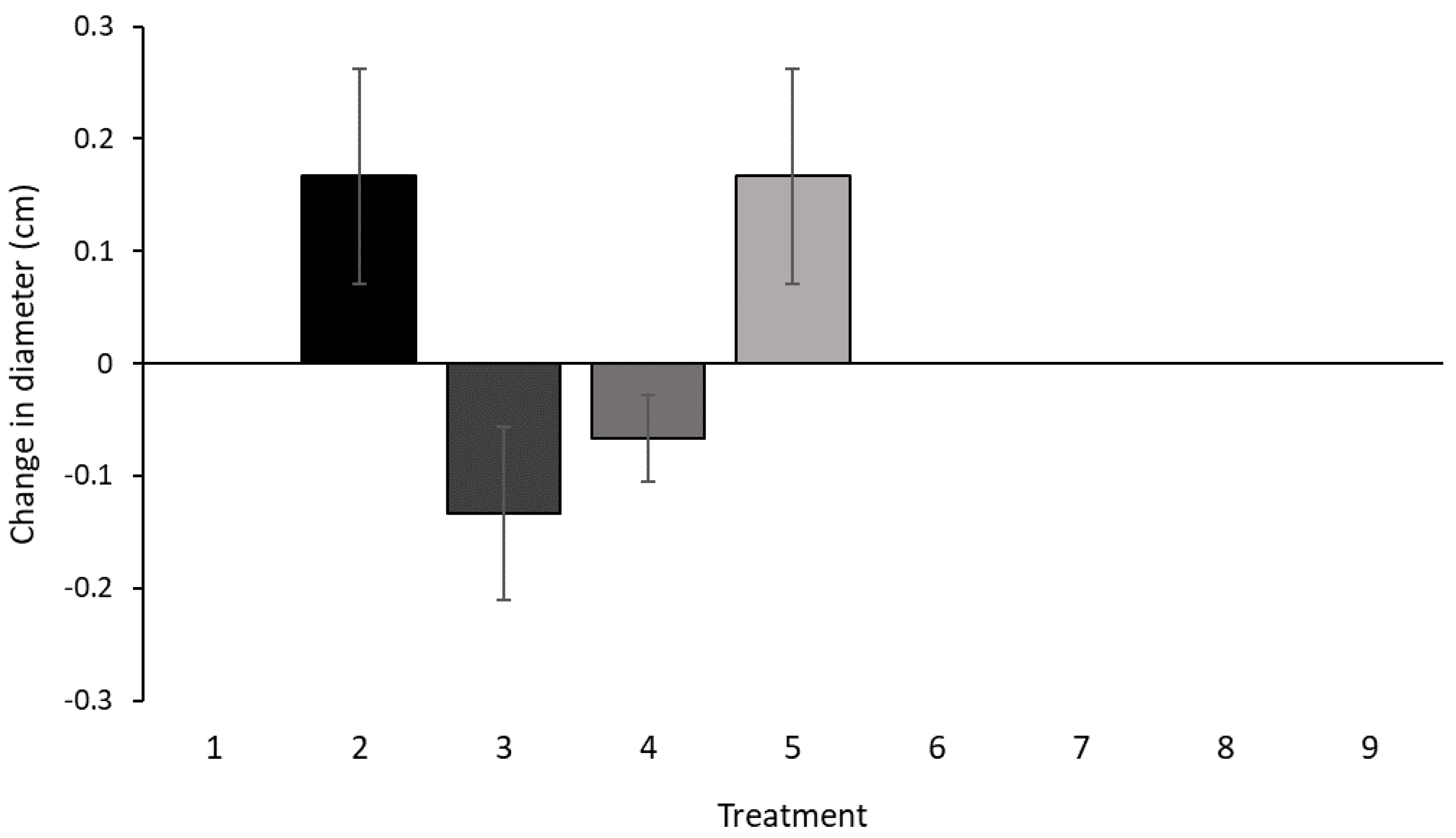
3.2. In Vitro Culture Assay

All 27 moss samples (9 treatments: 3 replicates) inoculated with fermentation extracts of USC-F426 and USC-F427 were observed for any change in heights, weights, diameters, and morphological characteristics. The mean and standard deviation (SD) were calculated for each treatment group. Many of the moss samples (97%) responded with a reduction in weight (Figure 4). Increases in the heights were observed for half of the treatments. No clear trend was observed for the change in diameters, regardless of the treatment (Figure 5 and Figure 6).
Two-way ANOVA analysis of the changes in weights and diameters as well as between treatments produced no significant differences (p > 0.05). There was, however, a significant (p < 0.05) difference in the heights between the treatments. The L. candidum in the in vitro cultures displayed some visible morphological responses. None of the L. candidum host specimens died and evidence of bright green gametophyte shoots, indicative of new growth, were present. Deposits of dark or bleached pigments on the outermost leaves often resembling drying or dying tissue (potential signs of parasitism) were present. Overall changes in colouration amongst all treatments represented by a loss or fade of original pigmentation were present.
Lastly, all 27 moss samples within the assay exhibited light to moderate contamination from non-inoculum fungi, which mostly presented on the agar. As all tissue was surface-sterilised, this contaminant was likely to come from either the intracellular or extracellular endophytes dwelling beneath the epidermis. In the repeated experiment, again, after 72 h, non-inoculant fungal contamination identical in morphology to the first contamination grew across all culture experiments, indicating the natural presence of such fungi within the moss tissue. This observation might indicate the presence of endophytes; however, future studies including Transmission Electron Microscopic (TEM) ones are needed for confirmation.

4. Discussion

This study identified a broad taxonomic diversity of the fungal isolates and confirmed the presence of previously unidentified fungal communities in association with this moss, which suggests that Leucobryum candidum is a promising micro-niche for a diverse array of fungi. The reportable orders included Amphisphaeriales, Botryosphaeriales, Capnodiales, Eurotiales, Hypocreales, Pleosporales, Sordariales, Trichosphaeriales, and Xylariales. Three of these orders (10%), Amphisphaeriales, Botryosphaeriales, and Trichosphaeriales, represent new reports of associations with mosses. The remaining orders (90%) are consistent with previous reports of bryophyte-associating taxa such as isolate USC-F436, which matched closely with Mycoshpaerella, a genus that has been previously described to form associations with Lunularia, a genus of thalloid liverwort [42]. Understanding the diversity and distribution of plant–symbiotic fungi across different environments is important for projecting responses to environmental changes such as the ones caused by climate change [43]. In total, 39% of isolates were cultured from L. candidum collected from forested environments, whilst coastal-collected samples yielded the highest number of isolates at 42%. Moss collected from urban environments produced the fewest number of culturable isolates and accounted for 18%. Urban sites were observed to be drier than both coastal and forested sites. The higher levels of fungal diversity, at the order level, amongst forest- and coastal-collected L. candidum, speculatively suggest fungal community phylogenetic diversity may be directly or indirectly impacted not only by biotic factors but also by abiotic and or anthropogenic factors. This would be consistent with well-established ecological concepts that the levels of biodiversity are higher in less fragmented forested environments [44,45]. Although this hypothesis is not thoroughly tested for fungi, studies have provided supporting evidence [27,45]. Bowman and Arnold (2017) [43] bring attention to concepts in fungal endophyte biology, which suggest plant-associated fungal communities will be impacted by minor environmental factors such as soil chemistry, rate of decomposition, plant productivity, and local hydraulic cycles. This is consistent with the well-established concept that the diversity of fungal communities is often directly or indirectly mediated by major environmental factors such as climate change, mean annual temperatures, and rainfalls and vegetation composition) [27].
One fungus within the Lecanicillium genus, isolated from the Mt. Mee sample, is associated with causing brown rot or wood rot, which is commonly found within rotting logs, the substrate of L. candidum in this study. As brown rot consumes woody material, the substrate becomes softer and spongier, can hold more moisture, and may become amenable and favourable for Leucobryum spore germination.
The BLAST match of isolates USC-F400 and USC-F434, Ophiostoma eucalyptigena (isolated from the host genus Eucalyptus), scored < 97% and thus cannot be confirmed past order level. Whilst unreportable, these isolates may be related to the Ophiostoma genus, particularly as the two isolates showed phylogenetic affinities with high (100%) bootstrap confidence to members of the Ophiostomatales order, which are often found within plant debris or soil [46] and are well-known associates of bark beetles and act as parasites of some plants [47], namely eucalypts [48]. Eucalypts were dominant in the overstory vegetation composition of most collection sites and may have been the species of rotting log substrate the mosses were sampled from.
No known studies have assessed the ability of L. candidum growth using in vitro cultures. Most studies that culture moss axenically allow for long growth periods, sometimes years for slower-growing mosses. When inoculating moss with fungi, observations will generally be reported more rapidly. Axenic in vitro culture experiments designed to determine if the fungal extracts from two isolates posed any beneficial or detrimental effects to the growth of L. candidum produced no discernible or significant host symptoms, other than a significant difference between the heights of the L. candidum between treatments. This is not entirely unusual as in some cases, interaction between moss and fungus simply does not generate any damage or reduction in growth to either organism. This result is consistent with the findings of Davey and Currah (2007) [49] who performed in vitro inoculation of axenic moss specimens with Cladophialophora fungi. They observed a lack of symptoms in the host and suggest that the fungi may have latent endophytic or saprophytic roles, which gradually lead to the degradation of the polyphenol-rich cell walls of mosses. Davey and Currah (2007) [49] suggest that the size and health of fungal associates within their host could be affected by micro-niche availability and chemical host defences, in addition to abiotic factors like moisture and nutrient availability. It is possible that the inoculated L. candidum was nutrient- and moisture-deprived in the axenic conditions without displaying symptoms of detriment. It has been suggested that for bryophytes to be colonised by diverse fungi, often simultaneously by several taxa, and not lose their vitality, they are likely to have genetically controlled defence mechanisms [16,50]. Observations of minute discolouration may have been a symptom of infection from fungal pathogens that are described to cause macroscopic, black, brown, or yellow necrotic and chlorotic patches in otherwise healthy-looking moss [14] like the ones observed in the inoculated L. candidum.

5. Conclusions

This study revealed a diversity of fungal associates, particularly from the Ascomycota phyla, to be present within the tissues of L. candidum moss within Southeast Queensland. Out of an original 70 isolates, 33 from two fungal divisions were cultured and characterised with molecular methods based on ITS sequence homology with archived sequences of type specimens. Those scoring < 99% were re-assessed for their homology to archived sequences including environmental isolates. According to BLAST search matches, a total of 10 orders and 17 families were represented amongst the six sample collection sites, with 3 orders being newly reported associations with bryophytes. The culturable fungi were represented by morphotypes that were mostly unique and site-specific as well as having high beta diversity over the differing environments. Whilst it is clear that L. candidum collected from SEQ serves as host to a biologically diverse array of fungi, the isolates within this study remain cryptic and are unable to be confirmed taxonomically past genus level, particularly as they represent biodiversity that is underrepresented in reference databases. Also, these isolates represent only a subset of the potential fungal associates of L. candidum as this study aimed to sub-culture and sequence fungi growing within the tissue, limiting phyllosphere-associated fungi. As such, a true estimate of the diversity of fungal associates of L. candidum would have to be more inclusive and combine both molecular and morphological techniques. This study provides further information within the field of bryomycology particularly as no known studies have yet focused on the fungal associates of L. candidum or its growth capabilities in artificial systems. Further data are presented in the Supplementary Section (Figures S1–S3 and Table S1).

Supplementary Materials

The following supporting information can be downloaded at: https://www.mdpi.com/article/10.3390/d17060370/s1, Figure S1: (A) fungal cultures isolated from and unique to D’Aguilar NP, (B) fungal cultures isolated from and unique to Bunyaville CP. Figure S2: Examples of successfully cultured isolates. (A) Fungal cultures isolated from and unique to Mt. Mee. (B) Fungal cultures isolated from and unique to Mt. Coolum NP. (C) Fungal cultures isolated from and unique to Noosa NP. (D) Fungal cultures isolated from and unique to Maroochy River CP; Figure S3. Preliminary UPGMA phylogenetic tree illustrating differences between fungal isolates in relation to other fungal reference strains. Table S1: summary of families and genera per site and putative categorisation of fungal isolates by site; Table S2: BLAST search results limited to type specimen sequence matching scoring ≥ 97% for ITS regions from fungi isolated from L. candidum at various sites within Southeast Queensland; Table S3: BLAST search results of isolates that scored < 99% matches to type specimens with updated search query to include environmental samples/unconfirmed isolate matches for ITS regions from fungi isolated from L. candidum at various sites within Southeast Queensland.

Author Contributions

Conceptualisation, L.V.M., D.İ.K. and A.S. methodology, L.V.M., D.İ.K., A.S. and A.J.F., validation, L.V.M., D.İ.K. and A.S., formal analysis, L.V.M.; investigation, L.V.M.; resources, D.İ.K. and A.S.; data curation, L.V.M.; writing—original draft preparation, L.V.M.; writing—review and editing, L.V.M. and D.İ.K.; visualisation, L.V.M.; supervision, D.İ.K. and A.S.; project administration, L.V.M. All authors have read and agreed to the published version of the manuscript.

Funding

This research received no external funding.

Institutional Review Board Statement

Not applicable.

Data Availability Statement

Data will be available on request to other interested researchers.

Acknowledgments

The authors acknowledge the traditional owners of the lands from which specimens were collected. The authors also gratefully acknowledge Pravech Ajawatanawong, Mahidol University, Thailand, for his support in the construction of the phylogenetic phylogram. Field samples were collected while the co-author (A.J. Franks) was a staff member of the Queensland Herbarium (BRI).

Conflicts of Interest

Andrew James Franks is employed by GHD Pty Ltd. The remaining authors of the paper declare that the research was conducted in the absence of any commercial or financial relationships that could be construed as a potential conflict of interest.

References

  1. During, H.J.; Tooren, B.F.V. Bryophyte interactions with other plants. Bot. J. Linn. Soc. 1990, 104, 79–98. [Google Scholar] [CrossRef]
  2. Schmid, E.; Oberwinkler, F.; Gomez, L. Light and electron microscopy of a host–fungus interaction in the roots of some epiphytic ferns from Costa Rica. Can. J. Bot. 1995, 73, 991–996. [Google Scholar] [CrossRef]
  3. Zuccaro, A.; Schulz, B.; Mitchell, J.I. Molecular detection of ascomycetes associated with Fucus serratus. Mycol. Res. 2003, 107, 1451–1466. [Google Scholar] [CrossRef]
  4. Boer, W.D.; Folman, L.B.; Summerbell, R.C.; Boddy, L. Living in a fungal world: Impact of fungi on soil bacterial niche development. FEMS Microbiol. Rev. 2005, 29, 795–811. [Google Scholar] [CrossRef]
  5. Dearnaley, J. Further advances in orchid mycorrhizal research. Mycorrhiza 2007, 17, 475–486. [Google Scholar] [CrossRef]
  6. Busby, P.E.; Ridout, M.; Newcombe, G. Fungal endophytes: Modifiers of plant disease. Plant Mol. Biol. 2016, 90, 645–655. [Google Scholar] [CrossRef]
  7. Beike, A.K.; Horst, N.A.; Rensing, S.A. Axenic bryophyte in vitro cultivation. J. Endocytobiosis Cell Res. 2010, 1, 102–108. [Google Scholar]
  8. Ligrone, R.; Duckett, J.G.; Renzaglia, K.S. Major transitions in the evolution of early land plants: A bryological perspective. Ann. Bot. 2012, 109, 851–871. [Google Scholar] [CrossRef]
  9. Bowen, W.R. The imperfect fungus Schizotrichella lunata on the moss Mnium cuspidatum. Bryologist 1968, 124–126. [Google Scholar] [CrossRef]
  10. Felix, H. Fungi on bryophytes: A review. Bot. Helv. 1988, 98, 239–269. [Google Scholar]
  11. Brown, D.H.; Bates, J.W. Bryophytes and nutrient cycling. Bot. J. Linn. Soc. 1990, 104, 129–147. [Google Scholar] [CrossRef]
  12. Read, D.J.; Duckett, J.G.; Francis, R.; Ligrone, R.; Russell, A. Symbiotic fungal associations in ‘lower’ land plants. Philos. Trans. R. Soc. Lond. B Biol. Sci. 2000, 355, 815–831. [Google Scholar] [CrossRef] [PubMed]
  13. Döbbeler, P. Microniches occupied by bryophilous ascomycetes. Nova Hedwig. 2002, 75, 275–306. [Google Scholar] [CrossRef]
  14. Davey, M.L.; Currah, R.S. Interactions between mosses (Bryophyta) and fungi. Botany 2006, 84, 1509–1519. [Google Scholar] [CrossRef]
  15. Ptasznska, A.; Mułenko, W.; Żarnowiec, J. Bryophytes microniches inhabited by microfungi. Ann. UMCS Biol. 2009, 64, 35. [Google Scholar] [CrossRef]
  16. Stenroos, S.; Laukka, T.; Huhtinen, S.; Döbbeler, P.; Myllys, L.; Syrjänen, K.; Hyvönen, J. Multiple origins of symbioses between ascomycetes and bryophytes suggested by a five-gene phylogeny. Cladistics 2010, 26, 281–300. [Google Scholar] [CrossRef]
  17. Halbwachs, H.; Vohnik, M.; Rentmeister, J.; Heinrichs, J. Structure and seasonality of fungal communities associated with bryophytes in temperate forests. Fungal Ecol. 2013, 6, 17–27. [Google Scholar]
  18. Pressel, S.; Bidartondo, M.I.; Ligrone, R.; Duckett, J.G. Fungal symbioses in bryophytes: New insights in the twenty first century. Phytotaxa 2014, 9, 238–253. [Google Scholar] [CrossRef]
  19. Gradstein, S.R.; Churchill, S.P.; Salazar-Allen, N.; Reiner-Drehwald, M.E. Guide to the Bryophytes of Tropical America; New York Botanical Garden Press: New York, NY, USA, 2001. [Google Scholar]
  20. Thieret, J.W. Bryophytes as economic plants. Econ. Bot. 1956, 10, 75. [Google Scholar] [CrossRef]
  21. Glime, J.M. Economic and ethnic uses of bryophytes. Flora N. Am. 2007, 27, 14–41. [Google Scholar]
  22. Furlan-Lopez, M.A.; Thomas, B.; Calatayud-Vernich, P.; Vidal, R.M. Natural and man-made disasters: Recovery in urban bryophyte communities. J. Bryol. 2022, 44, 45–59. [Google Scholar]
  23. Bais, H.; Sherrier, J. Advances in Botanical Research; Academic Press LTD-Elsevier Science: Cambridge, MA, USA, 2015. [Google Scholar]
  24. Osono, T. Role of phyllosphere fungi of forest trees in the development of decomposer fungal communities and decomposition processes of leaf litter. Can. J. Microbiol. 2006, 52, 701–716. [Google Scholar] [CrossRef] [PubMed]
  25. Grzesiak, B.; Wolski, G.J. Bryophilous species of the genus Galerina in peat bogs of Central Poland. Herzogia 2015, 28, 607–623. [Google Scholar] [CrossRef]
  26. Tosi, S.; Casado, B.; Gerdol, R.; Caretta, G. Fungi isolated from Antarctic mosses. Polar Biol. 2002, 25, 262–268. [Google Scholar] [CrossRef]
  27. Davey, M.L.; Heegaard, E.; Halvorsen, R.; Ohlson, M.; Kauserud, H. Seasonal trends in the biomass and structure of bryophyte-associated fungal communities explored by 454 pyrosequencing. New Phytol. 2012, 195, 844–856. [Google Scholar] [CrossRef]
  28. Tibpromma, S.; Hyde, K.D.; Maharachchikumbura, S.S.N.; Bhat, D.J.; Jones, E.B.G.; McKenzie, E.H.C.; Phillips, A.J.L. Fungal diversity notes 491–602: Taxonomic and phylogenetic contributions to fungal taxa. Fungal Divers. 2017, 83, 140–144. [Google Scholar] [CrossRef]
  29. Downing, A.; Marner, S. The first moss to be collected in Australia? Leucobryum candidum—Collected by William Dampier in 1699. J. Bryol. 1998, 20, 237–240. [Google Scholar]
  30. Michel, P.; Overton, J.M.; Mason, N.; Hurst, J.; Lee, W. Species–environment relationships of mosses in New Zealand indigenous forest and shrubland ecosystems. Plant Ecol. 2011, 212, 353–367. [Google Scholar] [CrossRef]
  31. El-Tarabily, K.A.; Hardy, G.E.S.J.; Sivasithamparam, K.; Hussein, A.M.; Kurtböke, D.I. The potential for the biological control of cavity-spot disease of carrots, caused by Pythium coloratum, by streptomycete and non-streptomycete actinomycetes. New Phytol. 1997, 137, 495–507. [Google Scholar] [CrossRef]
  32. Bakerspigel, A.; Miller, S.M. Fungi in soil and fungal diseases of plants. Am. J. Bot. 1953, 40, 201. [Google Scholar]
  33. Schoch, C.L.; Seifert, K.A.; Huhndorf, S.; Robert, V.; Spouge, J.L.; Levesque, C.A.; Chen, W.; Fungal Barcoding Consortium; Fungal Barcoding Consortium Author List; Bolchacova, E.; et al. Nuclear ribosomal internal transcribed spacer (ITS) region as a universal DNA barcode marker for Fungi. Proc. Natl. Acad. Sci. USA 2012, 109, 6241–6246. [Google Scholar] [CrossRef] [PubMed]
  34. Glass, N.L.; Donaldson, G.C. Development of primer sets designed for use with the PCR to amplify conserved genes from filamentous ascomycetes. Appl. Environ. Microbiol. 1995, 61, 1323–1330. [Google Scholar] [CrossRef] [PubMed]
  35. Sanger, F.; Nicklen, S.; Coulson, A.R. DNA sequencing with chain-terminating inhibitors. Proc. Natl. Acad. Sci. USA 1977, 74, 5463–5467. [Google Scholar] [CrossRef]
  36. Kauserud, H.; Mathiesen, C.; Ohlson, M. High diversity of fungi associated with living parts of boreal forest bryophytes. Botany 2008, 86, 1326–1333. [Google Scholar] [CrossRef]
  37. Raja, H.A.; Shearer, C.A.; Miller, A.N.; Oberlies, N.H. Fungal identification using molecular tools: A primer for the natural products research community. J. Nat. Prod. 2017, 80, 756–770. [Google Scholar] [CrossRef]
  38. Edgar, R.C. MUSCLE: Multiple sequence alignment with high accuracy and high throughput. Nucleic Acids Res. 2004, 32, 1792–1797. [Google Scholar] [CrossRef]
  39. Galtier, N.; Gouy, M.; Gautier, C. SEAVIEW and PHYLO_WIN: Two graphic tools for sequence alignment and molecular phylogeny. Comput. Appl. Biosci. 1996, 12, 543–548. [Google Scholar] [CrossRef]
  40. Gouy, M.; Guindon, S.; Gascuel, O. SeaView version 4: A multiplatform graphical user interface for sequence alignment and phylogenetic tree building. Mol. Biol. Evol. 2010, 27, 221–224. [Google Scholar] [CrossRef]
  41. Nilsson, R.H.; Kristiansson, E.; Ryberg, M.; Hallenberg, N.; Larsson, K.-H. Intraspecific ITS variability in the kingdom Fungi as expressed in the international sequence databases and its implications for molecular species identification. Evol. Bioinform. 2008, 4, 193–201. [Google Scholar] [CrossRef]
  42. Die Döbbeler, P.; Moosbewohnende Ascomyceten, I. Pyrenocarpen den Gametophyten besiedelnden Arten. Mitteilungen Bot. Staatssamml. München 1978, 14, 1–36. [Google Scholar]
  43. Bowman, E.A.; Arnold, A.E. Distributions of ectomycorrhizal and foliar endophytic fungal communities associated with Pinus ponderosa along a spatially constrained elevation gradient. Am. J. Bot. 2017, 105, 687–699. [Google Scholar] [CrossRef] [PubMed]
  44. Hawksworth, D.L.; Rossman, A.Y. Where are all the undescribed fungi? Phytopathology 1997, 87, 888–891. [Google Scholar] [CrossRef] [PubMed]
  45. Cannon, P.F.; Hawksworth, D.L. Fungal diversity in ecosystems. Mycol. Res. 1995, 99, 1057–1065. [Google Scholar]
  46. Zhang, Z.H.; Li, R.; Yang, D.; Wang, X.; Xiao, Y. Epiphytic bryophytes in Yunnan, China: Diversity, distribution, and conservation. J. Bryol. 2015, 37, 199–210. [Google Scholar]
  47. De Beer, Z.W.; Duong, T.A.; Dreyer, L.L.; Oberlander, K.C.; Roets, F. New species of Ophiostomatales from Scolytinae and Platypodinae beetles in the Cape Floristic Region, including the discovery of the sexual state of Raffaelea. Antonie Van Leeuwenhoek 2014, 108, 933–950. [Google Scholar]
  48. Crous, P.W.; Wingfield, M.J.; Burgess, T.I.; Hardy, G.E.S.J.; Crane, C.; Barrett, S.; Cano-Lira, J.F.; Le Roux, J.J.; Thangavel, R.; Guarro, J.; et al. Fungal Planet description sheets: 469–557. Persoonia Mol. Phylogeny Evol. Fungi 2015, 37, 218–403. [Google Scholar] [CrossRef]
  49. Davey, M.L.; Currah, R.S. A new species of Cladophialophora (hyphomycetes) from boreal and montane bryophytes. Mycol. Res. 2007, 111, 106–116. [Google Scholar] [CrossRef]
  50. Döbbeler, P. Polytrichadelphus magellanicus sensu lato (Musci) and its ascomycetes—Different fungi on different hosts. Sendtnera 1999, 7, 35–45. [Google Scholar]
Figure 1. Phylogenetic tree showing type specimen BLAST matches and closely related USC isolates.
Diversity 17 00370 g001
Figure 2. In vitro growth culture assay of L. candidum.
Diversity 17 00370 g002
Figure 3. Putative categories of fungal isolates in relation to the functions of their closest relatives.
Diversity 17 00370 g003
Figure 4. Comparison of the changes in in vitro L. candidum weights (g) after 42 days of incubation for each treatment. Footnote: Each bar represents mean change in weight of 3 replicates. Absent bars represent no change. For treatment type, refer to Table 2.
Diversity 17 00370 g004
Figure 5. Comparison of the changes in L. candidum height (cm) after 42 days of incubation for each treatment. Footnote: Each bar represents mean change in height of 3 replicates. Absent bars represent no change. For treatment type, refer to Table 2.
Diversity 17 00370 g005
Figure 6. Comparison of the changes in in vitro L. candidum diameters (cm) after 42 days of incubation for each treatment. Footnote: Each bar represents mean change in diameter of 3 replicates. Absent bars represent no change. For treatment type, refer to Table 2.
Diversity 17 00370 g006
Table 1. Location and environment type of L. candidum sample collection sites within SEQ.
SampleLocationLatitudeLongitudeEnvironment
Samples; 1A, 1BD’ Aguilar National Park (NP)−27.381297152.778036Forested
Samples; 2A, 2BBunyaville Conservation Park (CP)−27.368173152.953509Urban
Samples; 3A, 3BMt. Mee−27.097123152.702889Forested
Samples 4A, 4BMt. Coolum NP−26.563218153.089238Urban
Samples; 5A, 5BNoosa NP−26.384495153.102599Coastal
Samples; 6A, 6BMaroochy River CP−26.630635153.094886Coastal
Table 2. Broth types and the fungal inoculants.
BrothInoculant
PDBUSC-F427
PDBUSC-F426
PDBCombination; USC-F427, USC-F426
MBUSC-F427
MBUSC-F426
MBCombination; USC-F427, USC-F426
PDB = Potato Dextrose Broth; MB = moss extract broth.
Table 3. Summary of the selected fungal isolates cultured from each L. candidum collection site.
Site# Number of Fungal IsolatesHostSubstrateEnvironment
D’Aguilar NP10L. candidumRotting LogForest
Bunyaville CP2L. candidumRotting LogUrban
Mt. Mee3L. candidumRotting LogForest
Mt. Coolum NP4L. candidumRotting LogUrban
Noosa NP9L. candidumRotting LogCoastal
Maroochy River CP5L. candidumRotting LogCoastal
Total33
Disclaimer/Publisher’s Note: The statements, opinions and data contained in all publications are solely those of the individual author(s) and contributor(s) and not of MDPI and/or the editor(s). MDPI and/or the editor(s) disclaim responsibility for any injury to people or property resulting from any ideas, methods, instructions or products referred to in the content.

