Article

Health Status of Stranded Common Bottlenose Dolphins (Tursiops truncatus) and Contamination by Immunotoxic Pollutants: A Threat to the Pelagos Sanctuary—Western Mediterranean Sea

1
Istituto Zooprofilattico Sperimentale del Piemonte, Liguria e Valle d’Aosta, 10154 Torino, Italy
2
Department of Physical, Earth and Environmental Sciences, University of Siena, Via Mattioli, 4, 53100 Siena, Italy
3
Department of Integrative Marine Ecology, Stazione Zoologica Anton Dohrn, National Institute of Marine Biology, Ecology and Biotechnology, Genoa Marine Centre, Villa del Principe, via San Benedetto 2, 16126 Genoa, Italy
4
Delfini del Ponente APS, Via Regione Bussi 27, 18100 Imperia, Italy
5
DISTAV, University of Genoa, Corso Europa 26, 16132 Genoa, Italy
6
Faculty of Veterinary Medicine, University of Teramo, Strada Provinciale 18 Località Piano d’Accio, 64100 Teramo, Italy
7
Centro Interuniversitario per la Ricerca sui Cetacei (CIRCE), Department of Physical, Earth and Environmental Sciences, University of Siena, Strada Laterina 8, 53100 Siena, Italy
*
Author to whom correspondence should be addressed.
Diversity 2023, 15(4), 569; https://doi.org/10.3390/d15040569
Submission received: 31 January 2023 / Revised: 3 April 2023 / Accepted: 12 April 2023 / Published: 18 April 2023
(This article belongs to the Special Issue Monitoring, Threat Assessment, and Conservation Actions of Cetaceans)

Abstract

:
Between 2019 and 2021, 21 bottlenose dolphins were found stranded dead along the Ligurian Pelagos Sanctuary coast (Italy). For 11 animals, out of a total of 14 recovered, the cause of death was classified as natural, anthropic, or not determined based on gross and histological pathology and ancillary testing. Hexachlorobenzene (HCB), dichlorodiphenyltrichloroethanes (DDTs), and polychlorinated biphenyls (PCBs) were examined in their blubber, and results were discussed according to their toxicological properties. All specimens showed the following trend: PCBs > DDTs >> HCB, and the levels of cancerogenic, mutagenic, and teratogenic organochlorine compounds (T-OCs) were >50% of total OCs. Immunosuppressant organochlorine compound (IS-OC) levels in 10 out of 11 animals were above 50% of the total OCs. PCB levels always exceeded the threshold of 17 mg/kg lipid weight for PCB-induced adverse health effects. The results suggest that bottlenose dolphins living in the Pelagos Sanctuary undergo a high level of exposure to pathogens and OCs, betraying the designation of the Cetacean Sanctuary and, consequently, of a region created for their conservation. Immune dysfunction and infectious disease susceptibility appear to be highly connected with high levels of OC xenobiotics. These data are useful to understand health and mortality trends in cetacean populations, as well as for developing policies for cetacean conservation and management in this important protected area of the Mediterranean.

1. Introduction

The examination of stranded cetaceans provides valuable information on the health of marine mammal populations and, more generally, on the marine environment, increasing public awareness of the deterioration of marine ecosystems and the risks to animal and human health [1,2].
The causes of strandings are generally multifactorial and often act in conditions of general immunosuppression. Many anthropogenic chemical contaminants, such as most of the persistent organic pollutants (POPs), can contribute to increasing the susceptibility of marine mammals to diseases of infectious origin [3,4,5,6] due to their immunosuppressive characteristics [7,8]. Several emerging and reemerging viral, bacterial, protozoan, parasitic, and fungal diseases have been described in cetacean species [2,9]. At the moment, there is evidence that climate change impacts not only all marine ecosystems, but also cetaceans [10,11,12,13]. Given that climate change has the greatest effects on river and coastal ecosystems, making them more biologically and chemically polluted, coastal cetaceans, such as bottlenose dolphins (Tursiops truncatus), are most at risk [9,14]. Like many other cetaceans and large marine vertebrates, the bottlenose dolphin is a top predator [15,16,17]. Through the trophic chain, this species bioaccumulates POPs in its tissues and organs [8,18,19]. Numerous studies on bottlenose dolphins residing in the Mediterranean Sea, document the chemical stress from xenobiotic organochlorine compounds (OCs), in particular polychlorinated biphenyls (PCBs) and dichlorodiphenyltrichloroethane (DDT) [8,20]. These OCs have been investigated both in skin biopsies from free-ranging individuals [21,22,23] and in blubber samples from stranded specimens [22,24,25,26]. In the performed studies, it was found that dead stranded individuals have higher contamination levels than the living individuals monitored in the same study [8]. The post-mortem investigations in stranded specimens have shown an important correlation between high levels of these pollutants and the infectious diseases that were recognized as the most likely cause of death of these marine mammals [2,27]. This evidence is particularly documented in the Pelagos Sanctuary [28,29,30,31,32,33,34,35,36], an area for the protection of cetaceans covering 87,500 km2 and 2022 km of coast of the northwestern Mediterranean Sea, established under an international agreement between France, Italy, and the Principality of Monaco [37]. The area can be considered a biogeographically distinct sub-section of the Large Marine Ecosystem (LME) that is the Mediterranean and is characterized by markedly heterogeneous topography and hydrodynamic diversity and high levels of primary productivity related to the upwelling of nutrients, which are crucial for the presence of eight species of marine mammals on a relatively consistent basis. Unfortunately, the urbanization and industrialization of its coastal areas are increasing: some agricultural practices release fertilizers, fungicides, and pesticides; industries such as chemical, petrochemical, and metallurgical industries, oil and gas refineries, and drilling and desalination plants are responsible for contamination by new and old generation xenobiotics, metals, and polycyclic aromatic hydrocarbons [38]. This means that in this Marine Protected Area, contamination pressures on cetaceans are certainly not negligible [39,40].
To assess the impact of pollution on cetacean health, it is important to systematically investigate stranding events by means of a comprehensive post-mortem examination and thorough diagnostic investigations.
The present work was aimed at reporting the results of diagnostic and toxicological investigations in bottlenose dolphins stranded dead along the Ligurian coasts of the Pelagos Sanctuary between 2019 to 2021, in an attempt to assess the influence of pollution in the development of infectious diseases in this cetacean species living in the area. The possible correlations between sex, sexual maturity, age, pathologies, and levels of some OCs (PCBs, pp′DDT and its metabolites, and hexachlorobenzene (HCB)) were explored, as well the possible effects of the different OCs in relation to their toxicological characteristics and to the ratios between the different compounds.

2. Materials and Methods

2.1. Post-Mortem Examination

Within the total of 21 common bottlenose dolphins [14] stranded dead in the area under study between 2019–2021, thanks to the activities carried out by the National Reference Centre for Diagnostic Investigations on Stranded Marine Mammals (C.Re.Di.Ma.), at Istituto Zooprofilattico Sperimentale (IZS), Diagnostic Laboratory of Imperia, it was possible to carry out a complete post-mortem examination, according to internationally standardized guidelines [41], on 11 individuals (Figure 1, Table 1) [31,32,33].
On 3 additional carcasses recovered, which were in a very advanced state of decomposition, a limited sampling was carried out; therefore, these individuals were not included in the set.
The 7 remaining carcasses were not recovered, mainly for logistical reasons (individuals found offshore) or due to their very advanced decomposition status.
Each specimen was labelled with an IZS identification code and a code assigned by the Banca Dati Spiaggiamenti (BDS) (http://mammiferimarini.unipv.it, accessed on 6 December 2022). We reorganized the overall case, coding in chronological order, from Case 1 to Case 11.
The data collection included the date and location of stranding, sex, total body length (TBL) (cm), weight (kg), estimated age (years), estimated age class, sexual maturity, decomposition condition category (DCC), nutritional condition category (NCC), gastric contents, and the macroscopic lesions and helminths detected.
For specimens with docked tails, the total body length was estimated based on knowledge about the physiology of the species, considering that the length of the tail represents about 1/9–1/10 of the total body length (TBL) [42].
The age of the animals was calculated using the linear model for Mediterranean bottlenose dolphins of Butti et al. [43], based on TBL and two parameters of bone mineral density (BMD) from two different regions of interest (ROI) of the thoracic limb: (1) the global region of interest (GROI) between the proximal epiphysis of the humerus and the distal epiphysis of the radius and ulna and (2) the subregion of interest (SROI), the whole humerus. The model was: age = −14.8 + Length × 0.1 + [(rGROI × 48.3)/(−SROI × 29.4)] when the GROI and SROI were evaluated in terms of the right global region of interest (rGROI) and of the right subregion of interest (rSROI) of the dolphins examined.
Depending on the TBL and on the age estimated, the presumed age class was established based on three estimated age classes (newborns/calves, juveniles, and adults) [44,45,46], with the final differentiation between juveniles and adults made based on gonad maturation [47,48].
The decomposition condition category of the carcasses at the time of necropsy (DCC) was classified as code 1 (extremely fresh carcass, just dead), code 2 (fresh), code 3 (moderate decomposition), code 4 (advanced decomposition), or code 5 (mummified or skeletal remains) [41].
The nutritional status was assessed morphologically based on anatomical parameters such as the convexity of the dorsal profile, the rib prominence, and the amount of visceral fat and blubber thickness measured immediately anterior to the dorsal fin at three locations (dorsal, lateral, and ventral) [41]. Nutritional status was categorized as good, moderate, or poor (nutritional condition category-NCC).
Macroscopical findings of all cases were recorded, and gastric chambers were opened to evaluate pathological changes and the content’s features. The presence of helminths was estimated by macroscopic and microscopic examination of tissues. Endoparasites were preserved in 70% alcohol for microscopic identification, according to established morphological characteristics [49,50].
During necropsy, the tissue samples of all the major organs and lesions were collected and split into aliquots for subsequent analyses: one was kept frozen at −20 °C for microbiological and toxicological investigations, one was kept frozen at −80 °C for biomolecular analyses, and the other was preserved in 10% buffered formalin for histological and immunohistochemical (IHC) investigations.
Blood serum, intracardiac clot, aqueous humor, pericardial fluid, and cerebrospinal fluid (CSF) were collected, when available, and kept frozen at −20 °C for serological investigations.

2.2. Diagnostic Investigations

Table 1 gives an overview of the conducted analyses per case, arranged in chronological order.
For histological investigations, tissues samples of Cases 1, 2, 3, 4, 5, 6, 8, 10, and 11, including brain, lung, heart, liver, spleen, prescapular, pulmonary, tracheobronchial and mesenteric lymph nodes, tonsils, kidney, mammary gland, adrenal gland, intestine, muscle, stomach, bladder, reproductive system, blubber, and skin lesions, were collected and fixed in 10% neutral buffered formalin, embedded in paraffin, sectioned at 4 ± 2 μm, stained with hematoxylin and eosin (HE), and examined through a light microscope. With respect to the brain, following Giorda et al. [51] different areas were sampled and examined, including the basal nuclei, thalamus, mesencephalon, pons, obex, and frontal, parietal, occipital, and cerebellar cortex.
Immunohistochemistry (IHC) for Morbillivirus was performed on tissue sections of Cases 1, 2, 3, 4, 5, 6, 8, and 11, including the brain, which was systematically investigated, and the lung, spleen, lymph nodes, bladder, laryngeal tonsil, intestine and kidney, to confirm the pathological findings observed or the positivity found in biomolecular investigations. IHC was performed using a monoclonal anti Canine distemper virus (CDV) antibody (VMRD, Pullman, WA, USA) [52]. T. gondii IHC was carried out on the brain tissues of Cases 1, 3, 4, 5, 6, 8, and 11 using a polyclonal serum of caprine origin (VMRD, Pullman, WA, USA) [52].
For bacteriological investigations, tissue samples of Cases 1, 2, 3, 4, 5, 6, 7, 8, 10, and 11, including the brain, lung, prescapular lymph node, liver, spleen, tonsils, kidney, and bladder, were processed for standard aerobic, anaerobic, and microaerobic (5% CO2) bacterial culture and identification by biochemical and/or molecular analyses. Following international recommendations [53], samples from target tissues underwent specific bacteriological procedures to screen for Listeria spp., Salmonella spp. and Brucella spp.
Molecular detection of Dolphin morbillivirus (DMV) [54], Herpesvirus (HV) [55], Toxoplasma gondii [56], Brucella spp. [57] and Photobacterium damselae [58] was achieved on target tissues available in all cases, including the brain, lung, tonsils, prescapular, pulmonary, tracheobronchial and mesenteric lymph nodes, liver, spleen, kidney, and bladder for DMV; the brain, lung, prescapular, pulmonary, tracheobronchial lymph nodes, spleen, kidney, and skin lesions for HV; the brain, prescapular, pulmonary, tracheobronchial and mesenteric lymph nodes, liver, spleen, heart, and muscle for T. gondii; the brain, lung, tonsils, prescapular, pulmonary, tracheobronchial and mesenteric lymph nodes, spleen, kidney, testicle, skin lesions, and cerebrospinal fluid (CSF) for Brucella spp.; and the brain, lung, prescapular, pulmonary, tracheobronchial and mesenteric lymph nodes, liver, spleen, and kidney for Photobacterium damselae.
Moreover, molecular detection of Poxvirus [59] was attempted on skin lesions of Cases 1, 4, 6, and 11.
Finally, the presence of antibodies for T. gondii, Brucella spp., and Morbillivirus was investigated in Cases 1, 2, 3, 4, and 5 [52,60], specifically on cerebrospinal fluid (CSF) and pericardial fluid of Case 1; on serum of Case 2; on pericardial fluid, pleuric fluid, and serum of Case 3; on CSF, pericardial fluid, serum, and aqueous humor of Case 4; and on serum, intracardiac clot, and aqueous humor of Case 5.

2.3. Cause of Death Evaluation

A hypothesis on the cause of death was formulated considering biological and epidemiological data, macroscopic and microscopic findings, and the results of all investigations. The causes of death have been categorized into causes of natural origin (pathologies of infectious origin, neonatal/perinatal pathologies, traumatic intra-interspecific interactions, senescence, etc.) and anthropic origin (interaction with fishing, ship collisions, etc.) according to available bibliographic references [2,61] and the recently elaborated diagnostic framework “Evidence Based Diagnostic Assessment framework for cetacean necropsies on marine debris ingestion and common data collection” [62].
Moreover, starting from 2020, the Life DELFI’s harmonized protocols and frameworks (https://lifedelfi.eu/wp-content/uploads/2021/04/A3_Framework_Fishery_interaction-1.pdf, accessed on 8 February 2023) were adopted to better assess the cetacean post-mortem findings associated with fishery interaction. In this regard, evidence of direct signs (specific to each category of fishing interaction), other associated lesions, and aspecific findings were evaluated, taking into account the results of all ancillary analyses, including histology and IHC, such as for confirmation of capture myopathy [63,64].
According to the 6 Life DELFI framework categories, findings related to fishery interaction were categorized as “bycatch with active fishing gear”, “bycatch with passive fishing gear”, “chronic entanglement”, “larynx entanglement”, “ingestion”, and “intentional injury”; an extra category was added whenever the bycatch event could not be identified with active or passive fishing gear: i.e., “bycatch with not determined fishing gear”. For the assessment of the fishery interaction, most of the evidence requires a condition code of the carcass between 1 and 3. Thus, based on the DCC, the fishery interaction was categorized as confirmed (C), probable (P), or suspect (S).

2.4. Toxicological Analyses

Toxicological analysis was performed at the Department of Physical Sciences, Earth and Environment at the University of Siena according to the modified U.S. Environmental Protection Agency (EPA) 8081/8082 method for determination of hexachlorobenzene (HCB), polychlorinated biphenyls (PCBs), and dichlorodiphenyltrichloroethane and its metabolites (DDTs) [65]. Specifically, blubber samples (5–20 gr) were lyophilized in an Edwards freeze drier for three days and about 1 g, finely shredded, was extracted with n-hexane for pesticide residue analysis (PESTINORM, VWR Chemicals) in a Soxhlet apparatus. VWR cellulose thimbles (i.d. 25 mm, e.d. 27 mm, length 100 mm) used for extraction of the samples were preheated for about 30 min to 110 °C and pre-extracted for 9 h in a Soxhlet apparatus with n-hexane in order to remove any organochlorine contamination. Each sample was spiked with a surrogate compound (2,4,6-trichlorobiphenyl–IUPAC number 30; Ref. [66]) prior to extraction; then this compound was quantified, and its recovery calculated. After the 9-h extraction with n-hexane, samples were purified with sulphuric acid 95% (VWR Chemicals) and the extract underwent liquid chromatography on a column containing Florisil (60–100 mesh, Merck) that had been dried for 1 h at 110 °C. Decachlorobiphenyl (DecaPCB—IUPAC number 209) was used as an internal standard and was added to each sample prior to the analysis and included in the calibration standard (a mixture of Arochlor 1260, HCB and pp′—and op′—DDT, DDD, DDE).
High resolution capillary gas chromatography analyses were performed with an Agilent 6890N series gas chromatograph equipped with a 63Ni electron capture detector (ECD) and an automatic injector on-column. The column used was an SBP-5 bonded phase capillary column (30 m long, 0.25 mm internal diameter, film thickness 25 μm). The carrier gas was nitrogen with a head pressure of 15.5 psi (splitting ratio 50/1). The scavenger gas was argon/methane (95/5) at 40 mL/min. The oven temperature was 100 °C for the first 10 min, after which it was increased to 280 °C at 5 °C/min. The injector and detector temperatures were 200 and 280 °C, respectively. A mixture of specific isomers was used to calibrate the system, evaluate recovery, and confirm the results.
The standard injected was prepared with 50 ng/mL of HCB, 100 ng/mL of DDT (pp′DDT, pp′DDD, pp′DDE, op′DDD, op′DDE), 200 ng/mL of op′DDT, and 2 μg/mL of Arochlor 1260. In order to evaluate the linearity in the instrumental response and the instrumental sensitivity, the following quantities of standard were injected: 1, 2, and 4 μL. Capillary gas-chromatography revealed 30 PCB congeners (IUPAC no. 95, 99, 101, 118—pentachlorobiphenyls; 128, 135, 138, 141, 144, 146, 149, 151, 153, 156—hexachlorobiphenyls; 170, 171, 172, 174, 11, 178, 180, 183, 187—heptachlorobiphenyls; 194, 195, 196, 199, 201, 202—octachlorobiphenyls; 206—nonachlorobiphenyls). Total PCBs (PCBs) were quantified as the sum of all congeners. Total DDTs (DDTs) were calculated as the sum of the isomers op′DDT, pp′DDT, op′DDD, pp′DDD, op′DDE, and pp′DDE. The results were expressed in ng/g lipid weight (l.w.) unless otherwise specified. The limit of detection (LOD) was calculated by measuring replicates (n = 20) of blank samples, determining the mean value and standard deviation (SD), and calculating the LOD as the mean +2 SD. The LOD for all compounds analyzed was 0.1 ng/kg (ppt).
The extracted organic material (EOM%; lipid content) was calculated gravimetrically after extraction with n-hexane.
OCs were grouped according to their toxicological characteristics: immunosuppressant organochlorine compounds (IS-OCs); Endocrine Disrupting Chemicals (EDC-OCs); cancerogenic, mutagenic, and teratogenic organochlorine compounds (T-OCs); and the OCs showing qualities of both T-OCs and EDC-OCs and IS-OCs that were TEI-OCs (pp′DDE+PCB118+PCB153+pp′DDT) (Table 2).

2.5. Statistical Analysis

Statistical analysis was carried out with STATA 14 software [88]. In order to validate the use of the parametric approach, preliminary analyses were also conducted for checking the normality of the data. The Shapiro–Wilk test gave p-values greater than 0.05 (p > 0.05) for each variable [89]. Multiple regression was applied to verify the correlation between DDTs and PCBs with animal age and gender.

3. Results

3.1. Post-Mortem Examination

Considering the total of the 21 common bottlenose dolphins stranded in the area under study from 2019 to 2021, it was possible to recover 14 out of the 21 carcasses (66.66%) and to perform a complete necropsy on 11 animals (52.38% of the 21 carcasses), while on three carcasses, of which two were classified as DCC5 and one was classified as DCC4, only limited sampling, genetic investigations, and/or biometric data were carried out.
Of the total of the 11 common bottlenose dolphins completely examined, the majority were male (8/11) and adults (7/11); four carcasses were classified as “fresh” (DCC 2), four as in “moderate decomposition” (DCC 3), and three as in “advanced decomposition” (DCC 4). Regarding nutritional status, four animals were classified as NCC moderate, three NCC good, one NCC poor, and three not determined (ND).
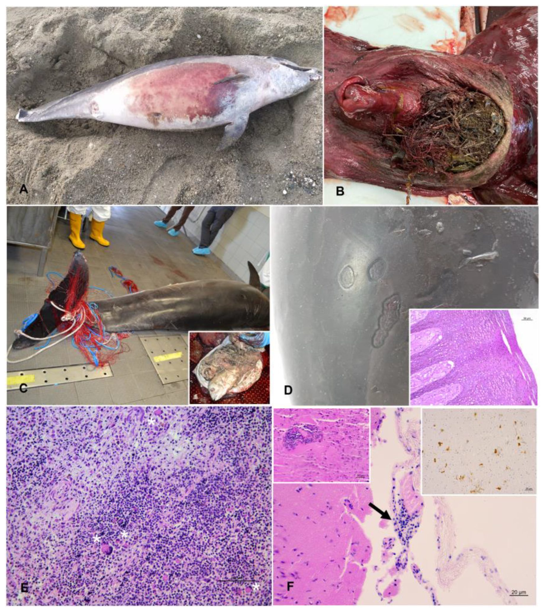
The main data (case number, date and location of stranding, sex, total body length (TBL), weight, estimated age, estimated age class, sexual maturity, DCC, NCC, gastric contents description, main gross and microscopic lesions, pathogens and helminths detected) of the 11 stranded bottlenose dolphins are reported in Table 3. Moreover, a selected collection of macroscopic findings of some of the animals under study are presented in Figure 2.

3.2. Diagnostic Investigations

3.2.1. Histopathological-Immunohistochemistry

Histopathological investigations were performed with a systematic approach on nine out of eleven animals (Cases 1, 2, 3, 4, 5, 6, 8, 10, and 11), including neuropathological investigation in all cases, except in Case 2, which, because of general tissue degradation, had a single tissue investigated (mesenteric lymph node) for its macroscopic appearance (reactive lymphadenopathy).
Immunohistochemistry (IHC) for Morbillivirus was performed on the central nervous system (CNS) and other tissues of eight out of eleven animals (Cases 1, 2, 3, 4, 5, 6, 8, and 11); T. gondii-IHC was carried out on the brain of seven out of eleven animals (Cases 1, 3, 4, 5, 6, 8, and 11). Specific Morbillivirus antigens were detected in three animals, while no specific T. gondii antigens were detected in the animals tested at the cerebral level.
Significant histopathological lesions likely caused by Morbillivirus were detected in three animals with virus antigens confirmed by biomolecular investigations, specifically in Cases 2, 4, and 11.
In Case 2, a marked lymphoid depletion was observed in the mesenteric lymph node; Morbillivirus-specific antigens were also detected by IHC performed ad hoc in the same organ. In Case 4, a marked lymphoid depletion was observed in the prescapular, pulmonary, and mesenteric lymph nodes and the laryngeal tonsil, along with lymphadenitis and tonsillitis with syncytia; moreover, a severe lymphoplasmacytic broncho-interstitial pneumonia with syncytia was observed. Morbillivirus-specific antigens were also detected by IHC in the CNS, lung, prescapular, tracheobronchial, and mesenteric lymph nodes, spleen, laryngeal tonsil, and kidney. In Case 11, a marked lymphoid depletion was observed in the spleen and mesenteric lymph node, along with lymphoplasmacytic meningoencephalitis; lymphoplasmacytic endometritis was also detected. Morbillivirus-specific antigens were detected by IHC in the CNS.
Regarding lesions likely associated with other pathogens of viral origin, significant histopathological lesions associated with poxviral aetiology were observed in multiple tattoo skin lesions in Case 6, with the Cetacean poxvirus antigen confirmed by biomolecular investigations and represented by the proliferation of dermal papilla cells, multifocal hydropic degeneration of Stratum spinosum keratinocites with hyperpigmentation, and rare intracytoplasmic eosinophilic inclusion bodies.
Moreover, histopathological lesions were observed in skin lesions in Case 11. The lesions showed a positive result to α -HV, were characterized by pyogranulomatous panniculitis, were composed of abundant neutrophils and macrophages and less abundant lymphocytes and plasma cells and were potentially associated with a mixed infection.
Histopathological lesions potentially associated with T. gondii were observed in Case 3, with parasite DNA detected by PCR in the cardiac muscle tissue characterized by a lymphoplasmacytic myocarditis.
The main lesions observed for each case are summarized in Table 3, while a more exhaustive description of microscopic details of all organs tested per individuals is reported in Supplementary Table S1.
A selected collection of microscopic findings of some of the animals under study are presented in Figure 2.

3.2.2. Bacteriological

Of the 10 animals from which tissues were submitted for bacteriological investigations, bacteria associated with significant pathological findings could be documented in three cases (1, 6, and 8).
Specifically, Enterococcus faecium was associated with a multicentric reactive lymphadenopathy in Case 1, in addition to being detected at a systemic level (lung, liver, and kidney); Carnobacterium spp. and Serratia spp. caused one abscess and were associated with a multicentric reactive lymphadenopathy in Case 6, in addition to being detected in the kidney; Enterococcus faecalis caused a suppurative pneumonia and reactive lymphadenopathy in Case 8, in addition to being detected at a systemic level (kidney, lung, and spleen).
Moreover, a systemic infection by Erysipelothrix rhusiopathiae was detected at a systemic level in Case 10 (in the prescapular lymph node, spleen, and kidney); although a reliable evaluation of the potential lesions associated could not be assessed because of tissue degradation, an acute septicemia cannot be excluded.
Listeria grayi and Clostridium perfringens were both detected in the CNS in Case 5, but a reliable evaluation of the potential association with the pathological finding detected (lymphoplasmacytic meningoencephalitis) could not be assessed, nor could a reliable evaluation be assessed for the detection of Clostridium perfringens in the lung, affected by lymphoplasmacytic interstitial pneumonia.
Another significant bacterium belonging to Listeria genus, Listeria seeligeri, was detected at the CNS level in Case 6, but a reliable evaluation of the potential lesions associated could not be assessed because of the numerous air bubbles in the cerebral tissue.
A mixed bacterial flora was cultured from skin lesions in Case 1.
No other significant bacteria, including Salmonella spp. and Brucella spp., were isolated. All results are shown in Table 3 and Supplementary Table S1.

3.2.3. Biomolecular

Biomolecular evidence of DMV was demonstrated in five animals (Cases 2, 4, 7, 10, and 11). More in detail, a systemic DMV infection was demonstrated in Cases 2 and 4, with biomolecular evidence of DMV in the spleen, mesenteric lymph node, lung, bladder, lung, tracheobronchial lymph node, spleen, kidney, CNS, and laryngeal tonsil, respectively. Moreover, in Case 2 the infection was associated with a severe mesenteric reactive lymphadenopathy, in the presence of a marked lymphoid depletion, and in Case 4 with lymphadenitis with syncytia in the presence of lymphoid depletion and with a severe interstitial broncho-pneumonia.
Biomolecular evidence of DMV was demonstrated in the spleen of Case 7 and in the lung and bladder of Case 10, without any associated lesions, while in Case 11 detection in the CNS and mesenteric lymph nodes was associated with NS meningoencephalitis and lymphoid depletion.
Biomolecular evidence of αHV was demonstrated in three animals, specifically in Case 1 (lung), Case 5 (mesenteric lymph node), and Case 11, which showed association with skin lesions. Moreover, Cetacean poxvirus was detected in Case 6, was associated with tattoo skin disease (TSD), and was subsequently sequenced as type 1 (CePV-1).
A T. gondii infection was demonstrated in six animals (Cases 3, 4, 5, 7, 8, and 10). More in detail, a systemic T. gondii infection was demonstrated in Cases 5 and 10, with biomolecular evidence of T. gondii in the pulmonary lymph node, spleen, CNS, muscle, heart, spleen, prescapular lymph node, liver, CNS, and muscle, respectively. Moreover, biomolecular evidence of T. gondii was demonstrated in the heart in Case 3, associated with a mild lymphoplasmacytic myocarditis, in the muscle in Case 4, in the spleen and heart in Case 7, and in the liver in Case 8.
No biomolecular evidence of Brucella spp. was demonstrated in the animals under study.
Biomolecular evidence of Photobacterium damselae subsp. damselae was demonstrated in Case 1 (lung, tracheobronchial lymph node), Case 2 (CNS), Case 4 (CNS, lung, liver, spleen, tracheobronchial lymph node), Case 5 (CNS, spleen, lung, pulmonary and mesenteric lymph nodes), and Case 10 (lung, prescapular lymph node). All results are shown in Table 3 and Table S1.

3.2.4. Serological

Antibodies for T. gondii were detected in three animals, out of five tested, specifically in the pericardial and pleuric fluids of Case 3 (1:40), in the serum, aqueous humor, and pericardial fluid of Case 4 (>1:40), and in the serum, aqueous humor, and intracardiac clot of Case 5 (1:40).
No evidence of Morbillivirus and Brucella spp. antibodies was demonstrated in samples from the dolphins investigated. All results are shown in Table 3 and Supplementary Table S1.

3.3. Cause of Death Evaluation

Hypotheses on the cause of death were formulated regarding five carcasses (Cases 4, 5, 6, 8, and 11) (45.45%), while for the remaining six cases (Cases 1, 2, 3, 7, 9, and 10) (54.54%) the cause of death was undetermined (ND). The cause of death was categorized as anthropic in two cases (2/5 = 40%), represented by bycatch with active fishing gear (a consequence of underlying pathologies) [32,33,90], and was associated with natural origin, specifically represented by infectious diseases, in the other three cases (3/5 = 60%) [31,33].
These results are shown in Table 3 and Supplementary Table S1.
Moreover, evidence of confirmed fishery interaction (ingestion) was observed in one case, and evidence of suspected interaction (bycatch with not determined fishing gear) was recorded in three cases. These results are shown in Supplementary Table S1.
In addition, the eleven specimens were divided into four subgroups depending on the different pathologies diagnosed; one specimen could be in several subgroups according to the results given in Table 3. The specimens in subgroup 1 included the five DMV-positive bottlenose dolphins (Cases 2, 4, 7, 10, and 11); in subgroup 2 were five bottlenose dolphins that presented lymphoid depletion (Cases 2, 4, 5, 6, and 11); and in subgroup 3 were three bottlenose dolphins that had both DMV and lymphoid depletion (Cases 2, 4, and 11). Each subgroup had its own control group: Control-subgroup 1 included all specimens without positive DMV (Cases 1, 3, 5, 6, 8, and 9); Control-subgroup 2 included all specimens without lymphoid depletion (Cases 1, 3, 7, 8, 9, and 10); and Control-subgroup 3 included all specimens without DMV and/or lymphoid depletion (Cases 1, 3, 8, and 9). Finally, subgroup 4 consisted of two DMV-positive bottlenose dolphins without lymphoid depletion (Cases 7 and 10).

3.4. Toxicological Analyses

The three studied POPs (HCB, PCBs, and DDTs) were detected in all of the bottlenose dolphin blubber samples analyzed. The blubber values of each animal are summarized in Table 4, which also shows the levels of OCs grouped according to their toxicological characteristics. All specimens showed the same pattern of relative abundance of the target contaminants: PCBs > DDTs >> HCB, and the levels of T-OCs were >50% of total OCs. Additionally, for IS-OCs, 10 out of 11 animals (except for the specimen with Case 11) showed levels that represented more than 50% of the total OCs. The representativeness of the EDC-OCs with values from 30.6 to 45.3% of the total OCs is also considerable. The four contaminants that show all of the toxicological characteristics considered, i.e., that belong to all three groups of immunosuppressants, toxicants, and EDCs, represented by pp′DDE, pp′DDT, PCB118, and PCB153, are summarized in the TEI-OCs group and have percentages ranging from 22.9% to 37.1% of the total OCs.
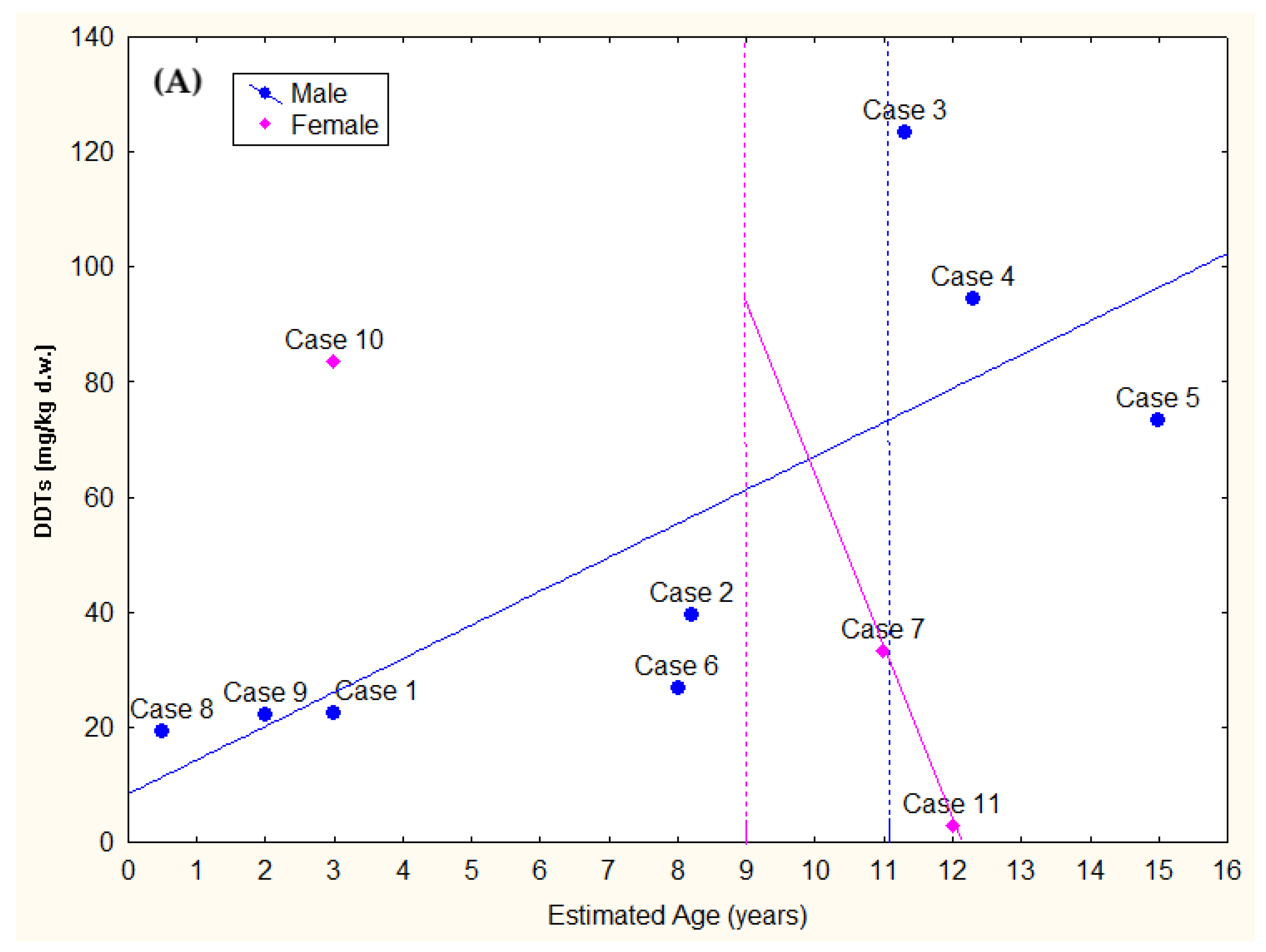
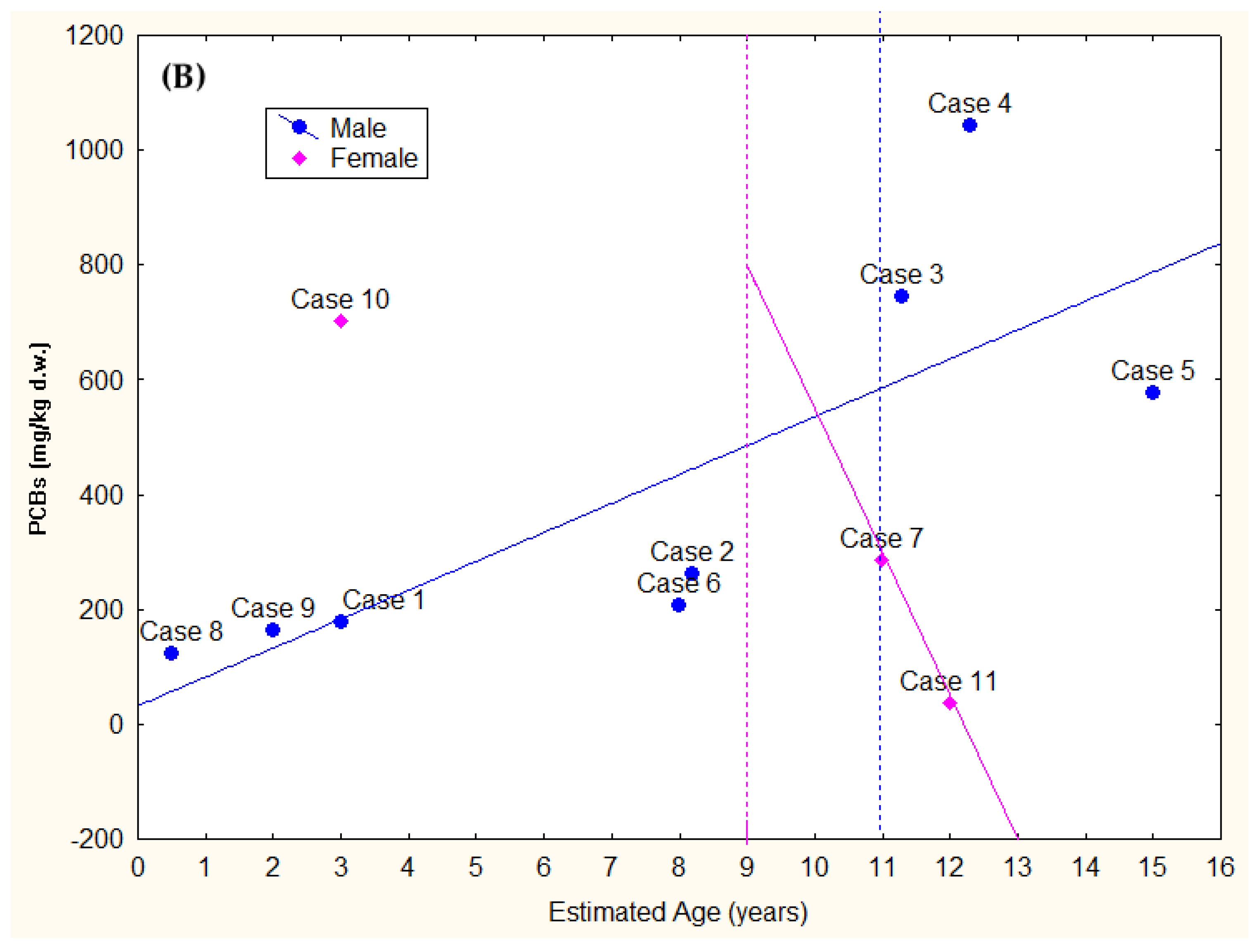
Levels of PCBs, DDTs, and HCB can be linked both to the physiological characteristics of the specimen (for example, age, sex, innate immunity, efficiency of the liver metabolic system, fat layer thickness), as well as to the environment in which they live and the prey they eat. In this regard, despite the small number of samples analyzed, it is really interesting to plot detected levels of DDTs and PCBs as a function of the sex of the specimens and their estimated age, knowing sexual maturity on the basis of necroscopic investigations and taking account of the fact that in the females sexual maturity is reached in approximately 9 years and in the males in approximately 11 years [91] (Figure 3A,B). It is evident that in the females the levels decrease according to the age of the specimens, while in the males the trend is the opposite, with an increase of the levels in the older specimens.
The pattern of PCBs, divided based on their chlorine content and compared with the values of some congeners in Arochlor 1260, is shown in Figure 4 and, from a descriptive point of view, reveals differences between specimens. To evaluate the magnitude of the change, the ranges (minimum and maximum) for each chlorination group and the coefficient of variation (CV) calculated as the ratio of the standard deviation to the mean, showing the extent of variability in relation to the mean of the population, were reported. The higher the CV, the greater the dispersion. The values were: %Penta-CBs range (3.6–14.1) and CV = 0.43; %Hexa–CBs range (33.9–50.8) and CV = 0.10; %Hepta–CBs range (28.2–43.3) and CV = 0.13; %Octa–CBs range (6.6–19.7) and CV = 0.36; %Nona–CBs range (0.3–3.4) and CV = 1.20. When the variability was low for Hexa–CBs and Hepta–CBs and medium–low for Penta–CBs and for Octa–CBs, it was very high for the Nona–CBs with an SD that was even higher than the mean. However, in all specimens except for the specimen from Case 11, the Hexa-CBs were the most represented contaminant, followed by the Hepta-CBs. In all animals, the smallest percentage was that of the Nona-CBs. Case 11 showed a percentage of Octa-CBs, which with Nona-CBs are the more superhydrophobic classes of evaluated PCBs, which was approximately double those found in Arochlor 1260 as well as other bottlenose dolphins.
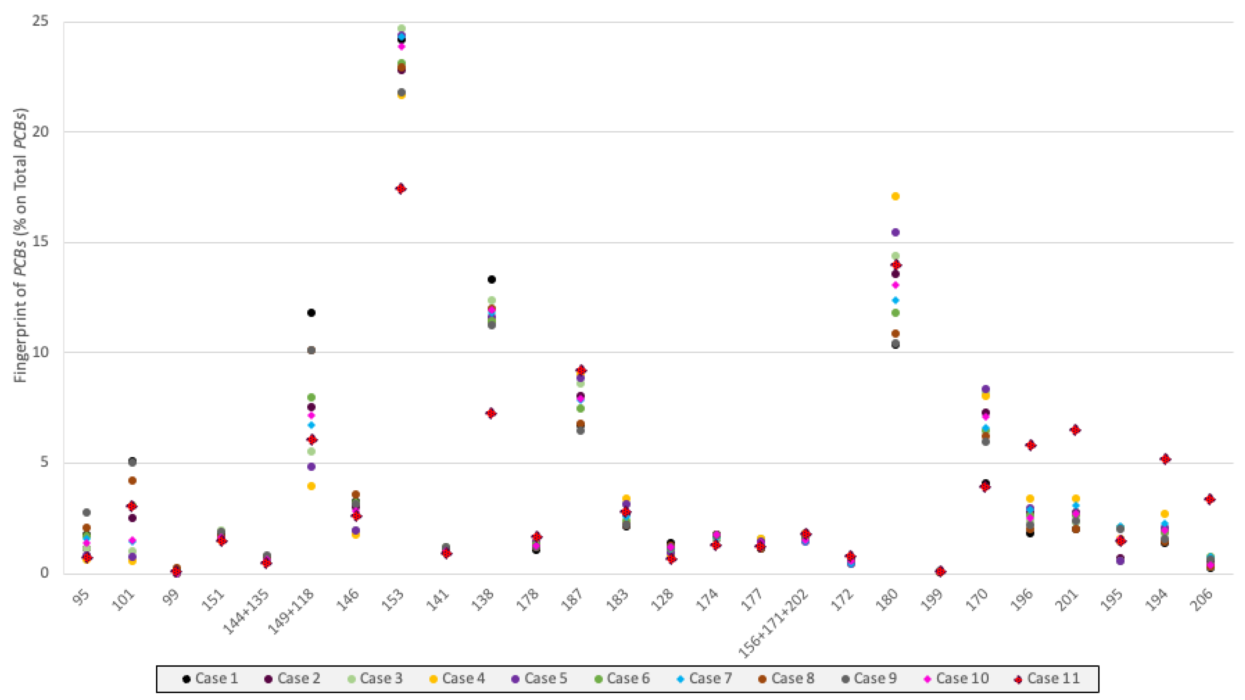
The fingerprints of thirty PCB congeners found revealed in the blubber of each animal are reported in Figure 5. The fingerprint of the specimen from Case 11 has been highlighted with a different indicator point (bigger and red-dotted) because it is the only one that has a completely different trend from the other ten, especially for the congeners 153, 138, 196, 201, 194, and 206. Specifically, the Octa-CBs PCB 201 (6.5%), PCB 196 (5.8%), and PCB 194 (5.2%) were higher than in the other 10 dolphins, in which the range was: PCB 201 (2.0–3.4%), PCB 196 (1.8–3.4%), and PCB 194 (1.4–2.7%). In addition to the PCB 153, the congeners most present in the specimens were PCB 180 (range 10.4–17.1%), PCB 138 (range 7.2–13.3%), PCB 170 (range 4.1–8.3%), PCB 187 (range 6.5–9.2%), and the mixture of PCB 149+118 (range 3.4–11.8%).
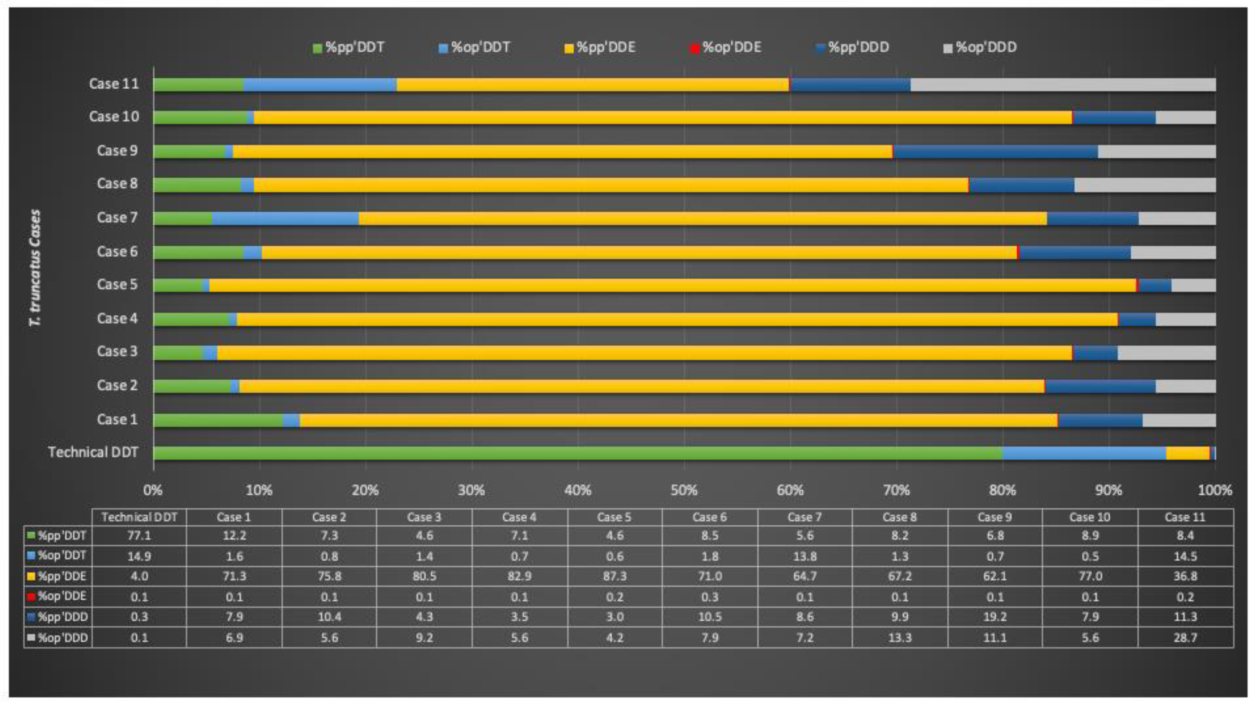
Regarding DDTs, a typical technical DDT is composed of pp′DDT (77.1%), the nearly inactive op′DDT (14.9%), pp′DDD (0.3%), op′DDD (0.1%), pp′DDE (4.0%), op′DDE (0.1%), and unidentified compounds (3.5%) [92]. DDE and DDD are also the main environmental metabolites and degradation products of DDT with similar characteristics. In the technical DDT the ratio of pp′DDE to pp′DDT is 0.05. The fingerprints of the DDT isomers in technical DDT and in the specimens is reported in Figure 6. It was only at the descriptive level that it was possible to show that the DDT isomers had different patterns among the specimens except for the pp′DDE, which was constantly the most present, although with a wide range from 36.8% to 87.3% (Figure 6). The CV was again used to assess the level of variability, and the range of individual isomers was also reported: %pp′DDT range (4.6–12.2) and CV = 0.29, %op′DDT range (0.5–14.5) and CV = 1.55, %pp′DDE range (36.8–87.3) and CV = 0.19, %op′DDE range (0.1–0.3) and CV = 0.60, %pp′DDD range (3.0–19.2) and CV = 0.52, %op′DDD range (4.2–28.7) and CV = 0.72. Very high variability was found for %op′DDT, low for %pp′DDE, medium-high for %op′DDE, %pp′DDD, and op′DDD, and medium-low for %pp′DDT. The high percentage of the op′DDD in the sample Case 11 (28.7%), which also has 14.5% of op′DDT, should be underlined. A high percentage of op′DDT (13.8%) is also present in specimen 7, while this isomer varies between 0.5% and 1.8% in other animals. Op′DDE is present in very low percentage in all samples.
The DDTs/PCBs ratio ranged between 0.08 and 0.17; the pp′DDE/DDTs ratio was always above 0.62, except for Case 11, which had a value of 0.37 and a pp′DDE/pp′DDT ratio ranging from 4.36 and 18.79. The Σop′DDT/DDTs ratio was the ratio that was above 0.20 for only two specimens: Case 7 (0.21) and Case 11 (0.43), which also had high values of the op′DDT/pp′DDT ratio (2.47 and 1.72, respectively). Finally, the ratio Σ2DDT/(Σ2DDE + Σ2DDD), with all o,p′- and p,p′-isomers included, showed a great variability between specimens, ranging between 0.06 and 0.30 (Table 5). In all bottlenose dolphins, the pp′DDE/pp′DDT ratio was higher than 0.05, with a high variability between specimens, with the minimum value in Case 11 (4.76), and with the maximum value in Case 3 (17.41).
Evaluating multiple regression for both DDTs and PCBs in males and females, we found that the two xenobiotics were correlated with age; however, while in males the correlation was significantly positive (DDTs Beta = 0.781 and p = 0.022; PCBs Beta = 0.784 and p = 0.021), in females the correlation, although not statistically significant because of the small number of samples, was negative and has been put forward for representative purposes only (DDTs Beta = −0.961; PCBs Beta = −0.962) (Figure 3A,B).

4. Discussion

Our results, obtained by means of a comprehensive post-mortem examination and diagnostic and toxicological investigations, contribute to understanding the multiple factors involved in the strandings and deaths of cetacean species living in the marine protected area Pelagos. Another aim of the study is to assess the influence of pollution in the development of infectious diseases in bottlenose dolphins (Tursiops truncatus) living in the area through the exploration of possible correlations between sex, sexual maturity, age, pathologies, and levels of OCs.
This species, of conservation interest due to its coastal habitat, is often considered fragile and highly threatened, and most at risk of biological and chemical pollution [9,14]. Notably, the species, exposed to the main anthropogenic stressors including overfishing, fishing interaction, chemical and noise pollution, collisions with boats, and habitat and shoreline degradation [93,94,95,96,97], has recently been reassessed as a species of ‘Least Concern’ on the IUCN Red List after a change of status in 2021 from ‘Vulnerable’ [98,99].
In the Ligurian Sea inside the Pelagos Sanctuary (northwestern Mediterranean Sea), impacts by a wide range of human activities (fisheries, coastal development, and marine traffic) and habitat degradation are present. The prevalence of the species has increased in recent years [100,101], and bottlenose dolphins are regularly seen all year round in coastal waters, used for feeding and nursing their calves, both in the eastern [102] and western sides of area [100].
Of the 21 bottlenose dolphins found stranded dead in the period between 2019 to 2021 along the Ligurian coast, 11 were submitted to a comprehensive post-mortem examination. On the basis of gross and histological pathology and ancillary testing, hypotheses on the cause of death were formulated for five animals.
Specifically, a natural cause of death, attributed to three animals (Cases 4, 8, and 11), was associated with infectious origin, specifically of sub-category viral (Case 11), bacterial (Case 8), and viral, bacterial, and parasitic (Case 4) origin. An anthropic cause of death was attributed in two cases (Cases 5 and 6) and was specifically associated with fishery interaction and with sub-category bycatch with active fishing gear (a consequence of underlying pathologies).
Despite the small sample size of animals suitable for a cause of death evaluation and in addition to the result that 3/5 dolphins had a recognized infectious-related cause of death, the impact of pathogen exposure in the two remaining dolphins should be taken into account. For these two dolphins, interaction with fishing (bycatch) was considered the ultimate cause of death, although the cause of death could be considered a consequence of the underlying pathologies of multifactorial etiology (bacterial, viral, and/or protozoan) that could have predisposed the animals to the fishery interaction.
Moreover, a noticeable pathogen exposure was also demonstrated in the other 5/6 dolphins with an undetermined cause of death (Cases 1, 2, 3, 7, and 10), in which the carcass condition strongly affected the diagnostic data collection or concomitant findings prevented a valid determination of an infectious-related cause.
Furthermore, in Cases 1, 2, 3, 7, 9, and 10, some pathological findings observed facilitated confirmation (Case 10) or suspicion (Cases 7 and 9) of a fishery interaction event, consistent in ingestion and bycatch with the category not determined fishing gear, respectively, that represented significant information to target specific conservation issues for the species in the area.
Considering the main pathogens involved in the animals under investigation, T. gondii, a zoonotic coccidian protozoan regarded as a primary pathogen for cetaceans, responsible for toxoplasmosis, a major emerging disease in these species [52], was detected in more than half of the animals (n = 6, 54.54%). Notably, considering the pathogenic role expressed, an inflammatory response was detected in only one animal (Case 3), with DNA detected at the heart level and T. gondii Ab titers, showing a lymphoplasmacytic myocarditis; moreover, the detection of DNA at a systemic level was demonstrated in two other animals (Case 5 and 10) for which, unfortunately, a reliable evaluation of signs of lesions suggestive of Toxoplasma morbidity or mortality (presence of cysts and/or IHC immunolabeling) could not be assessed (because of generalized autolysis that compromised histological investigations in Case 10) beyond the limited application of IHC routinely performed only on cerebral tissue that undoubtedly limits the overall diagnostic evaluation.
The low T. gondii Ab titers detected in three individuals with T. gondii molecular confirmation (Cases 3, 4, and 5), which are potentially suggestive of an inactive infection occurring despite the low number of animals tested (n = 5), confirm the exposition of bottlenose dolphins to the pathogen in the area; moreover, the low occurrence of inflammatory lesions [103,104,105], unlike in striped dolphins, as repeatedly reported in the area [2,27,35,52,105], seems to agree with what is expected in this coastal species, which experiences more frequent exposure to T. gondii without developing disease.
Dolphin morbillivirus (DMV), which has been associated with lethal disease outbreaks in cetaceans from the Mediterranean since 1990 and with single disease description along the Italian coastline reported by the C.Re.Di.Ma. in standard mortality rates [28,29,30,31,32,33,51,106], was detected in almost half of the animals (n = 5, 45.45%). Unlike the aforementioned pathogen, in three individuals (Cases 2, 4, and 11) DMV was associated with severe specific pathological findings [107], represented by lymphoid depletion (n = 3), suggestive of an immunocompromised host response and associated respectively with a severe mesenteric reactive lymphadenopathy (n = 1), lymphadenitis and tonsillitis with syncytia and a severe lymphoplasmacytic broncho-interstitial pneumonia with syncytia (n = 1), and lymphoplasmacytic meningoencephalitis (n = 1), along with specific immunoreactivity in the CNS and/or many other tissues tested. These results confirm a consistent circulation of the viral pathogen in the area, along with a strong correlation between the infection and the pathological microscopic changes [2,28,29,30,31,32,33,51,108].
Considering the results of toxicological investigations on the levels of dichlorodiphenyltrichloroethane (DDT) and its metabolites, some polychlorinated biphenyls (PCBs), and hexachlorobenzene (HCB) in the blubber of the animals under investigation, all specimens showed the pattern PCBs > DDTs >> HCB. This finding reflects the accumulation trend of these xenobiotics that has been reported in Mediterranean cetaceans in recent years, with PCBs always having the highest levels especially in the northwestern part of the basin [8,109,110].
It is of course important to highlight that the levels of PCBs exceeded in all specimens the threshold of 17 mg/kg lipid weight in blubber for PCB-induced adverse health effects, including immunosuppression [22,111]. Even using the highest reported PCB toxicity threshold for marine mammals (used for reproductive impairment in ring seals (Phoca hispida) in the Baltic Sea and equivalent to 41 mg/kg l.w. [112], all of the bottlenose dolphins examined in this study were above this level (Figure 7). These cut-off values are not used as an absolute value, but rather as a guide for determining whether PCB exposure levels in individual animals are likely to exert a significant biological (immunotoxic) effect based on empirical experimental data.
Furthermore, despite the worldwide reduction or prohibition on manufacture and usage of these POPs, their concentrations in the blubber of the studied bottlenose dolphins still show hazard levels. This finding, as mentioned above, may also be related to both the physiological characteristics of the specimen and the habitat it inhabits. Figure 3A,B can explain how levels of contaminants are related to gender and age. Males and females behave equally in accumulating contaminants until sexual maturity. In males, increasing levels of these POPs with age can be explained by the fact that this gender continuously accumulates contaminants in the blubber throughout life [113], as cetaceans have an inefficient detoxifying metabolic system in relation to these organochlorines and a very thick layer of fat, which, in addition to thermal insulation and energy reserve, functions as a target tissue for the accumulation of lipoaffin contaminants. Instead, in females, a decrease in contaminant levels after sexual maturity can be explained by the possibility of offloading (up to 90%) the total load of contaminants to the offspring during the gestation and lactation periods [65]. In bottlenose dolphins, pregnancy lasts about 12 months and usually a single offspring is released [114]. The mean interval between births is estimated to be 3.5 ± 1.6 years, ranging from 2 to 7 years [115]. However, the transfer of contaminants is particularly effective during lactation as the milk of these marine mammals is very rich in lipids dispersed in the form of droplets (milk fat globules) and packed into a membrane inherited from the mammary gland [116]. In a recent study [117], it was found that in bottlenose dolphin milk there is an average of 20.57% of lipids; the study also highlighted the abundance of unsaturated fatty acids (Unsaturated Fatty Acids/Saturated Fatty Acids ratio = 2.64). However, it should not be forgotten that the composition of fatty acids in milk is linked to food intake and mobilization from fat (blubber) deposits [118]. The mean age at weaning in free-ranging dolphins has been observed to be 3.2 years, although one calf may be weaned up to its fifth birthday and the withdrawal time varies between populations and individuals [115]. There is also some evidence of mortality of the first-born offspring, and this has been associated with the very high (possibly lethal) dose of PCBs that newborns may receive from their mothers [23,119,120]. Of the three females studied in this work, Case 10, based on gonadal appearance at gross investigation, resulted in an immature female, which was confirmed by the calculation of the age based on the length. This shows how theoretical models, even if taken with caution, can help to interpret many results. This should also be considered when discussing levels of DDTs and PCBs in males, as there can be individual variability of the specimens with respect to the total length of the body.
PCB and DDT concentrations do not appear to be closely correlated with the nutritional status of the specimens, evaluated in eight of the eleven animals. In fact, the only bottlenose dolphin with poor nutritional status (Case 3) showed the second highest level of PCBs and the first highest level of DDTs. Unfortunately, the cause of death of this specimen has not been determined, and at the infectious level it presented only T. gondii positivity. Case 5, which was diagnosed with DMV, had a moderate to good nutritional status. Only one case was determined as having an ND nutritional status. The interesting thing was that the three females studied all belonged to the DMV group. For one of these (Case 11), the viral infection was considered the cause of death. This was the oldest female (12 years old) with the lowest levels of OCs, but with a fingerprint for both PCBs and DDTs very different from other specimens, regardless of sex (Figure 4, Figure 5 and Figure 6). For PCBs, compared with other specimens, this female exhibited higher proportions of superhydrophobic class Octa-CBs and a lower percentage of recalcitrant PCBs 153 (17%). Like all other specimens, however, Case 11 had the highest percentages of Hexa-CBs and Hepta-CBs. This is consistent with other research demonstrating how Penta-CBs, Hexa-CBs, Hepta-CBs, and Octa-CBs are more slowly eliminated by organs and tissues than compounds with lower chlorine content [121,122]. Hexa-CBs are also typically predominant as PCB residues in cetaceans because of their widespread and persistent nature in the environment [123]. With respect to PCB 153, Case 11 confirmed the results of Fair et al. [123] in which the adult females had lower percentages of this congener than juvenile and adult males. All other specimens in this study (juveniles or adult males, except for Case 7) showed PCBs 153 ranging between 21.7% and 24.7%, and this congener was the most represented. The PCB 153 was the most abundant in all animals, probably since this congener is particularly persistent as it has chlorines in positions two, four and five of both rings of the biphenyl [124,125,126] and no adjacent, unsubstituted carbons in the ortho-meta position [127]. It is also very important in toxicology, being one of the four compounds belonging to the TEI-OCs showing qualities of both T-OCs and EDC-OCs as well as IS-OCs (Table 4). This congener, together with the other congeners most represented in dolphins, PCB 138 and PCB 180, is considered highly resistant to metabolization by marine mammals [128]. Furthermore, these three non-dioxin-like PCBs (NDL-PCBs), along with three others (PCB 28, PCB 52, and PCB 101), are considered “PCBs indicators” by the European Food Safety Authority due to their significant occurrence in environmental matrices and animal tissues but also in abiotic substrates [129,130,131,132]. In our study on bottlenose dolphin blubber, we also identified PCB 101, whereas PCB 28 and 52 were not evaluated. Detectable PCB 101 concentrations were present in all samples, but there was high variability between them (range: 0.6–5.1%). The toxicity of these PCBs is also dependent on differences in metabolism, absorption, and excretion, given that less chlorinated congeners such as PCB 28, PCB 52, and PCB 101 are more easily metabolized and excreted than more persistent congeners (PCB 138, PCB 153, and PCB 180) [133]. Furthermore, PCB 118 is included in the TEI-OCs group, as is PCB 153. Other research identified only four representative PCBs: PCB 153, PCB 138, PCB 118, and PCB 180, as these have been measured with minimal errors and are often used in the exploration of congener-specific effects [134]. For these two groups of PCBs, the first is represented only by four out of six PCBs (PCB 101, PCB 138, PCB 153, and PCB 180) and the second by PCB 118, PCB 138, PCB 153, and PCB 180; the mean percentage of total PCBs in the 11 specimens was 49.9% (SD = 3.0%) and 51.8% (SD = 3.5%), respectively. Therefore, also in this study, these PCB groups may be considered highly representative for the accumulation of these xenobiotics, accounting for approximately 50% of total PCB concentrations.
In addition, for DDTs, the specimen Case 11 also had abnormal percentages of op′DDT and op′DDD (Figure 5). These two compounds contribute significantly to the alteration of the ∑op′DDT/DDTs ratio, where an excess of these isomers greater than 20% suggests a non-insecticidal (or industrial) source of this xenobiotic [135]. In Case 11, the value was 0.43, and only one other specimen, Case 7, had a value exceeding the limit of 20% (ratio equal to 0.21). Indeed, wastes from the processing of technical DDT are generally enriched with op′ isomers compared to the pp′ DDT; the resulting compound is industrially applicable and is not subject to regulation for the use of DDT insecticide mixtures [109,136,137].
A high ratio of pp′DDE to pp′DDT in the analyzed samples (>0.05) has been associated with a lack of exposure to newly applied DDT [138,139] (and therefore there were no recent deposits of insecticide into that ecosystem), but also with the degradation of most of the active substance (pp′DDT) to pp′DDE [140]. In all bottlenose dolphins, the pp′DDE/pp′DDT ratio had a value much larger than 0.05, but there were many variables between specimens, with a minimum value in the specimen Case 11 (4.76) and a maximum value in the specimen Case 3 (17.41) (Table 5). The Case 11 sample value of 4.76 was above 0.05 of the technical DDT, but it was not too high to suggest an old pesticide introduction. The pp′DDE/DDTs ratio, as well as having a similar meaning to the pp′DDE/pp′DDT ratio, can also indicate the efficiency of metabolic processes [141]. In fact, the ppDDE/DDTs ratio indicates the relative abundance of metabolized forms of DDT. In the blubber of studied bottlenose dolphins, the ratio varies from 0.37 to 0.87 (Table 5). A value of this equal to 0.6 is considered critical, while higher values indicate that there are no new contamination inputs in the study area [142]. All our samples exceeded this limit except Case 11, with 0.37. The value of the Case 11 dolphin points to an alarming situation. This is also partly confirmed by the similarly significant Σ2DDT/(Σ2DDE + Σ2DDD) ratio (Table 5). Although there are no solid guidelines for what defines recent or old inputs, the value of one is normally taken as a reference to distinguish between legacy and recent inputs [143,144]. Values lower than one may be associated with historical DDT inputs, whereas higher values indicate new technical DDT inputs. The value found for all samples was <1. These results may indicate that there is no recent use of DDT in this region of the Mediterranean Sea, but for both females (Case 11 and Case 7) the ratio value was higher than for the other animals, suggesting a more recent intake of DDT [145]. In addition, the ratio op′DDT/pp′DDT is regarded as an indicator discriminating between the use of technical DDT and Dicofol [137,145]. Dicofol is a miticidal pesticide and acaricide synthesized from DDT, and therefore the isomers op′DDT, op′DDE, pp′DDT, or pp′-Cl-DDT (1,2,2,2-tetrachloro-1,1-bis (4-chlorophenyl) ethane—a chlorinated DDT intermediate that leads to Dicofol prior hydrolysis) are usually found in its formulations [146]. This pesticide, listed in Annex A of the Stockholm Convention on Persistent Organic Pollutants in 2019 [147], is very toxic to aquatic organisms, highly bioaccumulative, and extremely persistent. It is also known to be neurotoxic and possess endocrine disrupting properties, both as an original product and with its decomposition products [148]. Total DDT content ranged from 0.3% to 14.3% of Dicofol’s total weight [149], with some exceptions (average 20% of DDT in China) [137]. Since the technical DDT is usually 0.19 [150] and op′DDT shows a shorter half-life than pp′DDT in the environment [151], it appears reasonable to assume the influence of dicofol-type contamination if the ratio of op′DDT/pp′DDT is >0.2. In these samples, the values of this ratio ranged from 0.06 to 2.47, with only the female Case 7 having levels >2 (Table 5).
Finally, the ΣDDTs/ΣPCBs relationship determines which sector, agricultural or industrial, accounts for a larger part of the total concentration of organochlorine compounds. Ratios of less than one indicate an increased influence of industrial pollution or high population densities on organochlorine accumulation in samples. The ratios higher than one indicate that the agricultural sector would play an important role in the concentration of chlorinated hydrocarbons. In all the bottlenose dolphins analyzed, this ratio was much lower than one, confirming a significant impact due to industrial contaminants for the marine mammals in this part of the Mediterranean basin (Table 5) [65,122,152,153].
Considering all data about the concentration of POPs, the most interesting result is the percentage of OCs with immunosuppressant characteristics named IS-OCs (Table 4). In all specimens, except for the female Case 11, the IS-OCs represent more than 50% of the total POPs detected, reaching about 63% in the specimen Case 1. This young male, still immature, was apparently healthy due to a “good” nutritional condition code, yet nevertheless was rather compromised, having a high number of infections, even if it did not present with DMV or other diseases considered to cause death.
Based on this result, efforts were made to identify possible correlations between the main immunosuppression-related pathologies in the specimens and this group of OCs (IS-OCs), in addition to other groups of contaminants identified as having high toxicological value (Table 6). Four subgroups, depending on the different pathologies diagnosed, were considered (paragraph 3.3), but the adult female Case 11 has not been included in this data processing for the reasons already highlighted (being a specimen that differs from all the others probably due to a different physiology linked to sex and age, and therefore to its life history). The results, although of no statistical significance, are extremely interesting (Table 6). In fact, except for DDTs that do not seem to represent a particular stress for these marine mammals, the other toxicological groups considered have higher levels within the three subgroups 1, 2, and 3 compared to their control-subgroups. A comparison between the specimens in subgroup 3 and those in subgroup 4 showed that the simultaneous presence of DMV and lymphoid depletion occurred in specimens with the highest levels of immunosuppressive or highly toxic xenobiotics.

5. Conclusions

The present study provides novel data on the correlation between pathological evidence of infection and levels of OCs in bottlenose dolphin species living in the Pelagos Sanctuary, giving significant data on the possible effects of the different OCs in relation to their toxicological characteristics and to the ratios between the different compounds detected.
Considering the results of this survey, we confirm a high level of exposure to pathogens, with relevant infectious–associated pathological findings, and to concerning levels of organochlorine pollutants in the bottlenose dolphins under study, with high levels of PCBs in all animals investigated, usually associated with induced adverse health effects, including immuno-suppression.
These data highlight the need for continuous surveillance and monitoring studies on stranded cetaceans, maintaining a multidisciplinary and standardized approach, to improve knowledge of the impact of pathogens and pollutants and to better understand the interaction between pathogen, host, and environmental factors, in tight agreement with the “One Health” concept.
Moreover, our data are valuable for understanding health and mortality trends in cetacean populations and can provide information for establishing policies for cetacean conservation and management in such an important protected area of the Mediterranean basin.

Supplementary Materials

The following supporting information can be downloaded at: https://www.mdpi.com/article/10.3390/d15040569/s1, Table S1: Anamnestic data, main gross and microscopical findings, hypothesis of causa mortis, and most significant pathogens and parasites detected in the animals under study.

Author Contributions

Conceptualization, C.G. and L.M. (Letizia Marsili); methodology, C.G. and L.M. (Letizia Marsili); formal analysis, C.G., C.C., F.G. (Federica Giorda), L.M. (Lorenzo Minoia), G.C., F.C., E.F. and L.M. (Letizia Marsili); investigation, C.G., F.G. (Federica Giorda), C.C., G.C., F.C., I.C., D.A., F.G. (Fulvio Garibaldi), A.D., M.G., L.S., K.V., L.M. (Loretta Masoero), C.E.D.F., L.M. (Lorenzo Minoia) and L.M. (Letizia Marsili); data curation, C.G., F.G. (Federica Giorda), L.M. (Lorenzo Minoia) and L.M. (Letizia Marsili); writing—original draft preparation, C.G., L.M. (Lorenzo Minoia), F.G. (Federica Giorda) and L.M. (Letizia Marsili); writing—review and editing: C.G., F.G. (Federica Giorda), L.M. (Lorenzo Minoia), G.C., E.F., F.C. and L.M. (Letizia Marsili); funding acquisition, C.C. and C.G. All authors have read and agreed to the published version of the manuscript.

Funding

This research was funded by the Italian Ministry of Health (Ricerca Corrente 2018 IZS/PLV 09/18) and Ricerca Corrente 2021 IZS/PLV 06/21.

Institutional Review Board Statement

Not applicable. Ethical review and approval were waived for this study, as neither were animals sacrificed, nor were experiments performed with live animals. The permission for the management of dead stranded cetaceans was issued by the Italian Ministry of Health.

Data Availability Statement

The data presented in this study are available within the article and the Supplementary Materials.

Acknowledgments

The authors are grateful to Elena Fontanesi, for the valuable cooperation received in carcasses evaluation and assistance for sampling, to Guido Pietroluongo, for the collaboration in the assessment of findings related to fishery interaction in some animals under study and for necropsy investigations on one animal, along with Giorgia Corazzola, and to Cinzia Centelleghe, for the valuable assistance in the setup of specific IHC for capture myopathy confirmation. The authors are also grateful for the collaboration provided by the Department of Prevention, Local Veterinary Services (ASL 1 Imperiese).

Conflicts of Interest

The authors declare no conflict of interest.

References

  1. Aguirre, A.A.; Ostfeld, R.; Daszak, P. New Directions in Conservation Medicine: Applied Cases of Ecological Health; Oxford University Press: New York, NY, USA, 2012; ISBN 978-0-19-973147-3. [Google Scholar]
  2. Giorda, F.; Ballardini, M.; Di Guardo, G.; Pintore, M.D.; Grattarola, C.; Iulini, B.; Mignone, W.; Goria, M.; Serracca, L.; Varello, K.; et al. Postmortem findings in cetaceans found stranded in the Pelagos Sanctuary, Italy, 2007–14. J. Wildl. Dis. 2017, 53, 795–803. [Google Scholar] [CrossRef] [PubMed]
  3. Bossart, G.D. Marine Mammals as Sentinel Species for Oceans and Human Health. Vet. Pathol. 2011, 48, 676–690. [Google Scholar] [CrossRef] [PubMed]
  4. Centelleghe, C.; Da Dalt, L.; Marsili, L.; Zanetti, R.; Fernandez, A.; Arbelo, M.; Sierra, E.; Castagnaro, M.; Di Guardo, G.; Mazzariol, S. Insights Into Dolphins’ Immunology: Immuno-Phenotypic Study on Mediterranean and Atlantic Stranded Cetaceans. Front. Immunol. 2019, 10, 888. [Google Scholar] [CrossRef] [PubMed]
  5. Marsili, L.; Di Guardo, G.; Mazzariol, S.; Casini, S. Insights Into Cetacean Immunology: Do Ecological and Biological Factors Make the Difference? Front. Immunol. 2019, 10, 1219. [Google Scholar] [CrossRef]
  6. Mazzariol, S.; Marcer, F.; Mignone, W.; Serracca, L.; Goria, M.; Marsili, L.; Di Guardo, G.; Casalone, C. Dolphin Morbillivirus and Toxoplasma gondii Coinfection in a Mediterranean Fin Whale (Balaenoptera physalus). BMC Vet. Res. 2012, 8, 20. [Google Scholar] [CrossRef]
  7. Desforges, J.-P.W.; Sonne, C.; Levin, M.; Siebert, U.; De Guise, S.; Dietz, R. Immunotoxic Effects of Environmental Pollutants in Marine Mammals. Environ. Int. 2016, 86, 126–139. [Google Scholar] [CrossRef]
  8. Marsili, L.; Jiménez, B.; Borrell, A. Persistent Organic Pollutants in Cetaceans Living in a Hotspot Area: The Mediterranean Sea. In Marine Mammal Ecotoxicology; Fossi, M.C., Panti, C., Eds.; Academic Press: Cambridge, MA, USA, 2018; pp. 185–212. ISBN 978-0-12-812144-3. [Google Scholar]
  9. Van Bressem, M.F.; Raga, J.A.; Di Guardo, G.; Jepson, P.D.; Duignan, P.J.; Siebert, U.; Barrett, T.; de Oliveira Santos, M.C.; Moreno, I.B.; Siciliano, S.; et al. Emerging Infectious Diseases in Cetaceans Worldwide and the Possible Role of Environmental Stressors. Dis. Aquat. Org. 2009, 86, 143–157. [Google Scholar] [CrossRef]
  10. Hoegh-Guldberg, O.; Bruno, J.F. The Impact of Climate Change on the World’s Marine Ecosystems. Science 2010, 328, 1523–1528. [Google Scholar] [CrossRef]
  11. Sousa, A.; Alves, F.; Dinis, A.; Bentz, J.; Cruz, M.J.; Nunes, J.P. How Vulnerable Are Cetaceans to Climate Change? Developing and Testing a New Index. Ecol. Indic. 2019, 98, 9–18. [Google Scholar] [CrossRef]
  12. Sanderson, C.E.; Alexander, K.A. Unchartered Waters: Climate Change Likely to Intensify Infectious Disease Outbreaks Causing Mass Mortality Events in Marine Mammals. Glob. Chang. Biol. 2020, 26, 4284–4301. [Google Scholar] [CrossRef]
  13. Van Weelden, C.; Towers, J.R.; Bosker, T. Impacts of Climate Change on Cetacean Distribution, Habitat and Migration. Clim. Chang. Ecol. 2021, 1, 100009. [Google Scholar] [CrossRef]
  14. Reed, J.; Harcourt, R.; New, L.; Bilgmann, K. Extreme Effects of Extreme Disturbances: A Simulation Approach to Assess Population Specific Responses. Front. Mar. Sci. 2020, 7, 519845. [Google Scholar] [CrossRef]
  15. Fossi, M.C.; Marsili, L.; Casini, S.; Bucalossi, D. Development of New-Tools to Investigate Toxicological Hazard Due to Endocrine Disruptor Organochlorines and Emerging Contaminants in Mediterranean Cetaceans. Mar. Environ. Res. 2006, 62, S200–S204. [Google Scholar] [CrossRef] [PubMed]
  16. Green, A.; Larson, S. A Review of Organochlorine Contaminants in Nearshore Marine Mammal Predators. J. Environ. Anal. Toxicol. 2016, 6, 370. [Google Scholar] [CrossRef]
  17. Neri, A.; Sartor, P.; Voliani, A.; Mancusi, C.; Marsili, L. Diet of Bottlenose Dolphin, Tursiops truncatus (Montagu, 1821), in the Northwestern Mediterranean Sea. Diversity 2023, 15, 21. [Google Scholar] [CrossRef]
  18. Jones, K.C.; de Voogt, P. Persistent Organic Pollutants (POPs): State of the Science. Environ. Pollut. 1999, 100, 209–221. [Google Scholar] [CrossRef]
  19. Dron, J.; Wafo, E.; Boissery, P.; Dhermain, F.; Bouchoucha, M.; Chamaret, P.; Lafitte, D. Trends of Banned Pesticides and PCBs in Different Tissues of Striped Dolphins (Stenella coeruleoalba) Stranded in the Northwestern Mediterranean Reflect Changing Contamination Patterns. Mar. Pollut. Bull. 2022, 174, 113198. [Google Scholar] [CrossRef]
  20. Borrell, A.; Aguilar, A.; Tornero, V.; Sequeira, M.; Fernandez, G.; Alıs, S. Organochlorine Compounds and Stable Isotopes Indicate Bottlenose Dolphin Subpopulation Structure around the Iberian Peninsula. Environ. Int. 2006, 32, 516–523. [Google Scholar] [CrossRef]
  21. Gonzalvo, J.; Lauriano, G.; Hammond, P.S.; Viaud-Martinez, K.A.; Fossi, M.C.; Natoli, A.; Marsili, L. Chapter Nine—The Gulf of Ambracia’s Common Bottlenose Dolphins, Tursiops truncatus: A Highly Dense and yet Threatened Population. In Advances in Marine Biology; Notarbartolo Di Sciara, G., Podestà, M., Curry, B.E., Eds.; Mediterranean Marine Mammal Ecology and Conservation; Academic Press: Cambridge, MA, USA, 2016; Volume 75, pp. 259–296. [Google Scholar]
  22. Jepson, P.D.; Deaville, R.; Barber, J.L.; Aguilar, À.; Borrell, A.; Murphy, S.; Barry, J.; Brownlow, A.; Barnett, J.; Berrow, S.; et al. PCB Pollution Continues to Impact Populations of Orcas and Other Dolphins in European Waters. Sci. Rep. 2016, 6, 18573. [Google Scholar] [CrossRef]
  23. Genov, T.; Jepson, P.D.; Barber, J.L.; Hace, A.; Gaspari, S.; Centrih, T.; Lesjak, J.; Kotnjek, P. Linking Organochlorine Contaminants with Demographic Parameters in Free-Ranging Common Bottlenose Dolphins from the Northern Adriatic Sea. Sci. Total Environ. 2019, 657, 200–212. [Google Scholar] [CrossRef]
  24. Wafo, E.; Sarrazin, L.; Diana, C.; Dhermain, F.; Schembri, T.; Lagadec, V.; Pecchia, M.; Rebouillon, P. Accumulation and Distribution of Organochlorines (PCBs and DDTs) in Various Organs of Stenella coeruleoalba and a Tursiops truncatus from Mediterranean Littoral Environment (France). Sci. Total Environ. 2005, 348, 115–127. [Google Scholar] [CrossRef] [PubMed]
  25. Shoham-Frider, E.; Kress, N.; Wynne, D.; Scheinin, A.; Roditi-Elsar, M.; Kerem, D. Persistent Organochlorine Pollutants and Heavy Metals in Tissues of Common Bottlenose Dolphin (Tursiops truncatus) from the Levantine Basin of the Eastern Mediterranean. Chemosphere 2009, 77, 621–627. [Google Scholar] [CrossRef] [PubMed]
  26. Romanić, S.H.; Holcer, D.; Lazar, B.; Klinčić, D.; Mackelworth, P.; Fortuna, C.M. Organochlorine Contaminants in Tissues of Common Bottlenose Dolphins Tursiops truncatus from the Northeastern Part of the Adriatic Sea. Environ. Toxicol. Pharmacol. 2014, 38, 469–479. [Google Scholar] [CrossRef]
  27. Grattarola, C.; Giorda, F.; Iulini, B.; Pintore, M.D.; Pautasso, A.; Zoppi, S.; Goria, M.; Romano, A.; Peletto, S.; Varello, K.; et al. Meningoencephalitis and Listeria monocytogenes, Toxoplasma gondii and Brucella spp. Coinfection in a Dolphin in Italy. Dis. Aquat. Org. 2016, 118, 169–174. [Google Scholar] [CrossRef] [PubMed]
  28. C.Re.Di.Ma. Reports Strandings. C.Re.Di.Ma. Italian Diagnostic Report on Stranded Cetaceans (2016); Open Science Framework: Impera, Italy, 2016. [Google Scholar]
  29. C.Re.Di.Ma. Reports Strandings. C.Re.Di.Ma. Italian Diagnostic Report on Stranded Cetaceans (2017); Open Science Framework: Impera, Italy, 2017. [Google Scholar]
  30. C.Re.Di.Ma. Reports Strandings. C.Re.Di.Ma. Italian Diagnostic Report on Stranded Cetaceans (2018); Open Science Framework: Impera, Italy, 2018. [Google Scholar]
  31. C.Re.Di.Ma. Reports Strandings. C.Re.Di.Ma. Italian Diagnostic Report on Stranded Cetaceans (2019); Open Science Framework: Impera, Italy, 2019. [Google Scholar]
  32. C.Re.Di.Ma. Reports Strandings. C.Re.Di.Ma. Italian Diagnostic Report on Stranded Cetaceans (2020); Open Science Framework: Impera, Italy, 2020. [Google Scholar]
  33. C.Re.Di.Ma. Reports Strandings. C.Re.Di.Ma. Italian Diagnostic Report on Stranded Cetaceans (2021); Open Science Framework: Impera, Italy, 2021. [Google Scholar]
  34. Grattarola, C.; Gallina, S.; Giorda, F.; Pautasso, A.; Ballardini, M.; Iulini, B.; Varello, K.; Goria, M.; Peletto, S.; Masoero, L.; et al. First Report of Salmonella 1,4,[5],12:I:- In Free-Ranging Striped Dolphins (Stenella coeruleoalba), Italy. Sci. Rep. 2019, 9, 6061. [Google Scholar] [CrossRef]
  35. Giorda, F.; Di Guardo, G.; Varello, K.; Pautasso, A.; Sierra, E.; Pintore, M.D.; Grattarola, C.; Colella, E.M.; Berio, E.; Goria, M.; et al. Retrospective Immunohistochemical Investigation on Dolphin Morbillivirus Infection by Comparing the Performance of Heterologous Monoclonal and Polyclonal Antibodies—Short Communication. Acta Vet. Hung. 2021, 69, 204–210. [Google Scholar] [CrossRef]
  36. Romani-Cremaschi, U.; Zoppi, S.; Mattioda, V.; Audino, T.; Marsili, L.; Varello, K.; Iulini, B.; Marra, C.; Zoccola, R.; Battistini, R.; et al. Morganella Morganii Septicemia and Concurrent Renal Crassicaudiasis in a Cuvier’s Beaked Whale (Ziphius cavirostris) Stranded in Italy. Front. Mar. Sci. 2023, 9, 1058724. [Google Scholar] [CrossRef]
  37. Gazzetta Ufficiale della Repubblica Italiana. Available online: https://www.mase.gov.it/sites/default/files/archivio/normativa/legge_11102001_391_ratifica_accordo_santuario_mammiferi_marini.pdf (accessed on 19 January 2023).
  38. Pelagos Sanctuary. Available online: https://www.sanctuaire-pelagos.org/en/human-activities/coastal-urbanization (accessed on 20 March 2023).
  39. Panti, C.; Spinsanti, G.; Marsili, L.; Casini, S.; Frati, F.; Fossi, M.C. Ecotoxicological Diagnosis of Striped Dolphin (Stenella coeruleoalba) from the Mediterranean Basin by Skin Biopsy and Gene Expression Approach. Ecotoxicology 2011, 20, 1791–1800. [Google Scholar] [CrossRef]
  40. Fossi, M.C.; Panti, C.; Marsili, L.; Maltese, S.; Spinsanti, G.; Casini, S.; Caliani, I.; Gaspari, S.; Muñoz-Arnanz, J.; Jimenez, B.; et al. The Pelagos Sanctuary for Mediterranean Marine Mammals: Marine Protected Area (MPA) or Marine Polluted Area? The Case Study of the Striped Dolphin (Stenella coeruleoalba). Mar. Pollut. Bull. 2013, 70, 64–72. [Google Scholar] [CrossRef]
  41. IJsseldijk, L.; Brownlow, A.; Mazzariol, S. Best Practice on Cetacean Post Mortem Investigation and Tissue Sampling; ACCOBAMS ASCOBANS: Rotterdam, The Netherlands, 2019. [Google Scholar]
  42. Tanaka, H.; Li, G.; Uchida, Y.; Nakamura, M.; Ikeda, T.; Liu, H. Measurement of Time-Varying Kinematics of a Dolphin in Burst Accelerating Swimming. PLoS ONE 2019, 14, e0210860. [Google Scholar] [CrossRef]
  43. Butti, C.; Corain, L.; Cozzi, B.; Podestà, M.; Pirone, A.; Affronte, M.; Zotti, A. Age Estimation in the Mediterranean Bottlenose Dolphin Tursiops truncatus (Montagu 1821) by Bone Density of the Thoracic Limb. J. Anat. 2007, 211, 639–646. [Google Scholar] [CrossRef] [PubMed]
  44. Mann, J.; Smuts, B.B. Natal Attraction: Allomaternal Care and Mother–Infant Separations in Wild Bottlenose Dolphins. Anim. Behav. 1998, 55, 1097–1113. [Google Scholar] [CrossRef] [PubMed]
  45. Mann, J.; Connor, R.C.; Barre, L.M.; Heithaus, M.R. Female Reproductive Success in Bottlenose Dolphins (Tursiops Spp.): Life History, Habitat, Provisioning, and Group-Size Effects. Behav. Ecol. 2000, 11, 210–219. [Google Scholar] [CrossRef]
  46. Díaz López, B. Interactions between Mediterranean Bottlenose Dolphins (Tursiops truncatus) and Gillnets off Sardinia, Italy. ICES J. Mar. Sci. 2006, 63, 946–951. [Google Scholar] [CrossRef]
  47. Perrin, W.F.; Reilly, S.B. Reproductive Parameters of Dolphins and Small Whales of the Family Deiphinidae. Rep. Int. Whal. Comm. 1984, 6, 97–133. [Google Scholar]
  48. Cozzi, B.; Huggenberger, S.; Oelschläger, H. Urinary System, Genital Systems, and Reproduction. In Anatomy of Dolphins; Cozzi, B., Huggenberger, S., Oelschläger, H., Eds.; Academic Press: San Diego, CA, USA, 2017; Chapter 9; pp. 369–409. ISBN 978-0-12-407229-9. [Google Scholar]
  49. Anderson, R.C.; Chabaud, A.G.; Willmott, S. Keys to Genera of the Superfamily Metastrongyloidea. In C.I.H. Keys to the Nematode Parasites of Vertebrates; Commonwealth Agricultural Bureau: Farnham Royal, UK, 1978; Volume 5, pp. 1–40. [Google Scholar]
  50. Khalil, L.F.; Jones, A.; Bray, R.A. Keys to the Cestode Parasites of Vertebrates; CAB International: Wallingford, UK, 1994; p. 752. [Google Scholar]
  51. Giorda, F.; Crociara, P.; Iulini, B.; Gazzuola, P.; Favole, A.; Goria, M.; Serracca, L.; Dondo, A.; Crescio, M.I.; Audino, T.; et al. Neuropathological Characterization of Dolphin Morbillivirus Infection in Cetaceans Stranded in Italy. Animals 2022, 12, 452. [Google Scholar] [CrossRef]
  52. Di Guardo, G.; Proietto, U.; Di Francesco, C.E.; Marsilio, F.; Zaccaroni, A.; Scaravelli, D.; Mignone, W.; Garibaldi, F.; Kennedy, S.; Forster, F.; et al. Cerebral Toxoplasmosis in Striped Dolphins (Stenella coeruleoalba) Stranded Along the Ligurian Sea Coast of Italy. Vet. Pathol. 2010, 47, 245–253. [Google Scholar] [CrossRef]
  53. World Organisation for Animal Health (OIE). Manual of Diagnostic Tests and Vaccines for Terrestrial Animals (Terrestrial Manual), 8th ed.; World Organisation for Animal Health (OIE): Paris, France, 2018; ISBN 978-92-95108-18-9. [Google Scholar]
  54. Verna, F.; Giorda, F.; Miceli, I.; Rizzo, G.; Pautasso, A.; Romano, A.; Iulini, B.; Pintore, M.D.; Mignone, W.; Grattarola, C.; et al. Detection of Morbillivirus Infection by RT-PCR RFLP Analysis in Cetaceans and Carnivores. J. Virol. Methods 2017, 247, 22–27. [Google Scholar] [CrossRef]
  55. VanDevanter, D.R.; Warrener, P.; Bennett, L.; Schultz, E.R.; Coulter, S.; Garber, R.L.; Rose, T.M. Detection and Analysis of Diverse Herpesviral Species by Consensus Primer PCR. J. Clin. Microbiol. 1996, 34, 1666–1671. [Google Scholar] [CrossRef]
  56. Vitale, M. A High Sensitive Nested PCR for Toxoplasma Gondii Detection in Animal and Food Samples. J. Microb. Biochem. Technol. 2013, 5, 39–41. [Google Scholar] [CrossRef]
  57. Bounaadja, L.; Albert, D.; Chénais, B.; Hénault, S.; Zygmunt, M.S.; Poliak, S.; Garin-Bastuji, B. Real-Time PCR for Identification of Brucella Spp.: A Comparative Study of IS711, Bcsp31 and per Target Genes. Vet. Microbiol. 2009, 137, 156–164. [Google Scholar] [CrossRef]
  58. Osorio, C.R.; Romalde, J.L.; Barja, J.L.; Toranzo, A.E. Presence of Phospholipase-D (Dly) Gene Coding for Damselysin Production Is Not a Pre-Requisite for Pathogenicity in Photobacterium damselae Subsp. damselae. Microb. Pathog. 2000, 28, 119–126. [Google Scholar] [CrossRef] [PubMed]
  59. Barnett, J.; Dastjerdi, A.; Davison, N.; Deaville, R.; Everest, D.; Peake, J.; Finnegan, C.; Jepson, P.; Steinbach, F. Identification of Novel Cetacean Poxviruses in Cetaceans Stranded in South West England. PLoS ONE 2015, 10, e01243. [Google Scholar] [CrossRef] [PubMed]
  60. Hernández-Mora, G.; González-Barrientos, R.; Morales, J.-A.; Chaves-Olarte, E.; Guzmán-Verri, C.; Baquero-Calvo, E.; De-Miguel, M.-J.; Marín, C.-M.; Blasco, J.-M.; Moreno, E. Neurobrucellosis in Stranded Dolphins, Costa Rica. Emerg. Infect. Dis. 2008, 14, 1430–1433. [Google Scholar] [CrossRef] [PubMed]
  61. Arbelo, M.; de los Monteros, A.E.; Herráez, P.; Andrada, M.; Sierra, E.; Rodríguez, F.; Jepson, P.D.; Fernández, A. Pathology and Causes of Death of Stranded Cetaceans in the Canary Islands (1999−2005). Dis. Aquat. Org. 2013, 103, 87–99. [Google Scholar] [CrossRef] [PubMed]
  62. ASCOBANS/MOP9/Inf.6.2.3a. 2020. Available online: https://www.ascobans.org/sites/default/files/document/ascobans_mop9_inf6.2.3a_report-iwc-workshop-marine-debris.pdf (accessed on 19 January 2023).
  63. Bonsembiante, F.; Centelleghe, C.; Rossi, G.; Giglio, S.; Madeo, E.; Gelain, M.E.; Mazzariol, S. Clinico-Pathological Findings in a Striped Dolphin (Stenella Coeruleoalba) Affected by Rhabdomyolysis and Myoglobinuric Nephrosis (Capture Myopathy). J. Vet. Med. Sci. 2017, 79, 1013–1018. [Google Scholar] [CrossRef] [PubMed]
  64. Herráez, P.; Espinosa de los Monteros, A.; Fernández, A.; Edwards, J.F.; Sacchini, S.; Sierra, E. Capture Myopathy in Live-Stranded Cetaceans. Vet. J. 2013, 196, 181–188. [Google Scholar] [CrossRef]
  65. Marsili, L.; Focardi, S. Organochlorine Levels in Subcutaneous Blubber Biopsies of Fin Whales (Balaenoptera physalus) and Striped Dolphins (Stenella coeruleoalba) from the Mediterranean Sea. Environ. Pollut. 1996, 91, 1–9. [Google Scholar] [CrossRef]
  66. Ballschmiter, K.; Zell, M. Analysis of Polychlorinated Biphenyls (PCB) by Glass Capillary Gas Chromatography. Z. Anal. Chem. 1980, 302, 20–31. [Google Scholar] [CrossRef]
  67. Fishbein, L. Identification of Carcinogenic, Mutagenic and Teratogenic Substances in the Environment. Environ. Qual Saf. 1975, 4, 200–225. [Google Scholar]
  68. De Voogt, P.; Wells, D.E.; Reutergårdh, L.; Brinkman, U.A.T. Biological Activity, Determination and Occurence of Planar, Mono- and Di-Ortho PCBs. Int. J. Environ. Anal. Chem 1990, 40, 1–46. [Google Scholar] [CrossRef]
  69. Høyer, A.P.; Grandjean, P.; Jørgensen, T.; Brock, J.W.; Hartvig, H.B. Organochlorine Exposure and Risk of Breast Cancer. Lancet 1998, 352, 1816–1820. [Google Scholar] [CrossRef] [PubMed]
  70. Dorgan, J.F.; Brock, J.W.; Rothman, N.; Needham, L.L.; Miller, R.; Stephenson, H.E.; Schussler, N.; Taylor, P.R. Serum Organochlorine Pesticides and PCBs and Breast Cancer Risk: Results from a Prospective Analysis (USA). Cancer Causes Control 1999, 10, 1–11. [Google Scholar] [CrossRef] [PubMed]
  71. Porta, M.; Malats, N.; Jariod, M.; Grimalt, J.O.; Rifà, J.; Carrato, A.; Guarner, L.; Salas, A.; Santiago-Silva, M.; Corominas, J.M.; et al. Serum Concentrations of Organochlorine Compounds and K-Ras Mutations in Exocrine Pancreatic Cancer. Lancet 1999, 354, 2125–2129. [Google Scholar] [CrossRef]
  72. Aronson, K.J.; Miller, A.B.; Woolcott, C.G.; Sterns, E.E.; McCready, D.R.; Lickley, L.A.; Fish, E.B.; Hiraki, G.Y.; Holloway, C.; Ross, T.; et al. Breast Adipose Tissue Concentrations of Polychlorinated Biphenyls and Other Organochlorines and Breast Cancer Risk1. Cancer Epidemiol. Biomark. Prev. 2000, 9, 55–63. [Google Scholar]
  73. Holford, T.R.; Zheng, T.; Mayne, S.T.; Zahm, S.H.; Tessari, J.D.; Boyle, P. Joint Effects of Nine Polychlorinated Biphenyl (PCB) Congeners on Breast Cancer Risk. Int. J. Epidemiol. 2000, 29, 975–982. [Google Scholar] [CrossRef]
  74. Demers, A.; Ayotte, P.; Brisson, J.; Dodin, S.; Robert, J.; Dewailly, É. Plasma Concentrations of Polychlorinated Biphenyls and the Risk of Breast Cancer: A Congener-Specific Analysis. Am. J. Epidemiol. 2002, 155, 629–635. [Google Scholar] [CrossRef]
  75. Ferrante, M.C.; Mattace Raso, G.; Esposito, E.; Bianco, G.; Iacono, A.; Clausi, M.T.; Amero, P.; Santoro, A.; Simeoli, R.; Autore, G.; et al. Effects of Non-Dioxin-like Polychlorinated Biphenyl Congeners (PCB 101, PCB 153 and PCB 180) Alone or Mixed on J774A.1 Macrophage Cell Line: Modification of Apoptotic Pathway. Toxicol. Lett. 2011, 202, 61–68. [Google Scholar] [CrossRef]
  76. Adami, H.-O.; Lipworth, L.; Titus-Ernstoff, L.; Hsieh, C.; Hanberg, A.; Ahlborg, U.; Baron, J.; Trichopoulos, D. Organochlorine Compounds and Estrogen-Related Cancers in Women. Cancer Causes Control 1995, 6, 551–566. [Google Scholar] [CrossRef]
  77. Sohoni, P.; Sumpter, J. Several Environmental Oestrogens Are Also Anti-Androgens. J. Endocrinol. 1998, 158, 327–339. [Google Scholar] [CrossRef]
  78. Hilscherova, K.; Machala, M.; Kannan, K.; Blankenship, A.L.; Giesy, J.P. Cell Bioassays for Detection of Aryl Hydrocarbon (AhR) and Estrogen Receptor (ER) Mediated Activity in Environmental Samples. Environ. Sci. Pollut. Res. 2000, 7, 159–171. [Google Scholar] [CrossRef] [PubMed]
  79. Levin, M.; Morsey, B.; Mori, C.; Guise, S.D. Specific Non-Coplanar Pcb-Mediated Modulation of Bottlenose Dolphin and Beluga Whale Phagocytosis Upon in Vitro Exposure. J. Toxicol. Environ. Health Part A 2004, 67, 1517–1535. [Google Scholar] [CrossRef] [PubMed]
  80. Levin, M.; Morsey, B.; Mori, C.; Nambiar, P.R.; De Guise, S. Non-Coplanar PCB-Mediated Modulation of Human Leukocyte Phagocytosis: A New Mechanism for Immunotoxicity. J. Toxicol. Environ. Health Part A 2005, 68, 1977–1993. [Google Scholar] [CrossRef] [PubMed]
  81. Levin, M.; Morsey, B.; Mori, C.; Nambiar, P.R.; De Guise, S. PCBs and TCDD, Alone And In Mixtures, Modulate Marine Mammal But Not B6C3F1 Mouse Leukocyte Phagocytosis. J. Toxicol. Environ. Health Part A 2005, 68, 635–656. [Google Scholar] [CrossRef] [PubMed]
  82. Nesaretnam, K.; Corcoran, D.; Dils, R.R.; Darbre, P. 3,4,3′,4′-Tetrachlorobiphenyl Acts as an Estrogen in Vitro and in Vivo. Mol. Endocrinol. 1996, 10, 923–936. [Google Scholar] [CrossRef]
  83. Wong, W.; Pessah, N. Structural Specificity toward Skeletal- and Cardiac-Type Microsomal Calcium Release Channels. Mol. Pharmacol. 1996, 49, 740–751. [Google Scholar]
  84. Safe, S.; Astroff, B.; Harris, M.; Zacharewski, T.; Dickerson, R.; Romkes, M.; Biegel, L. 2,3,7,8-Tetrachlorodibenzo-p-Dioxin (TCDD) and Related Compounds as Antioestrogens: Characterization and Mechanism of Action. Pharmacol. Toxicol. 1991, 69, 400–409. [Google Scholar] [CrossRef]
  85. Guise, S.D. Effects of in Vitro Exposure of Beluga Whale Leukocytes to Selected Organochlorines. J. Toxicol. Environ. Health Part A 1998, 55, 479–493. [Google Scholar] [CrossRef]
  86. National Toxicology Program. Toxicology and Carcinogenesis Studies of a Binary Mixture of 3,3′,4,4′,5-Pentachlorobiphenyl (PCB 126) (Cas No. 57465-28-8) and 2,2′,4,4′,5,5′-Hexachlorobiphenyl (PCB 153) (CAS No. 35065-27-1) in Female Harlan Sprague-Dawley Rats (Gavage Studies). 2006; pp. 1–258. Available online: https://pubmed.ncbi.nlm.nih.gov/17160104/ (accessed on 20 January 2023).
  87. National Toxicology Program. Toxicology and Carcinogenesis Studies of 2,3′,4,4′,5-Pentachlorobiphenyl (PCB 118) (CAS No. 31508-00-6) in Female Harlan Sprague-Dawley Rats (Gavage Studies). 2010; pp. 1–174. Available online: https://pubmed.ncbi.nlm.nih.gov/21383778/ (accessed on 20 January 2023).
  88. Stata Corp Stata Treatment Effects Reference Manual. 202. Available online: https://www.stata.com/manuals/te.pdf (accessed on 20 January 2023).
  89. Peat, J.; Barton, B. Medical Statistics, a Guide to Data Analysis and Critical Appraisal. Annals 2005, 88, 603. [Google Scholar] [CrossRef]
  90. LifeDELFI. 2020. Available online: https://lifedelfi.eu/wp-content/uploads/2022/02/LifeDELFI.Deliverable_D2_First-annual-report-on-bycatch-of-stranded-dolphins_Part1.pdf (accessed on 8 February 2023).
  91. Wells, R.S.; Scott, M.D. The Bottlenose Dolphin. In Handbook of Marine Mammals: Small Cetaceans; Ridgway, S.H., Harrison, R.J., Eds.; Academic Press: London, UK, 1999; pp. 1–55. [Google Scholar]
  92. WHO. Environmental Health Criteria 9, DDT and Its Derivatives; Environmental health criteria; World Health Organization: Gland, Switzerland; Albany, NY, USA, 1979; ISBN 978-92-4-154069-8.
  93. Smith, B.D.; Crespo, E.A.; Notarbartolo di Sciara, G. Dolphins, Whales and Porpoises: 2002–2010 Conservation Action Plan for the World’s Cetaceans; Reeves, R.R., Ed.; IUCN: Gland, Switzerland; Cambridge, UK, 2003; ISBN 978-2-8317-0656-6. [Google Scholar]
  94. Derraik, J.G.B. The Pollution of the Marine Environment by Plastic Debris: A Review. Mar. Pollut. Bull. 2002, 44, 842–852. [Google Scholar] [CrossRef]
  95. Read, A.J.; Drinker, P.; Northridge, S. Bycatch of Marine Mammals in U.S. and Global Fisheries. Conserv. Biol. 2006, 20, 163–169. [Google Scholar] [CrossRef] [PubMed]
  96. López, B.D. Bottlenose Dolphins and Aquaculture: Interaction and Site Fidelity on the North-Eastern Coast of Sardinia (Italy). Mar. Biol. 2012, 159, 2161–2172. [Google Scholar] [CrossRef]
  97. Methion, S.; López, B.D. Abundance and Demographic Parameters of Bottlenose Dolphins in a Highly Affected Coastal Ecosystem. Mar. Freshw. Res. 2018, 69, 1355–1364. [Google Scholar] [CrossRef]
  98. Natoli, A.; Genov, T.; Kerem, D.; Gonzalvo, J.; Lauriano, G.; Holcer, D.; Labach, H.; Marsili, L.; Mazzariol, S.; Moura, A.E.; et al. The IUCN Red List of Threatened Species 2021: E.T16369383A215248781; International Union for Conservation of Nature (IUCN): Gland, Switzerland, 2021. [Google Scholar]
  99. Notarbartolo di Sciara, G.; Tonay, A.M. Conserving Whales, Dolphins and Porpoises in the Mediterranean Sea, Black Sea and Adjacent Areas; ACCOBAMS: Monaco City, Monaco, 2021; 160p, ISBN 978-2-9579273-1-9. [Google Scholar]
  100. Ascheri, D.; Fontanesi, E.; Ballardini, M.; Nani, B.; Alessi, J. Occurrence, Site Fidelity, and Abundance of Bottlenose Dolphins (Tursiops truncatus) in the Western Ligurian Sea. J. Cetacean Res. Manag. 2022, 23, 191–204. [Google Scholar] [CrossRef]
  101. De Santis, V.; Azzellino, A.; Lanfredi, C.; Airoldi, S. Are Bottlenose Dolphins (Tursiops truncatus) Increasing in the North–Westernpart of the Pelagos Sanctuary (Mediterranean Sea)? 2018. Available online: https://www.researchgate.net/publication/330937157_Are_bottlenose_dolphins_Tursiops_truncatus_increasing_in_the_North-Western_part_of_the_Pelagos_Sanctuary_Mediterranean_Sea (accessed on 24 January 2023).
  102. Gnone, G.; Bellingeri, M.; Dhermain, F.; Dupraz, F.; Nuti, S.; Bedocchi, D.; Moulins, A.; Rosso, M.; Alessi, J.; McCrea, R.S.; et al. Distribution, Abundance, and Movements of the Bottlenose Dolphin (Tursiops truncatus) in the Pelagos Sanctuary MPA (North-West Mediterranean Sea). Aquat. Conserv. Mar. Freshw. Ecosyst. 2011, 21, 372–388. [Google Scholar] [CrossRef]
  103. Corazzola, G.; Centelleghe, C.; Gallo, E.; Mazzariol, S. Anatomo- Pathological Findings on Encephalic Tissue of Stranded Cetaceans Affected by Toxoplasma gondii. 2017, p. 69. Available online: https://sief.it/data/documents/ATTI-IV-CONGRESSO-SIEF-2017.pdf (accessed on 24 January 2023).
  104. Di Guardo, G.; Falconi, A.; Di Francesco, A.; Mazzariol, S.; Centelleghe, C.; Casalone, C.; Pautasso, A.; Cocumelli, C.; Eleni, C.; Petrella, A.; et al. Western Blot Expression of 5-Lipoxygenase in the Brain from Striped Dolphins (Stenella coeruleoalba) and Bottlenose Dolphins (Tursiops truncatus) with or without Encephalitis/Meningo-Encephalitis of Infectious Nature. J. Biol. Regul. Homeost. Agents. 2015, 29, 245–250. [Google Scholar]
  105. Fernández-Escobar, M.; Giorda, F.; Mattioda, V.; Audino, T.; Di Nocera, F.; Lucifora, G.; Varello, K.; Grattarola, C.; Ortega-Mora, L.M.; Casalone, C.; et al. Toxoplasma gondii Genetic Diversity in Mediterranean Dolphins. Pathogens 2022, 11, 909. [Google Scholar] [CrossRef]
  106. Di Guardo, G.; Mazzariol, S. Toxoplasma gondii: Clues From Stranded Dolphins. Vet. Pathol. 2013, 5, 737. [Google Scholar] [CrossRef]
  107. Van Bressem, M.-F.; Duignan, P.J.; Banyard, A.; Barbieri, M.; Colegrove, K.M.; De Guise, S.; Di Guardo, G.; Dobson, A.; Domingo, M.; Fauquier, D.; et al. Cetacean Morbillivirus: Current Knowledge and Future Directions. Viruses 2014, 6, 5145–5181. [Google Scholar] [CrossRef]
  108. Pintore, M.D.; Mignone, W.; Di Guardo, G.; Mazzariol, S.; Ballardini, M.; Florio, C.L.; Goria, M.; Romano, A.; Caracappa, S.; Giorda, F.; et al. Neuropathologic findings in cetaceans stranded in Italy (2002–14). J. Wildl. Dis. 2018, 54, 295–303. [Google Scholar] [CrossRef]
  109. Minoia, L.; Consales, G.; Mazzariol, S.; Mancusi, C.; Terracciano, G.; Ceciarini, I.; Capanni, F.; Neri, A.; D’Agostino, A.; Marsili, L. Preliminary Assessment of Persistent Organic Pollutants (POPs) in Tissues of Risso’s Dolphin (Grampus griseus) Specimens Stranded along the Italian Coasts. Mar. Pollut. Bull. 2023, 186, 114470. [Google Scholar] [CrossRef] [PubMed]
  110. López-Berenguer, G.; Acosta-Dacal, A.; Luzardo, O.P.; Peñalver, J.; Martínez-López, E. POPs Concentrations in Cetaceans Stranded along the Agricultural Coastline of SE Spain Show Lower Burdens of Industrial Pollutants in Comparison to Other Mediterranean Cetaceans. Sci. Total Environ. 2023, 858, 159743. [Google Scholar] [CrossRef] [PubMed]
  111. Kannan, K.; Blankenship, A.L.; Jones, P.D.; Giesy, J.P. Toxicity Reference Values for the Toxic Effects of Polychlorinated Biphenyls to Aquatic Mammals. Hum. Ecol. Risk Assess. Int. J. 2000, 6, 181–201. [Google Scholar] [CrossRef]
  112. Helle, E.; Olsson, M.; Jensen, S. PCB Levels Correlated with Pathological Changes in Seal Uteri. Ambio 1976, 5, 261–262. [Google Scholar]
  113. Tanabe, S.; Tatsukawa, R.; Maruyama, K.; Miyazaki, N. Transplacental Transfer of PCBs and Chlorinated Hydrocarbon Pesticides from the Pregnant Striped Dolphin (Stenella coeruleoalba) to Her Fetus. Agric. Biol. Chem. 1982, 46, 1249–1254. [Google Scholar] [CrossRef]
  114. Robeck, T.R.; O’Brien, J.K.; Atkinson, S. Reproduction. In CRC Handbook of Marine Mammal Medicine; CRC Press: Boca Raton, FL, USA, 2018; pp. 169–207. [Google Scholar]
  115. Blasi, M.F.; Bruno, C.; Boitani, L. Female Reproductive Output in a Mediterranean Bottlenose Dolphin Tursiops truncatus Population. Aquat. Biol. 2020, 29, 123–136. [Google Scholar] [CrossRef]
  116. Martini, M.; Salari, F.; Altomonte, I. The Macrostructure of Milk Lipids: The Fat Globules. Crit. Rev. Food Sci. Nutr. 2016, 56, 1209–1221. [Google Scholar] [CrossRef]
  117. Martini, M.; Altomonte, I.; Sommer, M.F.; Gili, C.; Biancani, B.; Licitra, R.; Salari, F. Milk Composition, Fatty Acids Profile and Fat Globule Size of Bottlenose Dolphin (Tursiops truncatus, Montagu 1821) Milk at Early Lactation. Vet. Res. Commun. 2022, 46, 577–583. [Google Scholar] [CrossRef]
  118. Koopman, H.N. Phylogenetic, Ecological, and Ontogenetic Factors Influencing the Biochemical Structure of the Blubber of Odontocetes. Mar. Biol. 2007, 151, 277–291. [Google Scholar] [CrossRef]
  119. Schwacke, L.H.; Voit, E.O.; Hansen, L.J.; Wells, R.S.; Mitchum, G.B.; Hohn, A.A.; Fair, P.A. Probabilistic Risk Assessment of Reproductive Effects of Polychlorinated Biphenyls on Bottlenose Dolphins (Tursiops truncatus) from the Southeast United States Coast. Environ. Toxicol. Chem. 2002, 21, 2752–2764. [Google Scholar] [CrossRef]
  120. Wells, R.S.; Tornero, V.; Borrell, A.; Aguilar, A.; Rowles, T.K.; Rhinehart, H.L.; Hofmann, S.; Jarman, W.M.; Hohn, A.A.; Sweeney, J.C. Integrating Life-History and Reproductive Success Data to Examine Potential Relationships with Organochlorine Compounds for Bottlenose Dolphins (Tursiops truncatus) in Sarasota Bay, Florida. Sci. Total Environ. 2005, 349, 106–119. [Google Scholar] [CrossRef] [PubMed]
  121. Marsili, L.; Focardi, S. Chlorinated hydrocarbon (HCB, DDTs and PCBs levels in cetaceans stranded along the italian coasts: An overview. Environ. Monit Assess 1997, 45, 129–180. [Google Scholar] [CrossRef]
  122. Marsili, L.; Maltese, S.; Coppola, D.; Carletti, L.; Mazzariol, S.; Fossi, M.C. Ecotoxicological Status of Seven Sperm Whales (Physeter macrocephalus) Stranded along the Adriatic Coast of Southern Italy. Aquat. Conserv. Mar. Freshw. Ecosyst. 2014, 24, 103–118. [Google Scholar] [CrossRef]
  123. Fair, P.A.; Adams, J.; Mitchum, G.; Hulsey, T.C.; Reif, J.S.; Houde, M.; Muir, D.; Wirth, E.; Wetzel, D.; Zolman, E.; et al. Contaminant Blubber Burdens in Atlantic Bottlenose Dolphins (Tursiops truncatus) from Two Southeastern US Estuarine Areas: Concentrations and Patterns of PCBs, Pesticides, PBDEs, PFCs, and PAHs. Sci. Total Environ. 2010, 408, 1577–1597. [Google Scholar] [CrossRef]
  124. Safe, S.; Bandiera, S.; Sawyer, T.; Zmudzka, B.; Mason, G.; Romkes, M.; Denomme, M.A.; Sparling, J.; Okey, A.B.; Fujita, T. Effects of Structure on Binding to the 2,3,7,8-TCDD Receptor Protein and AHH Induction--Halogenated Biphenyls. Environ. Health Perspect. 1985, 61, 21–33. [Google Scholar] [CrossRef] [PubMed]
  125. Wolff, M.S.; Thornton, J.; Fischbein, A.; Lilis, R.; Selikoff, I.J. Disposition of Polychlorinated Biphenyl Congeners in Occupationally Exposed Persons. Toxicol. Appl. Pharmacol. 1982, 62, 294–306. [Google Scholar] [CrossRef] [PubMed]
  126. Bush, B.; Snow, J.; Koblintz, R. Polychlorobiphenyl (PCB) Congeners,p,p′-DDE, and Hexachlorobenzene in Maternal and Fetal Cord Blood from Mothers in Upstate New York. Arch. Environ. Contam. Toxicol. 1984, 13, 517–527. [Google Scholar] [CrossRef]
  127. Clarke, J.U. Structure-Activity Relationships in PCBs: Use of Principal Components Analysis to Predict Inducers of Mixed-Function Oxidase Activity. Chemosphere 1986, 15, 275–287. [Google Scholar] [CrossRef]
  128. Boon, J.P.; Van arnhem, E.; Jansen, S.; Kannan, N.; Petrick, G.; Schulz, D.; Duinker, J.C.; Reijnders, P.J.H.; Goksøyr, A. The Toxicokinetics of PCBs in Marine Mammals with Special Reference to Possible Interactions of Individual Congeners with the Cytochrome P450-Dependent Monooxygenase System: An Overview. In Persistent Pollutants in Marine Ecosystems; Walker, C.H., Livingstone, D.R., Eds.; Society of Environment Toxicology and Chemistry; Pergamon: Amsterdam, The Netherlands, 1992; pp. 119–159. ISBN 978-0-08-041874-2. [Google Scholar]
  129. Stenberg, M.; Andersson, P.L. Selection of Non-Dioxin-like PCBs for in Vitro Testing on the Basis of Environmental Abundance and Molecular Structure. Chemosphere 2008, 71, 1909–1915. [Google Scholar] [CrossRef]
  130. (EFSA) European Food Safety. Opinion of the Scientific Committee on a Request from EFSA Related to A Harmonised Approach for Risk Assessment of Substances Which Are Both Genotoxic and Carcinogenic. EFSA J. 2005, 3, 282. [CrossRef]
  131. (EFSA) European Food Safety. Results of the Monitoring of Non Dioxin-like PCBs in Food and Feed. EFSA J. 2010, 8, 1701. [CrossRef]
  132. Elnar, A.A.; Diesel, B.; Desor, F.; Feidt, C.; Bouayed, J.; Kiemer, A.K.; Soulimani, R. Neurodevelopmental and Behavioral Toxicity via Lactational Exposure to the Sum of Six Indicator Non-Dioxin-like-Polychlorinated Biphenyls (∑6 NDL-PCBs) in Mice. Toxicology 2012, 299, 44–54. [Google Scholar] [CrossRef] [PubMed]
  133. Van Larebeke, N.; Hens, L.; Schepens, P.; Covaci, A.; Baeyens, J.; Everaert, K.; Bernheim, J.L.; Vlietinck, R.; De, P.G. The Belgian PCB and Dioxin Incident of January-June 1999: Exposure Data and Potential Impact on Health. Environ. Health Perspect. 2001, 109, 265–273. [Google Scholar] [CrossRef] [PubMed]
  134. Rokoff, L.B.; Shoaff, J.R.; Coull, B.A.; Enlow, M.B.; Bellinger, D.C.; Korrick, S.A. Prenatal Exposure to a Mixture of Organochlorines and Metals and Internalizing Symptoms in Childhood and Adolescence. Environ. Res. 2022, 208, 112701. [Google Scholar] [CrossRef]
  135. Nowell, L.H.; Capel, P.D.; Dileanis, P.D. Pesticides in Stream Sediment and Aquatic Biota; CRC Press: Boca Raton, FL, USA, 1999; ISBN 978-0-429-10443-5. [Google Scholar]
  136. Viselli, R. La Contaminazione Da DDT e Dai Suoi Derivati Nei Comparti Abiotici Del Bacino Del Basso Toce e Del Lago Maggiore; ANPA Dipartimento Prevenzione e Risanamento Ambientali, Settore Applicazione Direttiva IPPC: Roma, Italy, 1999.
  137. Qiu, X.; Zhu, T.; Yao, B.; Hu, J.; Hu, S. Contribution of Dicofol to the Current DDT Pollution in China. Environ. Sci. Technol. 2005, 39, 4385–4390. [Google Scholar] [CrossRef]
  138. Sereda, B.; Bouwman, H.; Kylin, H. Comparing Water, Bovine Milk, and Indoor Residual Spraying as Possible Sources of DDTand Pyrethroid Residues in Breast Milk. J. Toxicol. Environ. Health Part A 2009, 72, 842–851. [Google Scholar] [CrossRef]
  139. Wong, M.H.; Leung, A.O.W.; Chan, J.K.Y.; Choi, M.P.K. A Review on the Usage of POP Pesticides in China, with Emphasis on DDT Loadings in Human Milk. Chemosphere 2005, 60, 740–752. [Google Scholar] [CrossRef]
  140. Aguilar, A. Relationship of DDE/ΣDDT in Marine Mammals to the Chronology of DDT Input into the Ecosystem. Can. J. Fish. Aquat. Sci. 1984, 41, 840–844. [Google Scholar] [CrossRef]
  141. Borrell, A.; Aguilar, A. Variations in DDE Percentage Correlated with Total DDT Burden in the Blubber of Fin and Sei Whales. Mar. Pollut. Bull. 1987, 18, 70–74. [Google Scholar] [CrossRef]
  142. Tsydenova, O.; Minh, T.B.; Kajiwara, N.; Batoev, V.; Tanabe, S. Recent Contamination by Persistent Organochlorines in Baikal Seal (Phoca sibirica) from Lake Baikal, Russia. Mar. Pollut. Bull. 2004, 48, 749–758. [Google Scholar] [CrossRef]
  143. Calamari, D.; Bacci, E.; Focardi, S.; Gaggi, C.; Morosini, M.; Vighi, M. Role of Plant Biomass in the Global Environmental Partitioning of Chlorinated Hydrocarbons. Environ. Sci. Technol. 1991, 25, 1489–1495. [Google Scholar] [CrossRef]
  144. Bosch, C.; Grimalt, J.O.; Fernández, P. Enantiomeric Fraction and Isomeric Composition to Assess Sources of DDT Residues in Soils. Chemosphere 2015, 138, 40–46. [Google Scholar] [CrossRef]
  145. Qiu, X.; Zhu, T.; Li, J.; Pan, H.; Li, Q.; Miao, G.; Gong, J. Organochlorine Pesticides in the Air around the Taihu Lake, China. Environ. Sci. Technol. 2004, 38, 1368–1374. [Google Scholar] [CrossRef]
  146. Qiu, X.; Zhu, T. Using the o,p′-DDT/p,p′-DDT Ratio to Identify DDT Sources in China. Chemosphere 2010, 81, 1033–1038. [Google Scholar] [CrossRef] [PubMed]
  147. European Union Commission Delegated Regulation (EU) 2022/643. 2022. Available online: https://eur-lex.europa.eu/legal-content/IT/TXT/PDF/?uri=CELEX:32022R0643&from=EN (accessed on 26 January 2023).
  148. Beasley, V.R. Direct and Indirect Effects of Environmental Contaminants on Amphibians. In Reference Module in Earth Systems and Environmental Sciences; Elsevier: Amsterdam, The Netherlands, 2020; ISBN 978-0-12-409548-9. [Google Scholar]
  149. Turgut, C.; Gokbulut, C.; Cutright, T.J. Contents and Sources of DDT Impurities in Dicofol Formulations in Turkey. Environ. Sci Pollut. Res. 2009, 16, 214–217. [Google Scholar] [CrossRef] [PubMed]
  150. Wong, F.; Alegria, H.A.; Bidleman, T.F.; Alvarado, V.; Angeles, F.; Galarza, A.Á.; Bandala, E.R.; de la Cerda Hinojosa, I.; Estrada, I.G.; Reyes, G.G.; et al. Passive Air Sampling of Organochlorine Pesticides in Mexico. Environ. Sci. Technol. 2009, 43, 704–710. [Google Scholar] [CrossRef]
  151. Li, J.; Zhang, G.; Qi, S.; Li, X.; Peng, X. Concentrations, Enantiomeric Compositions, and Sources of HCH, DDT and Chlordane in Soils from the Pearl River Delta, South China. Sci. Total Environ. 2006, 372, 215–224. [Google Scholar] [CrossRef]
  152. Marsili, L. Lipophilic Contaminants in Marine Mammals: Review of the Results of Ten Years’ Work at the Department of Environmental Biology, Siena University (Italy). Int. J. Environ. Pollut. 2000, 13, 416–452. [Google Scholar] [CrossRef]
  153. Fossi, M.C.; Marsili, L.; Lauriano, G.; Fortuna, C.; Canese, S.; Ancora, S.; Leonzio, C.; Romeo, T.; Merino, R.; Abad, E.; et al. Assessment of Toxicological Status of a SW Mediterranean Segment Population of Striped Dolphin (Stenella coeruleoalba) Using Skin Biopsy. Mar. Environ. Res. 2004, 58, 269–274. [Google Scholar] [CrossRef] [PubMed]
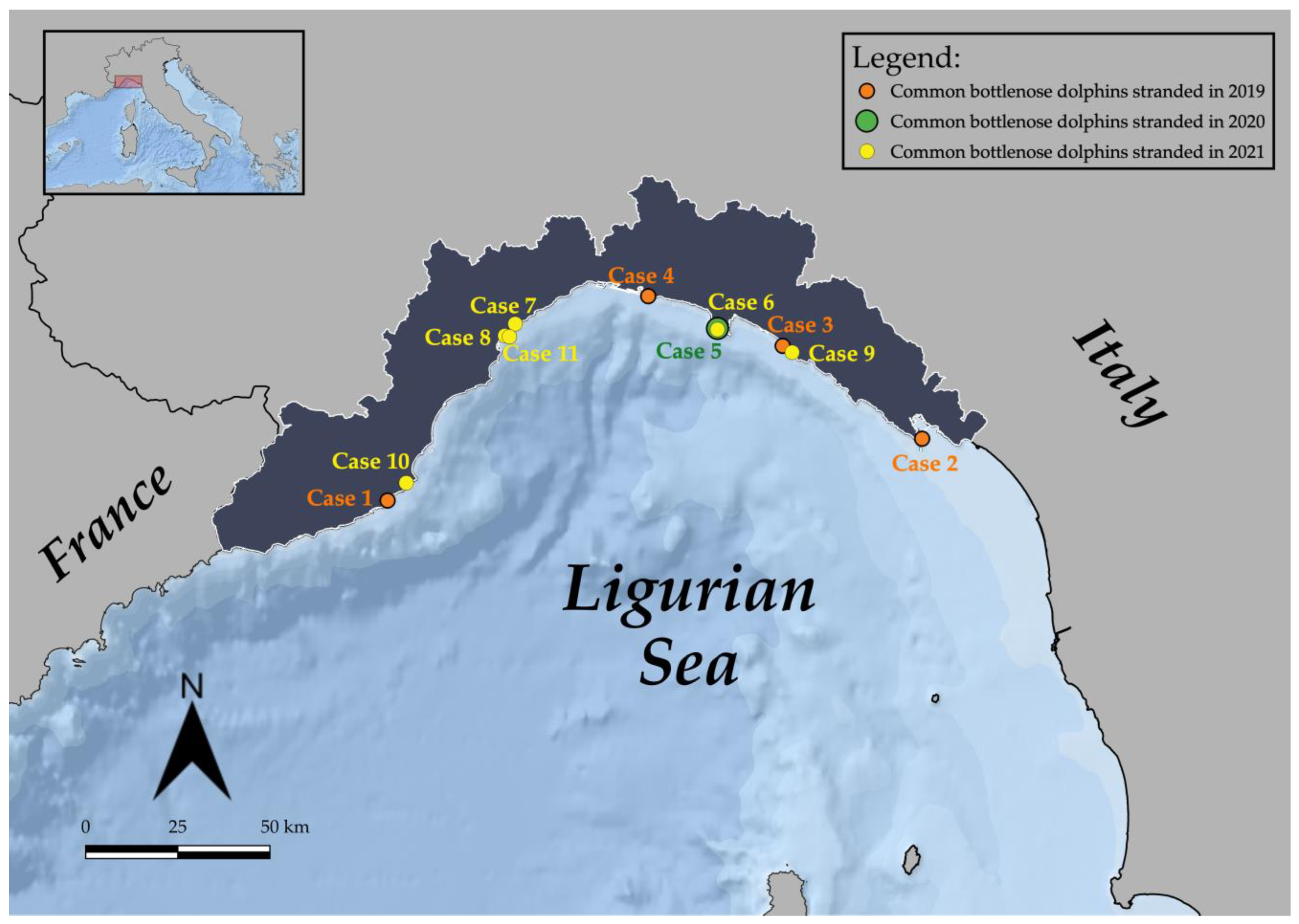
Figure 1. Map of the study area (Ligurian coastline), displaying the stranding locations of the 11 common bottlenose dolphins under study (QGIS Desktop 3.10.5 with GRASS 7.8.2).
Diversity 15 00569 g001
Figure 2. Macroscopic and microscopic images of some of the animals under study. (A) (Case 8) Amputation of the caudal fin, as a consequence of suspected fishery interaction (bycatch with not determined fishing gear). (B) (Case 10) Marine litter (fishing line agglomerate) in the esophagus. (C) (Case 5) Rope and nets at the tail. Lower right inset recent meal outcomes. (D) (Case 6) Tattoo skin lesions. Lower right inset multifocal areas of severe cytoplasmic vacuolation of the keratinocytes in the Stratum spinosum, with the formation of intraepithelial vesicles. Scale bar = 50 µm. (E) Prescapular lymph node (Case 4). Severe lymphoid depletion with several multinucleated giant cells (asterisks). Hematoxylin & Eosin (HE). Scale bar = 100 µm. (F) Occipital cortex (Case 11). Focal non suppurative meningitis (arrow). Upper left inset: frontal cortex (Case 11). Perivascular cuffing characterized by lymphocytes and plasma cells. HE. Scale bar = 20 µm. Upper right inset: Parietal cortex (Case 4). Positive labelling of endothelial and neuronal cells. Scale bar = 20 µm. IHC for CeMV.
Diversity 15 00569 g002
Figure 3. (A,B) Plotting of DDT (A) and PCB (B) levels in common bottlenose dolphins studied in relation to age estimated in relation to length. The pink dotted line indicates the age of sexual maturity for females (9 years) and blue for males (11 years). The blue continuous line represents the linear regression in males; the pink line shows the female trend after sexual maturity.
Diversity 15 00569 g003aDiversity 15 00569 g003b
Figure 4. Percentage composition of the polychlorinated biphenyls (PCBs) divided by chlorine content (Penta-CBs, Hexa-CBs, Hepta-CBs, Octa-CBs, Nona-CBs) on Total PCBs found in blubber of the different specimens of T. truncatus and present in Arochlor 1260.
Diversity 15 00569 g004
Figure 5. PCB fingerprint (% on Total PCBs) in the 11 specimens of bottlenose dolphin. The red dotted indicator point highlights the fingerprint of Case 11.
Diversity 15 00569 g005
Figure 6. Percentage composition of the op′ and pp′ forms of DDT, DDE, and DDD of total DDTs found in blubber of the different specimens of T. truncatus and present in Technical DDT.
Diversity 15 00569 g006
Figure 7. PCB levels in lipid weight basis (ppm l.w.) in the different specimens. The red dotted line represents the PCB threshold (17 ppm l.w.) indicated to produce deleterious effects in marine mammals. The black dotted line represents the highest reported PCB toxicity threshold (41 ppm l.w.) for marine mammals.
Diversity 15 00569 g007
Table 1. Overview of diagnostic investigations conducted per case. Cases are listed in chronological order; the IZS identification code is also reported for each specimen.
Case IDIZS CodeHistologicalImmuno-HistochemicalBacteriologicalBiomolecularSerological
118013XX (Morbillivirus; Toxoplasma gondii)XXX
242472 X *X (Morbillivirus)XXX
344599XX (Morbillivirus; Toxoplasma gondii)XXX
459260XX (Morbillivirus; Toxoplasma gondii)XXX
551352XX (Morbillivirus; Toxoplasma gondii)XXX
641716XX (Morbillivirus; Toxoplasma gondii)XX
750946 XX
853638XX (Morbillivirus; Toxoplasma gondii)XX
961838 X
1073951X XX
11177XX (Morbillivirus; Toxoplasma gondii)XX
* = limited.
Table 2. Analyzed compounds with their toxicological characteristics: cancerogenic, mutagenic, and teratogenic organochlorine compounds (T-OCs), Endocrine Disrupting Chemicals (EDC-OCs), immunosuppressant organochlorine compounds (IS-OCs), and the OCs showing qualities of both T-OCs and EDC-OCs as well as IS-OCs (TEI-OCs) [67,68,69,70,71,72,73,74,75,76,77,78,79,80,81,82,83,84,85,86,87]. The symbols: +, ×, •, * represent the indicators of contaminants belonging to the respective group.
CompoundsT-OCsEDC-OCsIS-OCsTEI-OCs
HCB ×
PCB95 ×
o,p′DDE+×
PCB101 ×
PCB99 ×
p,p′DDE+×*
o,p′DDD+×
PCB151
PCB144+135
PCB149
PCB118+×*
p,p′DDD+×
o,p′DDT+×
PCB146
PCB153+×*
PCB141
p,p′DDT+×*
PCB138+
PCB178
PCB187
PCB183
PCB128
PCB174
PCB177
PCB156+171+202+
PCB172
PCB180+
PCB199
PCB170+
PCB196
PCB201
PCB195
PCB194
PCB206
Table 3. T. truncatus stranded dead along the Ligurian coastline between 2019 and 2021 under investigation, arranged in chronological order. For each individual are detailed pieces of information on date and location of stranding, sex, total body length (TBL) (cm), weight (kg), estimated age (years), estimated age class, sexual maturity, decomposition condition category, nutritional condition category, gastric contents, main gross and microscopic lesions, pathogens and helminths detected, classification of the cause of death, origin, and sub-category.
Case
ID
Stranding DateStranding
Location
SexTotal Body Length (TBL)Weight
(kg)
Estimated Age [43]Estimated Age Class [44,45,46]Sexual Maturity (Gonad Maturation)DCCNCCGastric ContentMain Lesions (Gross and Microscopic) **Pathogens and Helminths Detected ***Classification of the Cause of DeathOriginSub-Category
123/02/
2019
Diano Marina (IM)M190903JuvenileImmature3GoScarce digested milkTwo skin ulcers at the right flank; prescapular, pulmonary and rectal reactive lymphadenopathy; meningeal fibrosis.α-HV (lung); Enterococcus faecium (lung, liver, kidney, and lns) Photobacterium damselae (lung and TB ln)ND
207/05/
2019
Isola Palmaria (SP)M243ND8AdultND4MoEvidence of recent mealMuscular extensive hematoma (right cervico-scapular area); severe mesenteric reactive lymphadenopathy, associated with marked lymphoid depletion.DMV (spleen, MES ln, lung, bladder; IHC + MES ln); Photobacterium damselae (CNS).ND
315/05/
2019
Sestri levante (GE)M27320011AdultMature (left: 18.5 cm)3PoScarce content (not recent meal)Two skin ulcers at the tip of the penis; gelatinous blubber edema; mild pulmonary edema with multifocal parenchymal hemorrhages and multifocal pulmonar parasitic granulomas; lymphoplasmacytic myocarditis and moderate cardiac valvular fibrosis; mesenteric reactive lymph adenomegaly; hemorrhagic CSF.T. gondii (heart); anti-T. gondii antibodies (1:40) (pericardial and pleuric fluid)ND
404/07/
2019
Genova (GE)M28423212AdultMature (left: 30 cm; right: 33 cm)2MoScarce content (not recent meal)Multifocal erosive parasitic skin lesions; generalized blubber gelatinous edema; serum hemorrhagic effusion in the peritoneal cavity; moderate to severe prescapular, pulmonary and mesenteric reactive lymphadenopathy, associated with lymphoid depletion and lymphadenitis with syncytia; multifocal hemorrhages in the esophageal mucosa; severe interstitial broncho-pneumonia; hemorrhagic CSF.DMV (lung, TB ln, spleen, kidney, CNS, laryngeal tonsil; IHC+ CNS, lung, PSC, TB, MES lns, spleen, tonsils, kidney); Photobacterium damselae (CNS, lung, liver, spleen, TB ln); T. gondii (muscle); anti-T. gondii antibodies (>1:40) (serum, HA, and pericardial fluid); Pennella sp. (skin)naturalinfectiousCoinfection (viral/bacterial/parasitic)
513/07/
2020
Camogli (GE)M310ND15AdultND2GoRecent meal outcomesNets and ropes around the caudal peduncle; abrasions; protrusion and eye bleeding; hepatic congestion; severe mesenteric and pulmonary reactive lymphadenopathy; severe pulmonary edema; gas embolism in the renal capsule and mesenteric vessels; subendocardial petechiae; mononuclear-eosinophilic interstitial pneumonia; splenic hypertrophy; generalized eosinophilic lymphadenitis and lymphoid depletion; lymphoplasmacytic enteritis; NS meningoencephalitis; muscular hyaline degeneration with wavy cells and atrophy of myocytes; atrioventricular valves fibrosis; mild aortic endocardiosis.T. gondii (PUL ln, spleen, CNS, muscle, heart); T. gondii antibodies (1:40) (serum, HA and intracardiac clot); α-HV (MES ln); Photobacterium damselae (l, lung, MES and PUL lfn, spleen, CNS); Listeria grayi (CNS); Cl. perfringens (lung, PUL ln, CNS)anthropicfishery interactionbycatch (consequence of underlying pathologies)
625/04/
2021
San Fruttuoso (GE)M241173.58AdultMature (average length: 9 cm)2MoRecent meal outcomesFive TSD, associated with proliferation of the dermal papillae and foci of hydropic degeneration of keratinocytes in the spinous layer; bilateral conjunctival hyperemia and hemorrhagic ocular discharge, blubber petechial hemorrhages associated with muscular hemorrhagic suffusions in the pre-scapular region; subcutaneous parasitization by larval cestodes; subcutaneous-muscular abscess in the right lumbar paravertebral region; multicentric reactive lymphadenopathy, associated with lymphoid depletion; pulmonary parasitic granulomas; splenomegaly; granulomatous gastritis; severe gas embolism in CNS, meningeal, mesenteric, and coronary vessels, renal capsule and lung; serum hemorrhagic effusion in the thorax cavity; abundant foamy fluids in trachea; pulmonary congestion associated with hemorrhagic lymphatic drainage to the marginal pulmonary lymph nodes; hemorrhagic CSF.Carnobacterium spp. (abscess, MES and TB lns, kidney); Serratia spp. (abscess, MES ln, kidney); Listeria seeligeri (CNS); Cetacean poxvirus 1 (skin lesion); Pholeter gastrophilus (stomach); Phyllobothrium delphini (blubber)anthropicfishery interactionbycatch (consequence of underlying pathologies)
706/06/
2021
Albissola (SV)F270ND11AdultND4NDAbsence of foodSevere dehydration; amputation of dorsal and caudal fins; fishing line around the thorax associated with a linear skin lesion; generalized advanced autolysis.T. gondii (spleen, heart); DMV (spleen)ND
812/06/
2021
Savona (SV)M140 *30<1Newborn
/Calf
Immature3NDScarce digested milkAmputation of the caudal fin; generalized blubber gelatinous edema, associated with hemorrhagic imbibition tracts; suppurative pneumonia associated with pulmonary reactive lymphadenopathy.Enterococcus faecalis (lung, CNS, kidney, spleen, PSC and TB lns); T. gondii (liver)naturalinfectiousbacterial
909/07/
2021
Sestri levante (GE)M170 *312JuvenileND4NDAbsence of foodAmputation of the caudal fin; circular cut injury of the peduncle; generalized advanced autolysis. ND
1010/09/
2021
Andora (SV)F190129.83JuvenileImmature3MoScarce content (not recent meal)Multifocal parasitic skin lesions; ventral blubber gelatinous edema; subcutaneous parasitization by larval cestodes; lung parasitic nodules; foreign body in the esophagus referable to marine litter (fishing line agglomerate); generalized moderate autolyisis.T. gondii (spleen, PSC ln, liver, CNS, muscle); DMV (lung, urinary bladder); Erysipelothrix rhusiopathiae (PSC ln, spleen, kidney); Photobacterium damselae (lung, PSC ln); Pennella sp (skin); Phyllobothrium delphini (blubber)ND
1124/12/
2021
Savona (SV)F28021112AdultMature (follicles at different development stage)2GoAbsence of foodFour sub-acute-chronic skin lesions; muscular and subcutaneous hematoma in the left prescapular region; hemothorax; moderate pulmonary oedema associated with foam in trachea; pyogranulomatous tonsillitis; granulomatous gastritis; lymphoplasmacytic endometritis; lymphoid depletion in spleen and mesenteric lymph nodes; NS meningoencephalitis.DMV (CNS and mesenteric lymph nodes; IHC + CNS); α-HV (skin lesions); Pholeter gastrophilus (stomach)naturalinfectiousviral
Legend: M = male; F = female; ND = not determined; DCC = decomposition condition category; NCC = nutritional condition category (Go, good; Mo, moderate; Po, poor); TSD: Tattoo skin disease; CNS: central nervous system; NS: non-suppurative; PSC ln, prescapular lymph node; TB ln, tracheobronchial lymph node; PUL ln, pulmonary lymph node; MES ln, mesenteric lymph node; LNs, lymph nodes; CSF, cerebrospinal fluid; AH, aqueous humor. * For these cases the length was estimated due to missing fluke/parts [42]. ** Pathological features of infectious disease are shown in bold. *** Suggested causative agent of pathological features, based on ancillary tests, are shown in bold.
Table 4. Levels of HCB, DDTs, PCBs, T-OCs, EDC-OCs, IS-OCs, and TEI-OCs in the blubber of different specimens of Tursiops truncatus (Case ID) expressed in ng/g dry weight (d.w.) For T-OCs, EDC-OCs, IS-OCs, and TEI-OCs, the respective percentages of total OCs are reported. EOM% = Extracted Organic Material Percentage.
Case IDEOM%HCBDDTsPCBsT-OCsEDC-OCsIS-OCsTEI-OCs
184.78280.78224,46.79177,609.93130,334.29 (65.1%)90,724.62 (45.3%)126,420.67 (63.1%)74,280.54 (37.1%)
283.91241.2139,621.54262,480.14200,443.90 (66.3%)121,294.01 (40.1%)178,423.27 (59.0%)104,702.02 (34.6%)
371.50158.45123,374.77743,861.65603,010.52 (69.5%)347,718.54 (40.1%)523,707.06 (60.4%)313,257.21 (36.1%)
424.7568.7694,466.531,043,474.63743,699.39 (65.4%)358,146.57 (31.5%)645,605.76 (56.7%)336,229.43 (29.6%)
570.21109.9473,303.76575,952.02443,256.75 (68.3%)240,098.94 (37.0%)388,104.78 (59.8%)224,646.44 (34.6%)
686.57208.2826,737.48206,617.95149,081.86 (63.8%)94,743.10 (40.6%)134,473.90 (57.6%)78,958.62 (33.8%)
746.05110.4333,104.91285,565.56206,284.90 (64.7%)122,933.62 (38.6%)178,991.43 (56.2%)104,268.77 (32.7%)
882.46306.7619,335.13121,898.2892,437.61 (65.3%)63,019.76 (44.5%)83,712.31 (59.1%)49,936.29 (35.3%)
973.31325.4322,276.64162,787.21115,577.15 (62.3%)80,968.49 (43.7%)105,309.60 (56.8%)60,827.45 (32.8%)
1049.71196.3383,376.02702,123.89517,643.34 (65.9%)302,423.38 (38.5%)459,438.61 (58.5%)269,589.47 (34.3%)
1179.7721.872935.1936,283.5920,358.92 (51.9%)12,018.26 (30.6%)18,004.87 (45.9%)8974.91 (22.9%)
Table 5. Mean values ± standard deviation (SD) and ratio values (i.e., DDTs/PCBs, pp′DDE/DDTs, pp′DDE/pp′DDT, Σ2DDT/(Σ2DDE + Σ2DDD), op′DDT/pp′DDT and Σop′DDTs/DDTs) investigated in blubber from T. truncatus.
Case IDDDTs/PCBspp′DDE/DDTspp′DDE/pp′DDTΣ2DDT/(Σ2DDE + Σ2DDD)Σop′DDTs/DDTsop′DDT/pp′DDT
10.130.715.860.160.090.13
20.150.7610.400.090.060.11
30.170.8017.410.060.110.31
40.090.8311.610.090.060.10
50.130.8718.790.060.050.14
60.130.718.370.110.100.21
70.120.6511.600.240.212.47
80.160.678.210.100.150.16
90.140.629.190.080.120.11
100.120.778.650.100.060.06
110.080.374.360.300.431.72
Table 6. Levels of PCBs, DDTs, IS-OCs, TEI-OCs, PCBs (101+138+153+180), and PCBs (118+138+153+180) (μg/g d.w.) in specimens with different statuses of immunosuppression-related pathologies.
PCBs
μg/g d.w.
DDTs
μg/g d.w.
IS-OCs
μg/g d.w.
TEI–OCs
μg/g d.w.
PCBs
(101+138+153+180)
μg/g d.w.
PCBs
(118+138+153+180)
μg/g d.w.
n° of SamplesMeanS.D.MeanS.D.MeanS.D.MeanS.D.MeanS.D.MeanS.D.
Control-subgroup 16331.5261.347.942.2227.0183.3133.7109.0171.3138.7178.1145.2
Subgroup 14573.4372.962.630.8365.6228.8203.7117.8289.8190.1302.6197.0
Control-subgroup 26365.6282.350.743.0246.3193.7145.4115.4186.9146.5195.5154.2
Subgroup 24522.1383.758.531.0337.7233.8186.1118.5266.5196.5276.5203.6
Control-subgroup 34301.5295.846.951.0209.8210.0124.6126.2156.1156.7162.3164.1
Subgroup 32653.0552.267.038.8412.0330.4220.5163.7331.3281.6343.4291.3
Subgroup 42493.8294.658.235.5319.2198.3186.9116.9248.4149.3261.8157.8
Subgroup 32653.0552.267.038.8412.0330.4220.5163.7331.3281.6343.4291.3
Disclaimer/Publisher’s Note: The statements, opinions and data contained in all publications are solely those of the individual author(s) and contributor(s) and not of MDPI and/or the editor(s). MDPI and/or the editor(s) disclaim responsibility for any injury to people or property resulting from any ideas, methods, instructions or products referred to in the content.