Article

First Record of Corallivorous Nudibranch Pinufius (Gastropoda: Nudibranchia) in the South China Sea: A Suspected New Species of Pinufius †

1
Third Institute of Oceanography, Ministry of Natural Resources, Xiamen 361005, China
2
Nansha Islands Coral Reef Ecosystem National Observation and Research Station, Guangzhou 510000, China
*
Authors to whom correspondence should be addressed.
urn:lsid:zoobank.org:pub:A9778147-079F-41CD-87A1-B11861D903DA.
Diversity 2023, 15(2), 226; https://doi.org/10.3390/d15020226
Submission received: 8 January 2023 / Revised: 31 January 2023 / Accepted: 31 January 2023 / Published: 4 February 2023
(This article belongs to the Special Issue Systematics, Phylogeography, Evolution and Conservation of Molluscs)

Abstract

:
A corallivorous nudibranch from the South China Sea reproduced explosively and caused extensive damage to Porites in our aquarium. In this study, morphological and molecular analyses of the nudibranch were conducted and described. Morphologically, this nudibranch was nearly consistent with Pinufius rebus in its characteristics intermediate between arminids and aeolids. The only detected difference was that the hook-like denticles on the masticatory border of P. rebus were absent in this nudibranch. In a molecular analysis, phylogenetic results based on the cytochrome oxidase subunit-I, 16S rRNA, and histone H3 gene sequences showed that this nudibranch and P. rebus form a well-supported sister clade under the superfamily Fionoidea, with significant interspecific divergence (0.18). Thus, we presumed that this nudibranch is a new species of Pinufius. Our results extend the distribution of Pinufius to the South China Sea, support the current taxonomic status of Pinufius under the superfamily Fionoidea, and imply that the species composition of Pinufius is more complex than previous records. Moreover, as a corallivorous nudibranch, the potential threat of Pinufius to coral health cannot be neglected.

1. Introduction

Coral reefs are among the most biologically diverse, productive, and fragile marine ecosystems on earth [1,2]. Coral reefs worldwide are suffering extensive deterioration due to extreme climates, anthropogenic disturbances, and biological factors such as competition, disease, and predation [3,4,5]. The crown-of-thorns starfish Acanthaster spp. and the muricid gastropods Drupella spp. are coral predators notorious for their substantial damage to corals [6,7]. Moreover, some flatworms and nudibranchs are also potential threats to coral reefs [8,9,10].
Nudibranchia is a diverse (approximately 2540 species) but taxonomically complex order of marine gastropod mollusks, among which there are many controversies on systematical relationships [11]. Initially, the order Nudibranchia was divided into the four suborders: Doridina, Aeolidina, Dendronotina, and Arminina [12]. More recently, Wägele and Willan [13] redistributed the order into two suborders: Anthobranchia and Cladobranchia. Cladobranchia contains seven superfamilies (Aeolidioidea, Arminoidea, Dendronotoidea, Doridoxoidea, Fionoidea, Proctonotoidea, and Tritonioidea) [14]. Among these, the superfamily Fionoidea has the richest variety of families [15]. Fionoidea, meanwhile, is the most controversial taxon, especially regarding disagreements between traditional morphology and emerging molecular identification [16,17].
As a morphologically special family in the superfamily Fionoidea, Pinufiidae, with its monotypic genus Pinufius, has only one species Pinufius rebus, which is very different from other members of Fionoidea [18]. The position of the anus is similar to that of some aeolids, but the external shape more resembles arminids; hence, Pinufius was initially classified into Arminina [18]. Subsequently, Rudman [19] redescribed the morphological and physiological features and feeding habits of Pinufius. According to distribution records in the literature, Pinufius inhabits the coral reefs of the Maldives [18], Australia [19], Indonesia [20], and the Philippines [21], and feeds exclusively on Porites spp. [22]. However, Rudman [19] did not give an opinion about the taxonomic status of Pinufius because of the similarity between P. rebus and some aeolids in radula and cerata. In the most recent classification of Gastropoda, Pinufiidae was classified into the superfamily Fionoidea [15].
Similarly, there is no consensus on the phylogenetic position of Pinufius. Pinufiidae once clustered with other ceras-bearing arminids including the species of Janolidae, Madrellidae, and Proctonotidae, under the superfamily Metarminoidea [23]. However, Metarminoidea was not named on the basis of existing genus-level taxa and, hence, cannot be valid [24]. In 2010, a phylogenetic analysis of multiple molecular datasets indicated that Pinufius clustered closely with Doto species of Dotoidae [21]. However, a morphological study showed that Pinufiidae was more closely related to Proctonotidae [11]. Mahguib and Valdés [25] provided a phylogenetic tree in which P. rebus clustered closely with Lomanotus spp. (Lonanotidae) and Eubranchus rustyus (Eubranchidae), and embedded into a large clade with some families of Aeolidioidea. In a study about host shifting, Pinufius was nested within a large clade with some Phestilla species of the family Trinchesiidae, which also feed exclusively on Porites spp. [20].
In China, 203 species were recorded in the order Nudibranchia [26]. Therein, only three coral-eating Phestilla (Phestilla melanobrachia, P. goniophaga, and P. fuscostriata) were reported [27,28,29], and Pinufius was never mentioned. In this study, a species of nudibranch was brought into our aquarium with Porites samples from Daya Bay in coastal waters of the northern South China Sea. After 2 months, while the population of this nudibranch broke out, they severely damaged these Porites samples and never left Porites to hurt other corals. According to primary observations under stereomicroscopes, we deduced that this species was P. rebus, according to the obligate association with Porites spp., as it had the appearance of this species including intermediate morphological features between aeolids and arminids [18]. However, there were significant differences in gene sequences between the nudibranch in this study and P. rebus online. In order to accurately identify the species and reveal the phylogenetic relationship with other relevant nudibranchs, we provide a detailed morphological description and comprehensive molecular phylogenetic analysis of this unknown nudibranch. This study is expected to provide a reference for Pinufius research and coral reef conservation in China.

2. Materials and Methods

2.1. Sample Collection

The tested specimens were collected from the surface of Porites samples in our aquarium using a pipette. Specimens for molecular analysis were preserved in 95% ethanol; specimens for morphological analysis were preserved in seawater temporarily and 4% formaldehyde. All specimens examined in this study were deposited in the Third Institute of Oceanography, Ministry of Natural Resources, Xiamen, Fujian, China (HYSS01 to HYSS75).

2.2. Morphological Analysis

2.2.1. External Morphology

Thirty-five specimens (HYSS06 to HYSS40) were used for external morphological analysis. They were placed in a petri dish with seawater, and magnesium sulfate solution was slowly added dropwise until the animals were anesthetized. External morphological characteristics were examined and photographed using a stereomicroscope (Leica M205FA; Leica Microsystems, Wetzlar, Germany).
Eggs and egg masses were examined and photographed under a biological microscope (Leica DM5000B; Leica Microsystems) and a stereomicroscope (Leica M205FA).

2.2.2. Internal Morphology

Twenty-three specimens (HYSS41 to HYSS63) were used for dissection of the reproductive system under a dissecting microscope (Leica S6D; Leica Microsystems) and a stereomicroscope (Leica M205FA). Identification of organs was aided by the relevant literature [18].
Buccal masses were extracted under a dissecting microscope (Leica S6D) and soaked in 25% hypochlorous acid for 20 min at 24 °C; they were then rinsed in pure water. The radula and jaw were removed and placed on filter paper (Φ = 5 μm) using forceps. The filter paper was mounted on a stub with liquid nitrogen and examined under a scanning electron microscope (Quanta 450; FEI, Portland, OR, USA).
For histological examination, specimens (HYSS75) were preserved in 4% paraformaldehyde for 24 h and dehydrated in ethanol. Dehydrated specimens were made transparent in the mixed solution of ethanol and xylene (1:1). Transparent specimens were embedded in melted wax and cooled to −20 °C. The wax-embedded specimens were cut into 4 μm slices. These slices were floated on the surface of 40 °C water to remove any wrinkles and dried on glass slides at 60 °C. The sections were cleared in xylene, a mixed solution of ethanol and xylene (1:1), and ethanol successively to remove the wax. Finally, the sections were stained with hematoxylin–eosin stain. Sections were examined under a biological microscope (Leica DM5000B).

2.3. Molecular Analysis

2.3.1. DNA Extraction and Sequencing

Four specimens (HYSS02–HYSS05) were used for molecular analyses. The genomic DNA of specimens was extracted using the Ezup Column Animal Genomic DNA Purification Kit (Sangon Biotech, Shanghai, China). DNA integrity was checked using electrophoresis on 1% agarose gel. Polymerase chain reactions (PCRs) were conducted to amplify the cytochrome oxidase I (COI), 16S ribosomal RNA (16S), and histone H3 gene sequences using a Veriti 96-Well Thermal Cycler (Thermo Fisher Scientific, St. Louis, MO, USA). The primers were HCO2198/LCO1498 for COI [30], 16SsarL/16SR for 16S [31,32], and H3AF/H3AR for histone H3 gene [33]. PCR products were purified with SanPrep Column DNA Gel Extraction Kit (Sangon Biotech) and sequenced using an ABI 3730XL Genetic Analyzer (Sangon Biotech).

2.3.2. Phylogenetic Analysis

Phylogenetic topology was constructed on the basis of concatenated COI–16S–histone H3 sequences of our specimens and 49 other nudibranch species (Supplementary Table S1). Among these species, 38 species belonged to the superfamily Fionoidea, including the genus Phestilla whose feeding habits and behaviors were similar to Pinufius; 10 species in the superfamily Poctonotoidea were morphologically similar to Pinufius. The best partition schemes and substitution models were estimated using a comparison of Akaike information criterion (AIC) scores with jModelTest v2.1.7 [34]. A Bayesian inference (BI) phylogenetic tree was constructed using Mrbayes v3.12 [35]; a set optimal model strategy was selected for different positions (GTR + I + G for COI and 16S and GTR + G for histone H3). Analyses were run for 10,000,000 generations with the Markov chains being sampled every 1000 generations. We determined the burn-in value of the first 2500 trees (25%), and the 50% majority-rule consensus tree was estimated. Pairwise genetic distances based on the COI gene were generated using MEGA X [36].

3. Results

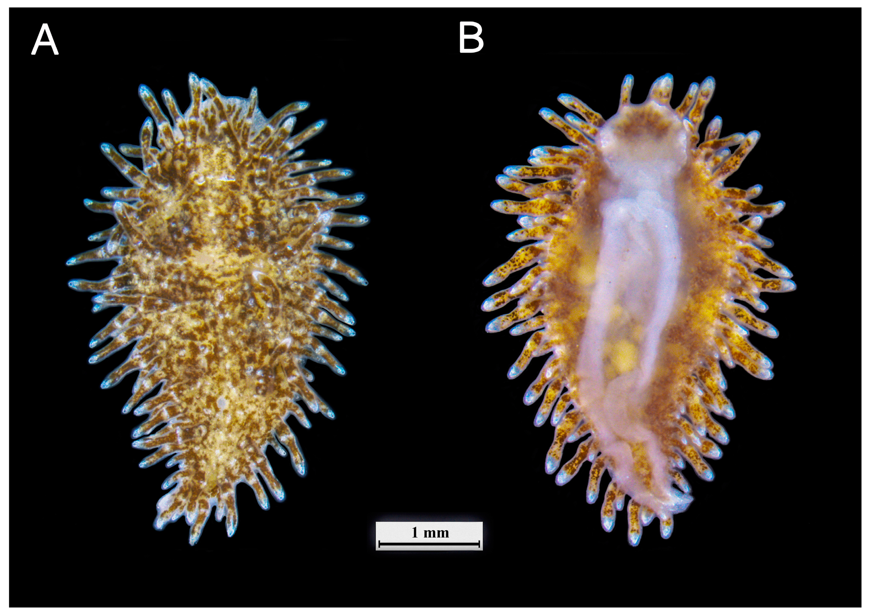
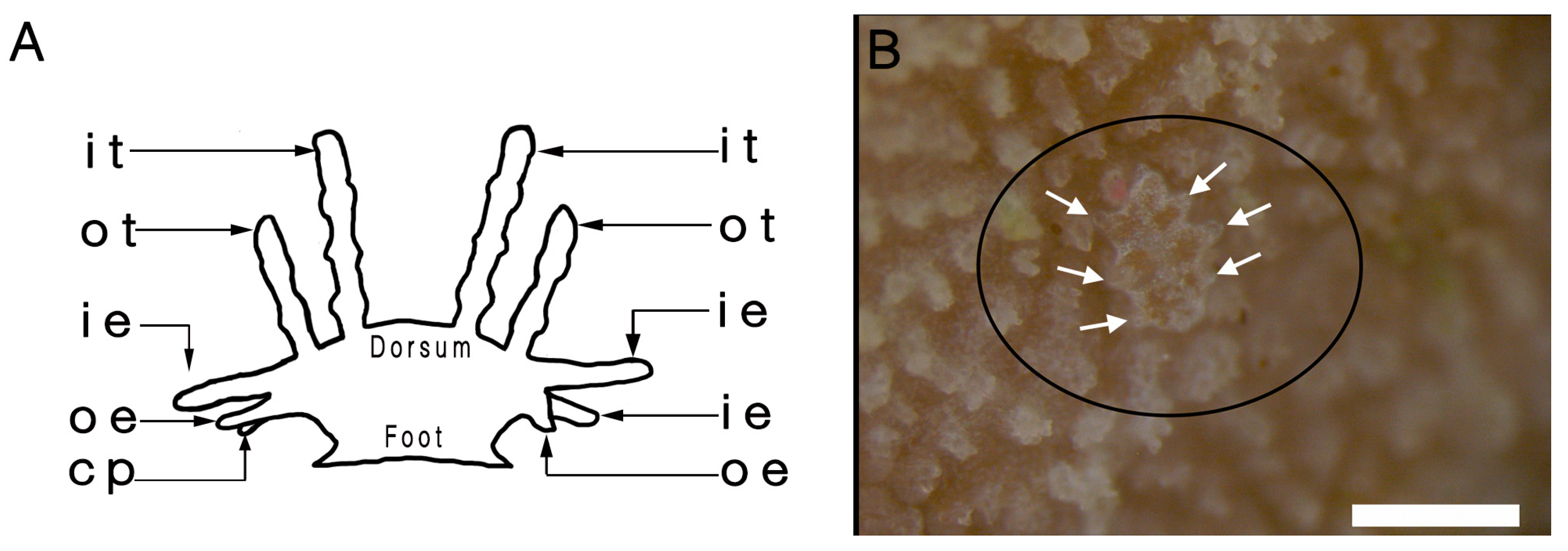
3.1. External Morphology

Mature specimens were 3–6 mm in length (Figure 1). Body color was usually light brown and changed with the color of hosts. The oral veil was round, about one-fourth the length of the body, slightly exceeded the anterior edge of dorsum, and contained brown speckling, without oral tentacles. The rhinophores were smooth and nonretractile, without a rhinophoral sheath. There was a white patch between the bases of rhinophores. The dorsum was flat and broad, with many small brown tubercles. When resting, the body was oval, and the foot and partial oral veil were shielded by the dorsum; when crawling, the body was elongated, and the foot usually extended behind the dorsum.
The cerata were variable in shape (Figure 2A). On the top of dorsum, they were rod-like and partly swollen, arranged symmetrically in five rows. Each row consisted of two pairs of cerata; the pair on the outside (Figure 2A: ot) was smaller than the pair on the inside (Figure 2A: it). Differing from the cerata on the top, the cerata around the edge of dorsum were irregular and dense in a double-row arrangement. The longest inner cerata around the edge of dorsum were on the anterior and usually misidentified as oral tentacles (Figure 2A: ie). The outside cerata around the edge of the dorsum were digitiform, as well as shorter and denser than the inner cerata (Figure 2A: oe). There were 1–2 ceras-like processes between the outside cerata around the edge of dorsum (Figure 2A: cp). The type and arrangement of the cerata in larvae were significantly different from those of adults (Figure 2B).
The anus was located on the right side of the midline between the third and fourth rows of cerata. The reproductive opening was located on the right side of the foot.
The egg masses were transparent belts (Figure 3A), with hundreds of eggs (Figure 3B). The eggs were 0.2–0.3 mm with transparent membranes (Figure 3B).

3.2. Internal Morphology

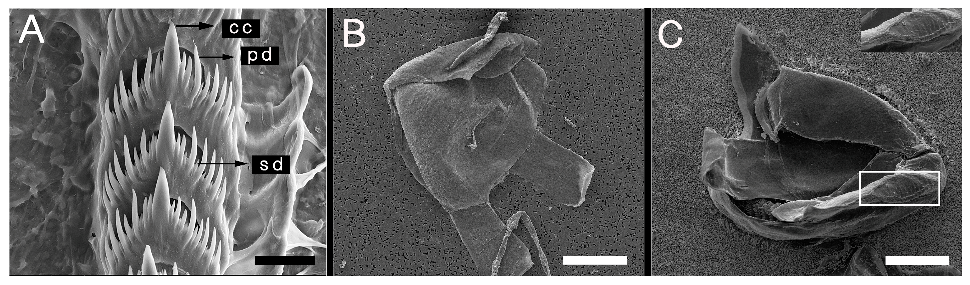
3.2.1. Radula and Jaw

In the buccal mass, a radula was enfolded in a pair of jaws. The radula formula was 17 × 0.1.0 (Figure 4A). The central cusp was strong (Figure 4A: cc), with 6–8 slenderer and shorter primary denticles on each side (Figure 4A: pd). Compared with the other primary denticles, the two pairs closet to central denticle looked runtish. Between the primary denticles, 1–2 secondary denticles were present (Figure 4A: sd). The jaw plates were transparent and triangular (Figure 4B,C). The masticatory border was rough and thick (Figure 4C), without the hook-like denticle of P. rebus as Marcus et al. [18] and Rudman [19] described.

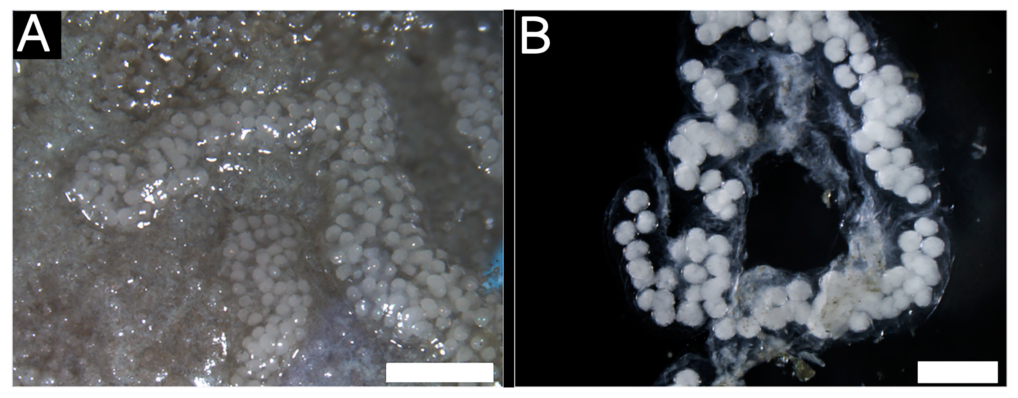
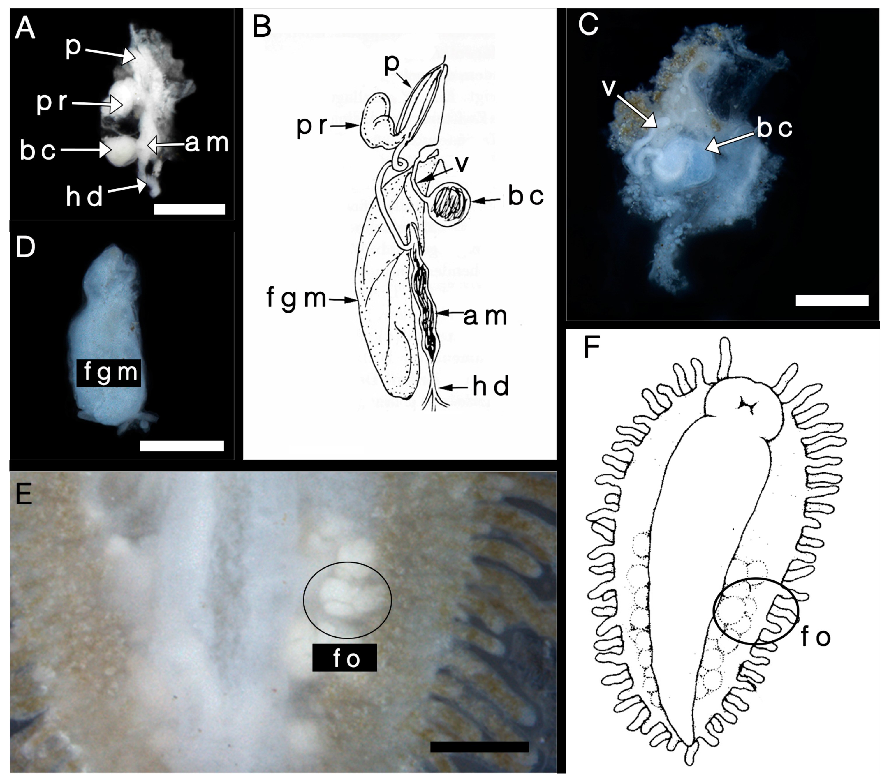
3.2.2. Reproductive System

The reproductive system (Figure 5) was diaulic, mainly consisting of a penis, a female gland mass, an ampulla, and a bursa copulatrix, as Marcus et al. [18] described (Figure 5B). The penis was slender and connected to an irregular, massive prostate (Figure 5A: p, pr). The bursa copulatrix with a transparent membrane was spherical and connected to an elongated vagina (Figure 5C: bc, v). The ampulla was slightly swollen (Figure 5C: am). The female gland mass was irregularly cylindrical (Figure 5D: fgm). Many follicles were visible in the posterior of the body through the ventral epidermis (Figure 5E: fo).

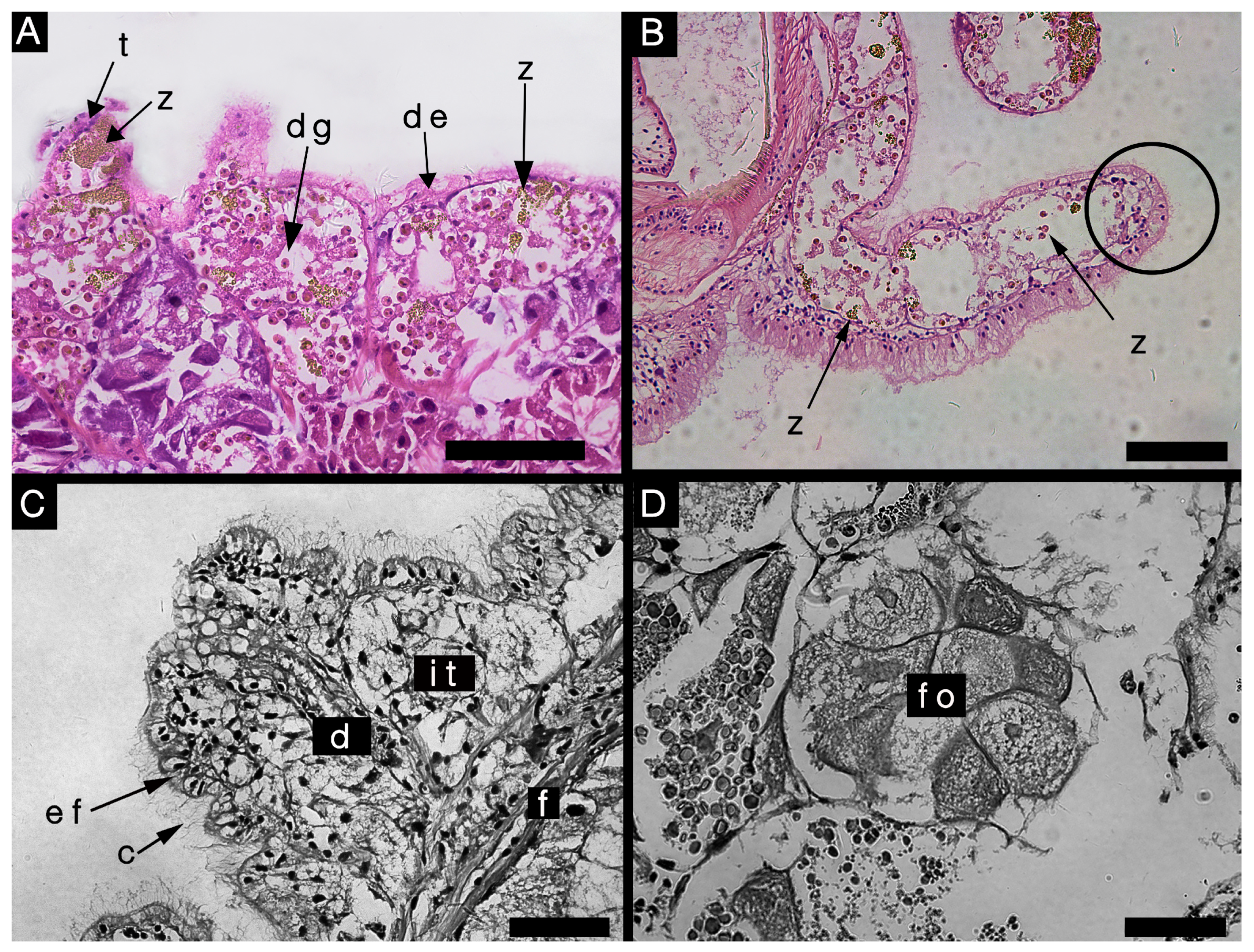
3.2.3. Histological Characteristics

The dorsal epidermis was thin and semitransparent (Figure 6A: de). Under the epidermis, zooxanthellae (Figure 6A: z) were mainly concentrated in the tubercles (Figure 6A: t) and digestive glands (Figure 6A: dg). The digestive gland extended to the cerata, with zooxanthellae (Figure 6B: z). There was no nematocyst in the apex of the cerata (Figure 6B). The epidermic cells of the foot (Figure 6C: ef) were ciliated and columnar (Figure 6C: c). The inner tissue (Figure 6C: it) was large and loose, and a narrow duct (Figure 6C: d) extended out to the epidermis. We believe this to be the foot gland described by Marcus et al. [18]. The connective tissue between the foot and cavity was filled with fibers (Figure 6C: f). The inner structure of the follicles (Figure 6D: fo) was multivesicular.

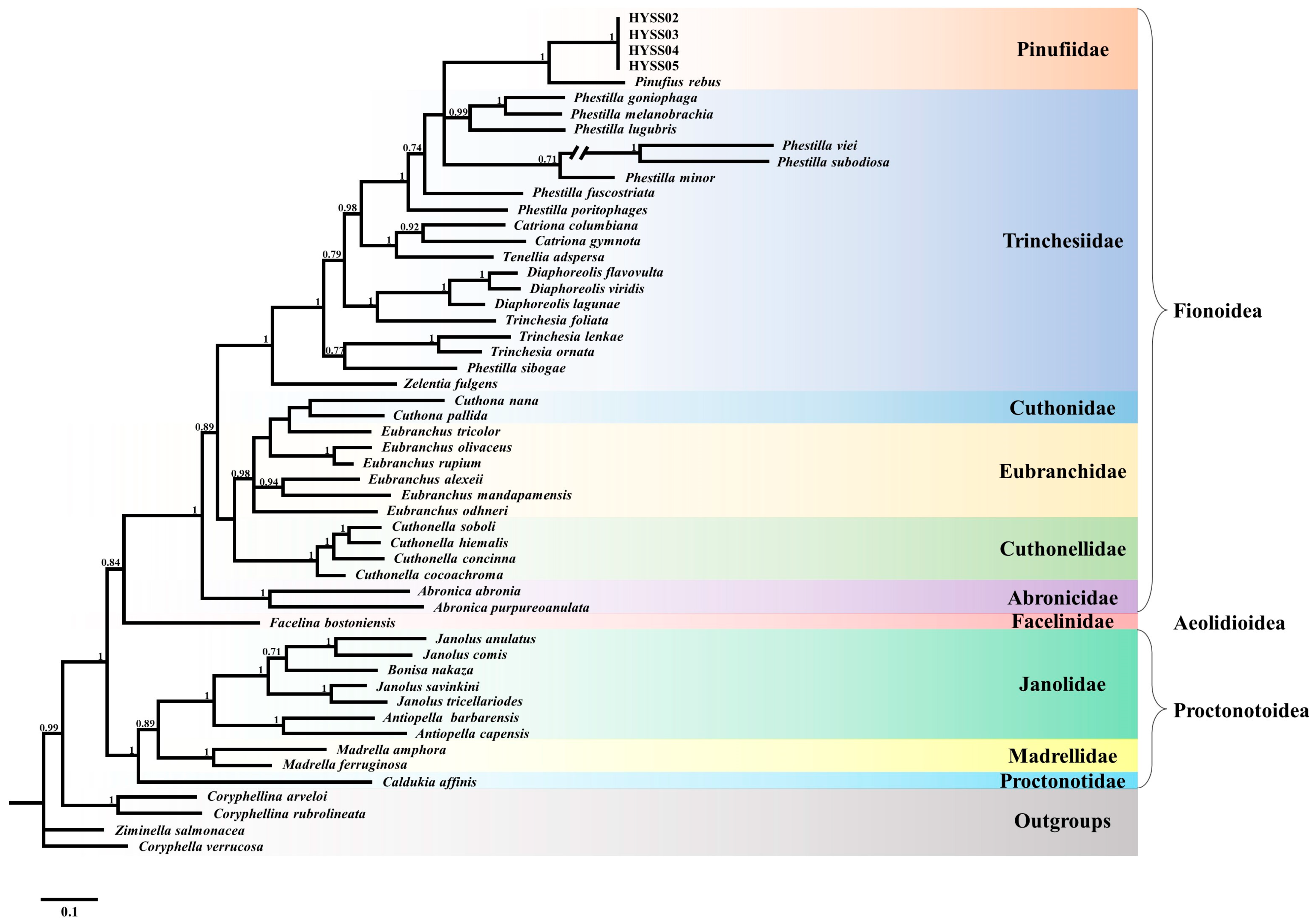
3.3. Phylogenetic Relationships

After alignment, we obtained three gene matrices including a 658 bp COI fragment, a 452 bp 16S rRNA fragment, and a 328 bp histone H3 fragment. The BI tree based on concatenated gene sequences indicated that all of the interspecific nodes were robust, with strong posterior probabilities (Figure 7). The resultant topology supported that the superfamilies Fionoidea and Proctonotidea formed monophyletic groups. With the exception of Phestilla sibogae, all other species of Phestilla clustered into a robust branch. The sequences of P. sibogae in this study were from Cella et al. [16], and its separation from other Phestilla spp. also happened in the studies of Hu et al. [27,28] and Ekimova et al. [37]. Our four nudibranch specimens (HYSS02-05) formed a well-supported monophyletic clade which was the sister species of Pinufius rebus. They were nested within the Phestilla clade of the superfamily Fionoidea, which had a significant phylogenetic distance from the superfamily Proctontidea.
Furthermore, a strong genetic divergence existed between the nudibranch specimens and P. rebus; the P-distance based on COI gene sequences between them was 0.18 (Supplementary Table S2).

4. Discussion

In this study, on the basis of morphological comparisons and molecular phylogenetic analyses, we reported a new distribution record for Pinufius in the South China Sea and confirmed the taxonomic status of Pinufius in the superfamily Fionoidea. Moreover, we put forward some new ideas about species composition and the ecological role of the genus Pinufius.

4.1. A New Distribution Record for Pinufius in the South China Sea

The genus Pinufius has been reported in coral reefs of the Maldives [18], Australia [19], Indonesia [20], and the Philippines [21]. This is the first record of Pinufius in the South China Sea.
As Marcus et al. [18] discussed, Pinufius is an interesting genus; “it is very aeolid-like in some ways, such as scleractinian parasitism, digestive gland in cerata, and single rachidian tooth in radula; however, in some ways, it is also arminid-like, such as general body shape and arrangement of gut”. Our results showed that the external morphological characteristics and feeding habits of the nudibranch specimens in this study were certainly consistent with the original description of Pinufius [18,19]. Morphologically, the dorsum of the Pinufius sp. in this study was well developed, bearing numerous cerata (Figure 1); these cerata contained digestive glands in which zooxanthellae were present but a nematocyst was absent (Figure 6B). In the anterior, there was a clear oral veil but no oral tentacle (Figure 1), and the radula was uniseriate (Figure 4A). Similarly, the Pinufius sp. in this study was remarkably consistent in feeding habits with P. rebus, the sole published Pinufius species, which also feeds exclusively on Porites spp. [19]. Genetic evidence suggested that the clade formed by our four specimens (HYSS02-05) was sister to P. rebus with a strong support (Figure 7). The above results are sufficient to support the conclusion that our specimens belonged to the genus Pinufius.
Considering the high dependence of Pinufius on Porites, as well as the wide distribution of Porites in the world [38], we speculated that Pinufius could inhabit a range as wide as Porites.

4.2. The Phylogeny of the Genus Pinufius

Under the superfamily Proctonotoidae, species of the families Janolidae, Madrellidae, and Proctonotidae are typical ceras-bearing arminids [23,24]. Their radulae are multiseriate, and their dorsal cerata contain digestive glands without zooxanthellae [39,40,41,42]. Pinufius was once classified into the same superfamily with the families Janolidae, Madrellidae, and Proctonotidae because of the similarities among them in shape [24]. However, the cerata and radula of Pinufius are very different from those of Proctonotoidea species (Supplementary Table S3). Furthermore, as an important and taxonomically available trait of some nudibranchs [43], their feeding preferences are fundamentally different; the members of Proctonotoidae prey on bryozoans, while Pinufius preys exclusively on Porites spp. [18,39]. Moreover, the phylogenetic analysis showed that the family Pinufiidae is a distant relative of the families under the superfamily Proctonotidea (Figure 7). In conclusion, our results did not support the previous status of the family Pinufiidae under the traditional ceras-bearing arminid superfamily.
The results sustain the phylogenetic relationship that the family Pinufiidae belongs to the superfamily Fionoidea. Within the superfamily Fionoidea, all Phestilla species, with the exception of Phestilla chaetopterus, feed on scleractinian corals [27,44,45]. Morphologically, the body of Phestilla species is elongated without notum edge, and the cerata contain digestive glands in which zooxanthella are present without nematocysts; the oral tentacles are obvious, and the radula is uniseriate with a formula of 0.1.0 [17,46]. Adult Phestilla with limited mobility stay on a coral colony for their entire adult stage [47]. On the basis of the above, it seems that there are many similarities between Phestilla and Pinufius in their radula, cerata, and the obligate association with scleractinian coral. The close relationship between Pinufius and Phestilla was also reflected in the present phylogenetic analysis where they formed a well-supported clade together (Figure 7). Fritts-Penniman et al. [20] previously considered that coral-eating nudibranchs had a common evolutionary history, and that Pinufius and Phestilla should be synonymous. However, the present results are not sufficient to revise the classification of Pinufiidae, because we cannot reasonably explain the significant differences in external morphology between Pinufius and Phestilla. Some experts considered that morphological synapomorphies possibly occur at earlier larval stages and disappear in the adult stages [17]. This viewpoint has inspired us to check more detailed morphological characteristics of early life history in future studies.

4.3. A Suspected New Species of the Genus Pinufius

The present molecular data regarding the COI gene showed that the Pinufius sp. in this study formed a sister clade to P. rebus, and the P-distance between them (0.18) was much higher than the intraspecific divergence typical of nudibranchs (0.02–0.06) according to prior studies [20]. Morphologically, compared to the descriptions of P. rebus by Marcus et al. [18] and Rudman [19], we found some differences on the masticatory border of the jaws. The denticles on the masticatory border of P. rebus are bifid with a pair of recurved hooks [18,19], and there was no any denticle on the masticatory border of the Pinufius sp. in this study (Figure 4C). Since its first discovery in 1960, P. rebus has been the only species in the genus Pinufius. In the present study, our specimens were presumed to be a new species in Pinufius on the basis of the above morphological differences and molecular evidence. However, further confirmation of the suspected new Pinufius species still faces the following impediments:
(1) The reference materials regarding the morphological features of Pinufius are limited. The existing textual descriptions and simple sketches cannot provide enough morphological details of the masticatory borders.
(2) The available sequences of P. rebus in the NCBI database were collected from the Philippines by Pola et al. [21]. They simply provided the sequences but no morphological depiction of their samples, which restricts us from explaining differences in our molecular results. Moreover, given the inconsistent sampling sites of morphological and molecular data, as well as the hardly perceptible interspecific difference in external morphology, the specimens of Pola et al. [21] may also be from a new species of Pinufius differing from P. rebus and our specimens. Future studies should focus on the above aspects to confirm the species composition of Pinufius.

4.4. Potential Threats to Local Coral Communities

In our aquarium, the outbreak of the Pinufius sp. caused serious injury to Porites. However, there are few reports of massive damage to natural corals by Pinufius and other nudibranchs [29]. This may be because there are many natural predators such as carnivorous fish and crustaceans in open water, but less in aquaria. In recent years, with the serious decline in coral reefs around the world, corallivorous predators have also largely been in decline. For example, many studies have indicated that outbreaks of Acanthaster planci were due to the overfishing of Cheilinus undulatus, Charonia tritonis, etc. [48,49,50]. In Daya Bay, affected by overfishing and habitat destruction, the numbers of coral reef fish and crustaceans have also been in decline [51,52]. Gochfeld et al. [47] highlighted that the outbreak of Phestilla might result in changes in coral compositions. Similarly, it is very possible that Pinufius sp. would threaten the stability of coral communities.

5. Conclusions

In this paper, we described a novel distribution of Pinufius in the South China Sea, supporting the systematic status of Pinufius under the superfamily Fionoidea, and deduced that the nudibranch in the present study might be a new species of the genus Pinufius. The morphological characteristics of this nudibranch are nearly identical with Pinufius rebus, including the arminid-like dorsum and oral veil, as well as aeolid-like cerata. However, the masticatory border without denticle is inconsistent with the descriptions of P. rebus. Molecular analysis showed that this nudibranch and P. rebus were clustered together, and their interspecific genetic divergence was significant. The present study demonstrates that the composition of Pinufius is more complex than previously recorded. Morphological and molecular data are needed to verify this new species. Enriching the information of Pinufius not only improves the knowledge about this animal but also provides references for relevant research on other corallivorous nudibranchs. Additionally, Pinufius and other small corallivorous invertebrates should be taken seriously as potential threats to coral reefs.

Supplementary Materials

The following supporting information can be downloaded at https://www.mdpi.com/article/10.3390/d15020226/s1: Table S1. GenBank accession numbers of the sequences used in this study; Table S2. Uncorrected COI p-distances of these species in molecular analysis; Table S3. Summary of the diagnostic characters of related genera [53,54,55,56,57,58,59].

Author Contributions

Conceptualization, Z.J. and W.N.; methodology, Z.J. and J.X.; formal analysis, Z.X. and B.C.; resources and investigation, P.T. and W.W.; software and data curation, J.X.; writing—original draft preparation, Z.J.; writing—review and editing and supervision, W.N.; funding acquisition, J.X. All authors have read and agreed to the published version of the manuscript.

Funding

This research was funded by the National Natural Science Foundation of China (grant numbers 42006098, 42006128, and 42106143), and the Scientific Research Foundation of Third Institute of Oceanography, Ministry of Natural Resources (grant number 2022024).

Institutional Review Board Statement

Not applicable.

Informed Consent Statement

Not applicable.

Data Availability Statement

Publicly available datasets were analyzed in this study. This data can be found at https://www.ncbi.nlm.nih.gov/ accessed on 14 December 2022.

Acknowledgments

The authors would like to express their gratitude to Li Gu for her assistance with scanning electron microscope analysis. The authors also thank the reviewers for their constructive and thorough comments that improved the manuscript.

Conflicts of Interest

The authors declare no conflict of interest. The funders had no role in the design of the study; in the collection, analyses, or interpretation of data; in the writing of the manuscript; or in the decision to publish the results.

References

  1. Hughes, T.P.; Baird, A.H.; Bellwood, D.R.; Card, M.; Connolly, S.R.; Folke, C.; Grosberg, R.; Hoegh-Guldberg, O.; Jackson, J.B.C.; Kleypas, J.; et al. Climate change, human impacts, and the resilience of coral reefs. Science 2003, 301, 929–933. [Google Scholar] [CrossRef] [PubMed]
  2. Smith, S.V. Coral-reef area and the contributions of reefs to processes and resources of the world’s oceans. Nature 1978, 273, 225–226. [Google Scholar] [CrossRef]
  3. Halpern, B.; Walbridge, S.; Selkoe, K.; Kappel, C.; Micheli, F.; D’Agrosa, C.; Bruno, J.; Casey, K.; Ebert, C.; Fox, H.; et al. A global map of human impact on marine ecosystems. Science 2008, 319, 948–952. [Google Scholar] [CrossRef] [PubMed]
  4. Rotjan, R.D.; Lewis, S.M. Impact of coral predators on tropical reefs. Mar. Ecol. Prog. Ser. 2008, 367, 73–91. [Google Scholar] [CrossRef]
  5. Selig, E.R.; Casey, K.S.; Bruno, J.F. New insights into global patterns of ocean temperature anomalies: Implications for coral reef health and management. Glob. Ecol. Biogeogr. 2010, 19, 397–411. [Google Scholar] [CrossRef]
  6. Morton, B.; Blackmore, G.; Kwok, C.T. Corallivory and prey choice by Drupella rugosa (Gastropoda: Muricidae) in Hong Kong. J. Molluscan Stud. 2002, 68, 217–223. [Google Scholar] [CrossRef]
  7. Pratchett, M.S.; Caballes, C.F.; Cvitanovic, C.; Raymundo, M.L.; Babcock, R.C.; Bonin, M.C.; Bozec, Y.M.; Burn, D.; Byrne, M.; Castro-Sanguino, C.; et al. Knowledge gaps in the biology, ecology, and management of the Pacific crown-of-thorns sea star Acanthaster sp. on Australia’s Great Barrier Reef. Biol. Bull. 2021, 241, 330–346. [Google Scholar] [CrossRef]
  8. Barton, J.A.; Bourne, D.G.; Humphrey, C.; Hutson, K.S. Parasites and coral associated invertebrates that impact coral health. Rev. Aquac. 2020, 12, 2284–2303. [Google Scholar] [CrossRef]
  9. Hume, B.C.C.; D’Angelo, C.; Cunnington, A.; Smith, E.G.; Wiedenmann, J. The corallivorous flatworm Amakusaplana acroporae: An invasive species threat to coral reefs? Coral Reefs 2014, 33, 267–272. [Google Scholar] [CrossRef]
  10. Rawlinson, K.A.; Stella, J.S. Discovery of the corallivorous polyclad flatworm, Amakusaplana acroporae, on the Great Barrier Reef, Australia—The first report from the wild. PLoS ONE 2012, 7, e42240. [Google Scholar] [CrossRef]
  11. Gosliner, T.M.; Fahey, S.J. Previously undocumented diversity and abundance of cryptic species: A phylogenetic analysis of Indo-Pacific Arminidae Rafinesque, 1814 (Mollusca: Nudibranchia) with descriptions of 20 new species of Dermatobranchus. Zool. J. Linn. Soc. 2011, 161, 245–356. [Google Scholar] [CrossRef]
  12. Odhner, N.H. Opisthobranchiate Mollusca from the western and northern coasts of Norway. K. Nor. Vidensk. Selsk. Skr. 1939, 1, 1–93. [Google Scholar]
  13. Wägele, H.; Willan, R.C. Phylogeny of the Nudibranchia. Zool. J. Linn. Soc. 2000, 130, 83–181. [Google Scholar] [CrossRef]
  14. MolluscaBase eds. Cladobranchia. Available online: http://www.molluscabase.org/aphia.php?p=taxdetails&id=827889 (accessed on 27 January 2023).
  15. Bouchet, P.; Rocroi, J.P.; Hausdorf, B.; Kaim, A.; Kano, Y.; Nützel, A.; Parkhaev, P.; Schrödl, M.; Strong, E.E. Revised classification, nomenclator and typification of gastropod and monoplacophoran families. Malacologia 2017, 61, 1–526. [Google Scholar] [CrossRef]
  16. Cella, K.; Carmona, L.; Ekimova, I.; Chichvarkhin, A.; Schepetov, D.; Gosliner, T.M. A radical solution: The phylogeny of the nudibranch family Fionidae. PLoS ONE 2016, 11, e0167800. [Google Scholar] [CrossRef]
  17. Korshunova, T.; Martynov, A.; Picton, B. Ontogeny as an important part of integrative taxonomy in tergipedid aeolidaceans (Gastropoda: Nudibranchia) with a description of a new genus and species from the Barents Sea. Zootaxa 2017, 4324, 1–22. [Google Scholar] [CrossRef]
  18. Marcus, E.; Marcus, E. Opisthobranchia aus dem Roten Meer und von den Malediven. Akad. Wiss. Lit. Abh. Math. Nat. Kl. 1960, 12, 873–933. [Google Scholar]
  19. Rudman, W.B. Further-studies on the anatomy and ecology of opisthobranch mollusks feeding on the scleractinian coral Porites. Zool. J. Linn. Soc. 1981, 71, 373–412. [Google Scholar] [CrossRef]
  20. Fritts-Penniman, A.L.; Gosliner, T.M.; Mahardika, G.N.; Barber, P.H. Cryptic ecological and geographic diversification in coral-associated nudibranchs. Mol. Phylogenetics Evol. 2020, 144, 106698. [Google Scholar] [CrossRef]
  21. Pola, M.; Gosliner, T.M. The first molecular phylogeny of cladobranchian opisthobranchs (Mollusca, Gastropoda, Nudibranchia). Mol. Phylogenetics Evol. 2010, 56, 931–941. [Google Scholar] [CrossRef]
  22. Rudman, W.B. The taxonomy and biology of further aeolidacean and arminacean nudibranch molluscs with symbiotic zooxanthellae. Zool. J. Linn. Soc. 1982, 74, 147–196. [Google Scholar] [CrossRef]
  23. Beesley, P.L.; Ross, G.J.B.; Wells, A. Mollusca: The Southern Synthesis. Fauna of Australia; CSIRO Publishing: Melbourne, Australia, 1998; Volume 5, pp. 1007–1011. ISBN 0643057560. [Google Scholar]
  24. Jensen, K.R. An outline of the systematics and classification of Nudibranchia (Gastropoda, Opisthobranchia). Phuket Mar. Biol. Cent. Spec. Publ. 2000, 21, 431–446. [Google Scholar]
  25. Mahguib, J.; Valdés, Á. Molecular investigation of the phylogenetic position of the polar nudibranch Doridoxa (Mollusca, Gastropoda, Heterobranchia). Polar Biol. 2015, 38, 1369–1377. [Google Scholar] [CrossRef]
  26. Liu, R. Checklist of Marine Biota of China Seas; China Science Publishing & Media Ltd. (CSPM): China, Beijing, 2008; pp. 536–545. ISBN 978-7-03-023722-4. [Google Scholar]
  27. Hu, J.; Zhang, Y.; Yiu, S.K.F.; Xie, J.Y.; Qiu, J.W. A new species of predatory nudibranch (Gastropoda: Trinchesiidae) of the scleractinian coral Goniopora. Zool. Stud. 2020, 59, 62. [Google Scholar] [CrossRef]
  28. Hu, J.; Zhang, Y.; Xie, J.Y.; Qiu, J.W. A new species of predatory nudibranch (Gastropoda: Trinchesiidae) of the Coral Pavona decussata. Zool. Stud. 2020, 59, 30. [Google Scholar] [CrossRef]
  29. Yiu, S.K.F.; Chung, S.S.W.; Qiu, J.W. New observations on the corallivorous nudibranch Phestilla melanobrachia: Morphology, dietary spectrum and early development. J. Molluscan Stud. 2021, 87, yab034. [Google Scholar] [CrossRef]
  30. Folmer, O.; Black, M.; Wr, H.; Lutz, R.; Vrijenhoek, R. DNA primers for amplification of mitochondrial Cytochrome Coxidase subunit I from diverse metazoan invertebrates. Mol. Mar. Biol. Biotechnol. 1994, 3, 294–299. [Google Scholar]
  31. Puslednik, L.; Serb, J.M. Molecular phylogenetics of the Pectinidae (Mollusca: Bivalvia) and effect of increased taxon sampling and outgroup selection on tree topology. Mol. Phylogenetics Evol. 2008, 48, 1178–1188. [Google Scholar] [CrossRef]
  32. Palumbi, S.R.; Kessing, B.; Martin, A. The Simple Fool’s Guide to PCR; Department of Zoology, University of Hawaii: Honolulu, HI, USA, 1991. [Google Scholar]
  33. Colgan, D.J.; McLauchlan, A.; Wilson, G.D.F.; Livingston, S.P.; Edgecombe, G.D.; Macaranas, J.; Cassis, G.; Gray, M.R. Histone H3 and U2 snRNA DNA sequences and arthropod molecular evolution. Aust. J. Zool. 1998, 46, 419–437. [Google Scholar] [CrossRef]
  34. Darriba, D.; Taboada, G.L.; Doallo, R.; Posada, D. jModelTest 2: More models, new heuristics and parallel computing. Nat. Methods 2012, 9, 772. [Google Scholar] [CrossRef]
  35. Ronquist, F.; Huelsenbeck, J.P. MrBayes 3: Bayesian phylogenetic inference under mixed models. Bioinformatics 2003, 19, 1572–1574. [Google Scholar] [CrossRef]
  36. Kumar, S.; Stecher, G.; Li, M.; Knyaz, C.; Tamura, K. MEGA X: Molecular evolutionary genetics analysis across computing platforms. Mol. Biol. Evol. 2018, 35, 1547–1549. [Google Scholar] [CrossRef]
  37. Ekimova, I.; Deart, Y.; Schepetov, D. Living with a giant parchment tube worm: A description of a new nudibranch species (Gastropoda: Heterobranchia) associated with the annelid Chaetopterus. Mar. Biodivers. 2017, 49, 289–300. [Google Scholar] [CrossRef]
  38. Kitano, Y.F.; Benzoni, F.; Arrigoni, R.; Shirayama, Y.; Wallace, C.C.; Fukami, H. A phylogeny of the family Poritidae (Cnidaria, Scleractinia) based on molecular and morphological analyses. PLoS ONE 2014, 9, e98406. [Google Scholar] [CrossRef]
  39. Mcdonald, G.; Nybakken, J. A worldwide review of the food of nudibranch mollusks. I. Introduction and the suborder Arminacea. Veliger 1997, 40, 157–159. [Google Scholar]
  40. Pola, M.; Hallas, J.M.; Gosliner, T.M. Welcome back Janolidae and Antiopella: Improving the understanding of Janolidae and Madrellidae (Cladobranchia, Heterobranchia) with description of four new species. J. Zool. Syst. Evol. Res. 2019, 57, 345–368. [Google Scholar] [CrossRef]
  41. Thompson, T.E.; Cattaneo, R.; Wong, Y.M. Eastern Mediterranean Opisthobranchia: Dotidae (Dendronotoidea), Arminidae and Madrellidae (Arminoidea). J. Molluscan Stud. 1990, 56, 393–413. [Google Scholar] [CrossRef]
  42. Burn, R.; Miller, M.C. A new genus, Caldukia and an extended description of the type species, Proctonotus? Affinis Burn, 1958 (Mollusca Gastropoda: Arminacea, Antiopellidae). J. Malacol. Soc. Aust. 1969, 1, 23–31. [Google Scholar] [CrossRef]
  43. Goodheart, J.A.; Bazinet, A.L.; Valdes, A.; Collins, A.G.; Cummings, M.P. Prey preference follows phylogeny: Evolutionary dietary patterns within the marine gastropod group Cladobranchia (Gastropoda: Heterobranchia: Nudibranchia). BMC Evol. Biol. 2017, 17, 221. [Google Scholar] [CrossRef]
  44. Faucci, A.; Toonen, R.J.; Hadfield, M.G. Host shift and speciation in a coral-feeding nudibranch. Proc. R. Soc. Biol. Sci. 2007, 274, 111–119. [Google Scholar] [CrossRef]
  45. Mehrotra, R.; Arnold, S.; Wang, A.; Chavanich, S.; Hoeksema, B.W.; Caballer, M. A new species of coral-feeding nudibranch (Mollusca: Gastropoda) from the Gulf of Thailand. Mar. Biodivers. 2020, 50, 36. [Google Scholar] [CrossRef]
  46. Miller, M.C. Aeolid nudibranchs (Gastropoda: Opisthobranchia) of the family Tergipedidae from New Zealand waters. Zool. J. Linn. Soc. 1977, 60, 197–222. [Google Scholar] [CrossRef]
  47. Gochfeld, D.J.; Aeby, G.S. Control of populations of the coral-feeding nudibranch Phestilla sibogae by fish and crustacean predators. Mar. Biol. 1997, 130, 63–69. [Google Scholar] [CrossRef]
  48. Cowan, Z.L.; Dworjanyn, S.A.; Caballe, C.F.; Pratchett, M. Benthic predators influence microhabitat preferences and settlement success of crown-of-thorns starfish (Acanthaster cf. solaris). Diversity 2016, 8, 27. [Google Scholar] [CrossRef]
  49. Cowan, Z.L.; Pratchett, M.; Messmer, V.; Ling, S. Known predators of crown-of-thorns starfish (Acanthaster spp.) and their role in Mitigating, if not preventing, population outbreaks. Diversity 2017, 9, 7. [Google Scholar] [CrossRef] [Green Version]
  50. Ormond, R.; Bradbury, R.; Bainbridge, S.; Fabricius, K.; Keesing, J.; de Vantier, L.; Medlay, P.; Steven, A. Test of a model of regulation of crown-of-thorns starfish by fish predators. In Proceedings of the Acanthaster and the Coral Reef: A Theoretical Perspective, Townsville, Australia, 6–7 August 1988; pp. 189–207. [Google Scholar]
  51. Guo, J.; Chen, Z.; Xu, Y.; Xu, S.; Li, C. Tempo-spatial distribution characteristics of fish resources in Daya Bay. Period. Ocean Univ. China 2018, 48, 47–55. [Google Scholar] [CrossRef]
  52. Liu, k.; Du, F.; Li, Y.; Wang, X.; Chen, H.; Zhang, J.; Li, C. Variation characteristics of microbenthic secondary productivity in Daya Bay of South China Sea for nearly 30 years. South China Fish. Sci. 2018, 14, 1–9. [Google Scholar] [CrossRef]
  53. Rudman, W.B. The ecology and anatomy of a new species of aeolid opisthobranch mollusc; A predator of the scleractinian coral Porites. Zool. J. Linn. Soc. 1979, 65, 339–350. [Google Scholar] [CrossRef]
  54. Goodheart, J.A.; Bleidißel, S.; Schillo, D.; Strong, E.E.; Ayres, D.L.; Preisfeld, A.; Collins, A.G.; Cummings, M.P.; Wägele, H. Comparative morphology and evolution of the cnidosac in Cladobranchia (Gastropoda: Heterobranchia: Nudibranchia). Front. Zool. 2018, 15, 43. [Google Scholar] [CrossRef]
  55. Camacho-García, Y.E.; Gosliner, T.M. A new species of the zephyrinid nudibranch genus Janolus (Mollusca: Nudibranchia) from North America and Costa Rica. Rev. Biol. Trop. 2006, 54, 1295–1305. [Google Scholar] [CrossRef]
  56. Alder, J.; Hancock, A. Notice of a collection of nudibranchiate Mollusca made in India by Walter Elliot Esq. with descriptions of several new genera and species. Trans. Zool. Soc. Lond. 1864, 5, 113–147. [Google Scholar] [CrossRef]
  57. Baba, K. Notes on a nudibranch, Madretla sanguinea (ANGAs), with reference to its papillary glands. Jpn. J. Malacol. 1935, 5, 181–187. [Google Scholar] [CrossRef]
  58. Alder, J.; Hancock, A. Description of a new genus of nudibranchiate Mollusca, with some new species of Eolis. Ann. Mag. Nat. Hist. 1844, 13, 161–167. [Google Scholar] [CrossRef]
  59. Behrens, D.W. Nudibranch Behavior; Humann, P., DeLoach, N., Eds.; New World Publications, Inc.: Jacksonville, FL, USA, 2005; p. 43. ISBN 1-878348-41-8. [Google Scholar]
Figure 1. The external morphology: (A) dorsal view; (B) ventral view. Scales: 1 mm.
Diversity 15 00226 g001
Figure 2. The arrangement of cerata. (A) The cross-sectional sketch of cerata: it, the inner ceras on the top; ot, the outer ceras on the top; ie, the inner ceras on the edge; oe, the outer ceras on the edge; cp, ceras-like process. (B) The larva with limited cerata (white arrow) under the microscope, scale: 500 μm.
Diversity 15 00226 g002
Figure 3. Egg masses under microscope: (A) Egg masses on the Porites sp.; (B) hundreds of eggs in transparent belts. Scales: (A,B) 1 mm.
Diversity 15 00226 g003
Figure 4. Radula and jaw under SEM. (A) Radula: cc, central cusp; pd, primary denticle; sd, secondary denticle. (B) The outside of jaw plate. (C) The inner side of jaw plate with a section of the masticatory border enlarged in the inset. Scales: (A,C) 100 μm; (B) 300 μm.
Diversity 15 00226 g004
Figure 5. Reproductive system. Comparison of digital photos (A,CE) and drawings by Marcus et al. [18] (B) and Rudman [19] (F): p, penis; pr, prostate; v, vagina; bc, bursa copulatrix; hd, hermaphroditic duct; am, ampulla; fo, follicle; fgm, female gland mass. Scales: (A,C,D) 500 μm; (E) 100 μm.
Diversity 15 00226 g005
Figure 6. Histological transections. (A) The dorsum: t, tubercle; z, zooxanthellae; dg, digestive gland; de, the dorsal epidermis. (B) Ceras: z, zooxanthellae. (C) The foot gland: ef, the epidermis of the foot; c, cilia; d, duct; it, the inner tissue of the foot; f, the fibers of connective tissue. (D) Follicle: fo, follicle. Scales: (A,B) 100 μm, (C) 70 μm, (D) 50 μm.
Diversity 15 00226 g006
Figure 7. Phylogenetic tree based on the concatenated sequences of COI, 16S rRNA and H3 genes, inferred by Bayesian inference (BI) analysis.
Diversity 15 00226 g007
Disclaimer/Publisher’s Note: The statements, opinions and data contained in all publications are solely those of the individual author(s) and contributor(s) and not of MDPI and/or the editor(s). MDPI and/or the editor(s) disclaim responsibility for any injury to people or property resulting from any ideas, methods, instructions or products referred to in the content.

