Communication

The Release and Spread of Basidiospores of Red-Listed Wood-Decay Fungus Fistulina hepatica in Oak Stands

by
Adas Marčiulynas
1,* and
Audrius Menkis
2
1
Institute of Forestry, Lithuanian Research Centre for Agriculture and Forestry, Liepų Str. 1, Girionys, 53101 Kaunas, Lithuania
2
Department of Forest Mycology and Plant Pathology, Uppsala BioCenter, Swedish University of Agricultural Sciences, P.O. Box 7026, SE-75007 Uppsala, Sweden
*
Author to whom correspondence should be addressed.
Diversity 2023, 15(11), 1110; https://doi.org/10.3390/d15111110
Submission received: 29 September 2023 / Revised: 23 October 2023 / Accepted: 24 October 2023 / Published: 25 October 2023

Abstract

:
The aim of this study was to obtain a better understanding on short-distance basidiospore dispersal of the wood-decay fungus Fistulina hepatica, thereby providing valuable knowledge for the conservation management of this protected species. Specifically, the study was expected to reveal site-specific patterns of basidiospore release and spread in oak stands during one fruiting season under north European conditions. The trapping of fungal spores was carried out between August and October 2022 using passive spore traps placed in three oak stands (>200-year-old) in central Lithuania. The average daily temperature was recorded throughout the period of spore trapping. Collected samples were analyzed using high-throughput sequencing of fungal ITS2 rDNA. The results showed that the relative abundance of F. hepatica reads was influenced by the time of fruitbody maturation, but not by the average daily temperature. Although there was a certain variation among different study sites, the results showed that a great majority of F. hepatica spores were deposited within 50 m from the fruitbody, showing that the fungus to a large extent is dependent on local habitats for colonization.

1. Introduction

Fungi represent one of the most diverse groups of organisms, accounting for a large proportion of species richness and being key players in ecosystem processes [1,2]. Until recently, fungi have been considered as a poorly studied group of organisms due to their mostly hidden nature and often sporadic and short-lived fruitbodies. However, a recent development of new research methods has provided new insights in fungal biology and ecology [3]. For example, DNA-based methods allow the detection and identification of fungal species directly in environmental samples. Such methods also open new possibilities for gathering new information needed for the conservation management of protected fungal species, which can be an important component of overall fungal biodiversity [4]. Recent observations suggest that the loss of fungal biodiversity can be as great as in other more well-studied groups of organisms [5]. The conservation of specific fungal species can be affected by their habitat requirements, which must be maintained and renewed to ensure their availability [6].
One example of such fungi is Fistulina hepatica, which is a wood-decay fungus that mainly develops on older trees of genera Quercus and Castanea. In rare cases, it is also found on trees of genera Acer, Betula, Corylus, or Fagus [7]. This fungus occurs in the temperate climate zone and follows the distribution of host trees [7,8,9]. Although the species is widespread, its abundance is not high due to the shortage of suitable habitats, i.e., old-growth trees. The species is red-listed and protected in many countries, including Poland, Lithuania, and some Scandinavian countries [10]. Fistulina hepatica is often found in forests with old and naturally dying host trees, but it may also be found in urban environments with suitable habitats [11]. Fistulina hepatica usually infects old living trees that have reached their maturity, where it develops as a pathogen at the same time, causing brown rot (Figure 1) [7]. Interestingly, this fungus was not found in stem wounds of mid-age oaks, showing that it may take many years for the fungus to establish in wood, cause the rot, and eventually produce fruitbodies [12]. Indeed, the fungus can develop in a tree for several decades before the wood structure is heavily affected, i.e., wood becomes brittle and falls into individual prismatic particles (Figure 1). Wood decay usually develops in the central part of the stem and, in rare cases, can reach up to six meters in height [9,13]. As decay progresses, hollow cavities can appear in the lower part of the tree. It can also colonize and persist in fallen trees or stumps as a wood-decaying saprotroph [13].
Fruitbodies are annual and appear on tree stems, in hollows or old wounds, on roots by emerging from the ground, and quite often on stumps in the later stages of decay. Fruitbodies often emerge in the second half of the summer and develop until late autumn. The fruitbody is soft, red in color, and secretes red juice. Pores are circular, creamy to yellowish in color, and often with gutta drops. Basidiospores are reddish brown, 4–5.5 × 3–4 μm in size [14], and represent the main source of fungal infections, which often takes place via exposed wood [15].
The production and release of spores of different fungal species has been widely discussed in the literature [16,17]. Depending on the fungal species and their adaptation, spores can be spread in the form of aerosols, with water droplets, or using vectors such as insects or animals [18]. Nevertheless, wind is the most common mean that fungi use for the spread of their spores. Although many fungal species release a large quantity of spores, these are usually deposited in a close vicinity to fruitbodies and only 0.8–2.2% of all spores are transported much further [19]. However, to estimate accurately how fungal spores disperse over long distances is often difficult [20]. For example, the spores of Fusarium, Mycosphaerella, and Lobaria were shown to travel distances of 40 to 400 m [21], while conidia of Aspergillus sydowi can travel thousands of kilometers [22]. The latter fungi are widespread and abundant in different environments, but the ability of protected and rare fungal species to spread over short and long distances can be more vital [23].
In many European countries, including Lithuania, there is only a limited number of old oaks suitable for the establishment of F. hepatica. Such oaks are often scattered both in forests and in agricultural landscapes, which limits the conservation management of F. hepatica. Even in larger oak stands, the distances between old oak trees can often reach more than a hundred meters, which may directly affect the spread and colonization of new habitats by this fungus. The aim of this study was to obtain a better understanding on short-distance basidiospore dispersal of the wood-decay fungus F. hepatica, which was expected to contribute to the conservation management of this protected species.

2. Materials and Methods

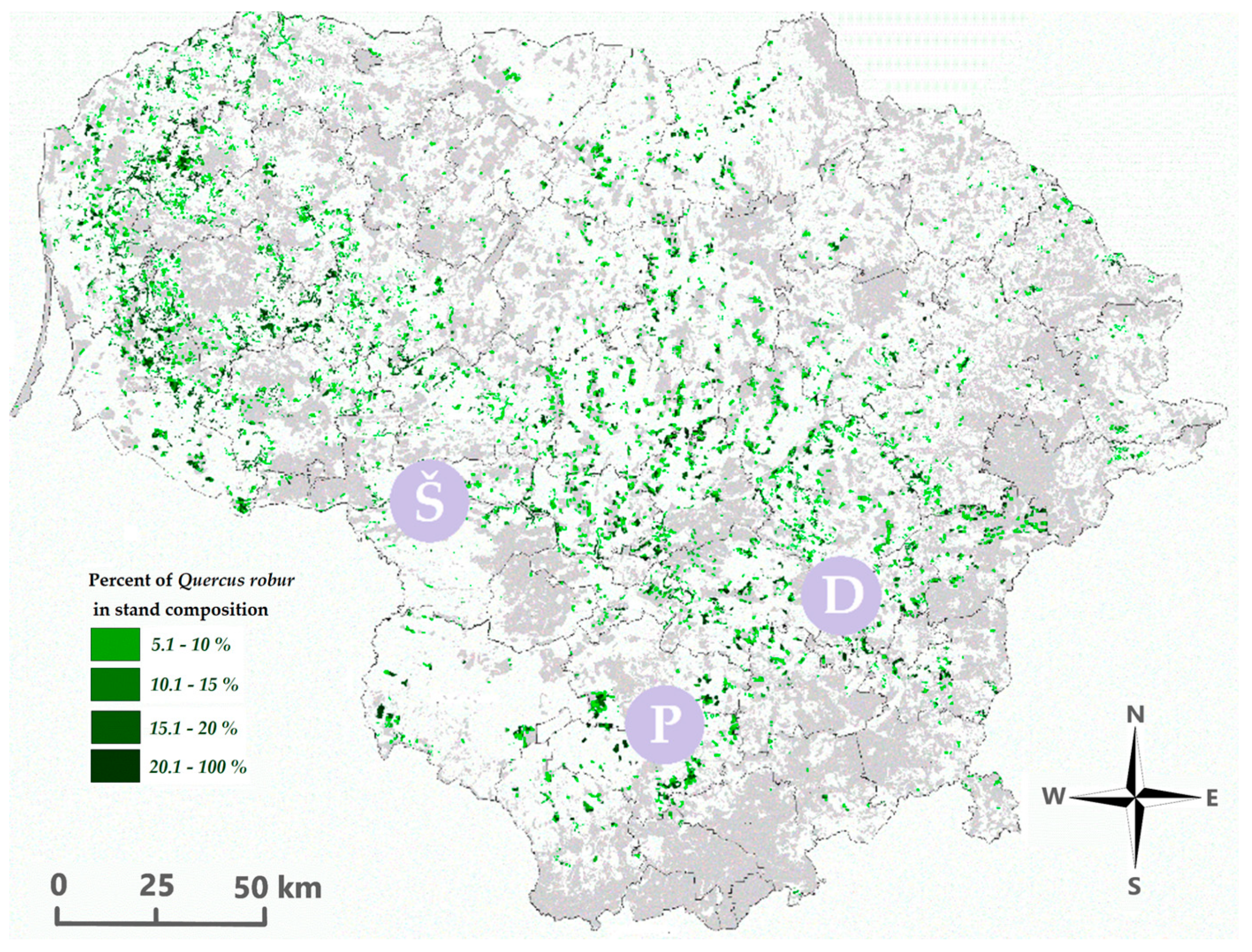
The spores of F. hepatica were trapped between August and October 2022, i.e., in the period when fruitbodies of F. hepatica are formed. The spore trapping was carried out in three oak stands, including Šilinė, Punia, and Dūkšos, which were known for the presence of F. hepatica (Figure 2).
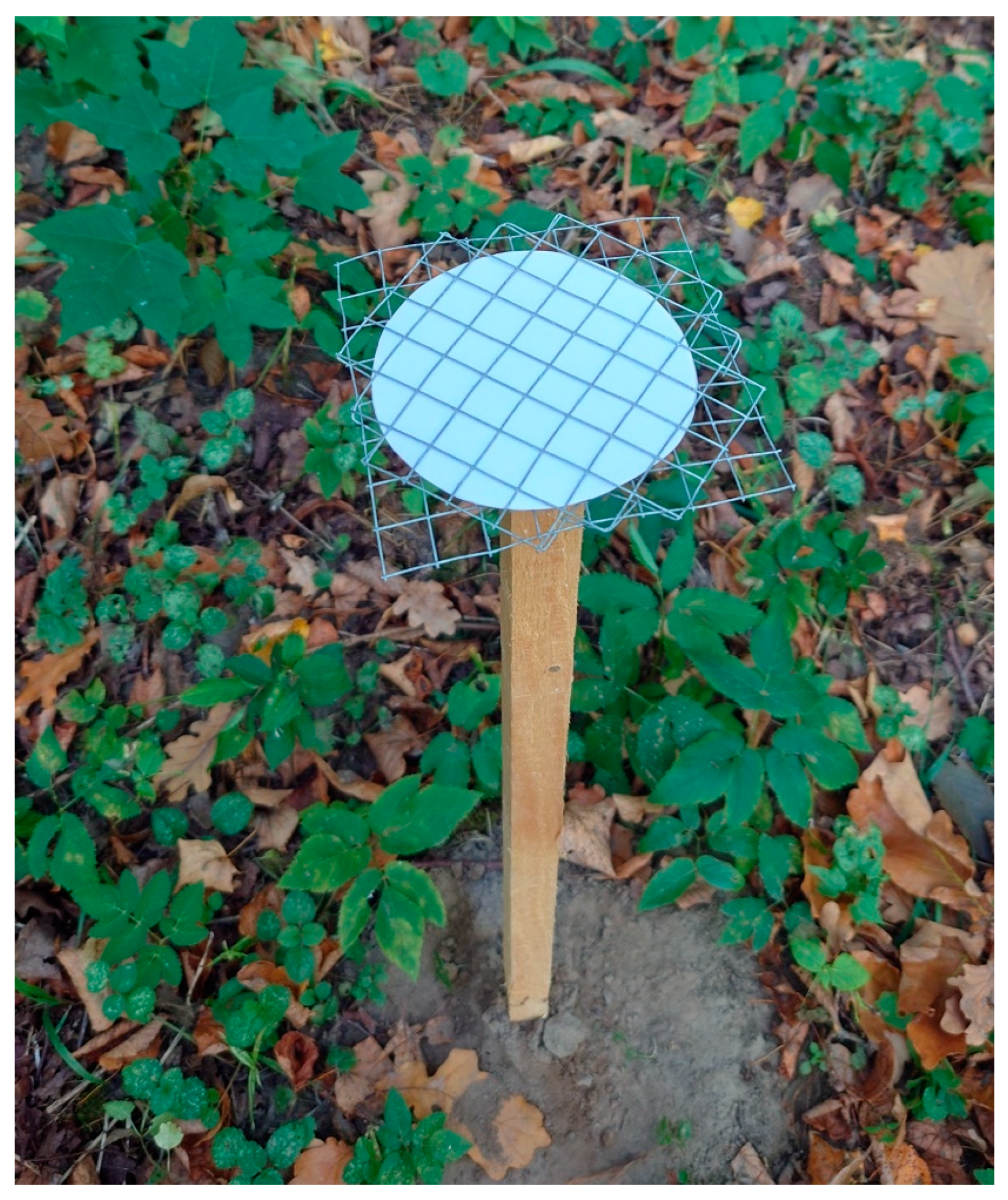
The selected oak stands were more than 200 years old. The oak stands at Punia and Dūkštos were similar in tree species composition as these were dominated by Q. robur with a small admixture of Picea abies and Alnus incana and with Corylus avellana, Sorbus sp., and Ulmus sp. in the understory. The ground vegetation consisted of Aegopodium podagraria, Urtica dioica, and Carex sp. At Šilinė, it was a pure old-growth oak stand with rare Prunus padus in the understory. The herbaceous cover consisted of Corex sp. Punia and Šilinė oak stands being on fertile binary soils of normal humidity, i.e., with groundwater deeper than 3 m from the surface. The Dūkštos oak stand was also on soils of normal humidity, but soil fertility was higher than in the other two oak stands of the study. The distance between individual study sites was >70 km (Figure 2). In all sites, the fruitbodies of F. hepatica were growing on the stems of living oaks at a height of 0.5–1.0 m from the ground. Fungal spores were collected using passive spore traps [24,25]. Each trap consisted of a 9 cm diameter Munktell filter paper (made of cotton stains, particle retention 5–6 μm, class 1F) (Ahlstrom-Munksjö, Stockholm, Sweden) that was sandwiched between two 10 × 10 cm stainless steel grills (mesh size 1 × 1 cm) that were attached horizontally to a 0.8 m long stick to anchor the trap to the ground (Figure 3 and Figure 4). Spore traps were placed at the end of August, i.e., just after the emergence of F. hepatica fruitbodies.
At each site, spore traps were placed at a distance of 5, 25, 50, 150, and 300 m from the fruitbody of F. hepatica (Figure 3 and Figure 4). Spore traps were placed in the direction of prevailing winds, i.e., from west to east. In all sites, spore trapping was initiated on 23 August 2022 and filters with fungal spores were collected once a week [23]. The spore trapping was continued until the end of the fruiting season of F. hepatica, which was different in different sites. In Šilinė, the last spore sample was collected on 5 October; in Punia, on 27 September; and in Dūkštos, on 26 October. In total, there were 30 filters with spore samples collected in Šilinė, 25 in Punia, and 45 in Dūkštos, resulting in 100 samples altogether. During the sampling period, each site was regularly monitored (within a radius of 500 m) for the presence of other fruitbodies of F. hepatica, but such fruitbodies were not detected. Collected samples with fungal spores were labeled, packed in plastic bags, transported to the laboratory, and stored at −20 °C before further processing. At each site, information on air temperature and relative humidity was collected at one-hour intervals near the fruitbodies using UT330C (Uni-Trend Technology, Dongguan, China) temperature and humidity loggers.

2.1. DNA Extraction, PCR Amplification, and Sequencing

In the laboratory, samples with fungal spores were freeze-dried (Labconco FreeZone Benchtop Freeze Dryer, Cole-Parmer, Vernon Hills, IL, USA) at −60 °C for 24 h. For the isolation of DNA, half of each filter paper was taken, cut into smaller pieces, and placed into three 2 mL screw-cap centrifugation tubes together with three sterile glass beads, which were 3 mm in diameter. The remaining materials were stored at −20 °C as a backup. The isolation of DNA followed the study by Marčiulynas et al. [26]. The concentration of DNA was measured using a NanoDrop One spectrophotometer (Thermo Scientific, Rodchester, NY, USA) and adjusted to 10 ng/µL.
Amplification of the ITS2 rDNA region was carried out using a fungal specific primer gITS7 [27] and a universal primer ITS4 [28], both containing sample identification barcodes. PCR reactions were performed in 50 μL reactions and included 0.25 ng/μL of template DNA, 200 μM of dNTPs, 750 μM of MgCl2, 0.025 μM of polymerase (5 U/μL), (DreamTaq Green, Thermo Scientific, Waltham, MA, USA), and 200 nM of each primer. The PCR reactions were performed using an Applied Biosystems 2720 thermal cycler (Waltham, MA, USA) and the following parameters: 95 °C for 5 min, which was followed by 27 cycles of denaturation at 95 °C for 30 s, annealing at 56 °C for 30 s, and an extension at 72 °C for 30 s. The final extension step was at 72 °C for 7 min. The PCR products were examined on a 1.5% agarose gel stained with Nancy-520 (Sigma-Aldrich, Stockholm, Sweden). The purification of PCR products was conducted using 3 M sodium acetate (pH 5.2) (Applichem GmbH, Darmstadt, Germany) and a 96% ethanol mixture (1:2). The quantification of purified PCR products was carried out using a Qubit fluorometer 4.0 (Life Technologies, Hägersten, Sweden). High-throughput sequencing of the amplified samples, which were pooled in an equimolar mix, was achieved using the PacBio Sequel platform and two SMRT cells (SciLifeLab, Uppsala, Sweden).

2.2. Bioinformatics and Statistical Analysis

Produced sequences were quality filtered and clustered using the SCATA NGS sequencing pipeline (http://scata.mykopat.slu.se, accessed on 2 September 2023). Short sequences (<200 bp) and low-quality sequences were removed, while homopolymers were collapsed to three base pairs (bp) prior to clustering. Sequences missing barcodes or primers were also removed. After quality filtering, sequences were clustered into different OTUs using single-linkage clustering based on a 98.5% similarity. For each cluster, the most common genotype (true read) was used to represent each OTU. A consensus sequence was generated for clusters with two sequences. The GenBank (NCBI) database and Blastn algorithm were used for taxonomic identification of different OTUs. The following parameters were used for identification: sequence coverage of at least 80%, species level—98–100%, and genus level—94–97%. Sequences that did not meet these criteria remained unidentified. Correlation analysis was carried out to reveal the relationship between the abundance of high-quality sequences of F. hepatica and air temperature, and the abundance of high-quality sequences of F. hepatica and the distance from the fruitbody using SAS v. 9.4 (Cary, NC, USA).

3. Results and Discussion

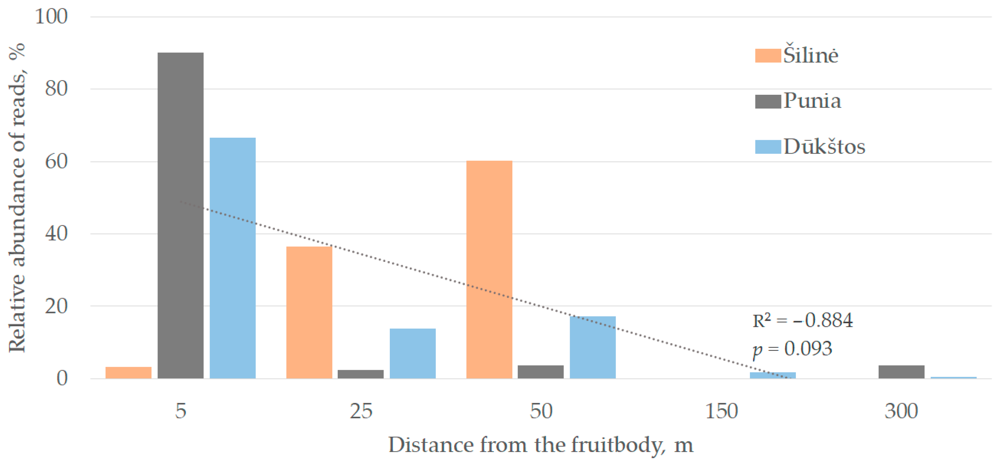
Although high-throughput sequencing generated a large number of reads, in the present study, only reads representing F. hepatica were used in the downstream analyses. Taken together, the absolute abundance of F. hepatica reads was as follows: in Dūkštos, 338 (62.4%) reads; in Šilinė, 123 (22.7%) reads; and in Punia, 81 (14.9%) reads. Although the number of F. hepatica reads varied among different sites, their abundance largely depended on the distance from the fruitbody (Figure 5). In the Punia and Dūkštos sites, 90.1% and 66.7% of all sequence reads were detected 5 m away from the fruitbody, respectively. In the Šilinė site, 60.2% of F. hepatica reads were detected at a distance of 50 m from the fruitbody, 36.6%—at a distance of 25 m, and 3.2%—at a distance of 5 m (Figure 5). At the distance of 150 m and 300 m from the fruitbody, there were only 0.6–3.7% of reads detected at the Dūkštos or Punia sites and no reads were detected at the Šilinė site. Such results may suggest that the structure of the stand and the density of the understory vegetation had only a minor effect on the dispersal of F. hepatica spores at each site. For example, in the Šilinė oak stand, which had no understory, sequence reads of F. hepatica were not found at a distance larger than 50 m from the fruitbody, while in the more dense oak stands of Dūkštos and Punia, sequence reads were found at much larger distances from the fruitbody (Figure 5). The latter may suggest that a great majority of F. hepatica spores were deposited within a distance of 50 m from their fruitbodies. This may also suggest that the spread of F. hepatica spores can be considered sufficient only within short distances from the fruitbody, i.e., within the same stand. In addition, as some spores are usually not viable, this may further limit their spread and colonization of new habitats [29].
Although there was no significant correlation between the number of sequence reads and the distance from the fruitbody (Figure 5), in agreement with other studies, the results showed that there is a gradient of spore deposition from the fruitbody [30]. Stenlid [31] showed that the gradient of spore deposition of Heterobasidion annosum (Fr) Bref., which is a common root-rot pathogen in temperate coniferous forests, is very steep within the first 100 m from the fruitbody. However, under extreme atmospheric conditions, its spores can travel up to 500 km.
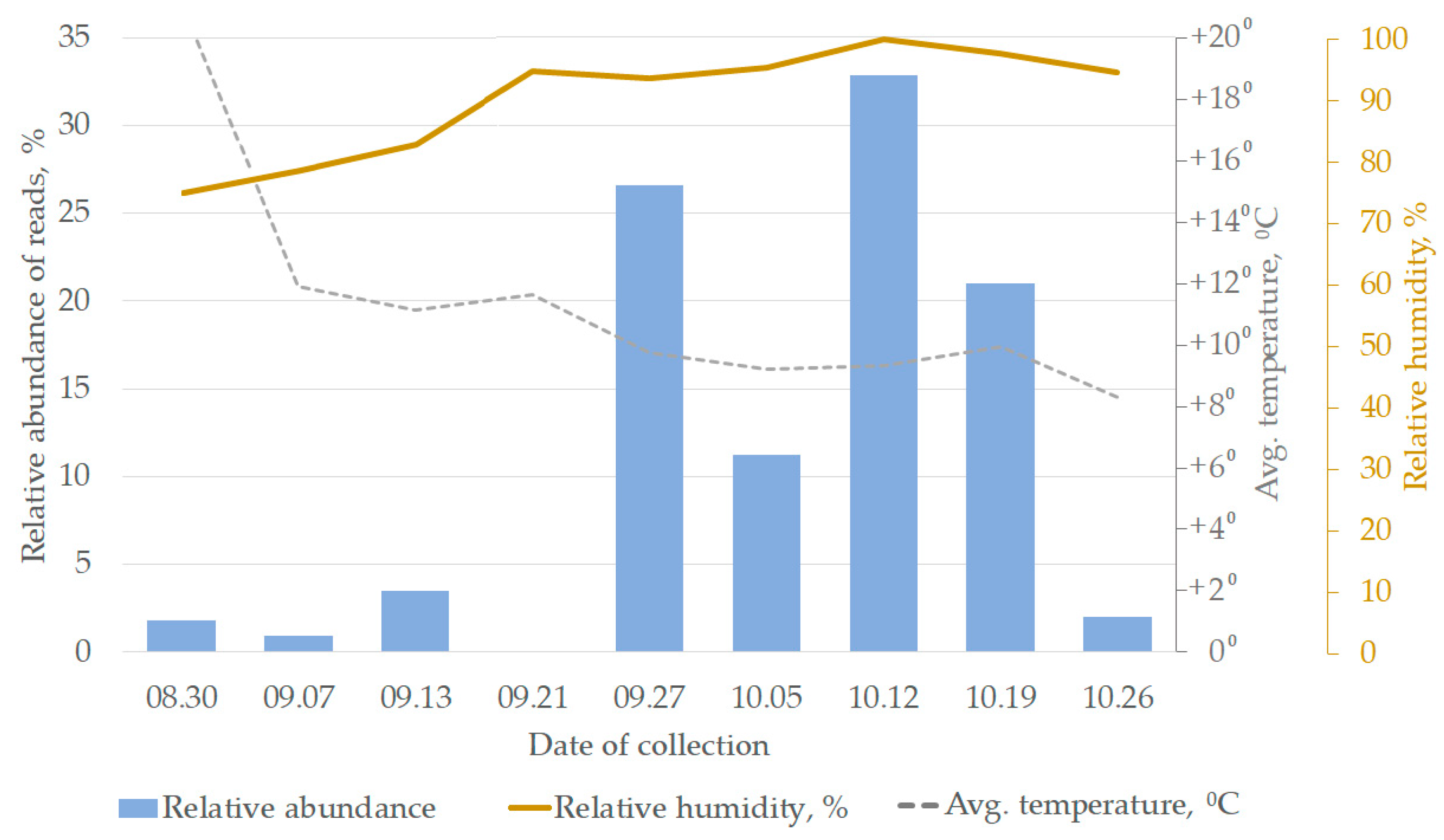
Sequence reads of F. hepatica were detected for the first time on 30 August, showing the release and spread of F. hepatica spores taking place in low abundances soon after the emergence of the fruitbody (Figure 6). In central Slovakia, the spores of F. hepatica were detected from the beginning of July until the first half of November, showing that sporulation takes place throughout the entire developmental period of the fruitbody [32]. We found that F. hepatica sporulated most intensively during the period of four weeks, i.e., from the end of September to the middle of October (Figure 6). However, there were certain differences at different study sites as at Šilinė and Punia, the largest number of F. hepatica sequence reads was detected on 27 September, while in Dūkštos, it was on 12 October. These differences were mainly due to the decay of fruitbodies as after 27 September, at both Šilinė and Punia, the fruitbodies decomposed and a further sampling of spores was not possible. At Dūkštos, fruitbodies of F. hepatica decomposed at the end of October, i.e., one month later. Nuss [33] showed that under favorable conditions, sporulation takes place for almost five weeks. Although Nuss [33] found that the period between the emergence of the fruitbody and its maturation and intensive sporulation was about two weeks, our observations showed that such a period was much longer, i.e., about 28 days (Figure 3 and Figure 6), which can probably be due to local environmental conditions. It was shown that intensive autumn sporulation of polypore fungi usually ends at average temperatures below 15 °C [33]. The results of the present study showed that the most intensive sporulation of F. hepatica occurred when the average daily temperature was around 10 °C (Figure 6). Nevertheless, there was no significant correlation between the number of detected sequence reads of F. hepatica and the average daily temperatures (p > 0.05).
In contrast to the average daily temperature, the relative air humidity showed a significant positive correlation (r = 0.62, p < 0.05) with the number of detected F. hepatica sequence reads (Figure 6). It was shown that air humidity may have a varying effect on the production and release of spores in different fungal species. For example, a high relative humidity favours the formation and release of Ganoderma sp. spores [34,35], but the opposite is observed for Aspergillus fumigatus and Penicillium sp. spores, which reach their minimum when the relative air humidity exceeds 70% [36]. The abundance of Cladosporium sp. spores in the air was shown to positively correlate with temperature but negatively correlate with relative air humidity and rainfall [37,38]. Taken together, the results suggest that the release of F. hepatica spores is most dependent on fruitbody maturation and relative air humidity as in our study, where the period of massive release of F. hepatica spores coincided with the period of fruitbody maturation and higher relative air humidity, but not with the changes in temperature. After reaching the peak, a sharp decrease in the abundance of F. hepatica sequence reads, and thus spores, is likely to be associated with a rapid decomposition of soft fruitbodies of F. hepatica [33,39].

4. Conclusions

The results demonstrated that a great majority of F. hepatica spores were deposited with a distance of 50 m from the fruitbody, showing that this protected species to a large extent depends on local habitats for colonization, while the long-distance dispersal is likely to be very occasional. The release of F. hepatica spores took place during the entire period of fruitbody development, but the extent of spore release was most dependent on the state of fruitbody maturation and a relative air humidity, but not on the average daily temperature. To increase the abundance of F. hepatica in oak stands, conservation measures should be directed to the creation of new habitats (e.g., exposed oak wood) in a close vicinity to its fruitbodies.

Author Contributions

Conceptualization, A.M. (Adas Marčiulynas) and A.M. (Audrius Menkis); methodology, A.M. (Adas Marčiulynas) and A.M. (Audrius Menkis); software, A.M. (Adas Marčiulynas) and A.M. (Audrius Menkis); validation, A.M. (Adas Marčiulynas) and A.M. (Audrius Menkis); formal analysis, A.M. (Adas Marčiulynas) and A.M. (Audrius Menkis); investigation, A.M. (Adas Marčiulynas); resources, A.M. (Adas Marčiulynas) and A.M. (Audrius Menkis); data curation, A.M. (Adas Marčiulynas) and A.M. (Audrius Menkis); writing—original draft preparation, A.M. (Adas Marčiulynas); writing—review and editing, A.M. (Audrius Menkis); visualization, A.M. (Adas Marčiulynas) and A.M. (Audrius Menkis); supervision, A.M. (Audrius Menkis). All authors have read and agreed to the published version of the manuscript.

Funding

This research was funded by the European Social Fund under the 09.3.3-LMT-K-712 “Development of Competences of Scientists other Researchers and Students through Practical Research Activities” measure.

Institutional Review Board Statement

Not applicable.

Informed Consent Statement

Not applicable.

Data Availability Statement

Not applicable.

Conflicts of Interest

The authors declare no conflict of interest.

References

  1. Blackwell, M. The fungi: 1, 2, 3 … 5.1 million species? Am. J. Bot. 2011, 98, 426–438. [Google Scholar] [CrossRef] [PubMed]
  2. Scheffers, B.R.; Joppa, L.N.; Pimm, S.L.; Laurance, W.F. What we know and don’t know about Earth’s missing biodiversity. Trends Ecol. Evol. 2012, 27, 501–510. [Google Scholar] [CrossRef]
  3. Arnold, A.E. Understanding the diversity of foliar endophytic fungi: Progress, challenges, and frontiers. Fungal Biol. Rev. 2007, 21, 51–66. [Google Scholar] [CrossRef]
  4. Heilmann-Clausen, J.; Barron, E.S.; Boddy, L.; Dahlberg, A.; Griffith, G.W.; Nordén, J.; Ovaskainen, O.; Perini, C.; Senn-Irlet, B.; Halme, P. A fungal perspective on conservation biology. Conserv. Biol. 2015, 29, 61–68. [Google Scholar] [CrossRef]
  5. Lofgren, L.A.; Stajich, J.E. Fungal biodiversity and conservation mycology in light of new technology, big data, and changing attitudes. Curr. Biol. 2021, 31, R1312–R1325. [Google Scholar] [CrossRef] [PubMed]
  6. Senn-Irlet, B.; Heilmann-Clausen, J.; Genney, D.; Dahlberg, A. Guidance for Conservation of Macrofungi in Europe; ECCF: Strasbourg, France, 2007. [Google Scholar]
  7. Ryvarden, L.; Gilbertson, R.L. European Polypores, Part 2; Fungiflora: Oslo, Norway, 1994; p. 507. [Google Scholar]
  8. Butin, H. Tree Diseases and Disorders; Oxford University Press: New York, NY, USA, 1995; p. 84. [Google Scholar]
  9. Schwarze, F.W.M.R.; Engels, J.; Mattheck, C. Fungal Strategies of Wood Decay in Trees; Springer: Berlin/Heidelberg, Germany, 2000; p. 193. [Google Scholar]
  10. Wojewoda, W. Checklist of Polish Larger Basidiomycetes. In Biodiversity of Poland, 1st ed.; Mirek, Z., Ed.; Instytut Botaniki im. W. Szafera, Polska Akademia Nauk: Krakow, Poland, 2003; Volume 7. [Google Scholar]
  11. Kujawa, A.; Michalak, M.; Konik, J. Dęby w krajobrazie rolniczym jak o siedlisko zastępcze dla ozorka dębowego Fistulina hepatica (Schaeff.) With. Przegląd Przyrodniczy. Chrońmy Przyr. Ojczystą 2018, 29, 21–27. Available online: https://docplayer.pl/61710618-Stanowiska-ozorka-debowego-fistulina-hepatica-schaeff-with-w-srodkowo-wschodniej-polsce.html (accessed on 25 September 2023).
  12. Marčiulynas, A.; Sirgedaitė-Šėžienė, V.; Menkis, A. Fungi Inhabiting Stem Wounds of Quercus robur following Bark Stripping by Deer Animals. Forests 2023, 14, 2077. [Google Scholar] [CrossRef]
  13. Yurkewich, J.I.; Castaño, C.; Colinas, C. Chestnut red stain: Identification of the fungi associated with the costly discolouration of Castanea sativa. For. Pathol. 2017, 47, e12335. [Google Scholar] [CrossRef]
  14. Niemelä, T. Suomen Kääpien Määritysopas; Guide to the Polypores of Finland: Valkeakoski, Finland, 2006; p. 16. [Google Scholar]
  15. Regué, A.; Bassié, L.; de-Miguel, S.; Colinas, C. Environmental and stand conditions related to Fistulina hepatica heart rot attack on Castanea sativa. For. Pathol. 2019, 49, e12517. [Google Scholar] [CrossRef]
  16. McCartney, A.; West, J. Dispersal of fungal spores through the air. In Food Mycology: A Multifaceted approach to Fungi and Food; Samson, R.A., Dijksterhuis, J., Eds.; CRC Press: Boca Raton, FL, USA, 2007; pp. 65–81. [Google Scholar]
  17. Money, N.P.; Fischer, M.W. Biomechanics of spore release in phytopathogens. Plant Relatsh. Mycota 2009, 5, 115–133. [Google Scholar] [CrossRef]
  18. Wyatt, T.T.; Wösten, H.A.; Dijksterhuis, J. Fungal spores for dispersion in space and time. Adv. Appl. Microb. 2013, 85, 43–91. [Google Scholar] [CrossRef]
  19. Tucker, K.; Stolze, J.L.; Kennedy, A.H.; Money, N.P. Biomechanics of conidial dispersal in the toxic mold Stachybotrys chartarum. Fungal Genet. Biol. 2007, 44, 641–647. [Google Scholar] [CrossRef]
  20. Golan, J.J.; Pringle, A. Long-distance dispersal of fungi. Microb. Spect. 2017, 5, 4–5. [Google Scholar] [CrossRef]
  21. Werth, S.; Wagner, H.H.; Gugerli, F.; Holderegger, R.; Csencsics, D.; Kalwij, J.M.; Scheidegger, C. Quantifying dispersal and establishment limitation in a population of an epiphytic lichen. Ecology 2006, 87, 2037–2046. [Google Scholar] [CrossRef] [PubMed]
  22. Shinn, E.A.; Smith, G.W.; Prospero, J.M.; Betzer, P.; Hayes, M.L.; Garrison, V.; Barber, R.T. African dust and the demise of Caribbean coral reefs. Geophys. Res. Lett. 2000, 27, 3029–3032. [Google Scholar] [CrossRef]
  23. Castaño Soler, C.; Bonet Lledos, J.A.; Oliva Palau, J.; Farré Martinez, G.; Martínez de Aragón, J.; Parladé Izquierdo, X.; Alday, J.G. Rainfall homogenizes while fruiting increases diversity of spore deposition in Mediterranean conditions. Fungal Ecol. 2019, 41, 279–288. [Google Scholar] [CrossRef]
  24. Schweigkofler, W.; O’Donnell, K.; Garbelotto, M. Detection and quantification of airborne conidia of Fusarium circinatum, the causal agent of pine pitch canker, from two California sites by using a real-time PCR approach combined with a simple spore trapping method. Appl. Environ. Microbiol. 2004, 70, 3512–3520. [Google Scholar] [CrossRef] [PubMed]
  25. Garbelotto, M.; Smith, T.; Schweigkofler, W. Variation in rates of spore deposition of Fusarium circinatum, the causal agent of pine pitch canker, over a 12-month-period at two locations in Northern California. Phytopathology 2008, 98, 137–143. [Google Scholar] [CrossRef]
  26. Marčiulynas, A.; Lynikienė, J.; Marčiulynienė, D.; Gedminas, A.; Menkis, A. Seasonal and site-specific patterns of airborne fungal diversity revealed using passive spore traps and high-throughput DNA sequencing. Diversity 2023, 15, 539. [Google Scholar] [CrossRef]
  27. Ihrmark, K.; Bodeker, I.T.M.; Cruz-Martinez, K.; Friberg, H.; Kubartova, A.; Schenck, J.; Strid, Y.; Stenlid, J.; Brandstrom-Durling, M.; Clemmensen, K.E.; et al. New primers to amplify the fungal ITS2 region–evaluation by 454-sequencing of artificial and natural communities. FEMS Microbiol. Ecol. 2012, 82, 666–677. [Google Scholar] [CrossRef]
  28. White, T.J.; Bruns, T.; Lee, S.; Taylor, J. Amplification and direct sequencing of fungal ribosomal RNA genes for phylogenetics. In PCR Protocols: A Guide to Methods and Applications; Innis, M.A., Gelfand, D.H., Sninsky, J.J., White, T.J., Eds.; Academic Press: San Diego, CA, USA, 1990; pp. 315–322. [Google Scholar]
  29. Edman, M.; Gustafsson, M.; Stenlid, J.; Ericson, L. Abundance and viability of fungal spores along a forestry gradient—Responses to habitat loss and isolation? Oikos 2004, 104, 35–42. [Google Scholar] [CrossRef]
  30. Gregory, P.H. Distribution of airborne pollen and spores and their long distance transport. Pure Appl. Geophys. 1978, 116, 309–315. [Google Scholar] [CrossRef]
  31. Stenlid, J. Regional differentiation in Heterobasidion annosum. In Proceedings of the International Conference on Root and Butt Rots, SLU, Uppsala, Sweden, 9–6 August 1993; Johansson, M., Stenlid, J., Eds.; pp. 243–248. [Google Scholar]
  32. Gáper, J. Discharge of basidiospores from Fistulina hepatica fruitbodies in the natural environment. Czech Mycol. 1996, 49, 41–48. Available online: http://www.czechmycology.org/_cm/CM49106.pdf (accessed on 19 September 2023). [CrossRef]
  33. Nuss, I. Ecology of polypores. Biblioth. Mycol. 1975, 45, 1–258. (In German) [Google Scholar]
  34. Martinez-Bracero, M.; Markey, E.; Clancy, J.H.; McGillicuddy, E.J.; Sewell, G.; O’Connor, D.J. Airborne Fungal Spore Review, New Advances and Automatisation. Atmosphere 2022, 13, 308. [Google Scholar] [CrossRef]
  35. Hasnain, S.M.; Al-Frayh, A.; Khatija, F.; Al-Sedairy, S. Airborne Ganoderma basidiospores in a country with desert environment. Grana 2004, 43, 111–115. [Google Scholar] [CrossRef]
  36. Pasanen, A.-L.; Pasanen, P.; Jantunen, M.J.; Kalliokoski, P. Significance of Air Humidity and Air Velocity for Fungal Spore Release into the Air. Atmos. Environ. Part A Gen. Top. 1991, 25, 459–462. [Google Scholar] [CrossRef]
  37. Katial, R.K.; Zhang, Y.; Jones, H.J.; Dyer, P.D. Atmospheric mold spore counts in relation to meteorological parameters. Int. J. Biometeorol. 1997, 41, 17–22. [Google Scholar] [CrossRef]
  38. Grinn-Gofroń, A.; Strzelczak, A. Hourly Predictive Artificial Neural Network and Multivariate Regression Tree Models of Alternaria and Cladosporium Spore Concentrations in Szczecin (Poland). Int. J. Biometeorol. 2009, 53, 555–562. [Google Scholar] [CrossRef] [PubMed]
  39. Soukup, F. A contribution to the knowledge of sporulation of some polypores I. Lesnictví 1988, 33, 145–158. Available online: http://www.czechmycology.org/_cm/CM421.pdf#page=3 (accessed on 22 September 2023).
Figure 1. Brown rot of Quercus robur wood caused by Fistulina hepatica.
Diversity 15 01110 g001
Figure 2. Map of Lithuania showing the position of study sites: P—Punia (N 54°30′50.1″, E 24°04′51.6″), D—Dūkštos (N 54°49′57.2″, E 24°57′15.0″), and Š—Šilinė (N 55°5′22.58″, E 22°57′ 13.85″). The map was reproduced with permission from FORESTGEN, www.forestgen.mi.lt (accessed on 5 September 2023).
Diversity 15 01110 g002
Figure 3. Development of Fistulina hepatica fruitbody. (A)—7 September 2022 (15-day-old fruitbody); (B)—13 September (21-day-old); (C)—21 September (29-day-old); (D)—27 September (35-day-old); (E)—5 October (43-day-old); (F)—12 October (50-day-old); and (G)—19 October (57-day-old). Plastic sheets in pictures were sticky traps used for collection of wood-colonizing insects (not this study). They did not interfere with the sampling of fungal spores.
Diversity 15 01110 g003
Figure 4. The passive spore trap with a filter paper in one of oak stands.
Diversity 15 01110 g004
Figure 5. Relative abundance of Fistulina hepatica sequence reads at different distances from the fruitbody. All sampling time points are combined.
Diversity 15 01110 g005
Figure 6. Relative abundance of Fistulina hepatica sequence reads, average daily temperature, and relative air humidity detected at different time points. All study sites and distances from the fruitbody are combined.
Diversity 15 01110 g006
Disclaimer/Publisher’s Note: The statements, opinions and data contained in all publications are solely those of the individual author(s) and contributor(s) and not of MDPI and/or the editor(s). MDPI and/or the editor(s) disclaim responsibility for any injury to people or property resulting from any ideas, methods, instructions or products referred to in the content.