Review

Unraveling Techniques for Plant Microbiome Structure Analysis

1
Institute of Molecular Biosciences, Mahidol University, Nakhon Pathom 73170, Thailand
2
Department of Sciences, Faculty of Science and Technology, Suan Sunandha Rajabhat University, Bangkok 10300, Thailand
*
Author to whom correspondence should be addressed.
Diversity 2022, 14(3), 206; https://doi.org/10.3390/d14030206
Submission received: 12 January 2022 / Revised: 4 March 2022 / Accepted: 9 March 2022 / Published: 11 March 2022

Abstract

:
Microbiome plays vital role in the life. Study the microbiome of plants with great impact in the planet can provide significant information to solve many problems. Therefore, finding structural population of plant microbiome needs scientific approach. Revealing the specific biochemical and genetical approaches towards identification of specific population provided the growing bodies of methods and procedures to study and analysis the plant microbiomes. Thus, this mini-review paper presents the summarized of scientific methods for study, identify and structural population analysis of plant microbiome.

1. Introductions: Agriculture, Food production and Problems

The present agricultural system is much dependent to many chemicals such as fertilizers and pesticides. The chemicals fate and destiny would be the accumulation toxic compounds and eventually have huge impact on health, environment and even soils as part of the food production system. Furthermore, the necessity to maintain or increase the system productivity leads to excess application of different chemicals thus providing the more harm to ecosystems, increasing pollutions and diseases, soil acidification, eutrophication and ultimately altered soil characteristics as well as inadequate management [1]. One particular example of excess chemical in agricultural system, is the diets with high nitrate food that caused the thyroid and diabetes and even increased the susceptibility of many cancers [2]. Additionally, chemical fertilizers have enormous effects on planet, for example, nitrogen fertilizers alter the global N-cycle and increase the greenhouse gases, damage the ozone and as a final point decreasing the soil organic matters and biodiversity [3,4]. To mitigate the harmful effect of chemicals in agriculture and food production; new food producing systems [5] and changing many practices in current system such as identification and application of ecofriendly alternatives, better cultivar, optimization of mechanized planting and application of microorganisms have been introduced. Microorganisms in this prospective play the huge role. Therefore, this mini-review paper presents the overview of techniques to studying the microorganisms and microbiome in plant microbe interactions and their global prospective in the larger framework to illuminate the correct and recent applications with respect to biochemical and genetical characterizations.

2. Microorganisms: Alternatives to Chemical Fertilizers and Pesticides

As it is necessary to find the alternatives for chemicals applied in agricultural system, especially for chemical fertilizers and pesticides, microorganisms showed great potentials to fulfil the alternative position [6]. The microorganisms in contact with plant and their distribution showed the great functionality in agriculture [7]. Their ecological niche is defined with different part of plants and their utilization has been shown to be very effective to plant growth promotion, disease suppression, toxin removal and assimilating nutrients.
All these advantages were highly dependent to the precise identification and finally bioformulation of them. The need to find the efficient way with specific process always was essential to find the most effective one which would modulate positively the agricultural yield. Therefore identification, diversity and distribution of plant microbe need precise methods. Hence, molecular methods find the outstanding position in plant microbe research and application.

3. Plant Microbiota

Plants have beneficial mutualistic interaction with different microbes in various parts [8]. Different types of microorganisms present on the surface as epiphytes, in the tissue endophyte, on the leaf phyllospheric and with root as rhizospheric microorganisms [9].The most dynamic and studied part was considered as the rhizospheric microorganisms with approved effect on plant growth [10,11,12]. Root and microbes (in the narrow zone around) provided specific microenvironment including roots, microorganisms and their secretions [7]. Microbiome as a scientific term describes all the microorganism’s genomes in this microenvironment. Other parts of the microenvironment include meta proteomics, meta transcriptomics and metametabolomics which are related to all proteins, expressed genes and metabolites respectively. The scope of this minireview presents the current overview of techniques for microbiome analysis with respect to history from single microbe to structural community population.
This interaction which involved countless microbial diversity had significant influence on plant growth and their survival against biotic and abiotic stresses [13,14]. The rhizospheric microbes which considered as the root microbiome is hugely diversifies and complex in their structural community. Two kinds of studies were done mostly in this microenvironment, the microbe-root interaction, and the plant response to different stress factors. Therefore the plant’s microbes present their functions as metaorganism or holobiont [15,16]. Understanding and analysis of the genome involved in this microenvironment helped to reveal the mechanisms involved in formation such a great interaction and utilize them to enhance the crop productivity and finally for sustainable food production [17,18]. Important part of this journey starts with specific method/s that utilized for microbe identification and microbial community analysis. Therefore, here, the summarized molecular techniques for plant microbiome analysis presented as biochemical and genetics methods.

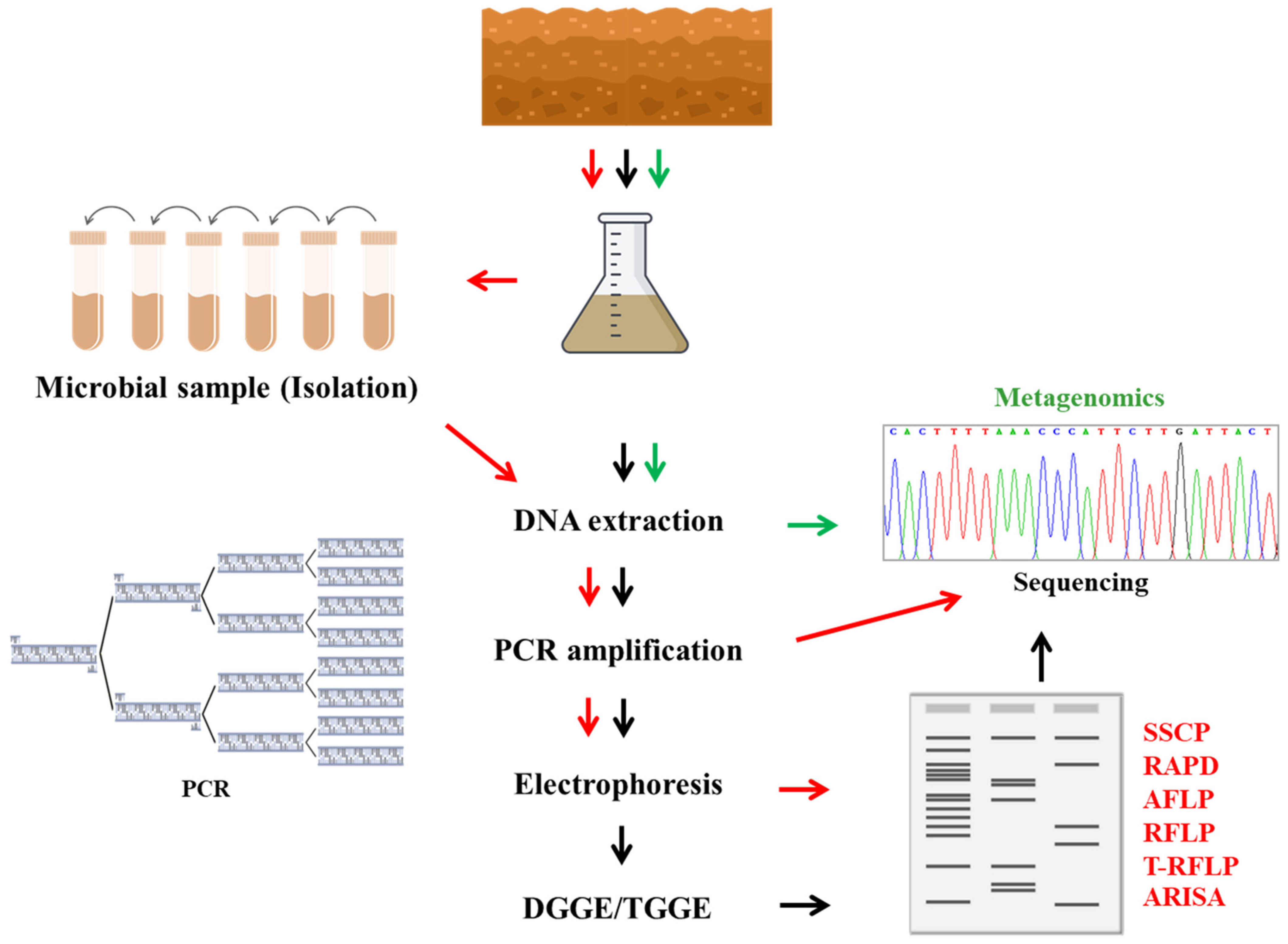
4. The Microbial Identification and Characterization Methods

Growing bodies of methods and procedures are now available for identification and community characterization of different microbes. However, these methods, from simple culturing and light microscope identification to specific molecular probe individually and together, provided much significant knowledge to understand the plant microbiome world, still need more scientific effort to shine the depth black box of agricultural microbiome. It is worth to mention specific strength and limitation for each method. Therefore, for each method here we provided examples for further study and correct application. The strategies always depended on the specific characterization of the microbial cell and utilizing these properties to find the microbial community structure. The divisions of the methods presented here were based on the utilization of microbial genome or phenotypic traits [19]. Scientifically, the diversity considered as the genetic multiplicity of species in the specific ecosystem and here the paper emphasize was on the composed plant microbe communities built the rhizospheric microenvironment. Species diversity consists of total presentation and distribution species. However, defining this term was with respect to eukaryotes therefore it should be used with caution [20,21]. Thus, diversity of microbe in the system or environment was specified as the number and density of specific microorganisms from specific taxa. This definition was respect to the microorganism’s community as functional groups.
It should be mention that in microbial ecology, diversity defined and elaborated with great concerns to the effect of stresses and disturbances from biotic or abiotic origins [22]. As mentioned earlier the difficulty of defining the species and genetic diversity raised due to the taxonomic and methodological limitation for prokaryotes. Then, dependability of phenotypic characterizations of microorganisms are influenced the capturability of them. The phenotypic characterizations of them are mostly related to their cell activities. Therefore, they are the main limitation of biochemical methods. Furthermore, most of the microorganisms cannot adapt themselves with laboratory conditions as their metabolic activities disrupted [23].
It is noteworthy to mentions here that however, traditional physiological and biochemical methods that used in the most biochemical test kit [23] had many limitations (which might be related to low metabolic activities under the experiment condition) still considered as useful methods such as specific fingerprint lipid biomarkers (LB), phospholipid fatty acids (PLFA) (Figure 1). As they showed that they could distinguish the microbial communities significantly based on the utilization of PLFA of specific bacteria as a biomarker [24].
Later, the nucleic acid technologies (molecular biology) provided more precise and reliable picture of microbial diversity and their structural communities. The precise application of different statistical analysis methods (to evaluate the variation of different data (fingerprints)) such as principal component analysis (PCA), canonical variant analysis (CVA) have had great advantages in finding the more bright picture of microbial diversity. Thus, biochemical and molecular based techniques [25] together usually provided better scientific overview for agricultural microbiology and helped more precisely to shed the light on structure–function insight characterizations.

5. Biochemical Techniques to Determine the Microbial Diversity

Traditionally the plate count considered as the fast and inexpensive method for diversity of culture-dependent microbes [26,27,28]. This method could present the information on the active species and culturable microorganisms which indicates the heterotrophic factors of the microorganism’s population [29]. Scientifically only 0.1% of microorganism was estimated to be cultured with different [30] laboratory techniques. It is noteworthy to mention that temperature, pH, and light conditions could be including in growth limitations of microorganisms with standard plate technique [31].
Physiological distributions presented in soils microorganisms would be investigated by Community Level Physiological Profiles (CLPP). The CLPP profiles showed the diversity of microbial community’s base on the utilization of carbon substrates [32,33,34].
Differences in these forms of utilizations could be analyzed and showed the different in the viable microorganisms and eventually microbial population. Many manual and commercial systems identify and characterize the microbe community with these methods [35,36,37].
The great advantage of this method is the automation features which by using an automated measuring machine, yielding large scientific results, and therefore dealing with huge information made simple. This method is very popular for understanding the importance of functionality attributes to the microbial communities even though the analysis of such data is sometimes complicated. Furthermore, this method was able to analyze the metabolic diversity of culturable microorganisms, but the slow-growing microbes have less influence on the presentation on final metabolic of community. It should be mentioned here that the pH of the plates is usually buffered which brings the different from reality of soil acidity or alkaline properties. Therefore, the results provided with this method lead to some limitations and should be analyzed with caution specially when dealing with specific microbes that strongly adapted to the environment specially pH (acidity or alkalinity). These conditions have considered disadvantages of this method and cannot represent the real community structure.
Another important method that worth to mention here is the results of fatty acids microbial community structure based that was presented by Fatty acid methyl ester (FAME) method [38,39,40,41]. Fatty acids constructed a relatively established fraction of the cell biomass (Figure 1). Fatty acids profile could discriminate the diversity between the major taxonomic populations within a microbial structure. Thus, by changing the fatty acid profiles, different microorganism’s community would present. The phospholipid fatty acid (PLFA) method revealed different strategies engaged by microorganism’s population to adapt to the environmental changes such as soil types, climatic origins, and different management [42,43,44].

6. Genetics Techniques

The density and metabolic activities of microbes in the environment have been analyzed by microbiologist with different molecular techniques for many years. Several techniques have been developed to detect, identify, and find the phylogenic relationship of plant microbe interactions. These methods can be separated based on polymerase chain reaction (PCR). The methods explain here, are nucleic acid hybridization [45,46,47], as well as length polymorphisms based on PCR and finally metagenomics approach.

6.1. Nucleic Acid Hybridization and Fluorescent In Situ Hybridization (FISH)

Based on the specific probes, the nucleic acid hybridization approach can be used as a quantitative toll in molecular microbiology. The method could be carried out on extracted DNA and RNA. In the early works, the biofilm spatial distribution of microorganism was studied by this method successfully [48]. With respect to many advantages, limitations of this method were unclear results for detection of cells contains low ribosome beside the lack of sensitivity for low copy number sequence. Thus, slow-growing bacteria cannot detect due to the low ribosome content per cell which was often linked with low physiological activity. Even though FISH with tyramime signal amplification method could analyze the slow-growing microbe [49,50].

6.2. Guanine/Cytosine (G + C)

Soil bacterial diversity can be investigated by differences in guanine/cytosine (G + C) rate of DNA [51]. 3% and 5% of differentiation of microorganisms G + C contents brought the clear result for this procedure. This technique presented a level of declaration based on this fact that different taxonomic levels have different percentage rate of G + C. The curves of microbial grouping reports used as factor for genetic differences. By low resolution, this approach could easily apply as a “change indicator” for population structure, particularly in the low diversity. No PCR bias, uncovering the rare member of the microbial populations and detecting some of the less detectable microorganism which couldn’t detect by PCR are the most advantages of this method. Conversely it needs large amounts of DNA [52].

6.3. PCR-Based Techniques

In agricultural microbiology the polymerase chain reaction (PCR) method has been applied to study the non-culture able microorganisms in the environment. This revealing approach can be extensively employed in many ecological and agricultural research too. PCR can be performed by the DNA extracted directly from the environmental system. PCR with the aim of 16S rDNA carried out comprehensively to study the microbe diversity and identification [53]. On the other hand, for fungal studies18S rDNA and internal transcribed spacer (ITS) regions are applied usually. In this method universal or specific primers for amplification of the 16S rDNA, 18S rDNA, or ITS have been done by many researchers, and PCR products are separated in different conditions of electrophoresis. Also, PCR product can be studied easily with specific probes to present definite data on the specific microorganism’s community [54].

6.3.1. Denaturing Gradient Gel Electrophoresis (DGGE) and Temperature Gradient Gel Electrophoresis (TGGE)

Denaturing gradient gel electrophoresis (DGGE) and temperature gradient gel electrophoresis (TGGE) are more precise techniques for exploring the microbial communities [55,56,57]. DGGE method can discrete DNA strength with only one specific base-pair variance however they are identical base on the length. In this method the gradient of denaturants electrophoretic system is based on the melting point [58]. With the same rule as DGGE method, in TGGE gradient temperature would be used rather than chemical denaturants. Detection of point mutations in DNA sequences could be achieved by these methods. Reliability, reproducibility to some extent and by some means inexpensive was the advantage of these methods which could provide simultaneous analysis of various samples; and present the fluctuations in microbial populations [59,60].

6.3.2. Length Polymorphisms

Randomly amplified polymorphic DNA polymerase chain reaction (RAPD-PCR) as useful genetic markers is an easy and fast technique for resolving of genetic diversity at various taxonomic levels [61,62]. In this method PCR product amplified with arbitrary primers; the PCR products were evaluated with electrophoresis stained; and the gel images were scrutinized with image documentation systems. The products as a band in the gel were scored and finalized as a matrix of randomly amplified polymorphic DNA bands. The size and percentage of specific bands was exploited to assess genomic multiplicity. Rapid and richness in polymorphic DNA were the advantages of this method compared to others (Figure 2).
Amplified fragment length polymorphisms (AFLP) were PCR-based markers for the fast detection of genetic diversity. Key feature of AFLP-PCR was its capacity for immediate and direct screening of many DNA regions distributed randomly throughout the genome. Detection of polymorphisms of genomic restriction fragments could be done by this method. This PCR based approach had accepted as suitable for evaluating genetic variations among individuals and populations [63,64]. The complexity in homologous markers (alleles), representing this method less applicable for accurate research. Conversely the promptness and simplicity with reliability marks which could be created by AFLPs were making this method as a powerful molecular tool kit for ecologists and evolutionary biologists [65,66].
Restriction fragment length polymorphism (RFLP) is another method that determining microbial mixture community with the help of a DNA sequence. PCR products such as rDNA were digested by restriction enzyme [67]. Different product’s lengths are distinguished by electrophoresis agarose or polyacrylamide gel electrophoresis (PCGE) [68]. Terminal restriction fragment length polymorphism (T-RFLP) follows the same rule could be used to evaluate spatial and temporal deviations in microbial community [32]. One of the most powerful techniques with high resolution and reproducibility for detecting the fungal diversity was automated ribosomal intergenic spacer analysis (ARISA) [69,70]. Ribosomal intergenic spacer analysis (RISA) is related to the length of the ribosomal intergenic spacer region between the 16S and 23S rRNA genes [71,72]. The method has been applied for characterization of different strains along with to classify groups of complexes populations [73,74].This method also can utilize length variability of rRNA genes to categorize trials into operational taxonomic units (OTUs).
Single-strand conformational polymorphism (SSCP) is based on separation of PCR of sequence variation of single-stranded DNA intra molecular folding [75,76,77]. Single-strand conformational polymorphism method was applied to distinguish the purity of microorganisms cultures and discriminate diversity of uncultivated microbial populations in various plants [78]. This method is easier than earlier methods as they don’t need the GC-clamp or specific device for their application. Like other PCR-base method it has the PCR bias limitation. Specific growth enrichment in particular microorganism led to identify the functional study of interest microorganisms is another useful method.
It should mention here that the amplicon sequencing that is based on the specific binding of the pair universal primers to the highly conserved region of microbial genome with the help of PCR and then sequencing, helped significantly to explore the microbial community [79,80]. The most famous one is 16S rDNA gene [81] which utilized commonly for identification of bacteria and microbial community based on the amplification of taxon specific hypervariable regions [80]. The accuracy of the method is highly dependent to the reference database quality and completeness. Sequencing procedures and analysis here should use with careful concern and microbiome results showed inferring and deducing with higher sequence similarity threshold (99%) [82].

7. Metagenomics Approach

Metagenomics approach which utilized the whole genome of the microbial community showed batter advantage compared to amplicon sequencing. As shown the whole genome and even extrachromosomal fragments such as plasmids provided complete information of specific microbe therefore present more depth and clear picture of microbial community [83]. Several quality control techniques and web-based tools helped to provide more approachable and precise illustration of microbial community with metagenomics approach. It should mention here that for detection and identification of effective microbe for further application in agricultural field, identification of bacterial in strain level which cannot achieved with 16S rDNA could be more approachable with metagenomics. Here for constructing the correct structural community the bioinformatics tools to find the inter-genomic differences would be very useful [84]. Furthermore, the functional level of microbial community could be explored with annotation of specific gene and targeting the targeted amplicon of that gene with metagenomics. Thus, gene prediction and annotation of results were two most important steps of functional microbial community structure analysis. It is noteworthy to mention here that prediction of gene doesn’t mean the expression of protein in this method which could bring its limitation [85]. For the more specific information about the whole functional activities of microbiome, many metaproteomic and metametabolomic approaches have been introduced recently that are outside the scope of this minireview and published recently elsewhere [86,87,88,89].

8. Conclusions

Microbiomes with very substantial impact on over life need accurate, precise, and rapid method/s to understand their key functions. This paper reviewed the methods and procedures in agricultural microbiology with emphasis on rhizosphere microbe. In near future with more scientific information on different microbiomes, correct application of them for better functionality would be more available for solving environmental pollutions. In the journey of finding the mechanism of microbiomes, it is important to know the microbial identification methods and their limitation for correct usage and annotations.

Author Contributions

The authors (S.S. and B.J.) contributed equally to this mini review paper. All authors have read and agreed to the published version of the manuscript.

Funding

This research received no external funding.

Institutional Review Board Statement

Not applicable.

Informed Consent Statement

Not applicable.

Acknowledgments

The authors would like to thank Chaisri Tharasawatdipipat, Dean of the Faculty of Science and Technology for his outstanding support. Wirongrong Thamyo is acknowledged for excellent secretarial assistance.

Conflicts of Interest

The authors declare no conflict of interest.

References

  1. Basosi, R.; Spinelli, D.; Fierro, A.; Jez, S. Mineral Nitrogen Fertilizers: Environmental Impact of Production and Use; Fernando Lòpez-Valdez, F.F.L., Ed.; NOVA Science Publishers: Hauppauge, NY, USA, 2014; pp. 3–43. [Google Scholar]
  2. Ahmed, M.; Rauf, M.; Mukhtar, Z.; Saeed, N.A. Excessive use of nitrogenous fertilizers: An unawareness causing serious threats to environment and human health. Environ. Sci. Pollut. Res. Int. 2017, 24, 26983–26987. [Google Scholar] [CrossRef] [PubMed]
  3. Hakeem, K.R.; Sabir, M.; Ozturk, M.; Akhtar, M.S.; Ibrahim, F.H. Nitrate and nitrogen oxides: Sources, health effects and their remediation. Rev. Environ. Contam. Toxicol. 2017, 242, 183–217. [Google Scholar] [CrossRef] [PubMed]
  4. Singh, B. Are nitrogen fertilizers deleterious to soil health? Agronomy 2018, 8, 48. [Google Scholar] [CrossRef] [Green Version]
  5. Chriki, S.; Hocquette, J.-F. The Myth of cultured meat: A review. Front. Nutr. 2020, 7, 7. [Google Scholar] [CrossRef] [PubMed] [Green Version]
  6. Santos, L.F.; Olivares, F.L. Plant microbiome structure and benefits for sustainable agriculture. Curr. Plant Biol. 2021, 26, 100198. [Google Scholar] [CrossRef]
  7. Dubey, S.; Sharma, S. Rhizospheric engineering by plant-mediated indirect selection of microbiome for agricultural sustainability. Crit. Rev. Plant Sci. 2021, 40, 379–397. [Google Scholar] [CrossRef]
  8. Jat, S.L.; Suby, S.B.; Parihar, C.M.; Gambhir, G.; Kumar, N.; Rakshit, S. Microbiome for sustainable agriculture: A review with special reference to the corn production system. Arch. Microbiol. 2021, 203, 2771–2793. [Google Scholar] [CrossRef] [PubMed]
  9. Kavamura, V.N.; Mendes, R.; Bargaz, A.; Mauchline, T.H. Defining the wheat microbiome: Towards microbiome-facilitated crop production. Comput. Struct. Biotechnol. J. 2021, 19, 1200–1213. [Google Scholar] [CrossRef] [PubMed]
  10. Bakker, P.A.; Berendsen, R.L.; Doornbos, R.F.; Wintermans, P.C.; Pieterse, C.M. The rhizosphere revisited: Root microbiomics. Front. Plant Sci. 2013, 4, 165. [Google Scholar] [CrossRef] [PubMed] [Green Version]
  11. Lakshmanan, V.; Selvaraj, G.; Bais, H.P. Functional soil microbiome: Belowground solutions to an aboveground problem. Plant Physiol. 2014, 166, 689–700. [Google Scholar] [CrossRef] [Green Version]
  12. Mendes, R.; Garbeva, P.; Raaijmakers, J.M. The rhizosphere microbiome: Significance of plant beneficial, plant pathogenic, and human pathogenic microorganisms. FEMS Microbiol. Rev. 2013, 37, 634–663. [Google Scholar] [CrossRef] [PubMed]
  13. Bandyopadhyay, P.; Bhuyan, S.K.; Yadava, P.K.; Varma, A.; Tuteja, N. Emergence of plant and rhizospheric microbiota as stable interactomes. Protoplasma 2017, 254, 617–626. [Google Scholar] [CrossRef] [PubMed]
  14. Panke-Buisse, K.; Poole, A.C.; Goodrich, J.K.; Ley, R.E.; Kao-Kniffin, J. Selection on soil microbiomes reveals reproducible impacts on plant function. ISME J. 2015, 9, 980–989. [Google Scholar] [CrossRef] [PubMed]
  15. Hacquard, S. Disentangling the factors shaping microbiota composition across the plant holobiont. New Phytol. 2016, 209, 454–457. [Google Scholar] [CrossRef] [PubMed] [Green Version]
  16. Hacquard, S.; Schadt, C.W. Towards a holistic understanding of the beneficial interactions across the Populus microbiome. New Phytol. 2015, 205, 1424–1430. [Google Scholar] [CrossRef] [PubMed]
  17. Knapp, D.G.; Németh, J.B.; Barry, K.; Hainaut, M.; Henrissat, B.; Johnson, J.; Kuo, A.; Lim, J.H.P.; Lipzen, A.; Nolan, M.; et al. Comparative genomics provides insights into the lifestyle and reveals functional heterogeneity of dark septate endophytic fungi. Sci. Rep. 2018, 8, 6321. [Google Scholar] [CrossRef] [PubMed] [Green Version]
  18. Quiza, L.; St-Arnaud, M.; Yergeau, E. Harnessing phytomicrobiome signaling for rhizosphere microbiome engineering. Front. Plant Sci. 2015, 6, 507. [Google Scholar] [CrossRef] [PubMed]
  19. Solbrig, O.T. The origin and function of biodiversity. Environ. Sci. Policy Sustain. Dev. 1991, 33, 16–38. [Google Scholar] [CrossRef]
  20. Godfray, H.C.J.; Lawton, J.H. Scale and species numbers. Trends Ecol. Evol. 2001, 16, 400–404. [Google Scholar] [CrossRef]
  21. Øvreås, L. Population and community level approaches for analysing microbial diversity in natural environments. Ecol. Lett. 2000, 3, 236–251. [Google Scholar] [CrossRef]
  22. Hughes, J.B.; Hellmann, J.J.; Ricketts, T.H.; Bohannan, B.J. Counting the uncountable: Statistical approaches to estimating microbial diversity. Appl. Environ. Microbiol. 2001, 67, 4399–4406. [Google Scholar] [CrossRef] [PubMed] [Green Version]
  23. Das, S.; Dash, H.R. Microbial Diversity in the Genomic Era; Academic Press: Cambridge, MA, USA, 2018. [Google Scholar]
  24. Rashid, M.; Stingl, U. Contemporary molecular tools in microbial ecology and their application to advancing biotechnology. Biotechnol. Adv. 2015, 33, 1755–1773. [Google Scholar] [CrossRef] [PubMed] [Green Version]
  25. Grządziel, J.; Gałązka, A. The molecular-based methods used for studying bacterial diversity in soils contaminated with PAHs (The Review). In Soil Contamination—Current Consequences and Further Solutions; Marcelo, L., Larramendy, S.S., Eds.; IntechOpen: London, UK, 2016; pp. 85–104. [Google Scholar]
  26. Elsayed, T.R.; Grosch, R.; Smalla, K. Potato plant spheres and to a lesser extent the soil type influence the proportion and diversity of bacterial isolates with in vitro antagonistic activity towards Ralstonia solanacearum. FEMS Microbiol. Ecol. 2021, 97, fiab038. [Google Scholar] [CrossRef] [PubMed]
  27. Li, X.P.; Li, J.H.; Qi, Y.H.; Guo, W.; Li, X.; Li, M.Q. Effects of naked barley root rot on rhizosphere soil microorganisms and enzyme activity. Acta Ecol. Sin. 2017, 37, 5640–5649. [Google Scholar] [CrossRef] [Green Version]
  28. Yu, M.; Wang, X.; Yan, A. Microbial profiles of retail pacific oysters (Crassostrea gigas) from Guangdong province, China. Front. Microbiol. 2021, 12, 689520. [Google Scholar] [CrossRef] [PubMed]
  29. Tabacchioni, S.; Chiarini, L.; Bevivino, A.; Cantale, C.; Dalmastri, C. Bias caused by using different isolation media for assessing the genetic diversity of a natural microbial population. Microb. Ecol. 2000, 40, 169–176. [Google Scholar] [CrossRef] [PubMed]
  30. Pace, N.R. A molecular view of microbial diversity and the biosphere. Science 1997, 276, 734–740. [Google Scholar] [CrossRef]
  31. van Elsas, J.D.; Duarte, G.F.; Keijzer-Wolters, A.; Smit, E. Analysis of the dynamics of fungal communities in soil via fungal-specific PCR of soil DNA followed by denaturing gradient gel electrophoresis. J. Microbiol. Methods 2000, 43, 133–151. [Google Scholar] [CrossRef]
  32. Escobar Ortega, J.S.; Aguilar Vásquez, N.N.; Ávila Alba, T.; García de Salamone, I.E. Impact of management of cover crop–soybean agroecosystems on rhizosphere microbial communities. Eur. J. Soil Sci. 2021, 72, 1154–1176. [Google Scholar] [CrossRef]
  33. Hu, H.; Li, X.; Wu, S.; Lou, W.; Yang, C. Effects of long-term exposure to oxytetracycline on phytoremediation of swine wastewater via duckweed systems. J. Hazard. Mater. 2021, 414, 125508. [Google Scholar] [CrossRef]
  34. Zuluaga, M.Y.A.; Milani, K.M.L.; Miras-Moreno, M.B.; Lucini, L.; Valentinuzzi, F.; Mimmo, T.; Pii, Y.; Cesco, S.; Rodrigues, E.P.; de Oliveira, A.L.M. The adaptive metabolomic profile and functional activity of tomato rhizosphere are revealed upon PGPB inoculation under saline stress. Environ. Exp. Bot. 2021, 189, 104552. [Google Scholar] [CrossRef]
  35. Ginting, R.; Solihat, N.; Hafsari, A. Potential bacteria capable of remediating mercury contaminated soils. In Proceedings of the IOP Conference Series: Earth and Environmental Science, Bogor, Indonesia, 16–18 September 2020; IOP Publishing: Bristol, UK, 2020; p. 012136. [Google Scholar]
  36. Kim, Y.S.; Balaraju, K.; Jeon, Y.H. Biological characteristics of Bacillus amyloliquefaciens AK-0 and suppression of ginseng root rot caused by Cylindrocarpon destructans. J. Appl. Microbiol. 2017, 122, 166–179. [Google Scholar] [CrossRef] [PubMed]
  37. Yinli, Z.; Jianwei, G.; Wei, Y.; Jianbo, B.; Xianqi, H. Study on isolation, identification and colonization ability of antagonistic bacterium M39 against pomegranate wilt pathogen. In Proceedings of the IOP Conference Series: Earth and Environmental Science, Guiyang, China, 14–16 June 2019; IOP Publishing: Bristol, UK, 2019; p. 052019. [Google Scholar]
  38. Khatri, K.; Mohite, J.A.; Pandit, P.S.; Bahulikar, R.; Rahalkar, M.C. Description of ‘Ca. Methylobacter oryzae’ KRF1, a novel species from the environmentally important Methylobacter clade 2. Antonie Van Leeuwenhoek 2020, 113, 729–735. [Google Scholar] [CrossRef] [PubMed]
  39. Nwigwe, C.; Fossey, A.; de Smidt, O. Characterisation of Eucalyptus rhizospheric communities using fatty acid methyl ester (FAME) profile analysis. S. Afr. J. Plant Soil. 2021, 38, 116–125. [Google Scholar] [CrossRef]
  40. Poblete-Morales, M.; Carvajal, D.; Almasia, R.; Michea, S.; Cantillana, C.; Levican, A.; Silva-Moreno, E. Pseudomonas atacamensis sp. nov., isolated from the rhizosphere of desert bloom plant in the region of Atacama, Chile. Antonie Van Leeuwenhoek 2020, 113, 1201–1211. [Google Scholar] [CrossRef] [PubMed]
  41. Shin, B.; Park, C.; Lee, B.H.; Lee, K.E.; Park, W. Bacillus miscanthi sp. nov., a alkaliphilic bacterium from the rhizosphere of Miscanthus sacchariflorus. Int. J. Syst. Evol. Microbiol. 2020, 70, 1843–1849. [Google Scholar] [CrossRef] [PubMed]
  42. Fu, Z.-D.; Li, Z.; Ping, C.; Qing, D.; Ting, P.; Chun, S.; Wang, X.-C.; Liu, W.-G.; Yang, W.-Y.; Yong, T.-W. Effects of maize-soybean relay intercropping on crop nutrient uptake and soil bacterial community. J. Integr. Agric. 2019, 18, 2006–2018. [Google Scholar] [CrossRef]
  43. Luo, Y.; Xiao, M.; Yuan, H.; Liang, C.; Zhu, Z.; Xu, J.; Kuzyakov, Y.; Wu, J.; Ge, T.; Tang, C. Rice rhizodeposition promotes the build-up of organic carbon in soil via fungal necromass. Soil Biol. Biochem. 2021, 160, 108345. [Google Scholar] [CrossRef]
  44. Wang, H.; Wang, J.; Ge, C.; Yao, H. Fungi dominated the incorporation of 13C-CO2 into microbial biomass in tomato rhizosphere soil under different CO2 concentrations. Microorganisms 2021, 9, 2121. [Google Scholar] [CrossRef]
  45. Liu, J.; Bao, Y.; Zhang, X.; Xu, S.; Qiu, J.; He, J. Rhodobacter kunshanensis sp. nov., a novel bacterium isolated from activated sludge. Curr. Microbiol. 2021, 78, 3791–3797. [Google Scholar] [CrossRef]
  46. Posada, L.F.; Álvarez, J.C.; Romero-Tabarez, M.; de-Bashan, L.; Villegas-Escobar, V. Enhanced molecular visualization of root colonization and growth promotion by Bacillus subtilis EA-CB0575 in different growth systems. Microbiol. Res. 2018, 217, 69–80. [Google Scholar] [CrossRef] [PubMed]
  47. Rasul, M.; Yasmin, S.; Zubair, M.; Mahreen, N.; Yousaf, S.; Arif, M.; Sajid, Z.I.; Mirza, M.S. Phosphate solubilizers as antagonists for bacterial leaf blight with improved rice growth in phosphorus deficit soil. Biol. Control 2019, 136, 103997. [Google Scholar] [CrossRef]
  48. Thurnheer, T.; Gmür, R.; Guggenheim, B. Multiplex FISH analysis of a six-species bacterial biofilm. J. Microbiol. Methods 2004, 56, 37–47. [Google Scholar] [CrossRef] [PubMed]
  49. Galaviz, C.; Lopez, B.R.; de-Bashan, L.E.; Hirsch, A.M.; Maymon, M.; Bashan, Y. Root growth improvement of mesquite seedlings and bacterial rhizosphere and soil community changes are induced by inoculation with plant growth-promoting bacteria and promote restoration of eroded desert soil. Land Degrad. Dev. 2018, 29, 1453–1466. [Google Scholar] [CrossRef]
  50. Zhang, J.; Guo, T.; Wang, P.; Tian, H.; Wang, Y.; Cheng, J. Characterization of diazotrophic growth-promoting rhizobacteria isolated from ginger root soil as antagonists against Ralstonia solanacearum. Biotechnol. Biotechnol. Equip. 2018, 32, 1447–1454. [Google Scholar] [CrossRef]
  51. Leal, C.; Fontaine, F.; Aziz, A.; Egas, C.; Clément, C.; Trotel-Aziz, P. Genome sequence analysis of the beneficial Bacillus subtilis PTA-271 isolated from a Vitis vinifera (cv. Chardonnay) rhizospheric soil: Assets for sustainable biocontrol. Environ. Microbiome 2021, 16, 3. [Google Scholar] [CrossRef]
  52. Chhetri, G.; Kim, J.; Kim, I.; Kang, M.; So, Y.; Seo, T. Oryzicola mucosus gen. nov., sp. nov., a novel slime producing bacterium belonging to the family Phyllobacteriaceae isolated from the rhizosphere of rice plants. Antonie Van Leeuwenhoek 2021, 114, 1925–1934. [Google Scholar] [CrossRef]
  53. Fazal, A.; Wen, Z.; Yang, M.; Liao, Y.; Fu, J.; He, C.; Wang, X.; Jie, W.; Ali, F.; Hu, D. Deciphering the rhizobacterial assemblages under the influence of genetically engineered maize carrying mcry genes. Environ. Sci. Pollut. Res. 2021, 28, 60154–60166. [Google Scholar] [CrossRef]
  54. Samaddar, S.; Schmidt, R.; Tautges, N.E.; Scow, K. Adding alfalfa to an annual crop rotation shifts the composition and functional responses of tomato rhizosphere microbial communities. Appl. Soil Ecol. 2021, 167, 104102. [Google Scholar] [CrossRef]
  55. Chu, H.; Wang, H.; Zhang, Y.; Li, Z.; Wang, C.; Dai, D.; Tang, M. Inoculation with ectomycorrhizal fungi and dark septate endophytes contributes to the resistance of Pinus spp. to pine wilt disease. Front. Microbiol. 2021, 12, 687304. [Google Scholar] [CrossRef]
  56. Elhady, A.; Abbasi, S.; Safaie, N.; Heuer, H. Responsiveness of elite cultivars vs. ancestral genotypes of barley to beneficial rhizosphere microbiome, supporting plant defense against root-lesion nematodes. Front. Plant Sci. 2021, 12, 721016. [Google Scholar] [CrossRef] [PubMed]
  57. Ran, L.; Li, J.; Xing, Y.; Zhang, J.; Zhou, X. Effects of p-Coumaric acid on the structure and abundance of soil Pseudomonas spp. community. Allelopath. J. 2021, 53, 211–218. [Google Scholar] [CrossRef]
  58. Zhao, X.; Miao, R.; Guo, M.; Zhou, Y. Effects of Fire Phoenix (a genotype mixture of Fesctuca arundinecea L.) and Mycobacterium sp. on the degradation of PAHs and bacterial community in soil. Environ. Sci. Pollut. Res. Int. 2021, 28, 25692–25700. [Google Scholar] [CrossRef] [PubMed]
  59. Dong, Z.; Guo, Y.; Yu, C.; Zhixian, Z.; Rongli, M.; Deng, W.; Li, Y.; Hu, X. The dynamics in rhizosphere microbial communities under bacterial wilt resistance by mulberry genotypes. Arch. Microbiol. 2021, 203, 1107–1121. [Google Scholar] [CrossRef] [PubMed]
  60. Pratiwi, E.; Satwika, T.; Agus, F. Analysis of peat bacterial diversity in oil palm plantations and a logged forest in Jambi, Indonesia, using PCR-DGGE technique. In Proceedings of the IOP Conference Series: Earth and Environmental Science, Bogor, Indonesia, 16–18 September 2020; IOP Publishing: Bristol, UK, 2020; p. 012200. [Google Scholar]
  61. González-García, S.; Álvarez-Pérez, J.M.; Sáenz de Miera, L.E.; Cobos, R.; Ibañez, A.; Díez-Galán, A.; Garzón-Jimeno, E.; Coque, J.J.R. Developing tools for evaluating inoculation methods of biocontrol Streptomyces sp. strains into grapevine plants. PLoS ONE 2019, 14, e0211225. [Google Scholar] [CrossRef] [Green Version]
  62. Mohkam, M.; Nezafat, N.; Berenjian, A.; Mobasher, M.A.; Ghasemi, Y. Identification of Bacillus probiotics isolated from soil rhizosphere using 16S rRNA, recA, rpoB gene sequencing and RAPD-PCR. Probiotics Antimicrob. Proteins 2016, 8, 8–18. [Google Scholar] [CrossRef]
  63. Keyser, C.A.; De Fine Licht, H.H.; Steinwender, B.M.; Meyling, N.V. Diversity within the entomopathogenic fungal species Metarhizium flavoviride associated with agricultural crops in Denmark. BMC Microbiol. 2015, 15, 249. [Google Scholar] [CrossRef] [Green Version]
  64. Zhang, Q.; Stummer, B.E.; Guo, Q.; Zhang, W.; Zhang, X.; Zhang, L.; Harvey, P.R. Quantification of Pseudomonas protegens FD6 and Bacillus subtilis NCD-2 in soil and the wheat rhizosphere and suppression of root pathogenic Rhizoctonia solani AG-8. Biol. Control 2021, 154, 104504. [Google Scholar] [CrossRef]
  65. Aserse, A.A.; Räsänen, L.A.; Aseffa, F.; Hailemariam, A.; Lindström, K. Diversity of sporadic symbionts and nonsymbiotic endophytic bacteria isolated from nodules of woody, shrub, and food legumes in Ethiopia. Appl. Microbiol. Biotechnol. 2013, 97, 10117–10134. [Google Scholar] [CrossRef]
  66. Moller, A.M.; Valle, M.C.E.; Teyer, L.F.S.; Rosales, G.V.; Bejar, A.A.G.; Hernandez, M.E.T. Genetic variability in Rhizoctonia solani Isolated from Vitis vinifera based on amplified fragment length polymorphism. Am. J. Agric. Biol. Sci. 2011, 6, 317–323. [Google Scholar] [CrossRef] [Green Version]
  67. Jacobs-Hoffman, I.; Hills, P.N. Effects of the commercial biostimulant BC204 on the rhizosphere microbial community of Solanum lycopersicum L. S. Afr. J. Bot. 2021, 143, 52–60. [Google Scholar] [CrossRef]
  68. Duan, Y.; Chen, R.; Zhang, R.; Jiang, W.; Chen, X.; Yin, C.; Mao, Z. Isolation, identification, and antibacterial mechanisms of Bacillus amyloliquefaciens QSB-6 and its effect on plant roots. Front. Microbiol. 2021, 12, 746799. [Google Scholar] [CrossRef] [PubMed]
  69. Johnston-Monje, D.; Lopez Mejia, J. Botanical microbiomes on the cheap: Inexpensive molecular fingerprinting methods to study plant-associated communities of bacteria and fungi. Appl. Plant Sci. 2020, 8, e11334. [Google Scholar] [CrossRef] [PubMed] [Green Version]
  70. Madigan, A.P.; Egidi, E.; Bedon, F.; Franks, A.E.; Plummer, K.M. Bacterial and fungal communities are differentially modified by melatonin in agricultural soils under abiotic stress. Front. Microbiol. 2019, 10, 2616. [Google Scholar] [CrossRef] [PubMed] [Green Version]
  71. Likar, M.; Stres, B.; Rusjan, D.; Potisek, M.; Regvar, M. Ecological and conventional viticulture gives rise to distinct fungal and bacterial microbial communities in vineyard soils. Appl. Soil. Ecol. 2017, 113, 86–95. [Google Scholar] [CrossRef]
  72. Ondreičková, K.; Gubišová, M.; Gubiš, J.; Klčová, L.; Horník, M. Rhizosphere bacterial communities of Arundo donax grown in soil fertilised with sewage sludge and agricultural by-products. Agriculture 2019, 65, 37–41. [Google Scholar] [CrossRef] [Green Version]
  73. Ondreičková, K.; Žofajová, A.; Piliarová, M.; Gubiš, J.; Hudcovicová, M. Monitoring of rhizosphere bacterial communities in soil with sewage sludge addition using two molecular fingerprinting methods: Do these methods give similar results? Agriculture 2016, 62, 52–61. [Google Scholar] [CrossRef] [Green Version]
  74. Wood, J.L.; Zhang, C.; Mathews, E.R.; Tang, C.; Franks, A.E. Microbial community dynamics in the rhizosphere of a cadmium hyper-accumulator. Sci. Rep. 2016, 6, 36067. [Google Scholar] [CrossRef]
  75. Abdeljalil, N.O.-B.; Vallance, J.; Gerbore, J.; Yacoub, A.; Daami-Remadi, M.; Rey, P. Combining potential oomycete and bacterial biocontrol agents as a tool to fight tomato Rhizoctonia root rot. Biol. Control 2021, 155, 104521. [Google Scholar] [CrossRef]
  76. Schmidt, C.S.; Alavi, M.; Cardinale, M.; Müller, H.; Berg, G. Stenotrophomonas rhizophila DSM14405T promotes plant growth probably by altering fungal communities in the rhizosphere. Biol. Fertil. Soils 2012, 48, 947–960. [Google Scholar] [CrossRef]
  77. Wang, X.-R.; Szmidt, A.E. Molecular markers in population genetics of forest trees. Scand. J. For. Res. 2001, 16, 199–220. [Google Scholar] [CrossRef]
  78. Vallance, J.; Le Floch, G.; Déniel, F.; Barbier, G.; Lévesque, C.A.; Rey, P. Influence of Pythium oligandrum biocontrol on fungal and oomycete population dynamics in the rhizosphere. Appl. Environ. Microbiol. 2009, 75, 4790–4800. [Google Scholar] [CrossRef] [PubMed] [Green Version]
  79. Armanhi, J.S.L.; de Souza, R.S.C.; de Araújo, L.M.; Okura, V.K.; Mieczkowski, P.; Imperial, J.; Arruda, P. Multiplex amplicon sequencing for microbe identification in community-based culture collections. Sci. Rep. 2016, 6, 29543. [Google Scholar] [CrossRef] [PubMed] [Green Version]
  80. D’Amore, R.; Ijaz, U.Z.; Schirmer, M.; Kenny, J.G.; Gregory, R.; Darby, A.C.; Shakya, M.; Podar, M.; Quince, C.; Hall, N. A comprehensive benchmarking study of protocols and sequencing platforms for 16S rRNA community profiling. BMC Genom. 2016, 17, 55. [Google Scholar] [CrossRef] [PubMed] [Green Version]
  81. Kittelmann, S.; Seedorf, H.; Walters, W.A.; Clemente, J.C.; Knight, R.; Gordon, J.I.; Janssen, P.H. Simultaneous amplicon sequencing to explore co-occurrence patterns of bacterial, archaeal and eukaryotic microorganisms in rumen microbial communities. PLoS ONE 2013, 8, e47879. [Google Scholar] [CrossRef] [PubMed]
  82. Kopylova, E.; Navas-Molina Jose, A.; Mercier, C.; Xu Zhenjiang, Z.; Mahé, F.; He, Y.; Zhou, H.-W.; Rognes, T.; Caporaso, J.G.; Knight, R.; et al. Open-Source Sequence Clustering Methods Improve the State Of the Art. mSystems 2016, 1, e00003–e00015. [Google Scholar] [CrossRef] [PubMed] [Green Version]
  83. Nayfach, S.; Pollard, K.S. Toward accurate and quantitative comparative metagenomics. Cell 2016, 166, 1103–1116. [Google Scholar] [CrossRef] [PubMed] [Green Version]
  84. Parab, S.; Bussolino, F. Multi-omics: Overview, challenges, and applications. In Microbiome-Host Interactions; CRC Press: Boca Raton, FL, USA, 2021; pp. 13–20. [Google Scholar]
  85. Boers, S.A.; Jansen, R.; Hays, J.P. Understanding and overcoming the pitfalls and biases of next-generation sequencing (NGS) methods for use in the routine clinical microbiological diagnostic laboratory. Eur. J. Clin. Microbiol. Infect. Dis. 2019, 38, 1059–1070. [Google Scholar] [CrossRef] [Green Version]
  86. Salvato, F.; Hettich, R.L.; Kleiner, M. Five key aspects of metaproteomics as a tool to understand functional interactions in host-associated microbiomes. PLoS Pathog. 2021, 17, e1009245. [Google Scholar] [CrossRef]
  87. Zhang, X.; Figeys, D. Perspective and guidelines for metaproteomics in microbiome studies. J. Proteome Res. 2019, 18, 2370–2380. [Google Scholar] [CrossRef]
  88. Levy, A.; Conway, J.M.; Dangl, J.L.; Woyke, T. Elucidating bacterial gene functions in the plant microbiome. Cell Host Microbe 2018, 24, 475–485. [Google Scholar] [CrossRef] [PubMed] [Green Version]
  89. Singh, D.; Raina, T.K.; Kumar, A.; Singh, J.; Prasad, R. Plant microbiome: A reservoir of novel genes and metabolites. Plant Gene 2019, 18, 100177. [Google Scholar] [CrossRef]
Figure 1. The phospholipid fatty acid (PLFA) method revealed different strategies engaged by microorganism’s population to adapt to the environmental changes such as soil types, climatic origins, and different management.
Diversity 14 00206 g001
Figure 2. Microbial diversity study from isolation of microbes to metagenomics. SSCP = Single-strand conformational polymorphism, RAPD = Randomly amplified polymorphic DNA, AFLP = Amplified fragment length polymorphisms, RFLP = Restriction fragment length polymorphism, T-RFLP = Terminal restriction fragment length polymorphism, ARISA = Automated ribosomal intergenic spacer analysis, DGGE = Denaturing gradient gel electrophoresis, TGGE = Temperature gradient gel electrophoresis.
Diversity 14 00206 g002
Publisher’s Note: MDPI stays neutral with regard to jurisdictional claims in published maps and institutional affiliations.

