3. Results
Figure 1,
Figure 2,
Figure 3,
Figure 4,
Figure 5,
Figure 6,
Figure 7,
Figure 8,
Figure 9,
Figure 10,
Figure 11,
Figure 12,
Figure 13 and
Figure 14.
MATERIAL: ♂, E Malaysia, Sabah, km 52 Rd. Kota-Kinabalu—Tambunan, 1700–1800 m, 3–8.VIII.2002, S. Kurbatov & S. Zimina leg., ‘Platerodrilus svetae sp.n., S. Kazantsev des. 2009′, ‘Paratype’ (ICM); ♀, E Malaysia: Sabah, Kinabalu Mt., S slopes, 6.02° N, 116.54° E, 1750–1800 m, S. Kazantsev leg., collected as last instar larva on 20.I.2018, turned into female on 6.IV.2018, died on 23.IV.2018 (MPGU); 2 larvae, ♀♀, E Malaysia: Sabah, Crocker Range, Gunung Alab Stn, 5.82° N, 116.34° E, 1850–2000 m, 23–28.I.2018, S. Kazantsev leg. (MPGU).
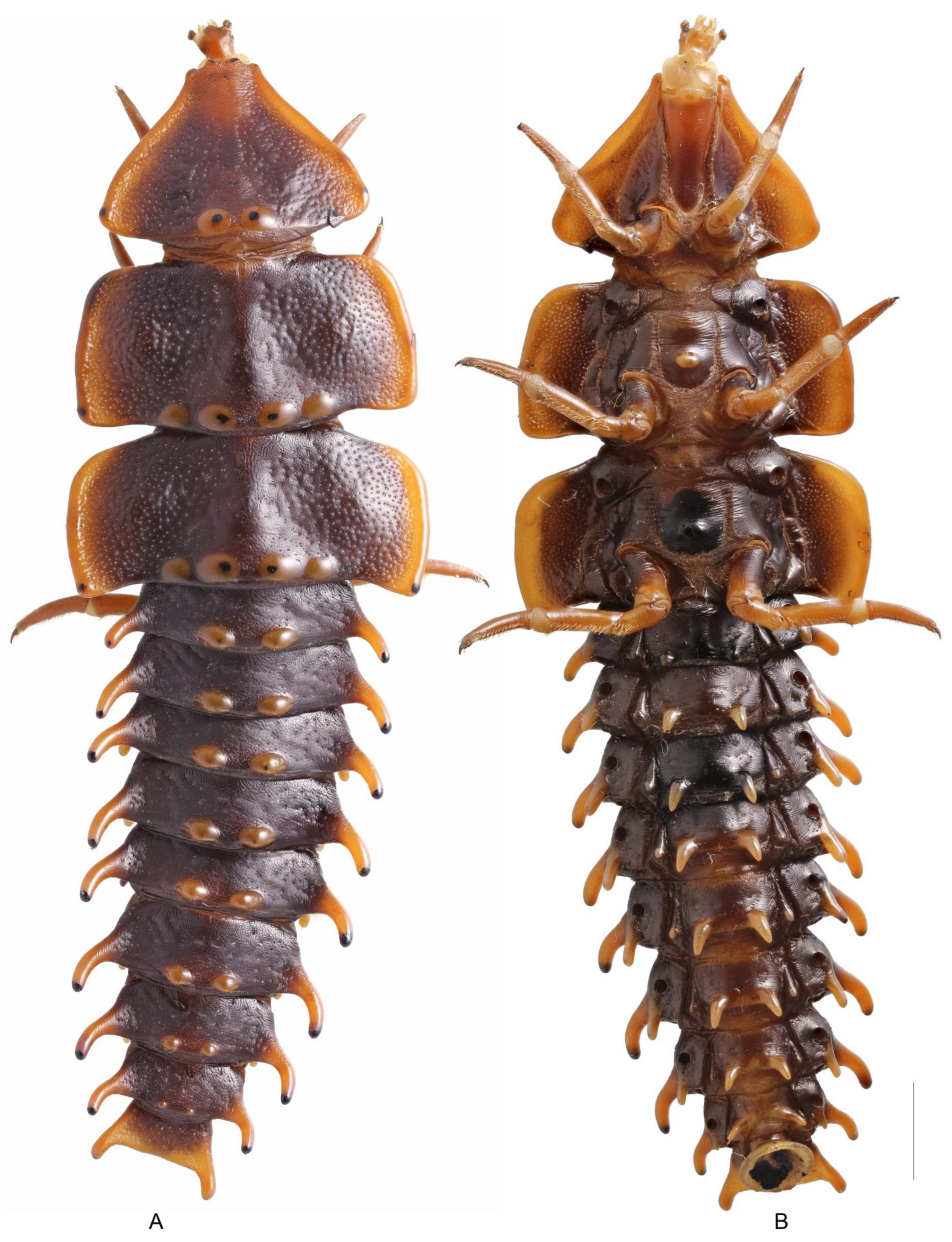
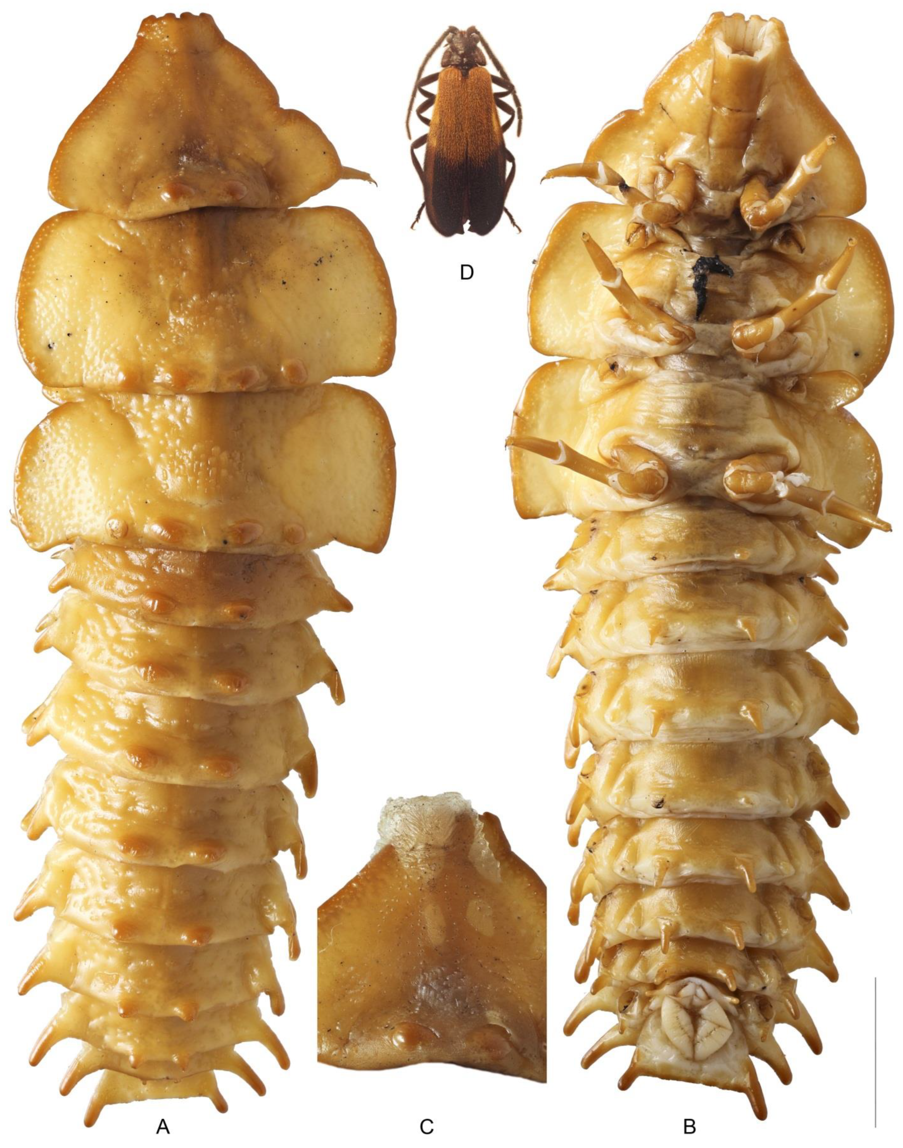
Larva. Last instar (female). Body length—ca. 72 mm (
Figure 2).
Body elongate, widest at thoracic segments, compressed, heavily sclerotized. Most part of the body dark brown, with head, thoracic sides, abdominal appendages and legs testaceous (
Figure 2).
Cuticle (
Figure 8A–C) compound, without spines, consisting of closed polygonal (usually penta- and hexagonal) areoles (
Figure 8C), relatively strongly pigmented and bearing numerous sensillae: in average 340 (CI95% 326.8–361.4;
n = 13) per square millimeter.
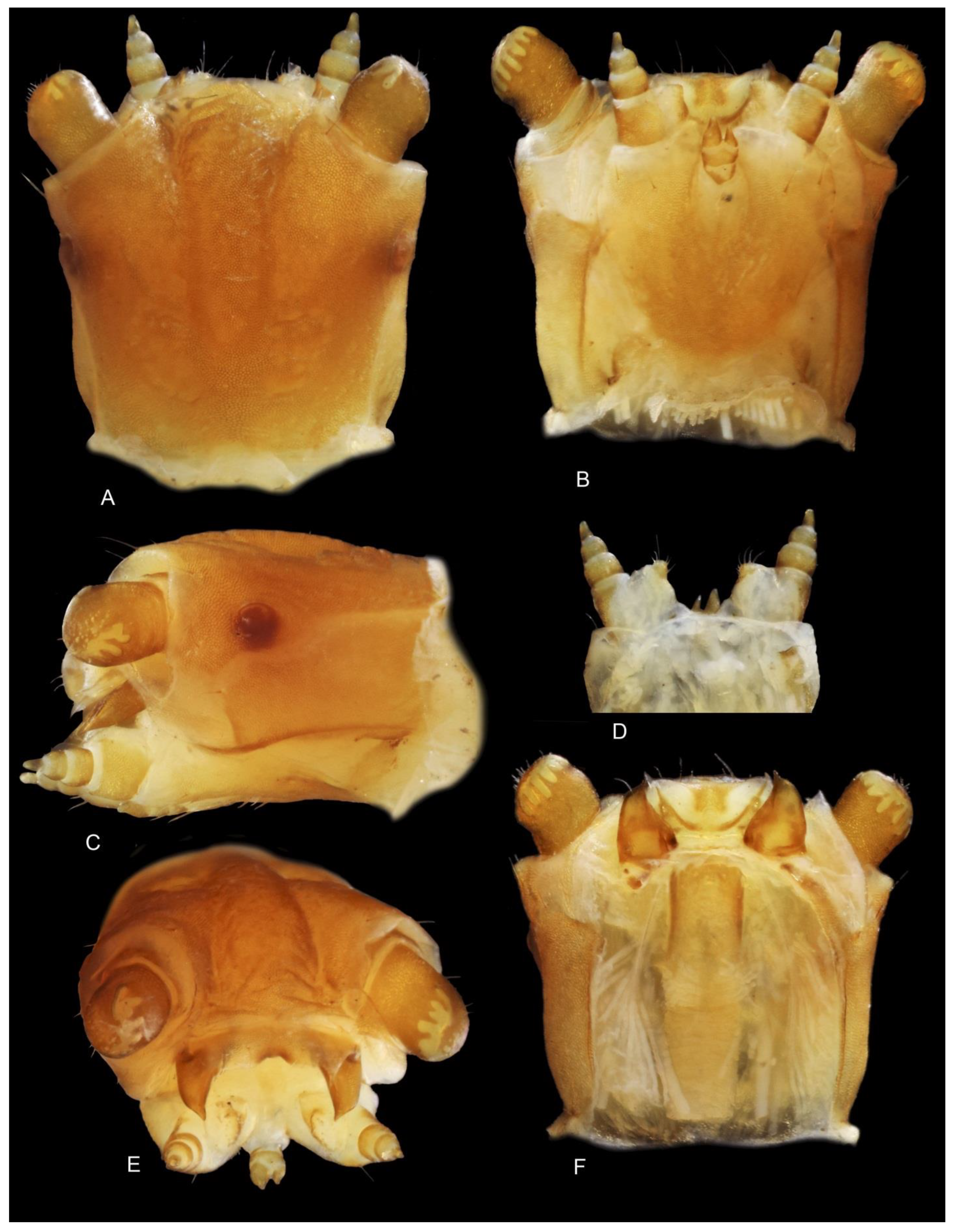
Head considerably smaller than prothorax, slightly longer than wide, completely retractable into prothorax, anterior margin rounded. Head capsule open ventrally, consisting of four sclerites: dorsal plate, with three pairs of long setae, a pair of lateral sclerites, each with two long setae, and semicircular ventral plate, with a pair of setae and a pair of narrow posterolateral processes. Frontal sutures absent (
Figure 2 and
Figure 3).
Antennae retractable, 2-segmented, antennomere 1 (antennifer?) represented by narrow annuliform sclerite located on membranous retractable tube; antennomere 2 large and robust, its apical membrane multi-lobed, with several petals ventrally and two pairs of petals dorsally, sclerotized parts bearing several short setae. Fossa antennalis complete. Dorsal plate with one pair of stemmata located at lateral edges posteriad of antennae. Mandibles approximate basally, tripartite, their dorsal plate formed by paired labrum and their distal part resting on galea. Maxillae with 3-segmented maxillary palps, tapering apically. Galea partly sclerotized, large, elongate, lying interoventrally with respect to maxillary palps, bearing several short setae distally. Labium with undivided prementum; labial palps 2-segmented; ligula absent (
Figure 3).
All thoracic terga considerably wider that abdominal terga, roughly punctuate. Pronotum trapezoidal, trisinuate anteriorly, posteriorly with a pair of distinct pupilled tubercles. Prosternum elongate, gradually narrowing posteriorly. Meso and metatergum with two pairs of tubercles posteriorly, of which only median pair have pupils, anteriorly with vestiges of longitudinal suture. Meso- and metanotum with distinct tubercle in posterior half, yellowish in mesonotum and black in metanotum. Thoracic pleuron consisting of well differentiated epipleurite and hypopleurite (the former absent on prothorax). Meso- and metathorax with spiracular plates located on epipleurites, each spiracular plate represented by prominent cavity with a spiracle at its edge and several spiracles of different size and shape at the bottom of the cavity. Both pairs of thoracic spiracles are similar in size and structure and functional (
Figure 2).
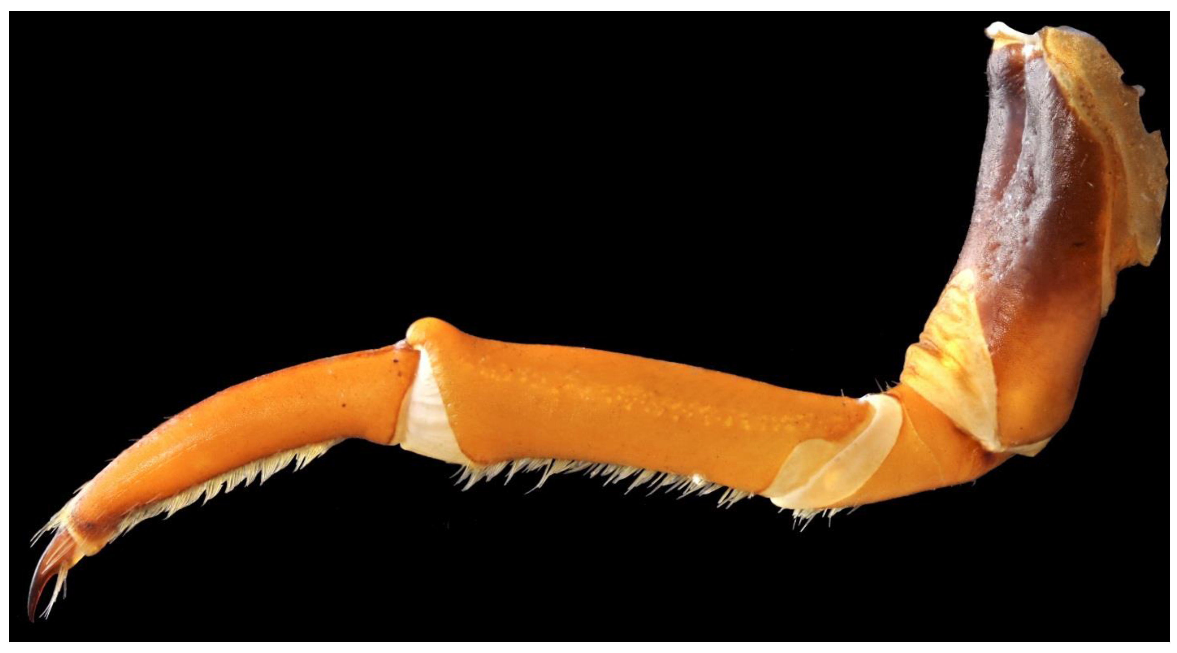
Legs 5-segmented, ventrally with a brush of dense short hairs. Coxa free, elongate. Trochanter divided into two parts by a membranous line bearing densely spaced sensillae (
Figure 4).
Abdominal tergites I–VIII transverse, scarcely punctured, with long, narrow, slightly curved lateral processes and a pair of distinct median tubercles, with tubercles on terga VI-VIII gradually diminishing in size. Tergite IX transverse, widening and truncate posteriorly, with paired short fixed urogomphi. Abdominal epipleurites prominent, with elongate backward-directed processes and spiracular plates. Hypopleurites differentiated, small, with small backward-directed processes. Abdominal sternites I–VIII entire, with a pair of backward-directed processes, these processes shortest on sternite 1. All thoracic spiracles are similar in size and structure to thoracic ones. Abdominal segment X short, tubular, distinctly widening posteriorly (
Figure 2).
Adult female. Exoskeleton. The paedomorphic adult female externally is similar to the last instar larva, surpassing the conspecific male many times in length and width, only weakly sclerotized and yellowish-white throughout, with the pygopodium (abdominal segment X) reduced to narrow non-functional appendix (
Figure 5).
However, two of its head structures are distinctly non-larval: the labrum is represented by a single sclerite, located between mandibles and noticeably emarginate anteriorly, and the mandibles are robust and uniform, distant from each other basally (
Figure 6E,F).
Furthermore, the membranous slit on the last antennomere has distinctly smaller petals (
Figure 6), galea is shorter and less sclerotized (
Figure 6B) and the labium seems to have obtained narrow semi-divided mentum (
Figure 6B).
Legs 5-segmented, structurally similar to the leg of a larva (
Figure 7).
Integument. The female cuticle is similar in structure to that of the larva, but thinner and less pigmented. Its distinguishing character is the presence of two layers cuticle, differing in their structure.
The outer cuticle somewhat thinner, with numerous areoles, bearing spines of different size on their surface (
Figure 8F). The shorter spines usually located close to the edge of the areole; the longer spines, with their length often subequal to areole’s width, directed at oblique angle to the surface and usually located farther away from the areole’s edge. Sensillae are considerably less numerous than in the larval cuticle: in average 14 (CI95% 11, 8–15, 8;
n = 25) per square millimeter.
The inner cuticle thicker, almost all areoles with a spine in their center, the length of the spine never exceed half the areole’s width (
Figure 8I). Sensillae are very scarce on most part of the surface, usually 1–2 (CI95% 0, 4–3, 1;
n = 17) per square millimeter.
Endoskeleton. Considerably simplified and not different in larva and female: phragmae, furcae and spinae absent, present only large pleural apodemes. Muscle fibers attached on tergites and sternites in a scattered manner, with the cuticle in attachment spots differing only in its sculpture. Compact sigillae formed only at segments’ borders; however, not on the sclerite, but on the intersegmental membrane (hereinafter referred to as phragmal area on intersegmental membrane). The pleural apodemes of all segments similar in shape, but those in meso- and metathorax are somewhat shorter.
Musculature. Due to the absence of many elements of the endoskeleton and relatively strong covers, the differentiation of muscles is fairly weak: many of them stem from extended cuticle zones in a way when their division into portions is impossible. This complicates the exact homologisation of muscles, and in some instances makes it impossible. Therefore, we had to use an original set of muscle designations, indicating, where possible, their probable homologues. The designation of each muscle consists of an abbreviation for the body part (a—abdomen; t—thorax, l—leg), indication of the origin of the muscle and its attachment point (O—occiput, T—tergum; S—sternum; Ph—phragmal area on intersegmental membrane) and its serial number. According to Snodgrass [
13], the origin of longitudinal muscles always corresponds to their anterior end and their attachment point—to their posterior end. This is the same for dorsoventral muscles, only with reference to the upper and lower ends of the muscle.
Description of body and leg muscles is given below, the head musculature has not been studied. For muscles whose homology is identifiable from topology of their attachment points, their designation in accordance with Friedrich and Beutel [
14] is given in parentheses. Owing to the remarkably extensive Appendix in the latter paper, defining the name of each muscle in the interpretation of most authors is fairly easy, except for the muscle designations in Geisthardt’s paper on skeleton and musculature of the larvae and imagoes of the firefly
Lamprohiza splendidula (L.) [
15]. Due to the importance Geisthardt’s paper for further discussion, we give designations separately.
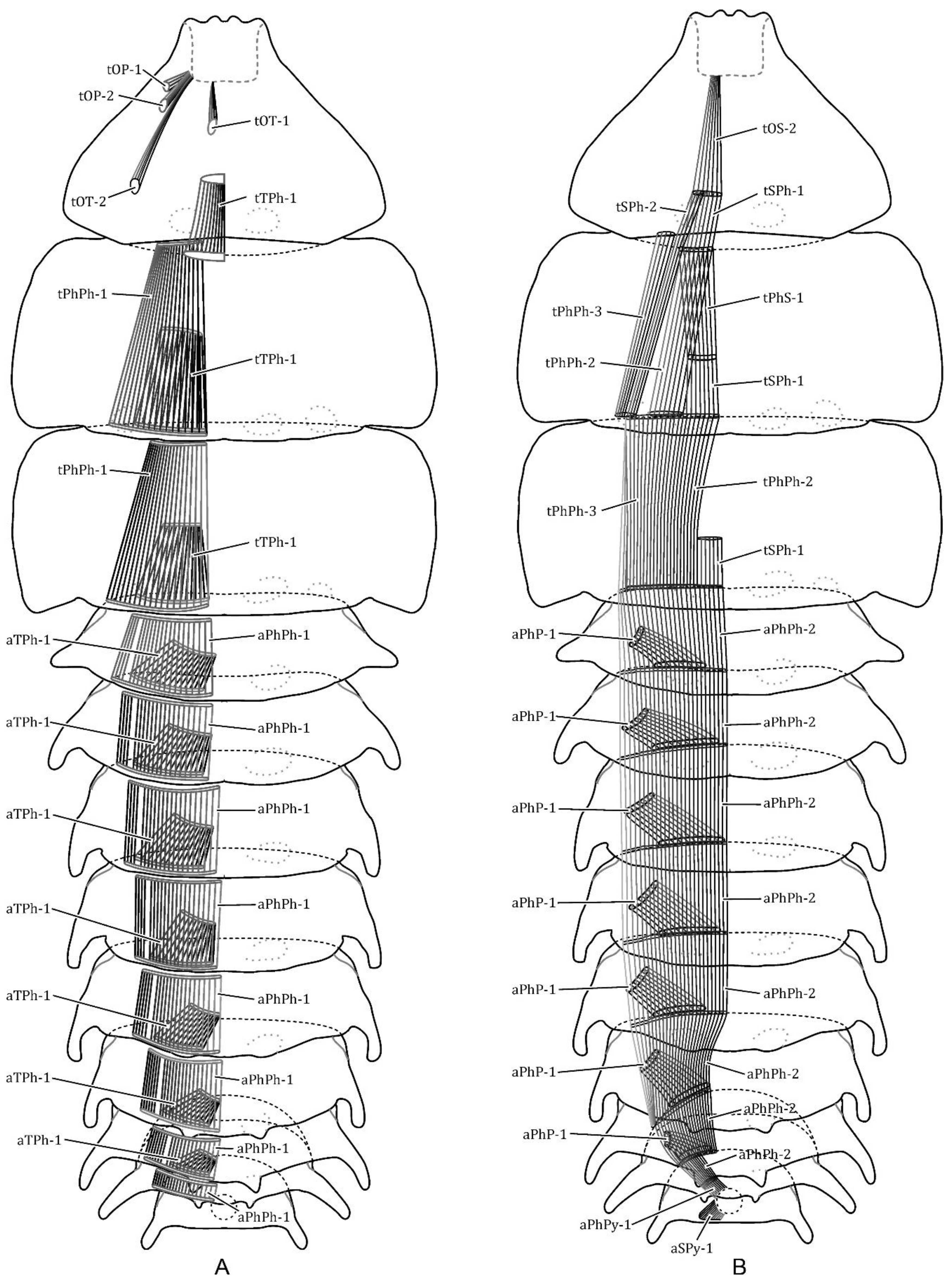
Thorax. Dorsal longitudinal muscles (
Figure 9A):
tOT-1, M. occiputo-tergalis medialis (O: dorsomedial occiput; I: median pronotum) (Friedrich and Beutel, 2007: Idml4; Geisthardt, 1979: ?M3, part).
tOT-2, M. occiputo-tergalis lateralis (O: dorsolateral occiput: I: lateral pronotum) (Friedrich and Beutel, 2007: Idml2; Geisthardt, 1979: M3).
tTPh-1, M. phragma-tergalis (O: medial portion of tergum; I: phragmal area on intersegmental membrane) (Friedrich and Beutel, 2007: IIdml2, IIIdml2; Geisthardt, 1979: M6).
tPhPh-1, M. pragma-phragmalis (O: phragmal area on intersegmental membrane; I: phragmal area on intersegmental membrane of next segment) (Friedrich and Beutel, 2007: IIdml1, IIIdml1; Geisthardt, 1979: M43).
tOS-1, M. occiputo-prosternalis lateralis (O: lateroventral occiput; I: anterior prosternum), not shown (Friedrich and Beutel, 2007: Idvm2/3; Geisthardt, 1979: M16).
tTS-1, M. tergo-sternalis lateralis (O: tergum, lateral; I: external margin of sternum). On the prothorax, this muscle consists of a series of bundles standing one behind the other from the posterior edge of the head almost to the coxal cavity (Friedrich and Beutel, 2007: ?IIdvm1, ?IIIdvm1; Geisthardt, 1979: no analogue).
tTS-2, M. tergo-sternalis intersegmentalis posterior (O: tergum, posterolateral; I: phragmal area on intersegmental membrane of next segment) (Friedrich and Beutel, 2007: ?Idvm11, ?IIdvm9; Geisthardt, 1979: ?M29, ?M48).
tOP-1, M. occiputo-pleuralis anterior (O: ventrolateral occiput; I: -anterior proepistern) (Friedrich and Beutel, 2007: Itpm2; Geisthardt, 1979: M17, part).
tOP-2, M. occiputo-pleuralis posterior (O: ventrolateral occiput; I: -posterior proepistern) (Friedrich and Beutel, 2007: Itpm1; Geisthardt, 1979: M17, part).
tTP-1, M. tergo-pleuralis intersegmentalis postertior (O: tergum, posterolateral; I: anterior epistern of next segment) (Friedrich and Beutel, 2007: ?IItpm12, ?IIItpm12; Geisthardt, 1979: M24, ?M47).
tTP-2, M. tergo-pleuralis anterior (O: tergum, mediolateral; I: episternum) (Friedrich and Beutel, 2007: no analogue; Geisthardt, 1979: M23, ?M52).
tTP-3, M. tergo-pleuralis anterior (O: tergum, mediolateral; I: membrane between epimer and sternum) (Friedrich and Beutel, 2007: no analogue; Geisthardt, 1979: M55).
tStP-1, M. stigma-pleuralis (O: stigma, ventral; I: anterior episternum) (Friedrich and Beutel, 2007: no analogue; Geisthardt, 1979: ?M53).
Ventral longitudinal muscles (
Figure 9B):
tOS-2, M. occiputo-prosternalis medialis (O: ventromedial occiput; I: posteromedian prosternum) (Friedrich and Beutel, 2007: Ivlm3; Geisthardt, 1979: M20).
tSPh-1, M. sterno-phragmalis medialis (O: posteromedian sternum; I: phragmal area on intersegmental membrane, medially) (Friedrich and Beutel, 2007: IIvlm1, IIIvlm1; Geisthardt, 1979: no analogue).
tSPh-2, M. sterno-phragmalis lateralis (O: posteromedian prosternum; I: phragmal area on intersegmental membrane, laterally), only on mesothorax (Friedrich and Beutel, 2007: Ivlm8; Geisthardt, 1979: no analogue).
tPhS-1, M. phragmo-sternalis (O: phragmal area on intersegmental membrane; I: posteromedian sternum), only on mesothorax; (Friedrich and Beutel, 2007: no analogue; Geisthardt, 1979: ?M18), absent in prothorax, replaced with internal fibres tPhPh-2 in metathorax.
tPhPh-2, M. phragmo-phragmalis medialis (O: phragmal area on intersegmental membrane; I: —same on next segment) (Friedrich and Beutel, 2007: Ivlm6, IIvlm6, IIIvlm6; Geisthardt, 1979: M66, part).
tPhPh-3, M. phragmo-phragmalis lateralis (O: phragmal area on intersegmental membrane; I: —same on next segment) (Friedrich and Beutel, 2007: Ivlm7, IIvlm7, IIIvlm7; Geisthardt, 1979: M66, part).
lTC-1, M. tergo-coxalis medialis (O: tergum, medially; I: medial coxal rim) (Friedrich and Beutel, 2007: ?Idvm16; Geisthardt, 1979: M33, M69).
lTC-2, M. tergo-coxalis anterior medialis (O: tergum, anterior of lTC-1; I: anteromedial coxal rim) (Friedrich and Beutel, 2007: Idvm15, ?IIdvm4, ?IIIdvm4; Geisthardt, 1979: M32, M68).
lTC-3, M. tergo-coxalis anterior lateralis (O: tergum, lateral of lTC-2; I: anterolateral coxal rim) (Friedrich and Beutel, 2007: Idvm13; Geisthardt, 1979: M31, M67).
lTC-4, M. tergo-coxalis posterior (O: tergum, laterad lTC-1; I: posterior coxal rim) (Friedrich and Beutel, 2007: Idvm15, ?IIdvm4, ?IIIdvm4; Geisthardt, 1979: M35).
lTTr-1, M. tergo-trochanteralis (O: tergum, lateral of lTC-1; I: trochanteral tendon) (Friedrich and Beutel, 2007: Idvm19, IIdvm7, IIIdvm7; Geisthardt, 1979: M41).
lPC-1, M. pleuro-coxalis anterior (O: epistern; I: anterolateral coxal rim) (Friedrich and Beutel, 2007: no analogue; Geisthardt, 1979: ?M36, M71).
lPC-2, M. pleuro-coxalis posterior (O: epimer; I: posterolateral coxal rim) (Friedrich and Beutel, 2007: no analogue; Geisthardt, 1979: ?M37, M73).
Sterno-coxal muscles:
lPhC-1, M. sterno-coxalis posterior (O: phragmal area on intersegmental membrane; I: posteromedial coxal rim) (Friedrich and Beutel, 2007: Iscm5, IIscm5, IIIscm5; Geisthardt, 1979: M40).
The prothorax, with coxal muscles counted, has 19 muscles; the meso- and metathorax have 19 and 20 muscles, respectively.
Abdomen. Dorsal longitudinal muscles (
Figure 9A):
aPhPh-1 M. pragma-phragmalis (O: phragmal area on intersegmental membrane; I: phragmal area on intersegmental membraneof next segment).
aTPh-1, M. tergo-phragmalis (O: medial portion of tergum; I: phragmal area on intersegmental membrane).
aTS-1, M. tergo-sternalis intersegmentalis anterior (O: tergum, anterolateral; I: phragmal area on intersegmental membrane of next segment).
aTS-2, M. tergo-sternalis intersegmentalis posterior (O: tergum, posterolateral; I: phragmal area on intersegmental membrane of next segment).
aTP-1, M. tergo-pleuralis anterior (O: anterolateral tergite; I: membranose band between sternum and pleurum).
aTP-2, M. tergo-pleuralis posterior (O: posterolateral tergite; I: membranose band between sternum and pleurum).
Ventral longitudinal muscles (
Figure 9B):
aPhPh-2, M. pragma-phragmalis (O: phragmal area on intersegmental membrane; I: phragmal area on intersegmental membraneof next segment).
aSPh-1, M. sterno-phragmalis (O: membranose band between sternum and pleurum; I: phragmal area on intersegmental membrane).
aPhPy-1, M. phragmo-pygidialis (O: phragmal area on intersegmental membrane; I: anterior rim of X segment), homologous with aPhPh-2, analogous with aPhP-1.
aPyS-1, M. pygidio-sternalis (O: posterior rim of X segment; I: poststernum of IX segment).
Every abdominal segment, except segment X, has 8 muscles.
Leg (
Figure 11). Muscles of the leg originating on the tergite and pleurite are described above in respective sections.
lSC-1, M. rotator coxalis (O: poststernum; I: anteromedial coxa).
lCTr-1, M. extensor trochanteris exterior (O: coxa, exterior; I: trochanter, interior).
ICTr-2, M. extensor trochanteris interior (O: coxa, interior; I: trochanter, interior).
lCTr-3, M. flexor trochanteris exterior (O: coxa, posterior; I: trochanter, exterior).
lCTr-4, M. flexor trochanteris interior (O: coxa, interior; I: I: trochanter, exterior).
lTrFE-1, M. rotator femoris (O: trochanter, ventroapical; I: femur, dorsoproximal).
lFeTi-1, M. flexor tibiotarsalis (O: tibia, dorsal; I: tibiotarsus, ventroproximal edge).
lTrPr-1, M. flexor pretarsalis (O: trochanter, ventroapical (a); femur, basilateral (b); femur, dorsoapical (c); tibiotarsus, dorsal (d); I: pretarsal tendon).
The leg musculature, excluding muscles originating on the tergite and pleurite, includes 8 muscles.
Heart (Cr) tubular, located medially in the dorsal part of abdominal segments I–VII (
Figure 12A), includes 7 chambers (decreasing in size caudally) and short aorta (Ao), extending to mesothorax.
Digestive system (
Figure 12A) includes short ectodermal foregut (Sd), slightly widened towards the midgut. Goiter and proventriculus not perceptible. Midgut (mE) tubular, significantly deformed by the hypertrophied uterus, not differentiated into sections. At the border of midgut and hindgut two pairs of Malpighian tubules (MT). Hindgut short, not forming loops, differentiated into wider section with plicate surface (large intestine, II) and rectum (Re), slightly shorter than previous section. Rectal pads absent.
The digestive system of the female is very similar to that of the larva, but the larval midgut is differentiated: its middle part is wider and riddled with dense network of tracheae. Furthermore, the larval hindgut is considerably longer and looped in segments IV–VI, and the larval Malpighian tubules are more numerous (11) in the single studied specimen).
Nervous system. Supra- and subesophageal ganglions displaced to prothorax and laying behind the occipital opening. Thoracic section represented by three subequal sized ganglions, in the abdominal section, by a chain of 8 ganglions. Posterior ganglion in abdominal section, synganglion by origin, approximate to preceding ganglion and located in segment 7; the long laterocaudal nerves stemming from it towards segments IX and X, are located asymmetrically, left ones lying over the reproductive system, right ones under it.
The female nervous system is similar to the larval one, except for two details: in the larva the metathoracic ganglion is noticeably larger than the preceding ones and the nerves stemming from synganglion are symmetrical.
Reproductive system (
Figure 12B and
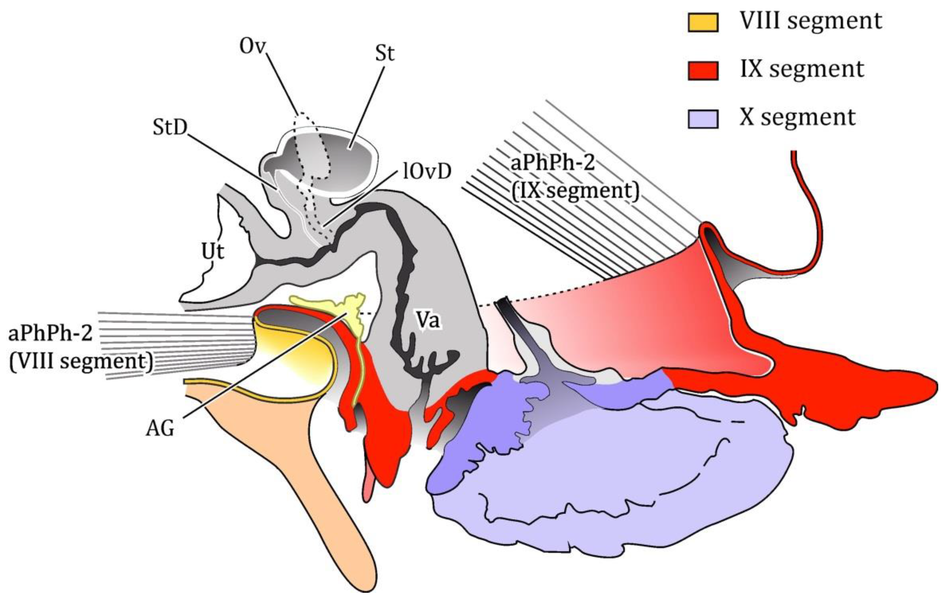
Figure 13). Ovaries (Ov) telotrophic, paired, small, not divided into ovarioles. Lateral oviducts (lOvD) short, connected to proximal part of the thick-walled vagina (Va). Vagina S-shaped (
Figure 13), with a roundish genital opening in the caudal third of sternite IX.
The genital opening is covered by paired roundish subtriangular appendages of segment IX (
Figure 7B), bearing duct openings of accessory glands (AG) at their base. These paired glands are formed by branched thick-walled tubes and partially covered by the vagina’s ascendant branch. It is possible that they are used for egg attachment to substrate (
Figure 14), as in many other insects [
16], although this is not characteristic of beetles [
17].
The oval unpaired spermatheca (St) lies on the dorsal side of the vagina, with its duct (StD) attached to the vagina in front of oviducts (
Figure 13). The vagina is proximally widened, forming a very large cylindrical uterus (Ut), filling all the inside of the body and reaching the prothorax (
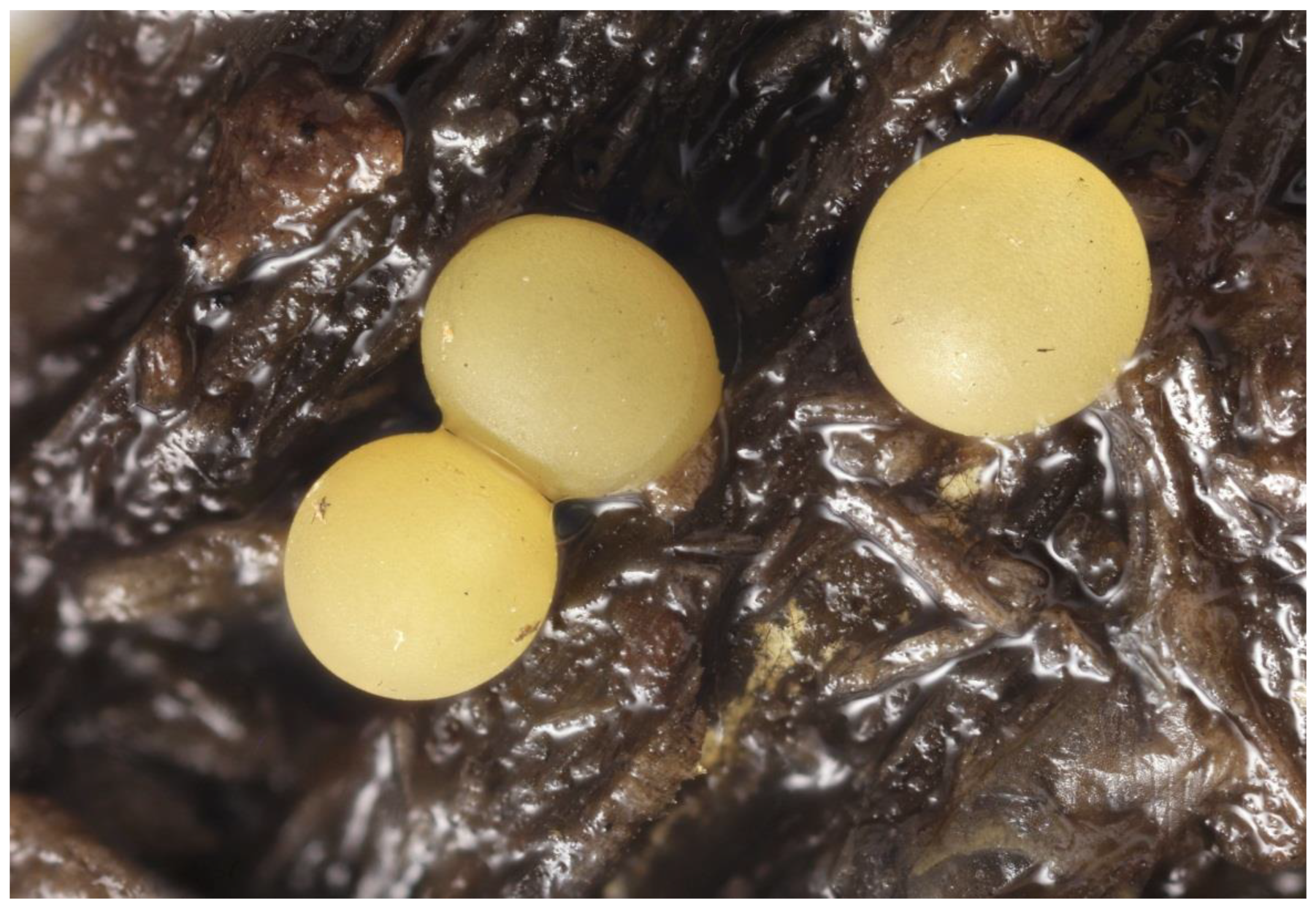
Figure 12B). In the studied female specimen, the uterus contained 496 round eggs of different sizes, the diameter of the considerably less numerous smaller ones about 2.1 times less than that of the large ones.
Egg. Large, circa 1 mm in diameter, round, whitish yellow; in uterus a smaller proportion of eggs were found to be smaller in size, about 0.5 mm in diameter (
Figure 12B and
Figure 14).
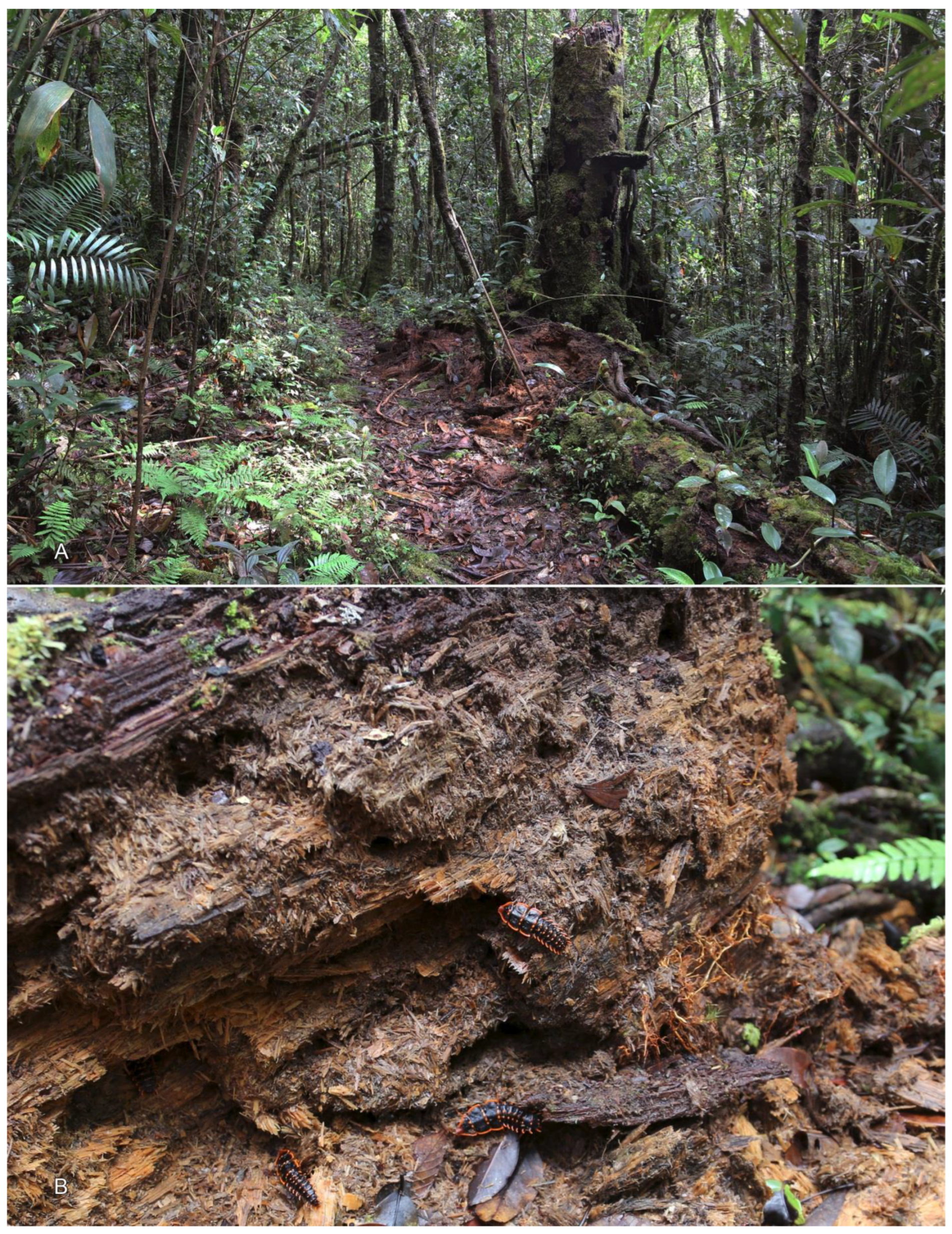
Biology. The last instar larvae were collected crawling on or near rotten logs in the primary rainforest in the Crocker Range Mountains at 1550–2000 m a.s.l. The rearing lasted 11 weeks, the larvae peacefully crawling and/or feeding on juices of decayed wood. Then one of the larvae, after 2 days of immobility, turned yellow and soon started laying eggs, from time to time exposing the genital opening upwards. After 2.5 weeks she passed away, having laid some eggs sparsely in small sticky clumps on the substrate, however, greater part of eggs remained inside the uterus, as the consequent dissection demonstrated. Not a single egg hatched.
4. Discussion
The “paedomorphic” female of Platerodrilus, described above, morphologically is not much different from a larva. It has larval antennae and a labiomaxillary complex, simple stemmata, one-claw tibiotarsus and one-condyle coxa-pleuron connection. The number and shape of its thoracic and abdominal sclerites also corresponds to the larval phase.
The larval anatomy in Lycidae has so far been very poorly studied, the only paper dating back to middle of the last century [
9]. Nevertheless, comparison of the studied female with conspecific larvae and the data on larval anatomy on
Lamprohiza Motschulsky, 1853, a genus from another elateroid family, Lampyridae [
15], reveals their close resemblance.
The resemblance is, above all, the absence of most elements of the endoskeleton, except for the pleural apodemes, whereas in adults the endoskeleton is well developed [
15]. It is worth mentioning that similar versions of the endoskeleton evolution are known from a number of families both in Adephaga [
18] and Polyphaga [
19]. In many cases, but not in all, the reduction of the endoskeleton is connected with the hard sclerotisation of larval covers. As lycid and lampyrid larvae generally have thin flexible covers, the resemblance of
Platerodrilus endoskeleton to that of the lampyrid larvae is probably better explained by this factor.
It is not clear if the smaller eggs that were discovered in the uterus are the ones that are supposed to develop into males, or just underdeveloped eggs still to reach the size of a mature one. In any case, all the eggs the female laid were large. On the other hand, the size of a mature/large egg is many times greater than the diameter of the lateral oviducts, so it is quite possible that all the eggs penetrating the uterus were small, and matured, i.e., grew in size, inside.
In addition, it is difficult to compare musculature: no exact data are available on lycid larvae, and of all related families only one species of fireflies has been studied [
15]. The general topology of muscles of the
Platerodrilus female is close to that of its larva, but Geisthardt [
15] demonstrated that in
Lamprohiza the larval thoracic musculature is much more diverse than in imago (33–42 muscles versus 15–22). It is partly because
Lamprohiza larvae have muscles that provide for the imaginal flight [15:531]. The studied
Platerodrilus female, too, has the same groups of muscles as the larva, but their diversity is significantly lower (see above).
Next, the digestive, circulatory and nervous systems of
Platerodrilus female are very similar to the larval ones. Especially important, in our opinion, is the absence of rectal pads, the development of which only at the pupal stage has been documented in many insect orders [
20,
21], including Coleoptera [K.V.M.’s pers. observation].
To sum it up, we may tentatively conclude that in terms of morphology, although the
Platerodrilus female is a larva with a developed reproductive system, referring to it as a female or a larviform female is quite correct. On the other hand, the reproductive system of the
Platerodrilus female is indeed noticeably different from that of non-paedomorphic females. The female imagoes of Cantharidae, Lampyridae and Lycidae do not have enlarged uteri, their ovaries are represented by numerous tubular ovarioles [
22,
23,
24] and their oviduct is unpaired [
25], whereas in the
Platerodrilus female the two ovaries are small and saccular, the tubular ovarioles are absent, the oviduct is paired and the uterus is enormous. The level of the morphological differentiation of the gonads of the
Platerodrilus female corresponds to the rudiments of the reproductory system observed in the last instar larvae of the phylogenetically close Elateridae [
26].
McMahon and Hayward [
27] designated the development of
Platerodrilus as “type II pedogenesis” and brought this developmental variant closer to neoteny. The appearance of sexual reproduction in larval stages is traditionally explained [
28,
29,
30] by two processes: progenesis and neoteny. Reliable distinction between these two options requires knowledge of the developmental parameters of ancestors. Our level of knowledge of Platerodrilini, however, does not allow for a reasoned choice between them and, broadly speaking, does not rule out any third point of view. It is obvious that the size of distribution area of a bisexual taxon is limited by the spreading capabilities of females. Based on the recent distribution range of Platerodrilini, we would assume that female ancestors were winged. Obviously, they could not be very large (flying net-winged beetles, both fossil and recent, are not greater than 25–30 mm in length). Therefore it would be logical to further assume that at a certain stage, circumstances favoured an increase in the size of females of the Platerodrilini ancestors, which led to the loss of their ability to fly. Undoubtedly, the nutritional characteristics of the larvae also played a role—low-calorie food limits the growth rate. Due to these considerations, it is rather difficult to diagnose the
Platerodrilus case as either neoteny or progenesis. On the one hand, the hypothesis of progenesis well explains the loss of the wing apparatus by insects, in which we agree with Gould’s opinion ([
30]:298). On the other hand, the presence of formed reproductive ducts and small gonads in Elateriformia larvae [
26] at the end of development simplifies the transition to neoteny. Using one of the most widespread interpretations, it seems correct to apply the term “paedomorphic” female for
Platerodrilus (which was the one used in the latest publication on a similar group of net-winged beetles [
10]). Due to the vagueness of certain terms, especially “neoteny”, we will limit ourselves to the more general term “paedomorphic female”.
However, three facts do not quite correlate with this seemingly well substantiated opinion:
The two layers of cuticle are of special importance, in our opinion. Their presence was noted already by Mjöberg, who called the outer cuticle the “postlarval skin” [
7]. Mjöberg, however, did not study its structure and thought it was just the regular larval cuticle. In fact, both layers differ principally from the larval cuticle in the fewer sensillae, to even almost rare solitary sensillae on the inner one. At the same time, the limited number of sensillae, up to their almost complete disappearance, is a characteristic feature of the pupa in many insect groups [
33,
34], personal observation.
Thus, we have to admit that the
Platerodrilus female has both larval and pupal characters. This fact was already noted by Mjöberg [
7]: “female of the ‘trilobite-larvae’ represents nothing more than a strongly condensed form of a larva and pupa and imago of a Lycid-female”. However, since then, new data on the development of the Coleoptera has been obtained, e.g., it was demonstrated that the complex reproductory system with developed ducts and external genitals are developed in larvae before wings and other imaginal appendages [
35]. Hinton [
36] designated this type as the “prepupa”, and we accept this term (having specified that this phase precedes “pharata pupa”). It is necessary to specify why it is “prepupa”, but not “pupa” that we should refer to this female as. All coleopterans in the pupal phase have wing rudiments, and the structure of appendages resemble imaginal appendages. In addition, the pupa’s mobility is limited to contraction of abdominal muscles (sometimes even the abdominal muscles do not function).
The interpretation of the
Platerodrilus female as the “prepupa” helps resolve most, if not all its morphological discrepancies, i.e., (a) presence of two layers of differently structured cuticle (the outer prepupal, the inner pupal); (b) retaining larval characters in segmentation and structure of the appendages, musculature and greater part of internal organs; (c) locomotion ability; (d) appearance of the first imaginal characters (mandibles, Malpighian tubules); (e) much weaker differentiation of external genitals than a pupa should have [
37,
38], personal observation; (f) dissimilarities in the reproductory system of the
Platerodrilus female and other net-winged beetles.
Hence, there are reasons to consider the
Platerodrilus female a free-living prepupa at the stage of its transformation into a pupa. As the imaginal structures are formed at the prepupal and pupal stages gradually [
39], it would be natural to assume that within the Lycidae we will come across different versions of paedogenetic females. The Lyropaeini females, for example, remain unknown, but the morphological characters of their larvae [
1,
40] allow expecting a version similar to
Platerodrilus. On the contrary, in Leptolycini the paedomorphic female was found to have more pupal characters (weakly differentiated maxillae and labium, abdominal segment X retracted into abdomen, hypopleurites fused with sternites), at the same time possessing purely larval split mandibles [
10].
