Short Note

8,8′-Dichloro-2,2,2′,2′-tetraethyl-4,4′-bibenzo[1,3,6,2]dioxazastannocinylidene

by
Irina N. Meshcheryakova
1,*,
Ilya A. Yakushev
2,
Anton V. Cherkasov
1,
Maxim V. Arsenyev
1,
Anastasiya V. Klimashevskaya
1,
Dmitriy S. Kolevatov
1 and
Alexandr V. Piskunov
1,*
1
G. A. Razuvaev Institute of Organometallic Chemistry of the Russian Academy of Sciences, 49 Tropinina Str., 603950 Nizhny Novgorod, Russia
2
Kurnakov Institute of General and Inorganic Chemistry of the Russian Academy of Sciences, 31 Leninskii Prosp., 119071 Moscow, Russia
*
Authors to whom correspondence should be addressed.
Molbank 2025, 2025(4), M2083; https://doi.org/10.3390/M2083
Submission received: 8 October 2025 / Revised: 22 October 2025 / Accepted: 30 October 2025 / Published: 4 November 2025

Abstract

Upon the reaction of glyoxal-bis(2-hydroxy-5-chlorophenyl)imine LH2 with diethyltin dichloride in the presence of a base (Et3N) in DMSO, the 1D-coordination polymer 1 was obtained, in which formally the L’(SnEt2)2 fragment acts as a monomeric unit. It was found that during the reaction, the initial ligand L undergoes transformation in the tin atom’s coordination sphere. This transformation results in the formation of a new ditopic 1,4-bis((5-chloro-2-oxidophenyl)imino)but-2-ene-2,3-bis(olate) ligand L’. The structure of the resulting complex 1 was examined by single-crystal X-ray diffraction analysis, elemental analysis, IR, and UV spectroscopy.

1. Introduction

This work focuses on the study of coordinating ability of glyoxal-bis(2-hydroxy-5-chlorophenyl)imine LH2 [1], which is a redox-active, potentially tetradentate ligand. Metal complexes based on substituted glyoxal-bis(2-hydroxyphenyl)imines [2,3,4,5,6,7] and, to a much greater extent, their closest analogue, N,N′-bis(3,5-di-tert-butyl-2-hydroxyphenyl)-1,2-phenylenediamine [8,9,10,11], have been actively studied in recent years. Such compounds exhibit remarkable optical and electrochemical properties [4,8,9,10,11], which makes them promising for applications as photonic materials and energy storage devices. Furthermore, several compelling examples demonstrate the efficacy of such derivatives as catalysts for the oxidation of primary alcohols [12], the disproportionation of 1,2-diphenylhydrazine [13], and the polymerization of lactides [14]. Despite the fact that substituted glyoxal-bis(2-hydroxyphenyl)imines were synthesized more than 40 years ago [1,2,3,15], only a few examples of their complexes have been reported in the literature to date. These include compounds of uranium(VI) [2,5,6], nickel(II) [3], tin(IV) [4], lead(IV) [4], and vanadium(IV) [7]. The previously studied tin(IV) and lead(IV) complexes [4] based on glyoxal-bis(2-hydroxy-3,5-di-tert-butylphenyl)imine contained a tetradentate-coordinated redox-active ligand in a dianionic state and were characterized by redox-amphoteric properties, as well as intense absorption in the near-IR region. The replacement of substituents in the phenolic ring of the ligand is expected to change the optical or electrochemical characteristics of the complexes, or their solubility, depending on the desired objective. Therefore, we attempted to synthesize a tin(IV) complex based on glyoxal-bis(2-hydroxy-5-chlorophenyl)imine (LH2).

2. Results and Discussion

It was found that the synthesized substituted glyoxal-bis(2-hydroxyphenyl)imines are isolated in the cyclic benzoxazine-benzoxazine form (Scheme 1a) [1,6,15]. An exception is the ligand bearing tert-butyl substituents at the 3- and 5-positions of the phenolic ring, which was successfully isolated in the glyoxal-bis-imine form [3], similar to that shown in Scheme 1b. However, in solution, this glyoxal-bis-imine undergoes intramolecular cyclization [7,16,17].
Studies of uranium compounds have shown that the complexation process involves the opening of the benzoxazine cycles of the ligands and conversion to the imine form [6].
A procedure described in reference [6] was tested for the synthesis of the tin complex based on LH2. It involves the direct interaction of the ligand and the metal salt in boiling methanol. However, the reaction between LH2 and Et2SnCl2 proceeded only in the presence of triethylamine. It results in the formation of a microcrystalline, nearly insoluble dark violet product that could not be identified. During the reaction in DMSO (Scheme 2), the solution color changes from colorless to dark blue, and the subsequent addition of methanol leads to the slow formation of a small amount of crystals of complex 1, suitable for X-ray diffraction analysis.
According to the SC XRD data, the complex 1 crystallizes in triclinic P-1 space group as solvate 1·2DMSO and forms a 1D-coordination polymer, where formally the L’(SnEt2)2 fragment acts as a monomeric unit (Figure 1). The initial ligand LH2 undergoes a transformation during the reaction, involving not only ring opening in the initial benzoxazine-benzoxazine form but also subsequent hydrolysis and condensation. This process leads to the formation of a new ditopic ligand L’, which is coordinated by two tin atoms. Thus, the ligand L’ consists of two O-Ph-N moieties joined by a C-C(-O)=C(-O)-C linker. The main geometric parameters of complex 1 are given in Table S2 (see Supplementary Materials). Each of the Sn4+ cations in L’(SnEt2)2 fragment is bound to two oxygen atoms and one nitrogen atom of the ligand and two carbon atoms of two ethyl groups (Figure 1). However, the metal centers in 1 are not equivalent, participate differently in the binding of the units to each other, and have different coordination environments. Thus, from the crystal structure point of view, the monomeric unit of 1 appears as the [L’(SnEt2)2]2 fragment (Figure 1). The chains pack in a linear fashion, stabilized by intermolecular contacts between the ligands of adjacent chains. These short contacts (3.45–3.58 Å) involve the chlorine atoms and carbon atoms of the C–C(–O)=C(–O)–C linker. Furthermore, multiple interactions between L’ and DMSO solvate molecules are also observed (see Supplementary Materials, Figure S2).
The Sn(1) and Sn(4) cations additionally coordinate two oxygen atoms of the neighboring ligand C-C(-O)=C(-O)-C linker, forming a flat SnOSnO metallocycle (Figure 1). The formal coordination number of these cations is seven; the coordination polyhedron is a distorted pentagonal bipyramid with Et- carbon atoms in the apical positions, and based on oxygen and nitrogen atoms of the ligands. The lengths of the covalent bonds Sn(1)-O(1) 2.195(3) Å, Sn(1)-O(2) 2.229(3) Å, Sn(4)-O(7) 2.249(3) Å, and Sn(4)-O(8) 2.164(3) Å are significantly shorter than the coordination bonds Sn(1)-O(2’) 2.392(3) Å, Sn(1)-O(3’) 2.581(3) Å, Sn(4)-O(6’) 2.584(3) Å, and Sn(4)-O(7’) 2.378(3) Å. In turn, the Sn(2) and Sn(3) cations additionally coordinate only one oxygen atom of the neighboring ligand O-Ph-N fragment (Figure 1). The formal coordination number of Sn(2) and Sn(3) is six, and the coordination environment can be described as distorted octahedral. The lengths of the Sn(2)-O(5) 2.560(3) Å and Sn(3)-O(4) 2.689(3) Å coordination bonds significantly exceed the lengths of the Sn(2)-O(3) 2.274(3) Å, Sn(2)-O(4) 2.135(3) Å, Sn(3)-O(5) 2.156(3) Å, and Sn(3)-O(6) 2.280(3) Å covalent bonds. It is interesting to note that the Sn-N distances for six-coordinate metal centers (2.178(3), 2.189(3) Å) are noticeably shorter than for seven-coordinate ones (2.293(3), 2.296(3) Å). It is also worth noting that for all metal centers, the Sn-O (from O-Ph-N fragment) bond lengths (2.135(3)–2.195(3) Å) are slightly shorter than the Sn-O (from linker) distances (2.229(3)–2.280(3) Å). The Sn-CEt covalent bond lengths for all cations in the monomeric unit of 1 lie within a narrow range of 2.120(5)–2.137(5) Å.
The values of the single C–O and C–N bonds in the O–Ph–N fragments are 1.307(6)–1.348(5) Å and 1.397(5)–1.400(6) Å, respectively. The distribution of C-C bond lengths in six-membered rings of the O–Ph–N moieties bound to the Sn(1) and Sn(4) atoms indicates partial alternation: the C-C distances lie in the wide range 1.369(7)–1.435(6) Å. In contrast, the C–C distances (1.381(6)–1.409(6) Å) in the C(15)–C(20) and C(29)–C(34) rings of the O–Ph–N fragments at the Sn(2) and Sn(3) atoms are equalized, indicating their aromatic nature. It should be noted that the C-O bond lengths for bridged O atoms (from O-Ph-N fragment) (1.340(5), 1.348(5) Å) are significantly longer than for terminal ones (1.307(6), 1.316(5) Å). Complex 1 is a rare example of tin compounds featuring an ONO ligand scaffold with this specific bond length distribution. The structurally closest known tin derivatives containing the aforementioned ONO fragment are characterized by similar bond length values [18,19]. The electron density delocalization is observed in the C-C(-O)=C(-O)-C linkers of the ligands: while single C-C bonds are significantly shortened (1.406(6)–1.439(6) Å) compared to 1.54 Å, double C=C bonds are noticeably elongated (1.406(6), 1.407 (7) Å) compared to 1.34 Å. The ligands in 1 are non-planar: the dihedral angles between the median planes of the O-Ph-N fragments are 159.8 and 167.8°.
The observed transformation of the ligand L into L’ could have occurred either within the coordination sphere of the tin atom or directly in solution before the interaction of LH2 with the metal salt. An additional experiment was carried out in an NMR tube, in which only LH2 and Et3N were initially present in the deuterated DMSO solution. It was found that even after heating the reaction mixture for 2 h at 60 °C, no changes occurred in the 1H NMR spectrum (see Supplementary Materials, Figures S3 and S4). That is, LH2 remains in the benzoxazine-benzoxazine form even in the presence of a base, and the transformation proceeds only upon the addition of diethyltin dichloride. The proposed mechanism of the ligand transformation is shown in Scheme S1 (see Supplementary Materials). We assume that this process must involve the stages of hydrolysis of the initial ligand L and condensation of the fragments formed as a result of hydrolysis. Therefore, the L’(SnEt2)2 unit is formed. A related process involving the hydrolysis of the initial ligand was previously observed for a cobalt complex based on the similar ligand-N,N′-bis(2-hydroxy-3,5-di-tert-butylphenyl)ethylenediamine [20].
Dark colored crystals of complex 1 exhibit a deep navy-blue color in transmitted light and display a golden luster in reflected light. Figure 2 shows photographs of the crystals (left) and a mechanically grinded sample (right), illustrating the difference in their coloration.
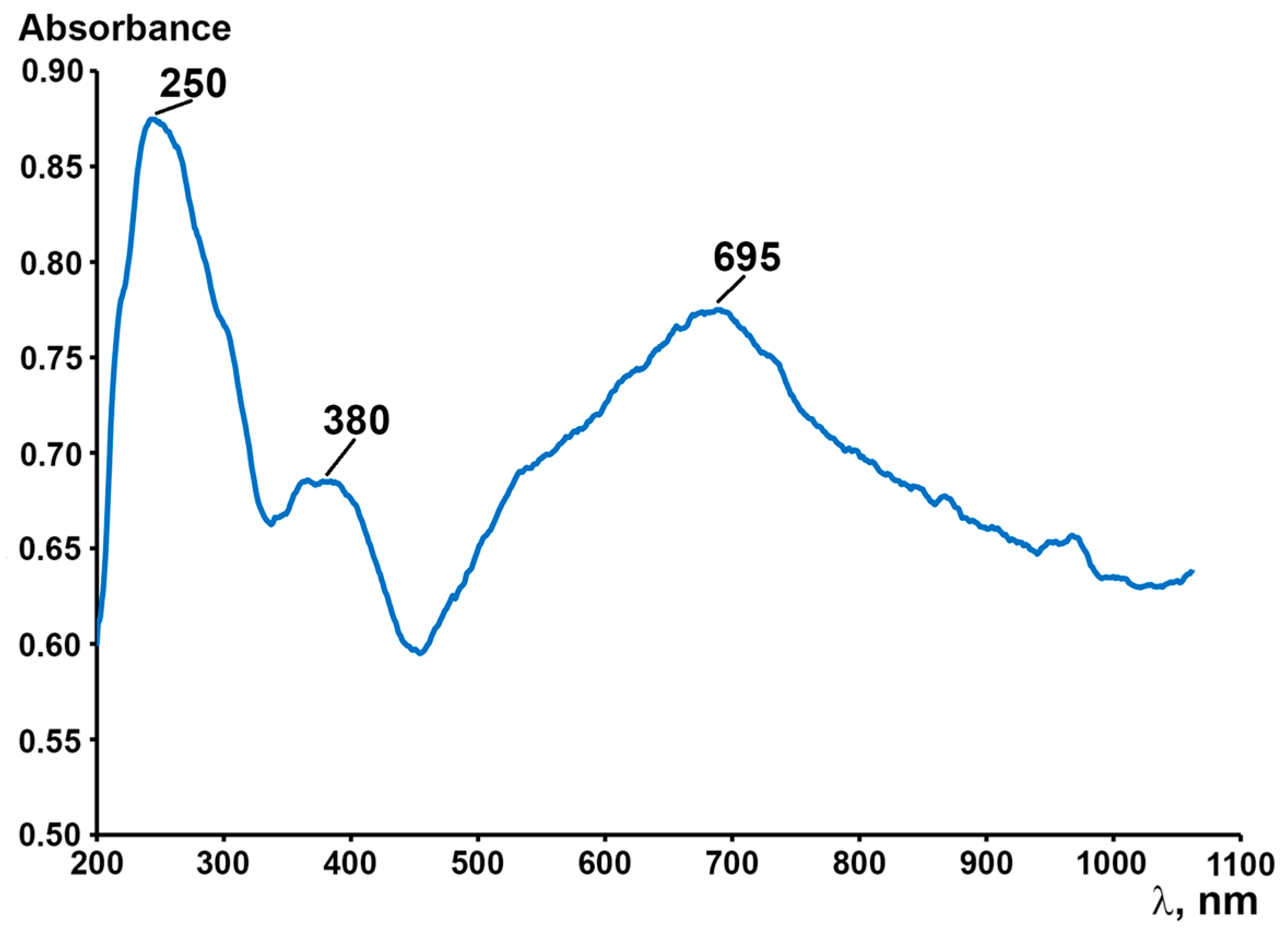
Due to the insolubility of the coordination polymer 1, we were unable to study its solution using UV spectroscopy. The absorption spectrum in the UV and the visible ranges of 1 was recorded in mineral oil and is presented in Figure 3. In the near-UV region, intense absorption bands with maxima at 250 and 380 nm are observed, which are assigned to σ→π* and π→π* transitions. The red and near-IR region of the spectrum features a broad absorption band ranging from 450 to 1000 nm with a maximum at 695 nm, attributed to an intraligand charge transfer.

3. Materials and Methods

3.1. General Information

Solvents were purified using standard methods [21]. The following commercially available reagents were used: 2-amino-4-chlorophenol, glyoxal (40% w/w aq.), Et2SnCl2, Et3N. Benzoxazino-benzoxazine LH2 was synthesized according to a known procedure [6], and its characteristics (NMR and IR spectra, elemental analysis data) are in full agreement with the literature data [1]. The elemental analysis was performed on a Vario el Cube instrument (Elementar, Okehampton, UK). IR spectra were recorded on an FSM-1201 Fourier transform spectrometer (Infraspek, St. Petersburg, Russia) in Nujol mulls (range 4000–400 cm−1) in a KBr cell. The solid-phase UV–vis spectrum of Nujol mull was registered on an SF-2000 spectrophotometer (ОKB Spectr, St. Petersburg, Russia) (range: 200–1100 nm).

3.2. Synthesis of [[L’(SnEt2)2·DMSO]2]n (1)

A solution of LH2 (0.2 g, 0.65 mmol) and Et2SnCl2 (0.16 g, 0.65 mmol) in 3 mL of DMSO was treated with 0.3 mL of Et3N. The resulting reaction mixture was kept at 60 °C for 3 h, during which the solution color turned intense blue. Then, 2 mL of methanol were added, and the reaction mixture was allowed to cool slowly to room temperature. Over 24 h, a dark blue crystalline precipitate with a golden luster formed. The precipitate was separated from the mother liquor by decantation, washed with methanol, and dried under reduced pressure (yield 34% (based on Et2SnCl2)). An elemental analysis of [[L’(SnEt2)2·DMSO]2]n was performed. The calculations (%) for the C52H68Cl4N4O10S2Sn4 are as follows: C 39.28; H 4.31. The obtained results were as follows (%): C 39.35, H 4.36. IR (Nujol, KBr, cm−1): 1597 (m), 1587 (m), 1551 (m), 1527 (s), 1485 (s), 1436 (m), 1415 (m), 1337 (w), 1327 (m), 1305 (s), 1280 (w), 1267 (w), 1240 (s), 1203 (w), 1173 (s), 1129 (m), 1077 (m), 1015 (m), 965 (s), 906 (w), 892 (w), 884 (w), 876 (w), 852 (m), 819 (m), 808 (m), 772 (w), 684 (m), 657 (m), 638 (m), 610 (w), 580 (s), 555 (s), 544 (s), 494 (m).

3.3. Single-Crystal X-Ray Diffraction Analysis

The single crystal X-ray data for complex 1 was obtained using synchrotron X-ray radiation at the “Belok” beamline [22] of the Kurchatov Synchrotron Radiation Source (National Research Center “Kurchatov Institute”, Moscow, Russia) in the ϕ-scan mode with the Rayonix SX165 CCD detector at 100 K. The experimental X-ray raw data for 1 were indexed, integrated, and scaled with the XDS data reduction program [23]. The structure was solved by direct methods and refined by full-matrix least-squares on F2 with anisotropic thermal parameters for all non-hydrogen atoms [24,25]. The sulfur atoms of one DMSO solvate molecule were disordered over two positions (occupancy ratio ca. 0.06 : 0.94) and refined using the SHELX EADP constraint. The hydrogen atoms were placed in calculated positions and refined using a riding model with dependent isotropic displacement parameters [Uiso(H) = 1.5Ueq(C) for methyl groups and Uiso(H) = 1.2Ueq(C) for all other H-atoms. All calculations were carried out using the SHELXTL program suite [26] and OLEX2 X-ray data visualization program package [27].
The crystallographic details are presented in Table S1, while the selected bond lengths and valence angles in complex 1 are listed in Table S2 (see Supplementary Materials). CCDC 2490335 contains the supplementary crystallographic data. These data can be obtained free of charge from The Cambridge Crystallographic Data Centre [28].
Crystal data for 1: C52H68Cl4N4O10S2Sn4, M = 1589.78, P-1, a = 13.1394(17) Å, b = 14.4927(14) Å, c = 18.189(2) Å, V = 2997.9(6) Å3, Z = 2, dcalc = 1.761 g/cm3. Grey prism single crystal with dimensions 0.13 × 0.05 × 0.02 mm was selected and intensities of 56638 reflections were collected (μ = 2.266 mm–1, θmax = 30.537°). After merging of equivalence reflections and absorption corrections, 14659 independent reflections (Rint = 0.0459) were used for the structure solution and refinement. Final R factors R1 = 0.0444 [for 11307 reflections with F2 > 2σ(F2)], wR2 = 0.1227 (for all reflections), S = 1.031, and largest diff. peak and hole are 1.466 and −1.466 e/Å3, respectively.

Supplementary Materials

The following materials are available online: crystallographic information; IR spectroscopy data of compound 1, 1H NMR spectra, the proposed mechanism of the ligand L transformation. Figure S1: IR spectrum of complex [[L’(SnEt2)2·DMSO]2]n (1); Figure S2: the fragment of crystal packing of complex 1; Figures S3 and S4: the 1H NMR spectra of LH2 in the absence and presence of Et3N; Figure S5: photograph of single crystals of complex 1; Table S1: Crystal Data and Structure Refinement for 1; Table S2: Selected bond lengths (Å) and angles (◦) in 1; Scheme S1: the proposed mechanism of the ligand L transformation.

Author Contributions

Investigation, project administration, data curation, I.N.M.; methodology, I.N.M. and M.V.A.; writing—original draft preparation, I.N.M., I.A.Y. and A.V.C.; formal analysis, I.A.Y. and A.V.K.; visualization, D.S.K.; supervision, writing—review and editing, A.V.P. All authors have read and agreed to the published version of the manuscript.

Funding

This research was financially supported by the Russian Science Foundation (Grant No. 24-23-00268), https://rscf.ru/project/24-23-00268/, accessed on 1 January 2024.

Data Availability Statement

CCDC 2490335 contains the supplementary crystallographic data. These data can be obtained free of charge from The Cambridge Crystallographic Data Centre [28].

Acknowledgments

This research was performed utilizing the analytical facilities of the G.A. Razuvaev Institute of Organometallic Chemistry’s Analytical Center, Russian Academy of Sciences. The experimental studies of the single crystal structure were performed using the unique scientific facility “Kurchatov synchrotron radiation source “KISI-Kurchatov” of the National Research Center “Kurchatov Institute”.

Conflicts of Interest

The authors declare no conflicts of interest.

References

  1. Ochoa, M.E.; Rojas-Lima, S.; Höpfl, H.; Rodríguez, P.; Castillo, D.; Farfán, N.; Santillan, R. Facile synthesis of 1,3,6-oxadiazepines from 2,2′-(1,2-ethanediyldiimino)bisphenols. Tetrahedron 2001, 57, 55–64. [Google Scholar] [CrossRef]
  2. Bandoli, G.; Clemente, D.A. Preparation and crystal structure of aqua[bis(2-hydroxyphenylimino)-ethanato-OONN′-]dioxouranium. J. Chem. Soc. Dalton Trans. 1975, 7, 612–615. [Google Scholar] [CrossRef]
  3. Min, K.S.; Weyhermüller, T.; Bothe, E.; Wieghardt, K. Tetradentate Bis(o-iminobenzosemiquinonate (1−)) π Radical Ligands and Their o-Aminophenolate (1−) Derivatives in Complexes of Nickel (II), Palladium (II), and Copper (II). Inorg. Chem. 2004, 43, 2922–2931. [Google Scholar] [CrossRef]
  4. Piskunov, A.V.; Trofimova, O.Y.; Fukin, G.K.; Ketkov, S.Y.; Smolyaninov, I.V.; Cherkasov, V.K. Tin(IV) and lead(IV) complexes with a tetradentate redox-active ligand. Dalton Trans. 2012, 41, 10970–10979. [Google Scholar] [CrossRef]
  5. Takao, K.; Tsushima, S.; Ogura, T.; Tsubomura, T.; Ikeda, Y. Experimental and theoretical approaches to redox innocence of ligands in uranyl complexes: What is formal oxidation state of uranium in reductant of uranyl(VI)? Inorg. Chem. 2014, 53, 5772–5780. [Google Scholar] [CrossRef]
  6. Niklas, J.E.; Hunter, K.M.; Gorden, A.E. Bonding Interactions in Uranyl α-Diimine Complexes: A Spectroscopic and Electrochemical Study of the Impacts of Ligand Electronics and Extended Conjugation. Inorg. Chem. 2019, 58, 15088–15100. [Google Scholar] [CrossRef]
  7. Meshcheryakova, I.N.; Kocherova, T.N.; Yakushev, I.A.; Arseniev, M.V.; Starikov, A.G.; Kazakov, G.G.; Protasenko, N.A.; Piskunov, A.V. Features of the Molecular and Electronic Structures of the Vanadium(IV) Complex Based on Glyoxal-bis(2-hydroxy-3,5-di-tert-butylphenyl)Imine. J. Struct. Chem. 2025, 66, 489–501. [Google Scholar] [CrossRef]
  8. Lehtonen, A. Metal Complexes of Redox Non-Innocent Ligand N,N′-Bis(3,5-di-tertbutyl-2-hydroxy-phenyl)-1,2-phenylenediamine. Molecules 2024, 29, 1088. [Google Scholar] [CrossRef] [PubMed]
  9. Meshcheryakova, I.N.; Cherkasov, A.V.; Arsenyev, M.V.; Aysin, R.R.; Belikov, A.A.; Bogomyakov, A.S.; Protasenko, N.A.; Kocherova, T.N.; Baryshnikova, S.V.; Piskunov, A.V. Stepwise transformation of a redox-active tetradentate ONNO ligand in the coordination sphere of tin(IV). Dalton Trans. 2025, 54, 7281–7293. [Google Scholar] [CrossRef] [PubMed]
  10. Karnbrock, S.B.; Köster, J.F.; Becker, I.; Golz, C.; Meyer, F.; Gimferrer, M.; Alcarazo, M. Bis(amidophenolate)-supported pnictoranides: Lewis acid-induced electromerism in a bismuth complex. Chem. Sci. 2025, 16, 14178–14185. [Google Scholar] [CrossRef]
  11. Karnbrock, S.B.; Köster, J.F.; Golz, C.; Mata, R.A.; Alcarazo, M. Isolation of a Square Pyramidal Bis(amidophenolate)-Supported As(III)-Cation: Coordination-Induced Electromerism at As. Angew. Chem. Int. Ed. 2025, 64, e202501439. [Google Scholar] [CrossRef]
  12. Chaudhuri, P.; Hess, M.; Müller, J.; Hildenbrand, K.; Bill, E.; Weyhermüller, T.; Wieghardt, K. Aerobic oxidation of primary alcohols (including methanol) by Copper (II)− and Zinc (II)− phenoxyl radical catalysts. J. Am. Chem. Soc. 1999, 121, 9599–9610. [Google Scholar] [CrossRef]
  13. Blackmore, K.J.; Lal, N.; Ziller, J.W.; Heyduk, A.F. Catalytic reactivity of a zirconium(IV) redox-active ligand complex with 1,2-diphenylhydrazine. J. Am. Chem. Soc. 2008, 130, 2728–2729. [Google Scholar] [CrossRef] [PubMed]
  14. Zelikoff, A.L.; Kopilov, J.; Goldberg, I.; Coates, G.W.; Kol, M. New facets of an old ligand: Titanium and zirconium complexes of phenylenediamine bis(phenolate) in lactide polymerisation catalysis. Chem. Commun. 2009, 44, 6804–6806. [Google Scholar] [CrossRef]
  15. Tauer, E.; Grellmann, K.H.; Kaufmann, E.; Noltemeyer, M. The condensation product of 2-aminophenol and glyoxal. Structure and photochemistry. Chem. Berichte 1986, 119, 3316–3325. [Google Scholar] [CrossRef]
  16. Abakumov, G.A.; Druzhkov, N.O.; Kurskii, Y.A.; Abakumova, L.G.; Shavyrin, A.S.; Fukin, G.K.; Poddel’skii, A.I.; Cherkasov, V.K.; Okhlopkova, L.S. Quinone imines and aminophenols as precursors of new heterocycles. Russ. Chem. Bull. 2005, 54, 2571–2577. [Google Scholar] [CrossRef]
  17. Bakaev, I.V.; Romashev, N.F.; Komlyagina, V.I.; Abramov, P.A.; Piskunov, A.V.; Gushchin, A.L. Palladium(II) complex with tetrahydrobenzoxazinobenzoxosine: Synthesis, electronic and molecular structures. J. Struct. Chem. 2022, 63, 1963–1972. [Google Scholar] [CrossRef]
  18. Camacho-Camacho, C.; Esparza-Ruiz, A.; Vásquez-Badillo, A.; Nöth, H.; Flores-Parra, A.; Contreras, R. Fused hexacyclic tin compounds derived from 3-(3,5-di-t-butyl-2-hydroxy-phenylimino)-3H-phenoxazin-2-ol. J. Organomet. Chem. 2009, 694, 726–730. [Google Scholar] [CrossRef]
  19. Baryshnikova, S.V.; Poddel’sky, A.I.; Bellan, E.V.; Smolyaninov, I.V.; Cherkasov, A.V.; Fukin, G.K.; Berberova, N.T.; Cherkasov, V.K.; Abakumov, G.A. Ferrocene-Containing Tin(IV) Complexes Based on o-Benzoquinone and o-Iminobenzoquinone Ligands. Synthesis, Molecular Structure, and Electrochemical Properties. Inorg. Chem. 2020, 59, 6774–6784. [Google Scholar] [CrossRef] [PubMed]
  20. Bruzeguini, C.E.T.; Bezerra, R.I.; Ribeiro, M.A. Synthesis and structure analysis of a cobalt(III) coordination compound obtained from a redox-active phenolate ligand and cobalt(II). Cryst. Struct. Commun. 2025, 81, 358–364. [Google Scholar] [CrossRef] [PubMed]
  21. Perrin, D.D.; Armarego, W.L.F.; Perrin, D.R. Purification of Laboratory Chemicals, 4th ed.; Butterworth-Heinemann: Oxford, UK, 1980. [Google Scholar]
  22. Svetogorov, R.D.; Dorovatovskii, P.V.; Lazarenko, V.A. Belok/XSA diffraction beamline for studying crystalline samples at Kurchatov Synchrotron Radiation Source. Cryst. Res. Technol. 2020, 55, 1900184. [Google Scholar] [CrossRef]
  23. Kabsch, W. XDS. Acta Crystallogr. Sect. D Biol. Crystallogr. 2010, 66, 125–132. [Google Scholar] [CrossRef]
  24. Sheldrick, G.M. A short history of SHELX. Acta Crystallogr. Sect. A Found. Crystallogr. 2008, 64, 112–122. [Google Scholar] [CrossRef]
  25. Sheldrick, G.M. SHELXT–Integrated space-group and crystal-structure determination. Acta Crystallogr. Sect. A Found. Crystallogr. 2015, 71, 3–8. [Google Scholar] [CrossRef] [PubMed]
  26. Sheldrick, G.M. Crystal structure refinement with SHELXL. Acta Cryst. C Cryst. Struct. Commun. 2015, 71, 3–8. [Google Scholar] [CrossRef] [PubMed]
  27. Dolomanov, O.V.; Bourhis, L.J.; Gildea, R.J.; Howard, J.A.; Puschmann, H. OLEX2: A complete structure solution, refinement and analysis program. Appl. Crystallogr. 2009, 42, 339–341. [Google Scholar] [CrossRef]
  28. The Cambridge Crystallographic Data Centre. Available online: http://www.ccdc.cam.ac.uk/data_request/cif (accessed on 22 September 2025).
Scheme 1. Forms of the LH2 ligand.
Molbank 2025 m2083 sch001
Scheme 2. Synthesis of complex 1.
Molbank 2025 m2083 sch002
Figure 1. Molecular structure view of the crystal unit of complex 1. Hydrogen atoms and the solvate DMSO molecules are omitted for clarity. Thermal ellipsoids are given at 30% probability level.
Molbank 2025 m2083 g001
Figure 2. Photographs of a single crystals (left) of complex 1 and the sample after grinding (right), taken using a microphoto setup at 10× magnification.
Molbank 2025 m2083 g002
Figure 3. Electronic absorption spectrum of solid sample of complex 1.
Molbank 2025 m2083 g003
Disclaimer/Publisher’s Note: The statements, opinions and data contained in all publications are solely those of the individual author(s) and contributor(s) and not of MDPI and/or the editor(s). MDPI and/or the editor(s) disclaim responsibility for any injury to people or property resulting from any ideas, methods, instructions or products referred to in the content.