Short Note

3-((Benzyloxy)carbonyl)bicyclo[1.1.1]pentane-1-carboxylic Acid

Department of Chemistry and Biochemistry and The Canadian Centre for Advanced Fluorine Technologies, University of Lethbridge, Lethbridge, AB T1K 3M4, Canada
*
Author to whom correspondence should be addressed.
Molbank 2025, 2025(4), M2075; https://doi.org/10.3390/M2075
Submission received: 26 September 2025 / Revised: 13 October 2025 / Accepted: 15 October 2025 / Published: 16 October 2025
(This article belongs to the Section Structure Determination)

Abstract

The compound 3-((benzyloxy)carbonyl)bicyclo[1.1.1]pentane-1-carboxylic acid was successfully synthesized. High-quality crystals were obtained, and its X-ray structure was solved and refined by Hirshfeld atom refinement using custom aspherical scattering factors with the Olex2/NoSphereA2 package. Hydrogen bonding interactions lead to head-to-head carboxylic acid dimer formation. A positional disorder for the bridging H-atom was detected and modeled to two parts in a 0.85:0.15 ratio. Detailed comparison with a neutron diffraction study of benzoic acid at the same temperature (100 K) demonstrates that the E–H-bond distances in the title compound are in excellent agreement (differing less than 1%) and the displacement ellipsoids volumes to the model are also in excellent agreement to the neutron diffraction structure. Moreover, both the variation in refined disorder occupancy and differences in C=O and C–O lengths of the disordered carboxylic acids in the two structures track well with their dimer O···O separations. This is longer by 0.023 Å in the structure of the title compound than in that of benzoic acid. A database search was conducted and used for comparison of the title compound to other high-quality structures of bicyclo[1.1.1]pentane-containing species.

1. Introduction

The bicyclo[1.1.1]pentane (BCP) group has received much attention in recent years due to the desirable properties it possesses in medicinal chemistry. It can be used as an sp3-rich bioisostere for one of the most commonly employed rings in bioactive compounds, 1,4-substituted arenes, whilst in many cases exhibiting superior pharmacological properties [1,2]. For example, Han et al. discovered that replacing the core phenyl ring of an inhibitor drug with a BCP led to greater tolerance towards hydrolysis of the neighboring benzamide moiety whilst retaining excellent potency and selectivity [3].
Although crystal structures of BCP-containing compounds are available, there are relatively few examples, especially when considering BCPs that are 1,3-disubstituted with carbonyl-based substituents and unsubstituted at the bridges. The BCP-containing title compound (2, Scheme 1) was synthesized, and its identity was confirmed by NMR spectroscopy and single-crystal X-ray diffraction. The high-quality diffraction data allowed for Hirshfeld atom refinement (HAR) with custom aspherical form factors, enabling H-atom positional refinement.

2. Results

2.1. Synthesis and Spectroscopy

The two-step synthesis of carboxylic acid 2 was carried out successfully. Initially, bicyclo[1.1.1]pentane-1,3-dicarboxylic acid underwent a bis-benzylation in the presence of BnBr and Li2CO3 in DMF to yield diester 1. Classic Fischer-type conditions utilizing BnOH and catalytic H2SO4 were also attempted, but performed poorly compared to basic conditions. Diester 1 was treated with LiOH·H2O in THF to yield the title compound 2 in a 63% yield. Monosaponification of 1 was achieved via careful control of the stoichiometry of the base (1.1 eq. vs. 2 eq.) as well as through precipitation of the carboxylate salt of 2 upon its formation. The free carboxylic acid 2 was then purified via flash chromatography and isolated as a white solid.

2.2. Crystal Structure Description

The title compound (2) crystallizes in the monoclinic space group P21/c, the most common space group (Figure 1). The C–C bonds within the BCP moiety have an average length of 1.557 ± 0.008 Å (Table 1), which is shorter than the overall Csp3–Csp3 bond length average of 1.530 Å (5777 exemplars [4]). However, the average for Csp3–Csp3 bonds in cyclobutane, a group which likely better reflects the degree of ring strain in a BCP, is 1.554 Å (679 exemplars [4]), which is indistinguishable from the average bond length in this work at a 99% confidence level.
When analyzing the exocyclic C–C bond at each bridgehead carbon (Table 1), it is interesting to note that the bond lengths of 2 are shorter than expected values for a general sp3-hybridized carbon bound to the carbonyl carbon of an acyclic ester or a carboxylic acid. These values add to the evidence that the bridgehead carbons contribute hybrid orbitals of greater s character than sp3 [5]. In fact, the C3–C4 bond is identical to the listed Csp2 value at the 99% confidence level and, although the value for the C1–C2 bond is higher than the average of the corresponding value, it is within the upper quartile.
The C4=O3 bond of the ester is 1.2083(9) Å, which is longer than the expected 1.196 Å average (551 exemplars [4]). Similarly, the C4–O4 bond of the ester is 1.3398(8) Å, longer than the average of 1.337 Å for esters of similar kind. The C1–C2 bond is 1.4865(9) Å, which is shorter than the 1.502 Å average expected for carboxylic acids (176 exemplars [4]). Likewise, the C3–C4 bond length of 1.4888(9) Å is shorter than the 1.497 Å expected for acyclic esters (553 exemplars [4]). The C1–O1 bond length is long if assumed to be the carbonyl of a carboxylic acid (Table 2). The C1–O2 bond is within expected values for the bond to the oxygen of the hydroxyl group; it is, however, in the lower quartile of this bond type. Considering that the bond lengths are derived from a refinement model built on the positional disorder of H2 and H1a, it would be sensical that the true bond length of C1–O1 is shorter and the length of C1–O2 longer than those reported herein. Indeed, we can define
Δ C O = d C O d ( C = O )
as the difference between the expected single and double bond lengths, influenced by resonance, in a carboxyl moiety. Compound 2 has a built-in comparison between the ester and carboxylic acid, only the latter of which is subject to a further reduction in ΔCO due to disorder of the proton in the bridging H-bond. Herein, ΔCO = 0.062 Å for the acid group vs. 0.132 Å for the ester. This may be compared to ΔCO = 0.022 Å for benzoic acid (BA) at 100 K from a detailed variable temperature neutron diffraction study [6], indicative of more extensive disorder in the latter. Indeed, our value is much closer to ΔCO = 0.058 Å for BA at 20 K [6].
The comparison to the BA neutron diffraction study is insightful at several levels. First, the H-atom placement in the refinement of 2 is strongly corroborated by the neutron study. Thus, the average C(Ar)–H for 2 is 1.095 ± 0.007 Å, just 0.64% longer than 1.088 ± 0.004 Å in BA at 100 K [6] and 0.94% longer than the average of 93 structures in the compilation by Allen and Bruno [7]. In 2, the average Z2–Csp3–H distance is found to be 1.093 ± 0.007 Å, just 0.35% shorter than was found by Allen and Bruno. This verifies that HAR using NoSpherA2 provides reliable H-atom structures that give meaningful results comparable to neutron diffraction [8]. Turning to displacement ellipsoids, the average Uiso value for the C and O atoms in 2 is 0.0143 ± 0.0015 Å3, compared to 0.0155 ± 0.0015 Å3 in BA. Moreover, for H-atoms 0.037 ± 0.004 Å3 in 2 vs. 0.039 ± 0.005 Å3 in BA at 100 K. That level of agreement adds confidence to the modeling of the H-bonding in 2 (related to the ΔCO mentioned above). In our disorder model, the ratio of major and minor occupancies (i.e., H2:H1a) is 0.85:0.15. In the BA neutron study, the ratio at 100 K is 0.67:0.33, whereas at 20 K it was reported to be 0.866:0.134, a much closer match to what we observe. In all other respects, the detailed description of the disorder modeling provided in [5], including the realization that H2 and H1a needed to be refined isotropically, map closely to our observations for 2. Additionally, it was necessary to restrain d(O–H) to the average values reported in [9] of 0.989 Å (Table 3).
A key insight to the difference in occupancies and the close relationships between occupancies and ΔCO comes from the follow-up neutron diffraction study of BA reported in [9]. This paper sought to bridge the gap between the data from RT down to 100 K in [5], which was at odds with the single 20 K result, by taking additional measurements at 6, 25, 60 and 75 K. This shows that, rather than a linear relationship, a smooth ‘S-curve’ describes the behavior from 0 K to ambient temperature. If the site occupancy ratio of 0.85:0.15 is mapped onto the d(C–O) plots (Figure 3 in [9]) a value of ΔCO = 0.065 Å is obtained, near enough identical to our result. A key to understanding this match in ΔCO but mismatch in temperature is a consideration of the separation distances in the bridging carboxylic acid (i.e., d(D–A) in Table 3). In BA at 100 K, d(D–A) is 2.615 Å, some 0.023 Å smaller. It has been shown by high pressure neutron diffraction studies that BA is strongly compressible and that reducing the O···O distance is a strong modulator for hydrogen bond symmetrization [10]. The H-bonding in 2, a bridgehead BCP carboxylic acid, is thus markedly weaker than in BA. Whether this is due to inherent differences in resonance effects (i.e., stabilization of the carboxylate ion in the aromatic acid) or due to less efficient solid-state packing in the barrel-shaped BCP (see Section 2.3) compared to the very efficient planar packing of BA [5] is a question for future investigation.

2.3. Structure Comparison

A database search (the Cambridge Structure Database, CSD, released 2025.2.0 [11]) was conducted with a focus on 1,3-disubstituted BCPs (with only H at the other sites). The search was restricted to structures with at least one –COOH or –COOR substituent directly attached to a bridgehead position. Additionally, only organic, non-ionic structures were considered. Out of the 19 hits, some were excluded due to high R-factor, significant disorder, or structures with consistently outlying values. The majority of these are found to be 100 K structure determinations. Key aspects of the geometry are highlighted for comparison, including the inner C–C–C angles at the bridges of the BCP fragment and the transannular distance between the bridgehead carbons (Table 4). We consider that the values for these parameters from the seven high-quality structures currently in the CSD make for an excellent comparison set to those in 2.
The inner angles of the BCP of compound 2 all lie within the minima and maxima of the other structures. The largest angle (angle 1) deviates most significantly from the average. Interestingly, 2 has the smallest difference between the three angle values; in other words, the BCP in this molecule appears especially symmetrical. The transannular distance between the bridgehead carbons is identical at the 99% confidence level to the average of the comparison set.

2.4. Lattice Structure

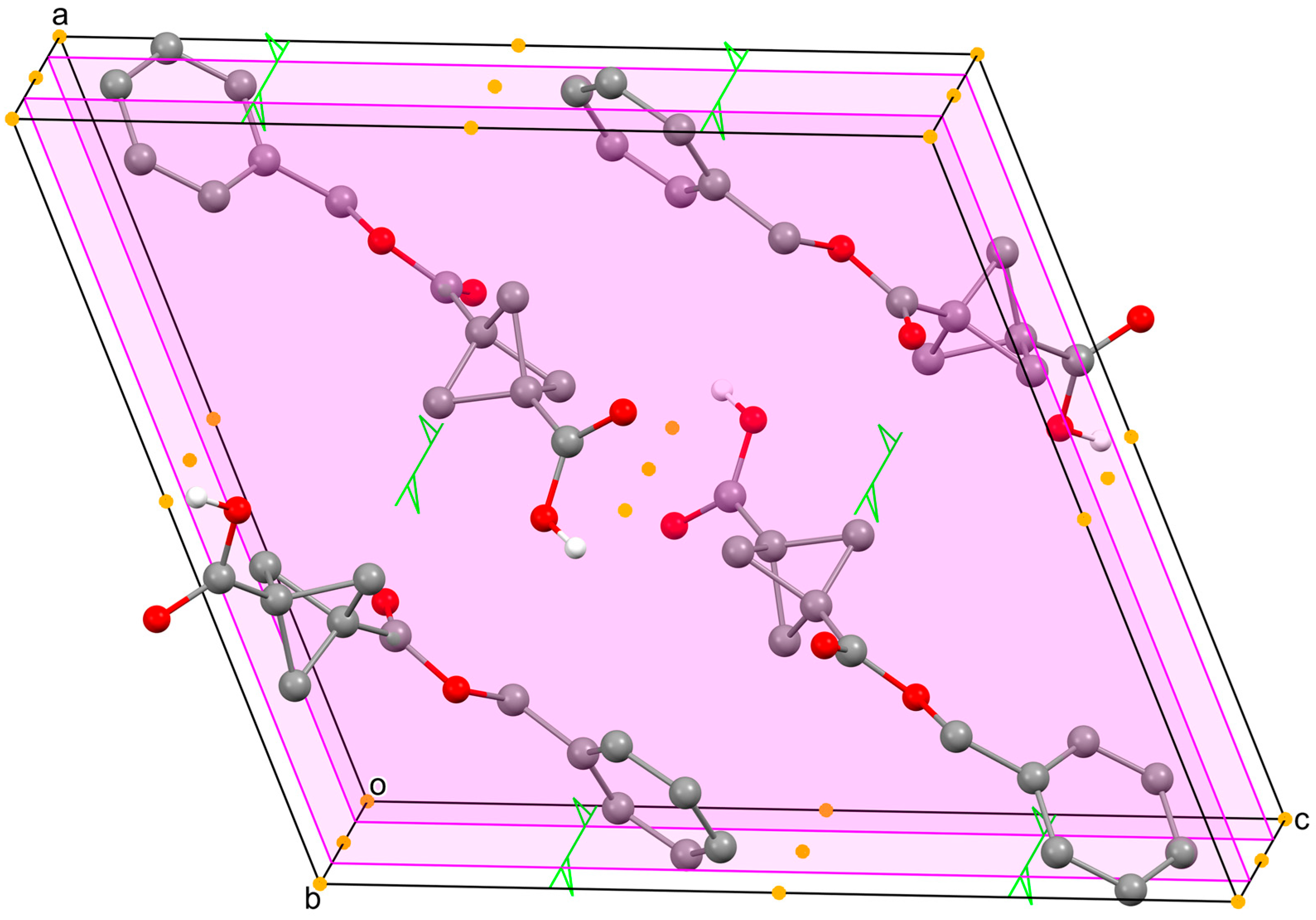
The compound appears to pack efficiently (Figure 2). The Dcalc for the compound is 1.323 g/cm3, which is lower than the average of 1.414 g/cm3 for BCPs disubstituted at the bridgehead positions (356 exemplars [11]). In this diagram, the two central molecules are not H-bonded to each other, e.g., through (½½½); rather, this occurs at (½0½) and (½1½) so that the dimer partners are not in view within the unit cell. The shortest intermolecular contact outside of the H-bonded pairs is that from O1 to ortho benzyl ring H7 at (x, 1/2 − y, −1/2 + z), that is, 0.275 Å < ∑rvdW. Thus, there are no secondary O···H interactions involving the carboxylic acid, whereas these are a prominent feature in the packing of BA [6].

3. Materials and Methods

3.1. Chemical Synthesis

3.1.1. General Materials and Procedures

Bicyclo[1.1.1]pentane-1,3-dicarboxylic acid was purchased from BLDpharm, LiOH·H2O from Thermo Fisher and all other compounds were acquired from Sigma-Aldrich. THF and DCM were purified using an MBraun solvent purification system (SPS), while all other compounds were used as received. 1H NMR spectra were recorded in CDCl3 at room temperature using a Bruker Avance II (300 MHz) instrument. The infrared spectrum was recorded using a Bruker Tensor 37 FT Infrared Spectrometer.

3.1.2. Dibenzyl Bicyclo[1.1.1]pentane-1,3-dicarboxylate (1)

Bicyclo[1.1.1]pentane-1,3-dicarboxylic acid (2.00 g, 12.8 mmol) and Li2CO3 (1.89 g, 25.6 mmol) were added to a round-bottom flask with a stir bar. The flask was affixed with a rubber septum and flushed with N2. DMF (51 mL, 0.25 M) was added to the flask, followed by benzyl bromide (4.6 mL, 38.4 mmol). The flask was affixed with a water condenser topped with a rubber septum, placed in an oil bath at 70 °C and the reaction mixture was stirred for 18 h under N2. The reaction mixture was cooled to room temperature and was transferred into an Erlenmeyer flask with water (400 mL), which caused precipitation of a solid. The precipitate was filtered over a frit and washed with additional water. The solid was collected and redissolved in DCM. The solution was dried over Na2SO4, filtered and concentrated under reduced pressure. The desired product (4.19 g, 97%) was isolated as a white solid by flash chromatography using 10% EtOAc in hexanes, producing the following results: 1H NMR (300 MHz, CDCl3) δ 7.40–7.30 (m, 10H), 5.12 (s, 4H), 2.35 (s, 6H). Spectroscopic data was in agreement with the literature [17].

3.1.3. 3-((Benzyloxy)carbonyl)bicyclo[1.1.1]pentane-1-carboxylic acid (2)

Compound 1 (4.19 g, 14.7 mmol), LiOH⋅H2O (679 mg, 16.2 mmol) and THF (29 mL, 0.5 M) were added to a Schlenk flask with a stir bar and the headspace was purged with nitrogen gas. All valves were closed, and the flask was placed in an oil bath at 60 °C upon which the mixture was stirred for 72 h. The reaction mixture was cooled to room temperature and was transferred to a separatory funnel with water. The aqueous layer was then washed with DCM (×3). The pH of the aqueous layer was adjusted to 2–3 using 12 M HCl and monitored with litmus paper. Next, the aqueous layer was added back to the separatory funnel, and the product was extracted with DCM (×3). The combined organic extracts were dried over Na2SO4, filtered and concentrated under reduced pressure. The desired product (2.28 g, 63%) was isolated as a white solid without any additional purification. IR (ATR, diamond) υ = 3005, 2930, 2889, 1724, 1697, 1436, 1299, 1218, 1176, 1025 cm−1; 1H NMR (300 MHz, CDCl3) δ 10.39 (bs, 1H), 7.34 (m, 5H), 5.13 (s, 2H), 2.36 (s, 6H). Spectroscopic data was in agreement with the literature [17].

3.2. Crystallography

Single crystals of 2 were obtained unexpectedly following product purification. The crystals were large and were manually broken into smaller pieces. A suitable crystal was selected under a microscope, placed on a nylon cryo-loop using Paratone™ oil and cooled directly on the goniometer using the Oxford Cryostream 800 cooling system of a Rigaku SuperNova dual source Cu/Mo SuperNova diffractometer equipped with a Pilatus 200K HPC detector. The crystal was kept at 100(1) K during data collection. Diffraction data was collected, corrected and processed using CrysAlisPro (1.171.44.85) [18]. Using Olex2 (version 1.5)[19], the structure was solved with the SHELXT (2019) [20] structure solution program using Intrinsic Phasing and refined with the olex2.refine [21] refinement package using Gauss–Newton minimization. After full refinement within the Independent Atom Method (IAM), the model was tested for stability towards Hirshfeld atom refinement using custom aspherical atomic scattering factors. The software environment of NoSpherA2, an implementation of NOn-PHERical Atom-form-factors in Olex2 [8] was employed. The ED is calculated from a Gaussian basis set single determinant SCF wavefunction with DFT using selected functionals, for a fragment of the crystal. This fragment can be embedded in an electrostatic crystal field by employing cluster charges. The following options were used: ORCA 5.0 [22], partitioning with NoSpherA2 [8], normal integration accuracy, R2SCAN/cc-pVTZ level of theory with a charge of 0 and a spin multiplicity of 1. The refinement continued to full convergence of both the scattering factors and atomic positions. Full details of the refinement are provided in the Supplementary Materials.
Crystal data for C14H14O4 (M = 246.265 g/mol): monoclinic, space group P21/c (no. 14), a = 13.5449(3) Å, b = 6.5288(1) Å, c = 15.0367(3) Å, β = 111.558(2)°, V = 1236.71(5) Å3, Z = 4, T = 100.00(10) K, μ(Cu Kα) = 0.806 mm−1, Dcalc = 1.323 g/cm3, 14371 reflections measured (7.02° ≤ 2Θ ≤ 160.78°), 2703 unique (Rint = 0.0365, Rsigma = 0.0274), which were used in all calculations. The final R1 was 0.0222 (I ≥ 2σ(I)) and wR2 was 0.0594 (all data).

Supplementary Materials

The following supporting information can be downloaded online: Figure S1. Labelled Diagram of 2; Table S1. Crystal data and structure refinement for 2; Table S2. Fractional Atomic Coordinates (×104) and Equivalent Isotropic Displacement Parameters (Å2×10³) for 2. Ueq is defined as 1/3 of the trace of the orthogonalised UIJ tensor; Table S3. Anisotropic Displacement Parameters (Å2×10³) for 2. The Anisotropic displacement factor exponent takes the form: -2π2[h2a*2U11+2hka*b*U12+…]; Table S4. Bond Lengths for 2; Table S5. Bond Angles for 2; Table S6. Hydrogen Bonds for 2; Table S7. Torsion Angles for 2; Table S8. Hydrogen Atom Coordinates (Å×104) and Isotropic Displacement Parameters (Å2×10³) for 2; Table S9. Atomic Occupancy for 2; Figure S2. Expanded packing diagram for 2 showing all intermolecular contacts 0.2 Å > ΣrvdW. The generator molecule (x, y, z) is colored purple and its direct H-bonded partner is rendered in mauve; Figure S3. IR spectrum of 3-((benzyloxy)carbonyl)bicyclo[1.1.1]pentane-1-carboxylic acid (2); Figure S4. 1H NMR spectrum (300 MHz, CDCl3) of dibenzyl bicyclo[1.1.1]pentane-1,3-dicarboxylate (1); Figure S5. 1H NMR Spectrum (300 MHz, CDCl3) of 3-((benzyloxy)carbonyl)bicyclo[1.1.1]pentane-1-carboxylic acid (2).

Author Contributions

Conceptualization, D.D.T., J.-D.H. and R.T.B.; experimental chemistry, S.K.N.; data processing and refinement D.D.T. and R.T.B.; writing—review and editing, J.-D.H. and R.T.B.; supervision, J.-D.H. and R.T.B.; funding acquisition, J.-D.H. All authors have read and agreed to the published version of the manuscript.

Funding

The authors acknowledge funding from the Natural Sciences and Engineering Research Council of Canada (NSERC), the Canada Research Chairs Program (CRC), the Canada Foundation for Innovation (CFI) and the University of Lethbridge. The diffractometer at the University of Lethbridge X-ray Diffraction Facility was purchased by the University and the Faculty of Arts and Science.

Data Availability Statement

Data available for this work is provided in the Supporting Information (detailed report of the SC-XRD diffraction study; 1H NMR spectra and IR spectrum). Raw SC-XRD data has been deposited via the joint CCDC/FIZ Karlsruhe deposition service and can be accessed by citing the data acquisition code 2490345.

Acknowledgments

The authors would like to thank Miriam van Hoeve for her assistance with X-ray data acquisition.

Conflicts of Interest

The authors declare no conflicts of interest.

Abbreviations

The following abbreviations are used in this manuscript:
AHydrogen acceptor
BABenzoic acid
BCPBicyclo[1.1.1]pentane; Bicyclo[1.1.1]pentyl
CSDCambridge structural database
DHydrogen donor
DcalcCalculated density from X-ray data
DCMDichloromethane
DMFDimethylformamide
EtOAcEthyl acetate
NMRNuclear magnetic resonance
SC-XRDSingle-crystal X-ray diffraction
THFTetrahydrofuran

References

  1. Taylor, R.D.; MacCoss, M.; Lawson, A.D.G. Rings in Drugs. J. Med. Chem. 2014, 57, 5845–5859. [Google Scholar] [CrossRef] [PubMed]
  2. Mykhailiuk, P.K. Saturated bioisosteres of benzene: Where to go next? Org. Biomol. Chem. 2019, 17, 2839–2849. [Google Scholar] [CrossRef] [PubMed]
  3. Pu, Q.; Zhang, H.; Guo, L.; Cheng, M.; Doty, A.C.; Ferguson, H.; Fradera, X.; Lesburg, C.A.; McGowan, M.A.; Miller, J.R.; et al. Discovery of Potent and Orally Available Bicyclo[1.1.1]pentane-Derived Indoleamine-2,3-dioxygenase 1 (IDO1) Inhibitors. ACS Med. Chem. Lett. 2020, 11, 1548–1554. [Google Scholar] [CrossRef] [PubMed]
  4. Allen, F.H.; Watson, D.G.; Brammer, L.; Orpen, A.G.; Taylor, R. Typical interatomic distances: Organic compounds. In International Tables for Crystallography Volume C: Mathematical, Physical and Chemical Tables; Springer: Dordrecht, The Netherlands, 2006; pp. 790–811. [Google Scholar]
  5. Levin, M.D.; Kaszynski, P.; Michl, J. Bicyclo[1.1.1]pentanes, [n]Staffanes, [1.1.1]Propellanes, and Tricyclo[2.1.0.02,5]pentanes. Chem. Rev. 2000, 100, 169–234. [Google Scholar] [CrossRef] [PubMed]
  6. Wilson, C.C.; Shankland, N.; Florence, A.J. A single-crystal neutron diffraction study of the temperature dependence of hydrogen-atom disorder in benzoic acid dimers. J. Chem. Soc. Faraday Trans. 1996, 92, 5051–5057. [Google Scholar] [CrossRef]
  7. Allen, F.; Bruno, I. Bond lengths in organic and metal-organic compounds revisited: X—H bond lengths from neutron diffraction data. Acta Crystallogr. B 2010, 66, 380–386. [Google Scholar] [CrossRef] [PubMed]
  8. Kleemiss, F.; Dolomanov, O.V.; Bodensteiner, M.; Peyerimhoff, N.; Midgley, L.; Bourhis, L.J.; Genoni, A.; Malaspina, L.A.; Jayatilaka, D.; Spencer, J.L.; et al. Accurate crystal structures and chemical properties from NoSpherA2. Chem. Sci. 2020, 12, 1675–1692. [Google Scholar] [CrossRef] [PubMed]
  9. Fillaux, F.; Cousson, A. A neutron diffraction study of the crystal of benzoic acid from 6 to 293K and a macroscopic-scale quantum theory of the lattice of hydrogen-bonded dimers. Chem. Phys. 2016, 479, 26–35. [Google Scholar] [CrossRef]
  10. Kurzydłowski, D. Potential energy barrier for proton transfer in compressed benzoic acid. RSC Adv. 2022, 12, 11436–11441. [Google Scholar] [CrossRef]
  11. Groom, C.R.; Bruno, I.J.; Lightfoot, M.P.; Ward, S.C. The Cambridge Structural Database. Acta Crystallogr. B 2016, 72 Pt 2, 171–179. [Google Scholar] [CrossRef] [PubMed]
  12. Grover, N.; Locke, G.M.; Flanagan, K.J.; Beh, M.H.R.; Thompson, A.; Senge, M.O. Bridging and Conformational Control of Porphyrin Units through Non-Traditional Rigid Scaffolds. Chem. Eur. J. 2020, 26, 2405–2416. [Google Scholar] [CrossRef]
  13. Kanazawa, J.; Maeda, K.; Uchiyama, M. Radical Multicomponent Carboamination of [1.1.1]Propellane. J. Am. Chem. Soc. 2017, 139, 17791–17794. [Google Scholar] [CrossRef] [PubMed]
  14. Yin, D. CCDC 2350394 Experimental Crystal Structure Determination. 2024. [CrossRef]
  15. Ripenko, V.; Sham, V.; Levchenko, V.; Holovchuk, S.; Vysochyn, D.; Klymov, I.; Kyslyi, D.; Veselovych, S.; Zhersh, S.; Dmytriv, Y.; et al. Light-enabled scalable synthesis of bicyclo[1.1.1]pentane halides and their functionalizations. Nat. Synth. 2024, 3, 1538–1549. [Google Scholar] [CrossRef]
  16. Luger, P.; Weber, M.; Szeimies, G.; Pätzel, M. 3-(tert-Butyl-oxy-carbonyl-amino)-bicyclo-[1.1.1]-pentane-carboxyl-ic acid at 293 K. Acta Crystallogr. C 2000, 56, 1170–1172. [Google Scholar] [CrossRef]
  17. Ripenko, V.; Vysochyn, D.; Klymov, I.; Zhersh, S.; Mykhailiuk, P.K. Large-Scale Synthesis and Modifications of Bicyclo[1.1.1]pentane-1,3-dicarboxylic Acid (BCP). J. Am. Chem. Soc. 2021, 86, 14061–14068. [Google Scholar] [CrossRef]
  18. Agilent. CrysAlis PRO; Agilent Technologies Ltd.: Yarnton, UK, 2014. [Google Scholar]
  19. Dolomanov, O.; Bourhis, L.; Gildea, R.; Howard, J.; Puschmann, H. OLEX2: A complete structure solution, refinement and analysis program. J. Appl. Crystallogr. 2009, 42, 339–341. [Google Scholar] [CrossRef]
  20. Sheldrick, G. SHELXT—Integrated space-group and crystal-structure determination. Acta Crystallograp. A 2015, 71, 3–8. [Google Scholar] [CrossRef] [PubMed]
  21. Bourhis, L.J.; Dolomanov, O.V.; Gildea, R.J.; Howard, J.A.; Puschmann, H. The anatomy of a comprehensive constrained, restrained refinement program for the modern computing environment—Olex2 dissected. Acta Crystallogr. A 2015, 71 Pt 1, 59–75. [Google Scholar] [CrossRef] [PubMed]
  22. Neese, F. Software update: The ORCA program system—Version 5.0. Wiley Interdiscip. Rev. Comput. Mol. Sci. 2022, 12, e1606. [Google Scholar] [CrossRef]
Scheme 1. Synthetic sequence for the preparation of carboxylic acid 2.
Molbank 2025 m2075 sch001
Figure 1. Displacement ellipsoids plot (40% probability) of 2 showing the atom numbering of the atoms in the asymmetric unit. First, atoms of the symmetry-related carboxylate H-bonded dimer are labeled (′); dimerization is centrosymmetric with respect to (½ 0 ½) and H-bonding is indicated by dashed magenta lines. The minor component of the bridging H2 atom is omitted for clarity.
Molbank 2025 m2075 g001
Figure 2. Packing ball and stick diagram (H-atoms on C omitted) showing the symmetry relationships in the unit cell: 1 ¯ as yellow circles; 21 as green arrows; c–glides as magenta planes.
Molbank 2025 m2075 g002
Table 1. Key bond lengths of the BCP moiety for 2.
BondLength (Å)Csp3 (Å) aCsp2 (Å) a
C–C bonds in the BCP moiety
C2–C211.5485(9)
C2–C221.5655(9)
C2–C231.5504(9)
C3–C211.5623(9)
C3–C221.5448(9)
C3–C231.5626(9)
Average a1.557 ± 0.0081.530
Bridgehead to substituent
C1–C21.4866(9)1.5021.476
C3–C41.4888(9)1.4971.489
a Values taken from [4].
Table 2. C–O bond lengths for 2.
Bond Length (Å)Lower Quartile (Å)Upper Quartile (Å)ΔCO (Å)
Carbonyl (C=O)
C1-O11.2385(9)0.062
C4-O31.2083(9)
C–C(=O)–OH1.214 a1.203 a1.224 a
Bond to the hydroxyl group (C–O)
C1-O21.3001(8)0.132
C4-O41.3398(8)
C–C(=O)–OH1.308 a1.308 a1.298 a
a Standard values obtained from International Tables for Crystallography (2006) [4].
Table 3. Hydrogen bonds for 2.
D  H  Ad(D–H) (Å)d(H–A) (Å)d(D–A) (Å)D–H–A (°)
O2 H2 O10.9900(19)1.650(3)2.6376(7)174.5(14)
O1 H1a O2′0.990(2)1.654(10)2.6376(7)172(7)
Table 4. Comparison of internal BCP angles and transannular distance between bridgehead carbons.
RefcodeAngle 1 (°) aAngle 2 (°) aAngle 3 (°) aC2···C3 (Å) bRef.
DIGGOT73.8(2)73.3(1)73.8(2)1.859(2)[12]
SUJKIU73.8(1)73.8(1)73.5(1)1.866(2)[12]
SUJKOA73.8(1)73.4(1)73.1(1)1.861(2)[12]
WERXOJ73.78(7)73.20(7)73.00(7)1.853(1)[13]
YOMPAV73.9(1)73.7(1)73.7(1)1.868(2)[14]
DUFWOU74.5(2)73.8(2)73.8(2)1.871(3)[15]
WOCXIV0174.00(4)73.81(4)73.49(4)1.868(9)[16]
Average73.9 ± 0.273.6 ± 0.273.4 ± 0.31.864 ± 0.006
273.60(5)73.58(5)73.52(5)1.863(1)This work
a Angles represent inner ∠C–C–C at the bridges of the BCP moiety and are ordered according to 1 > 2 > 3 for each structure. b Transannular distance between bridgehead carbons.
Disclaimer/Publisher’s Note: The statements, opinions and data contained in all publications are solely those of the individual author(s) and contributor(s) and not of MDPI and/or the editor(s). MDPI and/or the editor(s) disclaim responsibility for any injury to people or property resulting from any ideas, methods, instructions or products referred to in the content.