Communication

Three Hypoxanthine Derivatives from the Marine Cyanobacterium Okeania hirsuta

1
Department of Ocean Sciences, Tokyo University of Marine Science and Technology, Tokyo 108-8477, Japan
2
Fisheries Technology Institute, Japan Fisheries Research and Education Agency, Yokohama 236-8648, Japan
3
Department of Chemistry, The University of Tokyo, Tokyo 113-0033, Japan
*
Author to whom correspondence should be addressed.
Molbank 2025, 2025(3), M2051; https://doi.org/10.3390/M2051
Submission received: 25 July 2025 / Revised: 14 August 2025 / Accepted: 20 August 2025 / Published: 21 August 2025
(This article belongs to the Section Natural Product Chemistry)

Abstract

Three novel hypoxanthine derivatives (13) were obtained from the Okinawan cyanobacterium Okeania hirsuta. The structures of these compounds were elucidated mainly based on the spectroscopic data, including 1D and 2D NMR, as well as high-resolution mass spectrometry. In particular, the amounts of obtained compounds 2 and 3 were only 200 μg and much less than 50 μg, respectively. Therefore, some carbons signals could not be observed on 13C NMR spectra of these compounds. However, the detailed analysis of HSQC and HMBC spectra allowed us to elucidate their structures. For NMR measurements of compound 3, it was found that using an 800 MHz NMR machine equipped with a cryogenic probe and acetic acid-d4 as a solvent is essential. Compounds (13) were N-3′-carbonylbutyl group-connected hypoxanthines.

1. Introduction

In July 2010, an outbreak of the toxic cyanobacterium Okeania hirsuta [1] occurred on Kuba Beach in Okinawa, Japan [2]. The cyanobacterium had been misidentified as Moorea producence at first and re-identified as O. hirsuta [2]. We have collected the cyanobacterium Okeania hirsuta samples at the site [2]. Cyanobacteria are recognized as promising sources of novel natural bioactive products [3]. Furthermore, the genus Okeania is known as one of the most diverse compound-producing cyanobacteria [4,5,6,7,8,9,10]. Therefore, we conducted bioprospecting studies for biologically active compounds in our O. hirsuta samples and succeeded in finding some novel compounds [2,11,12,13]. So far, studies on compounds derived from the genus Okeania have been mainly concerned with low-polarity compounds [2,4,5,6,7,8,9,10,11,12,13]. Thus, we have tried to isolate polar compounds from the O. hirsuta extracts. As a result, three novel hypoxanthine derivatives (13) were obtained, along with the known nucleosides, thymidine (4), adenosine (5), and deoxyadenosine (6), from the cyanobacterial sample. This paper reports the purification and structural elucidation of these new compounds (13).

2. Results

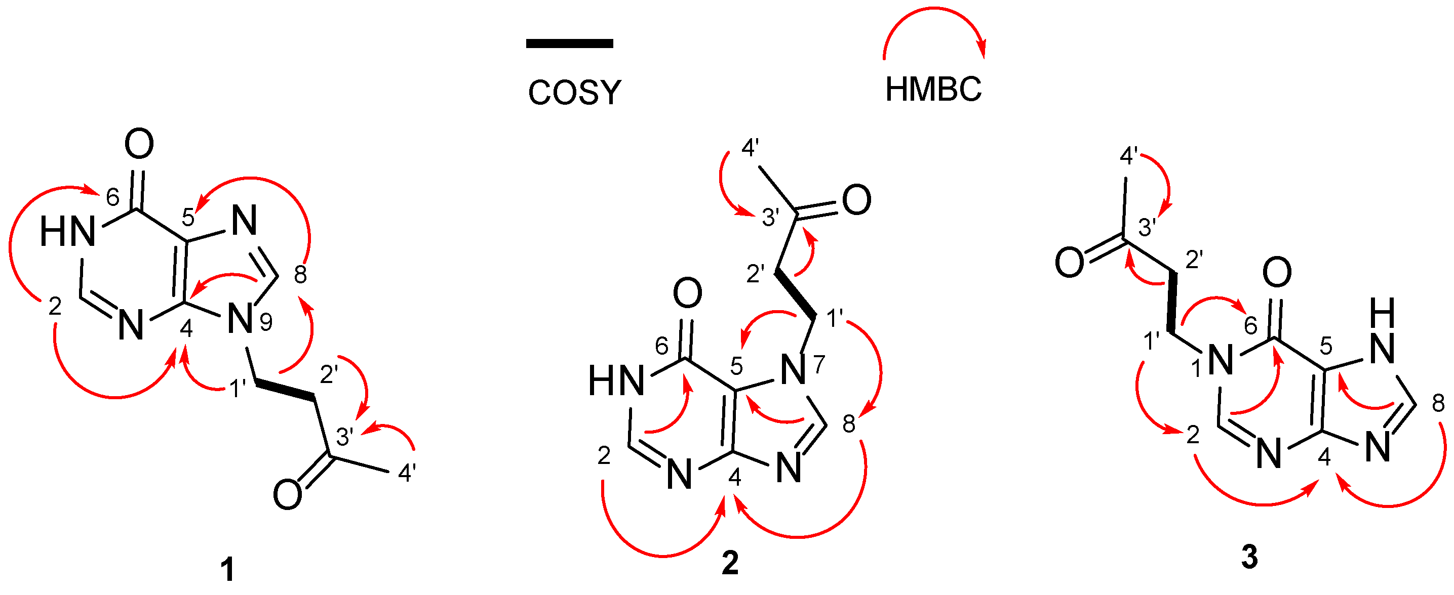
Compound 1 (Figure 1) was isolated as a pale-yellow amorphous solid (0.4 mg). High-resolution electrospray ionization mass spectrometry (HR-ESI-MS) analysis of compound (1) showed an [M + H]+ ion peak at m/z 207.0855 (calcd. for C9H11N4O2 m/z 207.0877), indicating a molecular formula of C9H10N4O2 with seven degrees of unsaturation. 13C (nuclear magnetic resonance) NMR and heteronuclear single quantum correlation (coherence) (HSQC) spectra revealed the presence of one methyl, one amide, one ketone, two aliphatic methylene, and four aromatic carbons (Table 1). Heteronuclear multiple-bond coherence (HMBC) correlations from H-2 (δH 8.01) to C-4 (δc 148.3) and C-6 (δc 157.1), and from H-8 (δH 7.98) to C-4 (δc 148.3) and C-5 (δc 123.9) confirmed the hypoxanthine structure (Figure 2). A correlation spectroscopy (COSY) correlation from H-1′ (δH 4.28) to H-2′ (δH 3.08) and HMBC correlations from H-2′ (δH 3.08) to C-3′ (δc 206.2) as well as from H-4′ (δH 2.10) to C-3′ (δc 206.2) revealed the existence of a CH3-CO-CH2-CH2- unit. Finally, HMBC correlations from H-1′ (δH 4.28) to C-4 (δc 148.3) and C-8 (δc 140.2) confirmed that the CH3-CO-CH2-CH2- unit was connected to N-9 of hypoxanthine (Figure 2).
Compound 2 (Figure 1) was obtained as a pale-yellow amorphous solid (0.2 mg). Compound 2 contained impurities (Figure S12). However, further purification was not performed because there was a possibility of losing the compound. In fact, the presence of impurities was a nuisance in structural determination by NMR analysis. Furthermore, due to the insufficient amount of the compound, some carbon signals could not be directly observed in the 13C NMR spectra. However, detailed analysis of HSQC and HMBC spectra enabled the structural determination of compound 2. HR-ESI-MS analysis of compound (2) showed an [M + H]+ ion peak at m/z 207.0837 (calcd. for C9H11N4O2 m/z 207.0877), indicating a molecular formula of C9H10N4O2, which is the same as that for compound 1. NMR analysis of compound 2 revealed that 2 also has a CH3-CO-CH2-CH2- unit and a hypoxanthine unit. HMBC correlations from H-1′ (δH 4.45) to C-5 (δc 114.8) and C-8 (δc 144.1) confirmed that the CH3-CO-CH2-CH2- unit was connected to N-7 of hypoxanthine (Figure 2).
Compound 3 (Figure 1) was obtained as a pale-yellow amorphous solid (much less than 50 μg). Compound 3 contained impurities (Figure S20). However, further purification was not performed because there was a possibility of losing the compound. These conditions were the same as for compound 2. Furthermore, since compound 3 was present in much smaller quantities, it was necessary to measure NMR using an 800 MHz NMR machine equipped with a cryogenic probe. HR-ESI-MS analysis of compound (3) showed an [M + H]+ ion peak at m/z 207.0834 (calcd. for C9H11N4O2 m/z 207.0877), indicating a molecular formula of C9H10N4O2, which is the same as that for compounds 1 and 2.
Interestingly, one of the two methine protons (H-2 and H-8) in the hypoxanthine unit could not be detected on the 1H-NMR spectrum with dimethyl sulfoxide (DMSO)-d6. Thus, acetic acid-d4 was examined as the solvent instead of DMSO. As a result, it was found that acetic acid-d4 gave a good resolution for two methine protons at δH 8.41 and δH 8.57 in the NMR spectra. It is unclear why acetic acid-d4 showed a good resolution in the 1H- NMR spectrum for these methine protons, while DMSO did not. However, this phenomenon may be related to the already reported tautomeric effect [14]. Once two methine protons were observed, the corresponding carbons could be determined by HSQC spectra as δc 150.1 (δH 8.57) and δc 143.5 (δH 8.41). Detailed NMR analysis (COSY and HMBC) of compound 3 revealed that 3 also has a CH3-CO-CH2-CH2- unit and a hypoxanthine unit as 1 and 2. Then, HMBC correlations from H-1′ (δH 4.37) to C-2 (δc 150.1) and C-6 (δc 156.8) confirmed that the CH3-CO-CH2-CH2- unit was connected to N-1 of hypoxanthine (Figure 2).
In this study, three new hypoxanthine derivatives (13) were isolated along with the known nucleosides, thymidine (4), adenosine (5), and deoxyadenosine (6), from the polar fraction of the cyanobacterial sample.
To the best of our knowledge, only two purine alkaloids with the N-3′-carbonylbutyl group have been reported to have been observed in nature so far. These are 1-(3′-carbonylbutyl)purine-6,8-dione and 9-(3′-carbonylbutyl)purine-6,8-dione from the marine gorgonian Subergorgia suberosa [15]. Hypoxanthine is a naturally occurring purine derivative. Hypoxanthine is found in nucleic acids and is present in the anticodon of tRNA in the form of nucleoside inosine [16,17]. Compounds 13 are the first hypoxanthines with the N-3′-CARBONbylbutyl group.

3. Materials and Methods

3.1. General Experimental Procedures

NMR spectral data were recorded on a Bruker AVANCE III 800 MHz spectrometer equipped with a 5 mm TCI cryogenic probe (Bruker, Billerica, MA, USA) or a Bruker AVANCE III 600 MHz (Bruker, Billerica, MA, USA) spectrometer and referenced to residual solvent signals (δH 2.50, δC 39.5, at 300 K for DMSO-d6 and acetic acid-d4 at δH 2.04, δC 20.0, 298 K). DMSO-d6 and acetic acid-d4 were purchased from Kanto Chemical Co., Tokyo. Japan. The HR-ESI- MS data were acquired using a Bruker micrOTOF QII mass spectrometer (Bruker, Billerica, MA, USA). Ultraviolet (UV) spectra were measured using a U-3000 spectrophotometer (Hitachi High-Tech Science Co., Tokyo, Japan). High-performance liquid chromatography (HPLC) purification was performed using a Hitachi Chromaster HPLC system equipped with a 5110 pump and a 5430 diode array detector (Hitachi High-Tech Science Co., Tokyo, Japan).

3.2. Collection of Cyanobacteria

An outbreak of the toxic cyanobacterium Okeania hirsuta occurred on Kuba Beach in Okinawa, Japan, in July 2010. The cyanobacterial sample was collected at the site. O. hirsuta was the dominant cyanobacterial specimen in all samples [2]. The identification of O. hirsuta was performed based on 16S rRNA sequence analysis [2]. A voucher specimen (20100713-a) was deposited at the Tokyo University of Marine Science and Technology.

3.3. Isolation of Compounds

A frozen sample of O. hirsuta (wet weight: 9.7 kg) was extracted three times with methanol (MeOH). After filtration, the extract was partitioned between 80% aqueous MeOH and hexane. The 80% aqueous MeOH layer was then partitioned between ethyl acetate (EtOAc) and distilled water (H2O), and the H2O layer was further extracted with 1-butanol (BuOH). The 1-BuOH layer (4.3 g, dry weight) was subjected to stepwise fractionation using 30%, 50%, 70%, 85%, and 100% MeOH on an open glass column (40 × 170 mm) packed with ODS-7512-12A resin (Senshu Scientific Co., Tokyo, Japan). The 30% MeOH eluted fraction (2.3 g, dry weight) was subjected to stepwise fractionation using 0%, 50%, and 100% MeOH on an open glass column (40 × 400 mm) packed with Diaion HP-20SS (Mitsubishi Chemical Co. Tokyo, Japan). The 50% MeOH eluted fraction (440 mg, dry weight) was subjected to stepwise fractionation using 5%, 10%, 50%, and 100% MeOH on an open glass column (40 × 400 mm) packed with cosmosil 75C18-OPN) (Nacalai Tesque Co., Kyoto, Japan). The 10% MeOH eluted fraction (23 mg, dry weight) was then subjected to HPLC [column, Develosil C30-UG-5 (Nomura Chemical Co., Aichi, Japan) with the following conditions: flow rate, 2.0 mL/min; detection, 210 nm; temperature, 27 °C; solvent, MeOH/H2O; gradient, 15% MeOH isocratic elution]. The purified fraction was re-chromatographed with HPLC using a column, Develosil XG-C30-M5 (Nomura Chemical Co., Aichi, Japan). Consequently, compound 1 (0.4 mg), compound 2 (0.2 mg), compound 3 (less than 50 μg), thymidine (4, 0.9 mg), adenosine (5, 3.6 mg), and deoxyadenosine (6, 1.4 mg) were isolated.
Compound 1: 9-(3-oxobutyl)-1,9-dihydro-6H-purin-6-one. Spectroscopic data for compound 1: UV (H2O) λmax (ε) 203 nm (34990), 250 nm (12420). HR-ESI-MS (Figure S3): C9H10N4O2 ([M + H]+ m/z 207.0855, calcd. for C9H11N4O2, 207.0877). NMR data are shown in Table 1 and Figures S4–S8.
Compound 2: 7-(3-oxobutyl)-1,7-dihydro-6H-purin-6-one. Spectroscopic data for compound 2: UV (H2O) λmax (ε) 194 nm (35612), 256 nm (12937). HR-ESI-MS (Figure S11): C9H10N4O2 ([M + H]+ m/z 207.0837, calcd. for C9H11N4O2, 207.0877). NMR data are shown in Table 1 and Figures S12–S16.
Compound 3: 1-(3-oxobutyl)-1,7-dihydro-6H-purin-6-one. Spectroscopic data for compound 3: HR-ESI-MS (Figure S19): C9H10N4O2 ([M + H]+ m/z 207.0834, calcd. for C9H11N4O2, 207.0877). NMR data are shown in Table 1 and Figures S20–S23.
Spectroscopic data for thymidine (4): Figures S24–S25.
Spectroscopic data for adenosine (5): Figures S26–S27.
Spectroscopic data for deoxyadenosine (6): Figures S28–S29.

4. Conclusions

In this study, three novel hypoxanthine derivatives (13) were obtained along with known nucleosides, thymidine (4), adenosine (5), and deox-yadenosine (6), from the polar fraction of Okinawan cyanobacterium Okeania hirsuta extracts. The amounts of compounds 2 and 3 were very small. Therefore, some carbons signals for these compounds could not be observed on 13C NMR spectra. However, the detailed analysis of HSQC and HMBC spectra allowed us to elucidate their structures. In particular, the amount of compound 3 was much lower than that of 50 μg. Thus, some protons of compound 3 could not be observed with the usual NMR measurements. For NMR measurements of compound 3, it was found that using an 800 MHz NMR machine equipped with a cryogenic probe and acetic acid-d4 as a solvent is essential. Compounds (13) were novel hypoxanthine derivatives.

Supplementary Materials

Figure S1: Final HPLC chromatogram of (1); Figure S2: Positive-mode HR-ESI-MS spectrum of (1); Figure S3: Enlarged positive-mode HR-ESI-MS spectrum of (1); Figure S4: 1H NMR spectrum of (1) in DMSO-d6 (600 MHz); Figure S5: 13C NMR spectrum of (1) in DMSO-d6 (150 MHz); Figure S6: COSY spectrum of (1) in DMSO-d6 (600 MHz); Figure S7: HSQC spectrum of (1) in DMSO-d6 (600 MHz); Figure S8: HMBC spectrum of (1) in DMSO-d6 (600 MHz); Figure S9: Final HPLC chromatogram of (2); Figure S10: Positive-mode HR-ESI-MS spectrum of (2); Figure S11: Enlarged positive-mode HR-ESI-MS spectrum of (2); Figure S12: 1H NMR spectrum of (2) in DMSO-d6 (600 MHz); Figure S13: 13C NMR spectrum of (2) in DMSO-d6 (150 MHz); Figure S14: COSY spectrum of (2) in DMSO-d6 (600 MHz); Figure S15: HSQC spectrum of (2) in DMSO-d6 (600 MHz); Figure S16: HMBC spectrum of (2) in DMSO-d6 (600 MHz); Figure S17: Final HPLC chromatogram of (3); Figure S18: Positive-mode HR-ESI-MS spectrum of (3); Figure S19: Enlarged positive-mode HR-ESI-MS spectrum of (3); Figure S20: 1H NMR spectrum of (3) in acetic acid-d4 (800 MHz); Figure S21: COSY spectrum of (3) in acetic acid-d4 (800 MHz); Figure S22: HSQC spectrum of (3) in acetic acid-d4 (800 MHz); Figure S23: HMBC spectrum of (3) in acetic acid-d4 (800 MHz); Figure S24: Positive-mode HR-ESI-MS spectrum of (4); Figure S25: 1H NMR spectrum of (4) in D2O (600 MHz); Figure S26: Positive-mode HR-ESI-MS spectrum of (5); Figure S27: 1H NMR spectrum of (5) in D2O (600 MHz); Figure S28: Positive-mode HR-ESI-MS spectrum of (6); Figure S29: 1H NMR spectrum of (6) in D2O (600 MHz).

Author Contributions

Conceptualization, H.N. and M.S.; isolation, R.K.; structural analysis, R.K., B.Z., R.W. and H.U.; writing—draft preparation, R.K., M.S. and H.N. All authors have read and agreed to the published version of the manuscript.

Funding

This work was funded by the JSPS KAKENHI (grant number 22K05817 for H.N.; grant numbers 23H01962 and 24K08613 for M.S.) and JST SPRING (grant number JPMJSP2147 for B.Z.).

Data Availability Statement

The authors confirm that the data supporting the findings of this study are available within the article and/or its Supplementary Materials.

Conflicts of Interest

The authors declare no conflicts of interest.

References

  1. Engene, N.; Paul, V.J.; Byrum, T.; Gerwick, W.H.; Thor, A.; Ellisman, M.H. Five chemically rich species of tropical marine cyanobacteria of the genus Okeania gen. nov. (Oscillatoriales, Cyanoprokaryota). J. Phycol. 2013, 49, 1095–1106. [Google Scholar] [CrossRef] [PubMed]
  2. Kanda, N.; Zhang, B.T.; Shinjo, A.; Kamiya, M.; Nagai, H.; Uchida, H.; Araki, Y.; Nishikawa, T.; Satake, M. 7-Epi-30-methyloscillatoxin D from an Okinawan cyanobacterium Okeania hirsuta. Nat. Prod. Commun. 2023, 18, 1–5. [Google Scholar]
  3. Tidgewell, K.; Clark, B.R.; Gerwick, W.H. 2.06-The natural products chemistry of cyanobacteria. In Comprehensive Natural Products, 2nd ed.; Liu, H.W., Mander, L., Eds.; Elsevier: Oxford, UK, 2010; pp. 141–188. [Google Scholar]
  4. Umeda, K.; Kurisawa, N.; Jeelani, G.; Nozaki, T.; Suenaga, K.; Iwasaki, A. Isolation and structure determination of a new analog of polycavernosides from marine Okeania sp. cyanobacterium. Beilstein J. Org. Chem. 2024, 20, 645–652. [Google Scholar] [CrossRef] [PubMed]
  5. Ebihara, A.; Taguchi, R.; Jeelani, G.; Nozaki, T.; Suenaga, K.; Iwasaki, A. Kagimminols A and B, cembrene-type diterpenes from an Okeania sp. marine cyanobacterium. J. Nat. Prod. 2024, 87, 1116–1123. [Google Scholar] [CrossRef] [PubMed]
  6. Watanabe, N.; Jeelani, G.; Nozaki, T.; Iwasaki, A. Amantamide C, an antitrypanosomal linear lipopeptide from a marine Okeania sp. cyanobacterium. ACS Omega 2024, 9, 36795–36801. [Google Scholar] [CrossRef] [PubMed]
  7. Parker, S.A.J.; Hough, A.; Wright, T.; Lax, N.; Faruk, A.; Fofie, C.K.; Simcik, R.D.; Cavanaugh, J.E.; Kolber, B.J.; Tidgewell, K.J. Isolation and bioassay of linear veraguamides from a marine cyanobacterium (Okeania sp.). Molecules 2025, 30, 680. [Google Scholar] [CrossRef] [PubMed]
  8. Niiyama, M.; Kurisawa, N.; Umeda, K.; Jeelani, G.; Agusta, A.L.; Nozaki, T.; Suenaga, K. Ouhanamide, an antiparasitic linear lipopeptide from a marine Okeania sp. cyanobacterium. J. Nat. Prod. 2025, 88, 1360–1368. [Google Scholar] [CrossRef] [PubMed]
  9. Wakai, K.; Kurisawa, N.; Umeda, K.; Jeelani, G.; Agusta, A.L.; Nozaki, T.; Suenaga, K. Sealgamide: An antitrypanosomal lipopeptide isolated from a marine Okeania sp. cyanobacterium. J. Nat. Prod. 2025, 88, 1048–1056. [Google Scholar] [CrossRef] [PubMed]
  10. Tojo, S.; Natsume, N.; Niino, T.; Iwasaki, A.; Suenaga, K.; Ohno, O.; Teruya, T. Hedopeptolide, a NO production inhibitor from the marine cyanobacterium Okeania sp. J. Nat. Prod. 2025, 88, 1023–1029. [Google Scholar] [CrossRef] [PubMed]
  11. Zhang, B.T.; Nishino, H.; Kawabe, R.; Kamio, M.; Watanabe, R.; Uchida, H.; Satake, M.; Nagai, H. N-Desmethylmajusculamide B, a lipopeptide isolated from the Okinawan cyanobacterium Okeania hirsuta. Biosci. Biotech. Biochem. 2024, 88, 517–521. [Google Scholar] [CrossRef] [PubMed]
  12. Zhang, B.; Kawabe, R.; Kamio, M.; Uchida, H.; Matsuura, N.; Mori-Yasumoto, K.; Satake, M.; Nagai, H. Okeaniazole A: Thiazole-containing cyclopeptide from the marine cyanobacterium Okeania hirsuta with antileishmanial activity. Tetrahedron Lett. 2024, 150, 155284. [Google Scholar] [CrossRef]
  13. Nishino, H.; Zhang, B.T.; Uchida, H.; Kamio, M.; Nagai, H.; Satake, M. (10 E, 15 Z)-12-(Dimethylsulfonio)-9, 13-dihydroxyoctadeca-10, 15-dienoate. Molbank 2024, 2024, M1784. [Google Scholar] [CrossRef]
  14. Bartl, T.; Zacharová, Z.; Sečkářová, P.; Kolehmainen, E.; Marek, R. NMR quantification of tautomeric populations in biogenic purine bases. Eur. J. Org. Chem. 2009, 2009, 1377–1383. [Google Scholar] [CrossRef]
  15. Qi, S.H.; Zhang, S.; Huang, H. Purine alkaloids from the South China Sea gorgonian Subergorgia suberosa. J. Nat. Prod. 2008, 71, 716–718. [Google Scholar] [CrossRef] [PubMed]
  16. Schmalle, H.W.; Hänggi, G.; Dubler, E. Structure of hypoxanthine. Acta. Cryst. C. 1988, 44, 732–736. [Google Scholar] [CrossRef]
  17. Srinivasan, S.; Torres, A.G.; Ribas de Pouplana, L. Inosine in biology and disease. Genes 2021, 12, 600. [Google Scholar] [CrossRef] [PubMed]
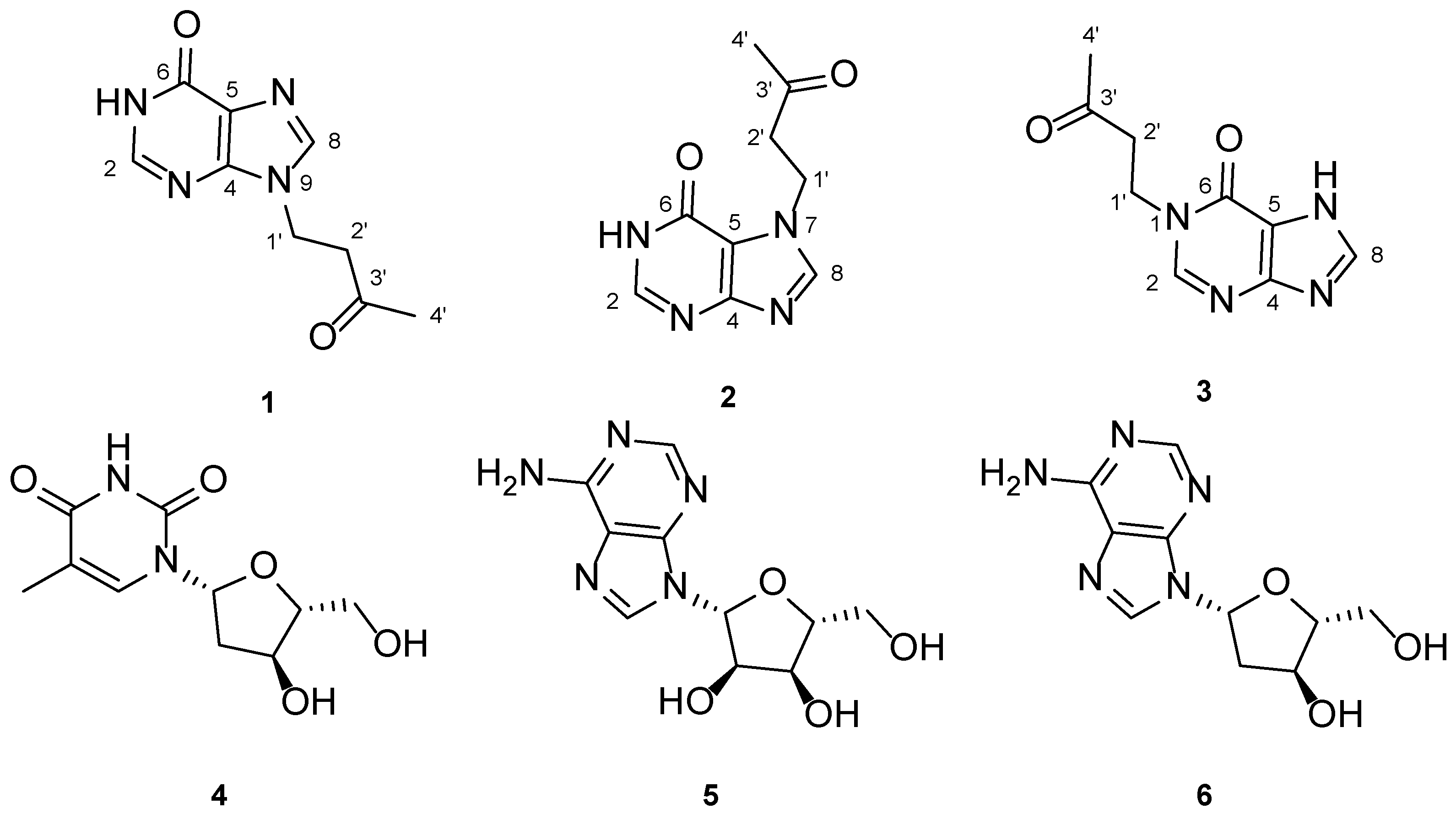
Figure 1. Structures of isolated compounds from the cyanobacterium Okeania hirsuta.
Molbank 2025 m2051 g001
Figure 2. Key 1H-1H COSY and HMBC correlations of 13.
Molbank 2025 m2051 g002
Table 1. NMR data for compounds (13).
1 (in DMSO-d6)2 (in DMSO-d6)3 (in Acetic Acid-d4)
No.δH a (J in Hz)δC b, typeδH a (J in Hz)δC b, typeδH c (J in Hz)δC d, type
1′4.28, dd (6.7, 6.7)38.3, CH24.45, dd (6.7, 6.7)41.4, CH24.37, dd (6.4, 6.4)f 43.6, CH2
2′3.08, dd (6.7, 6.7)42.3, CH23.11, dd (6.7, 6.7)43.5, CH23.12, dd (6.4, 6.4)f 42.0, CH2
3′ 206.2, C e 206.2, C e 209.1, C
4′2.10, s29.8, CH32.09, s29.8, CH32.16, sf 29.7, CH3
28.01, s145.8, CH7.95, s144.4, CH8.57, sf 150.1, CH
4 148.3, C e 157.3, C e 153.7, C
5 123.9, C e 114.8, C e 117.6, C
6 157.1, C e 154.3, C e 156.8, C
87.98, s140.2, CH8.15, s144.1, CH8.41, sf 143.5, CH
a Measured at 600 MHz. b Measured at 150 MHz. c Measured at 800 MHz. d Measured at 200 MHz. e Assigned by HMBC. f Assigned by HSQC.
Disclaimer/Publisher’s Note: The statements, opinions and data contained in all publications are solely those of the individual author(s) and contributor(s) and not of MDPI and/or the editor(s). MDPI and/or the editor(s) disclaim responsibility for any injury to people or property resulting from any ideas, methods, instructions or products referred to in the content.