Short Note

3′H-Spiro[dibenzo[c,h]xanthene-7,1′-isobenzofuran]-3′-one

EaStCHEM School of Chemistry, University of St Andrews, North Haugh, St Andrews, Fife KY16 9ST, UK
*
Author to whom correspondence should be addressed.
Molbank 2025, 2025(3), M2033; https://doi.org/10.3390/M2033
Submission received: 31 May 2025 / Revised: 2 July 2025 / Accepted: 3 July 2025 / Published: 7 July 2025
(This article belongs to the Section Structure Determination)

Abstract

Target compound 3′H-spiro[dibenzo[c,h]xanthene-7,1′-isobenzofuran]-3′-one (1) has long been known to be a by-product obtained from the preparation of naphtholphthalein. The structure of compound 1 was elucidated in the early 20th century; however, this compound has not previously been fully characterized using modern techniques. In this report, 1H NMR and 13C NMR spectra are provided. X-ray crystallography is also used to characterize the title compound for the first time.

1. Introduction

Phenolphthalein is readily prepared by heating phenol with phthalic anhydride under acidic conditions [1,2]. In contrast, naphtholphthalein (2, Scheme 1) is known to be harder to prepare under these conditions, so the careful control of the reaction temperature is needed to optimize the isolated yield [3,4]. In the late 19th century, Grabowski heated a mixture of 1-naphthol and phthalic anhydride to 280 °C, instead of isolating naphtholphthalein, and an unidentified material referred to as an “anhydride of naphtholphthalein” was obtained [5]. Unlike naphtholphthalein, the isolated material was insoluble in aqueous sodium hydroxide, and the characteristic blue/green colour of naphtholphthalein (in alkaline conditions) was not observed. It was known at this time, and confirmed in a later report, that “Grabowski’s anhydride” had the chemical formula C28H16O3 rather than the C28H18O4 required for naphtholphthalein [4]. Work by Copisarow concluded that the identity of “Grabowski’s anhydride” was the fluoran derivative 3′H-spiro[dibenzo[c,h]xanthene-7,1′-isobenzofuran]-3′-one (1) [4]. Copisarow’s synthesis protocol involved thoroughly mixing finely powdered samples of 1-naphthol, phthalic anhydride, and zinc chloride. The resulting mixture was then heated to 100 °C for 3 h to afford naphtholphthalein (2) and fluoran 1. In addition, a small quantity of naphtholphthalein isomer 3 was also isolated (Scheme 1). Furthermore, Copisarow demonstrated that heating naphtholphthalein 2 did not lead to any of the starting material being converted to fluoran 1 [4,6]. In contrast, samples of naphtholphthalein isomer 3 were found to undergo cyclization and the loss of water to afford fluoran 1 [6]. It was concluded that during the reaction of 1-naphthol with phthalic anhydride, both compounds 2 and 3 are formed initially, but compound 3 is not stable under the reaction conditions and is largely converted to fluoran 1 (Scheme 2).
A short study by Werner in 1917 involved heating mixtures of 1-naphthol with phthalic anhydride in concentrated sulfuric acid at temperatures between 60 °C and 90 °C [3]. Werner found that fluoran 1 was the only significant product at higher temperatures and that reducing the reaction temperature to a maximum of 65 °C increased the yield of naphtholphthalein (although a small quantity of fluoran 1 was also isolated).
In this paper, we describe the results from a recent study to examine whether fluoran 1 or naphtholphthalein (2) was the major product from the reaction of phthalic anhydride and 1-naphthol when the reaction was attempted in methanesulfonic acid [1]. Our findings broadly align with the reactions conducted by Werner in 1917 (Scheme 3), since fluoran 1 was isolated in 47% yield when methanesulfonic acid was used in place of 95% sulfuric acid (Scheme 4). Werner found that heating the reaction mixture in 95% sulfuric acid above 65 °C favoured the formation of fluoran 1, and we found that this is also the case when methanesulfonic acid is used. Older studies of compound 1 predate IR and NMR spectroscopy and modern X-ray crystallography, and as a result, literature characterization data for compound 1 is limited. Our experiments allowed us to acquire hitherto unreported IR spectra and NMR spectra for fluoran 1. Samples of 1 prepared by Werner’s method were also compared with those made using the newer protocol reported in this study. Furthermore, compound 1 was characterized using X-ray crystallography for the first time.

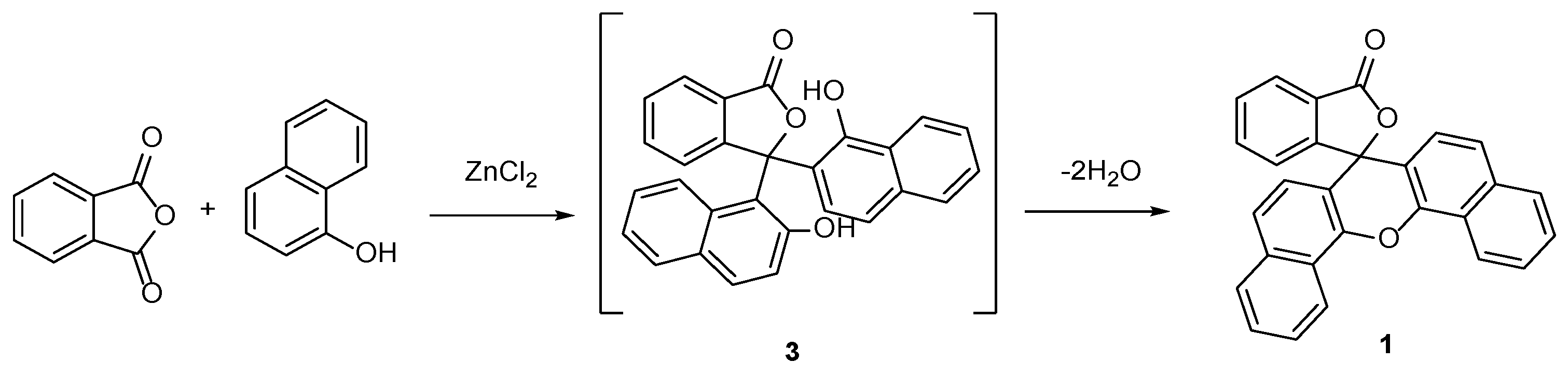
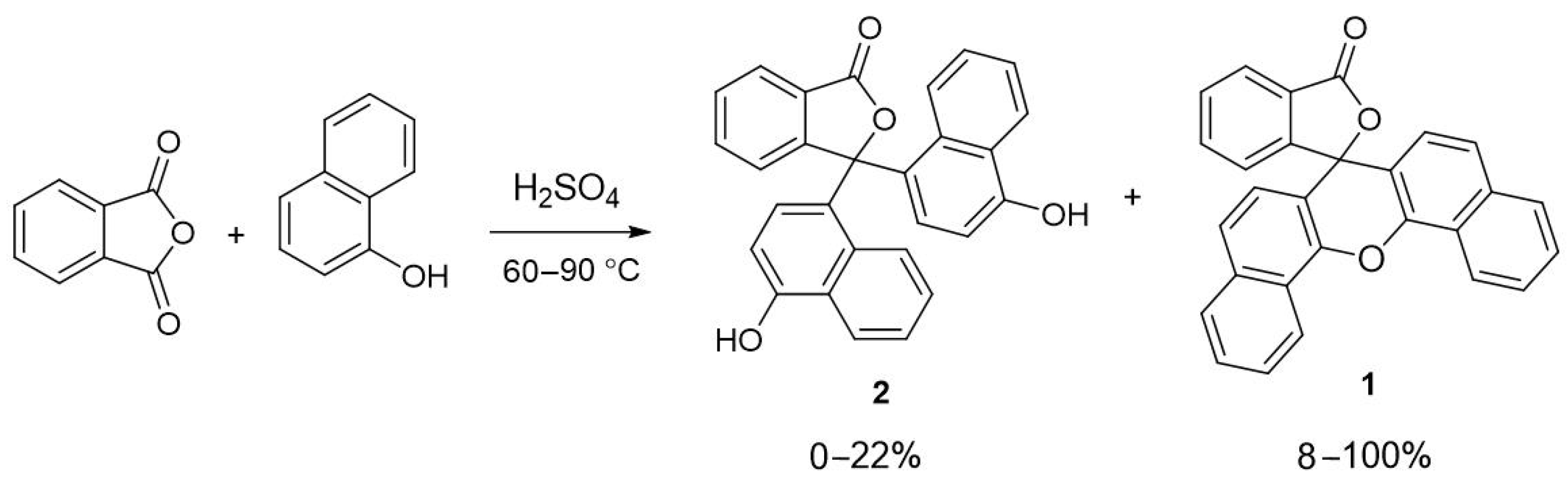
2. Results and Discussion

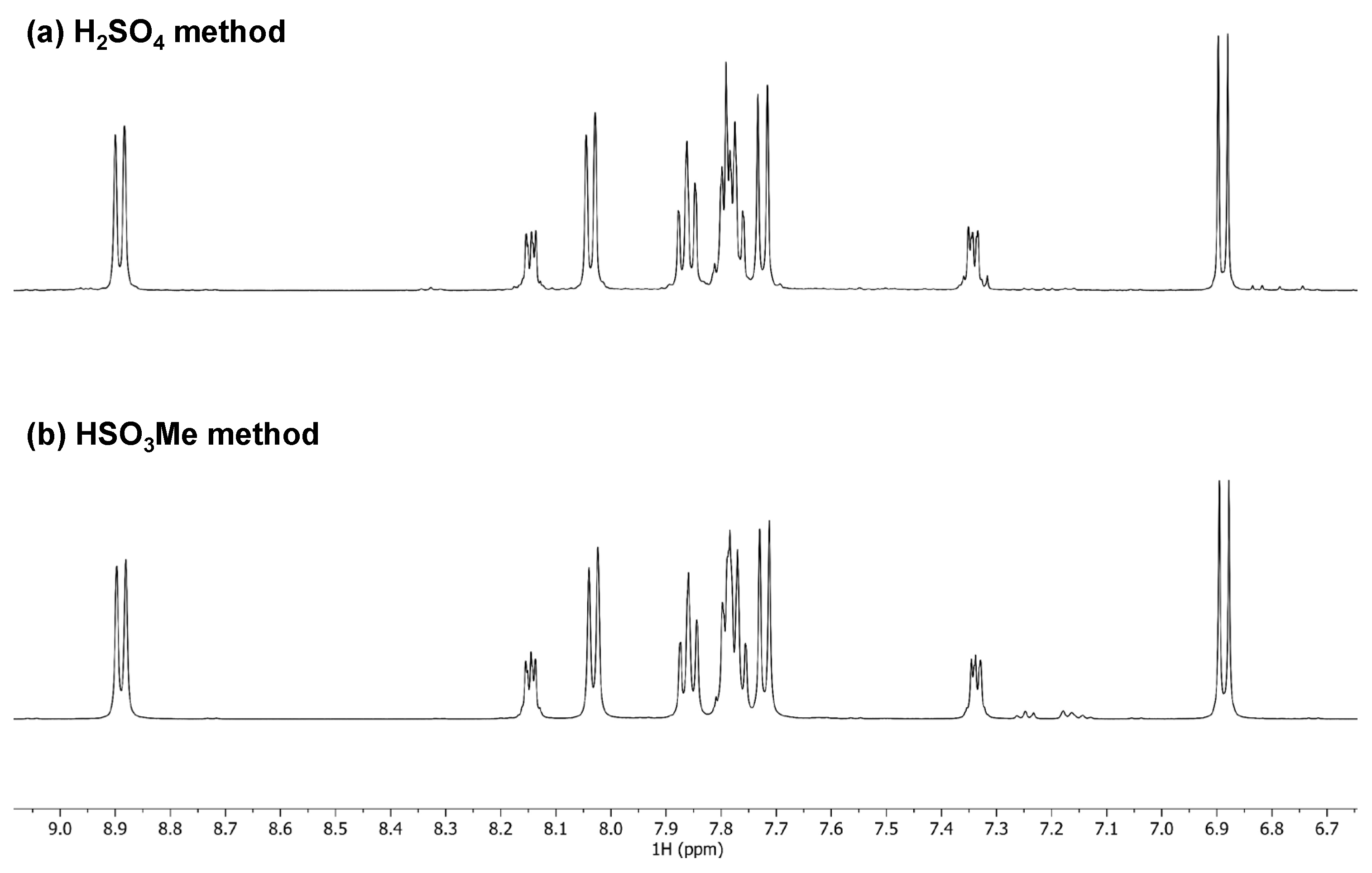
2.1. Synthesis and Spectroscopy

The reaction of 1-naphthol with phthalic anhydride in hot methanesulfonic acid or hot 95% sulfuric acid (90 °C) proceeds over 3 h. The crude product was then easily isolated by filtration after the reaction was quenched with water. The crude material was obtained as a tan-coloured solid; however, this contained unreacted 1-naphthol. The removal of unreacted 1-naphthol was achieved by mixing the crude product in hot ethanol and filtering off the remaining solid. After treatment with ethanol, the product was then purified by recrystallization from hot toluene. A total of 1–2 mg of crude product was removed from the reaction mixture and added to a 10 mL aliquot of 0.5 M sodium hydroxide to check whether any naphtholphthalein was present. In each case, the distinctive blue/green colour of naphtholphthalein was not observed when crude material was treated with aqueous base. These observations align with those of Grabowski, Werner, and Copisarow [3,4,5].
Most previous reports of fluoran 1 were published in the late 19th century and early 20th century, and in this period, modern spectroscopic and advanced X-ray crystallography techniques did not yet exist. As part of this study, the 1H NMR spectra of samples of fluoran 1 prepared by Werner’s method and our own were acquired and compared. An overlay of the 500 MHz 1H NMR spectrum is presented (Figure 1) and shows that the products from both reactions have the same structure. A full assignment of the structure was also made using 2D NMR techniques, and a full set of assigned spectra is provided in the Supporting Information (Supporting Information Figures S1–S5). The ATR-IR spectrum of fluoran 1 was also obtained (Supporting Information Figure S6), and this shows a distinctive signal at 1752.5 cm−1 that is consistent with the γ-lactone carbonyl group within the isobenzofuranone moiety [7]. The melting point was determined to be 302–303 °C, and this observation is very similar to those of earlier studies [4,6].

2.2. The Crystal Structure of 1

Crystals of 1 suitable for X-ray diffraction were obtained by the slow cooling of a solution of 1 in toluene. The data confirm that the structure of 1 is in agreement with the spectroscopic results.
The structure (Figure 2) shows only slight distortion from ideal tetrahedral angles at the spiro carbon (C3), ranging from 101.3(3)° in the constrained isobenzofuranone ring (C4−C3−O2) to 114.7(3)° (C4-C3-O10), which are comparable to the observed angles (101.6(8) and 112.7(9)°, respectively) of the known dihydroxy-aldehyde analogue 4 (CSD code MUMPUH) [8]. The C−O bond lengths in the xanthene moiety are of typical length, 1.370(5) and 1.383(4) Å, and the C=O distance is typical of isobenzofuranones at 1.209(4) Å. The xanthene and isobenzofuranone rings are linked via the spiro carbon (C3). Unlike in the structure of analogue 4, where the xanthene adopts a planar conformation (angle between xanthene benzo-rings, 1.1(4)°), the xanthene group of compound 1 adopts a bowed conformation (angle between xanthene benzo-rings, 15.06(14)°). The interplanar angle between the xanthene and isobenzofuranone rings is 87.72(6)°, slightly less than that of dihydroxy-aldehyde analogue 4 (89.2(4)°).
The structure displays several non-classical intermolecular CH···O hydrogen bonds. Adjacent isobenzofuranone moieties form weak reciprocal CH···O hydrogen bonds from an aromatic C8-H8 to the carbonyl O1 (H···O 2.559(3) Å, CH···O 170.2(2)°) in an R 2 2 10 fashion, forming intermolecular dimers. These dimers link into chains (Figure 3) propagating down [100] through additional bifurcated non-classical CH···O hydrogen bonds from C6-H6 to both O1 and O2 (H···O 2.695(3) and 2.675(3) Å, CH···O 125.5(2) and 151.0(3)°, respectively). Dimers of molecules are also formed through weak π-stacking (Figure 4) (centroid··· centroid 3.757(3) Å). These extend into chains down [100] through very weak π-stacking (centroid··· centroid 3.995(3) Å). A similar motif is seen in dihydroxy-aldehyde analogue 4 with alternating stronger (centroid··· centroid 3.587(7) Å) and weaker (centroid··· centroid 3.949(6) Å) interactions, both at slightly shorter distances [8]. The cupping of xanthene results in the furanone moieties, of the π-stacked molecules, being pulled closer together (O1···O1 11.905(4) Å; 13.340(16) Å in compound 4) and displaying less horizonal slip than is observed in dihydroxy-aldehyde analogue 4 (Figure 4) (furanone···furanone offset 0.053(16) and 1.214(16) Å; 0.85(5) and 1.87(5) Å in compound 4). The combination of the non-classical hydrogen bonded chains and weak π-stacking expand the chains down [100] into sheets across (011). Further non-classical hydrogen bonds from the C15 and C16 of the xanthene moiety to the carbonyl O1 (H···O 2.644(3) and 2.517(2) Å, CH···O 142.8(3) and 161.7(3), respectively) link sheets into the overall 3D structure.
In summary, the first X-ray crystal structure of 3′H-spiro[dibenzo[c,h]xanthene-7,1′-isobenzofuran]-3′-one (1) was obtained, and full 1H and 13C NMR assignments were made. The title compound was prepared by heating a mixture of phthalic anhydride and 1-naphthol in methanesulfonic acid or 95% sulfuric acid. This study confirms the structure of “Grabowski’s anhydride” as being fluoran 1, aligning with the proposal made by Copisarow in the early 20th century.

3. Experimental Section

Melting points were recorded on a Stuart SMP3 melting point apparatus (tolerance ±1.5 °C at 300 °C) (Mettler Toledo Ltd., Leicester, UK). The melting point apparatus was calibrated using samples of potassium nitrate (mp 333–334 °C). IR spectra were recorded on a Nicolet Summit FTIR instrument with an Everest diamond ATR accessory (Fisher Scientific, Loughborough, UK). NMR spectra were obtained for 1H at 500.13 MHz and for 13C at 125.77 MHz using a Bruker AVIII_HD 500 instrument (Bruker UK Ltd., Coventry, UK). Spectra were run at 25 °C in DMSO-d6. Chemical shifts are reported in ppm with respect to the reference, and coupling constants J are reported in Hz. The HRMS data were acquired from the University of St Andrews Mass Spectrometry Service.

3.1. 3′H-spiro[dibenzo[c,h]xanthene-7,1′-isobenzofuran]-3′-one (1)

3.1.1. Method 1—Methanesulfonic Acid

A stirred mixture of 1-naphthol (0.48 g, 3.3 mmol) and phthalic anhydride (0.30 g, 2.0 mmol) in methanesulfonic acid (1.00 g) was heated to 90 °C in a boiling tube for 3 h. Upon cooling to room temperature, water (20 mL) was added, and the resulting mixture was agitated and scratched with a glass rod until the crude product was precipitated as a tan-coloured solid. The crude product was filtered off under suction, washed with water (2 × 5 mL), and then stirred in boiling ethanol for 15 min. The solid material was recovered by filtration under suction and recrystallized from toluene. The crystalline product was filtered off under suction and washed with ice-cold toluene (2 × 2 mL) to afford product 1 (0.31 g, 47%).

3.1.2. Method 2—95% Sulfuric Acid

A stirred mixture of 1-naphthol (0.48 g, 3.3 mmol) and phthalic anhydride (0.30 g, 2.0 mmol) in 95% sulfuric acid (1.00 g) was heated to 90 °C in a boiling tube for 3 h. Upon cooling to room temperature, water (20 mL) was added, and the resulting mixture was triturated with a glass rod until a tan-coloured solid was formed. The crude product was filtered off under suction, washed with water (2 × 5 mL), and then stirred in boiling ethanol for 15 min. The solid material was recovered by filtration under suction and recrystallized from toluene. The crystalline product was filtered off under suction and washed with ice-cold toluene (2 × 2 mL) to afford product 1 (0.15 g, 23%).
mp 302–303 °C (lit. [4] 300 °C). IR (ATR) 3051.4, 2987.9 (ArCH), 1752.5 (C=O), 1567.8, 1374.8, 1096.6 cm−1; 1H NMR (500 MHz, DMSO-d6); 8.90 (2H, d, J = 8.3 Hz, ArH), 8.14–8.15 (1H, m, ArH), 8.03 (2H, d, J = 8.1 Hz, ArH), 7.85–7.88 (2H, m, ArH), 7.76–7.80 (4H, m, ArH), 7.72 (2H, d, J = 8.7 Hz, ArH), 7.33–7.35 (1H, m, ArH), 6.89 (2H, d, J = 8.7 Hz, ArH); 13C NMR (126 MHz, d6-DMSO); 169.3 (C=O), 153.8 (ArCq), 146.1 (ArCq), 136.5 (ArCH), 134.4 (ArCq), 131.1 (ArCH), 128.8 (ArCH), 128.4 (ArCH), 127.9 (ArCH), 125.9 (ArCq), 125.5 (ArCH), 124.8 (ArCH), 124.1 (ArCH), 123.8 (ArCq), 122.5 (ArCH), 112.8 (ArCq), 82.9 (Cq). HRMS (ESI+) m/z (%) Calcd. for C28H17O3 401.1172, found 401.1181 [M+1] (100).
Colourless X-ray-quality crystals of 1 were grown by the slow cooling of a sample dissolved in toluene.

3.2. X-Ray Structure Determination of 1

X-ray diffraction data were collected at 100 K using a Rigaku MM-007HF High Brilliance Microfocus RA generator/confocal optics with an XtaLAB P200 diffractometer [Cu Kα radiation (λ = 1.54187 Å)]. Data were collected (using a calculated strategy) and processed (including correction for Lorentz, polarization, and absorption) using CrysAlisPro [9]. The structure was solved by dual-space methods (SHELXT) [10] and refined by full-matrix least-squares against F2 (SHELXL-2019/3) [11]. Non-hydrogen atoms were refined anisotropically, and hydrogen atoms were refined using a riding model. All calculations were performed using the Olex2 interface [12].
Crystal data for C28H16O3, M = 400.41 g mol−1, colourless needle, crystal dimensions 0.18 × 0.02 × 0.02 mm, triclinic, a = 7.8473(3), b = 10.7485(7), c = 11.3596(7) Å, α = 89.856(5), β = 83.900(4), γ = 75.109(4) °, U = 937.54(9) Å3, T = 100 K, space group P 1 ¯ (no. 2), Z = 2, 15936 reflections measured, 3715 unique (Rint = 0.0924), which were used in all calculations. The final R1 [I > 2σ (I)] was 0.0924, and wR2 (all data) was 0.2801. Data were deposited at the Cambridge Crystallographic Data Centre as CCDC 2453093. The data can be obtained free of charge from the Cambridge Crystallographic Data Centre via http://www.ccdc.cam.ac.uk/getstructures (accessed on 2 July 2025).

Supplementary Materials

The following are available online. Figure S1: 1H-NMR spectrum of 1; Figure S2: 13C DEPTQ NMR spectrum of 1; Figure S3: 1H-1H COSY 2D H-H correlation NMR spectrum of 1; Figure S4: 1H-13C HSQC 2D H-C correlation NMR spectrum of 1; Figure S5: 1H-13C HMBC 2D H-C correlation NMR spectrum of 1; Figure S6: IR spectrum of 1; Figure S7: ESI mass spectrum of 1; CIF file for compound 1.

Author Contributions

Synthetic steps, crystallization trials, and preliminary analysis were conducted by N.V., I.A.S. and I.L.J.P.; A.P.M. and D.B.C. collected the X-ray data and solved the structure; B.A.C., D.B.C., A.P.M. and I.A.S. wrote this paper. All authors have read and agreed to the published version of the manuscript.

Funding

This research received no external funding.

Data Availability Statement

CCDC 2453093 contains the supplementary crystallographic data for this paper. These data can be obtained free of charge from The Cambridge Crystallographic Data Centre via www.ccdc.cam.ac.uk/structures (accessed on 2 July 2025).

Acknowledgments

The authors express gratitude to the University of St Andrews School of Chemistry for allowing the authors to use their laboratory facilities and for the provision of materials.

Conflicts of Interest

The authors declare no conflicts of interest.

References

  1. Sabnis, R.W. A facile synthesis of phthalein dyes. Tetrahedron Lett. 2009, 50, 6261–6263. [Google Scholar] [CrossRef]
  2. Sabnis, R.W. Developments in the chemistry and applications of phthalein dyes. Part 1: Industrial applications. Color. Technol. 2018, 134, 187–205. [Google Scholar] [CrossRef]
  3. Werner, E.A. III-The Preparation of α-Naphtholphthalein. J. Chem. Soc. Trans. 1918, 113, 20–21. [Google Scholar] [CrossRef]
  4. Copisarow, M. XXVI-Phthaleins and Fluorans. J. Chem. Soc. Trans. 1920, 117, 209–218. [Google Scholar] [CrossRef]
  5. Baeyer, A. Ueber die Phenolfarbstoffe. Ber. Dtsch. Chem. Ges. 1871, 4, 658–665. [Google Scholar] [CrossRef]
  6. Copisarow, M.; Weizmann, C. XCVII-Phthalides of the Benzene, Naphthalene and Carbazole Series. Part 1. J. Chem. Soc. Trans. 1915, 107, 878–886. [Google Scholar] [CrossRef]
  7. Jones, R.N.; Gallagher, B.S. The infrared spectra of steroid lactones. J. Am. Chem. Soc. 1959, 81, 5242–5251. [Google Scholar] [CrossRef]
  8. Xu, X.; Strongin, R.M.; Fronczek, R.F. CCDC 1404764; CSD Communication: Omagh, UK, 2015. [Google Scholar] [CrossRef]
  9. CrysAlisPro v1.171.42.94a & 109a; Rigaku Oxford Diffraction, Rigaku Corporation: Oxford, UK, 2024.
  10. Sheldrick, G.M. SHELXT–Integrated space-group and crystal structure determination. Acta Crystallogr. Sect. A Found. Adv. 2015, 71, 3–8. [Google Scholar] [CrossRef] [PubMed]
  11. Sheldrick, G.M. Crystal structure refinement with SHELXL. Acta Crystallogr. Sect. C Struct. Chem. 2015, 71, 3–8. [Google Scholar] [CrossRef] [PubMed]
  12. Dolomanov, O.V.; Bourhis, L.J.; Gildea, R.J.; Howard, J.A.K.; Puschmann, H. OLEX2: A complete structure solution, refinement and analysis program. J. Appl. Crystallogr. 2009, 42, 339–341. [Google Scholar] [CrossRef]
Scheme 1. Reaction of phthalic anhydride with 1-naphthol mixed with ZnCl2 and heated for 3 h.
Molbank 2025 m2033 sch001
Scheme 2. Reaction of phthalic anhydride 1-naphthol to initially form compound naphtholphthalein 3 and subsequent loss of water to afford fluoran 1.
Molbank 2025 m2033 sch002
Scheme 3. Reaction of phthalic anhydride with 1-naphthol mixed with hot 95% sulfuric acid for 4 h.
Molbank 2025 m2033 sch003
Scheme 4. Reaction of phthalic anhydride with 1-naphthol in methanesulfonic acid to form fluoran 1.
Molbank 2025 m2033 sch004
Figure 1. Comparison of 500 MHz 1H NMR spectra of samples of fluoran 1 prepared in (a) 95% sulfuric acid and (b) methanesulfonic acid.
Molbank 2025 m2033 g001
Figure 2. The molecular structure of 1. The anisotropic displacement ellipsoids are displayed at the 50% probability level. Oxygen atoms are depicted in red.
Molbank 2025 m2033 g002
Figure 3. A view of the structure of 1 showing non-classical hydrogen-bonded chains running along the crystallographic a-axis. Oxygen atoms are depicted in red.
Molbank 2025 m2033 g003
Figure 4. A view of the π-stacking dimers in 1 (left) and 4 (right) which further stack into chains running along the crystallographic a-axis. Oxygen atoms are depicted in red.
Molbank 2025 m2033 g004
Disclaimer/Publisher’s Note: The statements, opinions and data contained in all publications are solely those of the individual author(s) and contributor(s) and not of MDPI and/or the editor(s). MDPI and/or the editor(s) disclaim responsibility for any injury to people or property resulting from any ideas, methods, instructions or products referred to in the content.

