Communication

Polymorphism of an Nα-Aroyl-N-Aryl-Phenylalanine Amide: An X-ray and Electron Diffraction Study

1
Institut für Pharmazie, Martin-Luther-Universität Halle-Wittenberg, Wolfgang-Langenbeck-Straße 4, 06120 Halle, Germany
2
Max-Planck-Institut für Kohlenforschung, Kaiser-Wilhelm-Platz 1, 45470 Mülheim an der Ruhr, Germany
3
Center for Discovery and Innovation, Hackensack Meridian Health, 111 Ideation Way, Nutley, NJ 07110, USA
4
Department of Medical Sciences, Hackensack Meridian School of Medicine, 123 Metro Blvd, Nutley, NJ 07110, USA
5
Department of Microbiology and Immunology, Georgetown University, 3900 Reservoir Road N.W., Washington, DC 20007, USA
*
Author to whom correspondence should be addressed.
Molbank 2024, 2024(3), M1851; https://doi.org/10.3390/M1851
Submission received: 30 April 2024 / Revised: 9 July 2024 / Accepted: 12 July 2024 / Published: 17 July 2024
(This article belongs to the Section Structure Determination)

Abstract

:
In view of the rise of drug-resistant tuberculosis and difficult-to-treat related diseases caused by non-tuberculous mycobacteria, there is an urgent need for antimycobacterial drug discovery. Nα-aroyl-N-aryl-phenylalanine amides (AAPs) have been identified as antimycobacterial agents and are subject to lead optimization. The aim of the present study is to evaluate the impact of N-aryl ortho cyano substitution in a lead compound on the crystal and molecular structure and its in vitro activity against Mycobacterium abscessus. The title AAP can be conveniently synthesized from N-Boc-protected d-phenylalanine in two amide coupling steps using a previously established racemization-free method. Two polymorphic forms in the solid-state are described, as discovered by X-ray and electron diffraction. The introduction of a cyano group in the ortho position of the AAP N-aryl ring, however, leads to loss of in vitro activity against M. abscessus subsp. abscessus.

1. Introduction

Mycobacterial infections are a serious public health threat of global concern. They can manifest as pulmonary or extrapulmonary tuberculosis (TB) [1,2], localized or disseminated non-tuberculous mycobacterial (NTM) diseases [3], leprosy and chronic ulcers (Buruli ulcer) [4]. The rise of drug-resistant TB [5,6] and hard-to-treat NTM pulmonary disease [7] necessitate drug discovery and development efforts in this field [8]. Nα-Aroyl-N-aryl-phenylalanine amides (AAPs) have been identified as antimycobacterial agents that specifically and stereoselectively inhibit the mycobacterial RNA polymerase with a low probability of cross-resistance to rifamycins [9]. Their mechanism of action was unveiled in a seminal work by Lin et al. [10]. In vitro activity of compound 1 against Mycobacterium tuberculosis, the pathogen causing TB, and the clinically relevant NTM species Mycobacterium avium and Mycobacterium abscessus has been discovered in several phenotypic compound screenings [11,12,13,14,15]. Hit-to-lead optimization driven by whole-cell activity against M. abscessus subsp. abscessus resulted in lead compound 2 (Figure 1) [16]. While retaining the phenylalanine side chain proved beneficial, replacement of the 2-thiophenoyl moiety in 1 by a 2-fluorobenzoyl moiety and the morpholine ring by thiomorpholine 1,1-dioxide lowered the minimum inhibitory concentration (MIC) against M. abscessus subsp. abscessus by about an order of magnitude to the submicromolar level. The most recent work on AAPs focused on improving their metabolic stability [17]. In the present contribution, we disclose the influence of a cyano group in ortho position on the AAP N-aryl ring instead of the morpholino group, while retaining the phenylalanine side chain the 2-fluorobenzoyl moiety as in the lead compound 2.

2. Results and Discussion

2.1. Synthesis

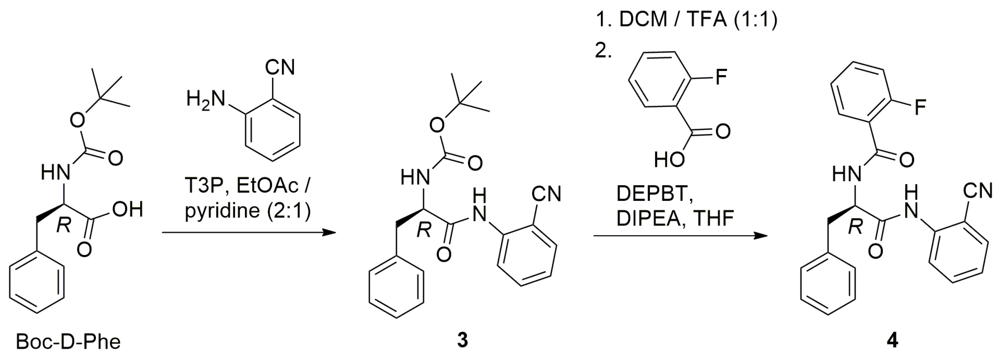
Compound 4 was obtained in good yield by applying a previously published racemization-free method [16], shown in Scheme 1. In brief, commercially available N-tert-butyloxycarbonyl (Boc) d-phenylalanine (Boc-d-Phe) was reacted with 2-aminobenzonitrile (2-cyanoaniline) in ethyl acetate to form anilide 3, using T3P® (propanephosphonic acid anhydride) [18] as coupling reagent in the presence of pyridine [19]. T3P® is known to cause a low degree of racemization of racemization-prone carboxylic acids [20], and the presence of the N-bound carbamate group further helps prevent racemization in the first amide coupling step [21]. After removal of the Boc protecting group, the 2-fluorobenzoyl moiety was introduced in the second amide coupling step using 3-(diethoxyphosphoryloxy)-1,2,3-benzotriazin-4(3H)-one (DEPBT) [22,23] as a coupling reagent in the presence of Hünig’s base (N,N-diisopropylethylamine, DIPEA). For a brief discussion of advantages and disadvantages of DEPBT in the synthesis of AAPs, we refer the interested reader to a recent review [9]. Compound 4 was identified by 1H and 13C-APT NMR spectroscopy, FT-IR spectroscopy and high-resolution mass spectrometry (see Supplementary Materials).

2.2. Structural Elucidation

Crystallization of 4 from ethanol afforded fine needle-shaped crystals, hereafter referred to as 4-I. X-ray crystallography at 100 K unambiguously confirmed the molecular structure (Figure 2a). Both amide groups adopt the Z conformation. The molecular conformation of the solid form 4-I is nonetheless distinctly different from that of 2 (CSD refcode: KODROO) [17] and related thiomorpholinoanilide AAP derivatives [16], whose crystal structures feature an intramolecular N–H···O hydrogen bond between the amide groups and intermolecular N–H···O hydrogen bonds to form pseudo centrosymmetric R 2 2 ( 10 ) hydrogen-bonded dimers [9]. In contrast, the solid-state structure of 4-I lacks a comparable intramolecular N–H···O hydrogen bond. Instead of forming dimers, the molecules in the crystal structure of 4-I form polymeric stacks through two N–H···O classical hydrogen bonds with an R 2 2 ( 12 ) motif [24], assisted by C–H···N weak hydrogen bonds (Figure 2b). These stacks extend by translational symmetry in the crystallographic a-axis direction (Figure 2b). Table 1 lists the corresponding hydrogen bond parameters. The packing index of 4-I is 72.0% [25]. Determination of the unit cell parameters of 4-I from the single-crystal at room temperature indicated that there was no phase change upon cooling to 100 K (Section 3.3).
To shed light on the crystal and molecular structure of 4 in the powder as synthesized, hereafter 4-II, we performed electron diffraction on a crystallite. Figure 3a shows the molecular structure in 4-II. As in 4-I, both amide groups exhibit the Z conformation. In contrast to 4-I, the anilide group forms an intramolecular N–H···O hydrogen bond to the benzoyl oxygen atom (O2) with an S(7) motif, as previously observed in the crystal structures of 2 and related AAPs [9]. Nonetheless, hydrogen-bonded dimers, as in 2 and related AAPs, are not encountered in 4-II. Instead, 4-II features N–H···O hydrogen-bonded chains formed by a single N–H···O hydrogen bond formed between the Nα N2–H2A moiety and the benzoyl oxygen atom (O2). The hydrogen-bonded chains so formed extend by translational symmetry in the a-axis direction (Figure 3b). Table 2 lists the corresponding hydrogen bond parameters. Notably, the carbonyl oxygen atom of the anilide group (O1) and the nitrile nitrogen atom (N3) do not act as hydrogen bond acceptors. The packing index calculated for 4-II is 70.2%, which suggests that the crystal packing in 4-II is slightly less dense than in 4-I. The calculated densities of 4-I and 4-II at room temperature (see Section 3.3 and Section 3.4) are similar. A powder X-ray diffraction (PXRD) analysis of the bulk material of 4 as synthesized suggests that the sample did not exclusively consist of 4-II (see Supplementary Materials).

2.3. Hirshfeld Surface Analysis and Density Functional Theory (DFT) Study

In order to compare the crystal structures of 4-I and 4-II and to gain insight into the environments of the molecules in the two polymorphs, we generated and visualized the Hirshfeld surfaces, which represent the shortest distances between molecules in a crystal and their neighbors [26]. Figure 4 shows the Hirshfeld surface mapped with the normalized contact distance (dnorm) for the molecules in 4-I and 4-II and their fingerprint plots, which are a summary of the corresponding interatomic distances. A red color in the dnorm plot indicates contacts shorter than, white equal to and blue longer than the sum of the van der Waals radii of the nearest atoms to a point on the Hirshfeld surface.
The N–H···O and C–H···N intermolecular hydrogen bond donor and acceptor sites, as described in Section 2.2, show up as red areas. Minor red areas resulting from a C–H···F contact in 4-I and a C–H···O contact in 4-II are also visible. Inspection of the Hirshfeld surface mapped with dnorm also confirmed that the carbonyl oxygen atom O1 in 4-II remains without a short contact resulting from hydrogen bonding. This may be regarded as an exception to Etter’s first general hydrogen bond rule for organic compounds, which states that all good proton donors and acceptors are used in hydrogen bonding [27].
The corresponding fingerprint plots (Figure 4b,d) reveal distinct differences between the two polymorphs. Both plots show the spikes indicative of the N–H···O hydrogen bonds. The shortest hydrogen bond distance (di plus de from the tips of the spikes) is shorter in 4-I than in 4-II (cf. Table 1 and Table 2). Wings characteristic of C···H contacts, resulting from C–H···π interactions, are observed for both structures. A triangular feature on the diagonal, typical of C···C contacts from π···π stacking, is not pronounced in either 4-I or 4-II. Both structures have in common a feature characteristic of H···H contacts from close packing. In contrast to 4-II, the fingerprint plot for 4-I (Figure 4b) exhibits small spikes resulting from a weak C–H···N hydrogen bond associated with the nitrile nitrogen atom (see Section 2.2) and a feature resulting from short C–F···H contacts. The asymmetry in the fingerprint plot for 4-II suggests packing inefficiency [28], which appears to be consistent with a lower packing index and the lower calculated density of 4-II at room temperature compared with 4-I at 100 K (see Section 2.2).
To shed light on the preferred molecular conformation of the free molecule of 4, we performed DFT calculations. Figure 5 shows a structure overlay plot of the molecular structures in the polymorphs 4-I and 4-II and the minimum energy structure from the DFT structure optimization. The results show that the conformation of the DFT-optimized free molecule is similar to the molecular structure encountered in 4-II and features a similar intramolecular N–H···O hydrogen bond. The absence of a comparable intramolecular N–H···O hydrogen bond in form 4-I is presumably a result of the favorable formation of additional N–H···O and C–H···N intermolecular hydrogen bonds and close packing in this polymorph.

2.4. Antimycobacterial Evaluation

We subjected compound 4 to antimicrobial susceptibility testing against M. abscessus subsp. abscessus using the broth microdilution method (Middlebrook 7H9 medium supplemented with 10% albumin-dextrose-saline). Up to a compound concentration of 100 µM, we did not observe growth inhibition of the reference strain M. abscessus ATCC 19977. Likewise, the compound proved inactive (MIC > 50 µM) against M. abscessus Bamboo, a clinical isolate [29].

3. Materials and Methods

3.1. General

All chemicals were purchased and used as received. Solvents were distilled before use. HPLC analysis was conducted on a Shimadzu instrument equipped with two LC-10AD pumps, an SPD-M10A VP photodiode array detector, and a Waters XTerra® RP18 column (3.5 μm, 3.9 mm × 100 mm), using gradient elution with methanol/water containing 0.05% trifluoroacetic acid. NMR spectra were recorded on an Agilent Technologies VNMRS 400 MHz NMR spectrometer. Chemical shifts are reported relative to the residual solvent signal of chloroform-dH = 7.26 ppm, δC = 77.23 ppm). Abbreviations: s = singlet, d = doublet, dd = doublet of doublets, m = multiplet, td = triplet of doublets, and qd = quartet of doublets. The IR spectrum was recorded on a Bruker Alpha FT-IR spectrometer with a resolution of 4 cm−1, using the ATR technique; a total of 16 scans were accumulated. The APCI mass spectrum was measured on an Advion Expression compact mass spectrometer. The HRMS analysis was performed on a Thermo Scientific Q ExactiveTM Plus Orbitrap mass spectrometer using methanol as a solvent.

3.2. Synthesis

(R)-Boc-2-amino-N-(2-cyanophenyl)-3-phenylpropanamide (3): The synthesis of 3 was carried out essentially following “General Procedure A” in ref. [16] Boc-d-Phe (1.24 g, 4.66 mmol) and 2-aminobenzonitrile (0.50 g, 4.23 mmol) were dissolved in a mixture of ethyl acetate (10 mL) and pyridine (5 mL). After cooling to −20 °C, T3P® (2.69 g, 8.46 mmol) was added with stirring. The reaction mixture was then allowed to slowly warm up to room temperature and stirred for 48 h. Subsequently, ca. 25 mL of ethylacetate was added, and the mixture was washed three times each with an equal volume of a 0.25 M aqueous KH2PO4 solution. The organic phase was then dried over anhydrous Na2SO4, and the solvent was removed under reduced pressure. The product was purified by flash chromatography (silica gel, heptane/ethyl acetate gradient). Yield: 1.32 g (3.61 mmol, 85%). 1H NMR (400 MHz, chloroform-d) δ 8.59–8.31 (m, 2H), 7.63–7.51 (m, 2H), 7.36–7.21 (m, 6H), 7.17 (td, J = 7.6, 1.1 Hz, 1H), 4.94 (d, J = 7.2 Hz, 1H), 4.62–4.46 (m, 1H), 3.25 (dd, J = 14.0, 6.2 Hz, 1H), 3.16 (dd, J = 14.1, 7.6 Hz, 1H), 1.43 (s, 9H). MS (APCI+) m/z 366.2 [M + H]+, 310.1 [M − C4H8 + H]+, and 266.0 [M − Boc + H]+ 266.0.
(R)-N-(1-((2-cyanophenyl)amino)-1-oxo-3-phenylpropan-2-yl)-2-fluorobenzamide (4): The synthesis of 4 was performed essentially following “General Procedure B” in ref. [16] Compound 3 (1.32 g, 3.61 mmol) was dissolved in 20 mL dichloromethane, and trifluoroacetic acid (20 mL) was added with stirring. The reaction mixture was stirred for 1 h at room temperature, and subsequently, dichloromethane/trifluoroacetic was removed under reduced pressure. The residue was taken up with ca. 30 mL of ethyl acetate, and the solution was washed three times with an equal volume of saturated aqueous NaHCO3 solution. The organic phase was dried over anhydrous Na2SO4, and the solvent was removed with a rotary evaporator to obtain unprotected (R)-2-amino-N-(2-cyanophenyl)-3-phenylpropanamide in virtually quantitative yield (0.96 g, 3.60 mmol), which was used without further purification.
An amount of 88 mg (0.33 mmol) of the residue was dissolved in 5 mL of THF, and 2-fluorobenzoic acid (51 mg, 0.36 mmol) and DEPBT (298 mg, 1.00 mmol) were added with stirring. After the addition of DIPEA (0.17 mL, 1.00 mmol), the mixture was stirred for 4 h at room temperature. Subsequently, ca. 25 mL of brine was added, and the mixture was extracted thrice with an equal volume of ethyl acetate. The combined organic phases were washed each once with 0.25 M aqueous KH2PO4 solution, water, saturated aqueous NaHCO3, water and, finally, brine. After drying over anhydrous Na2SO4, the solvent was removed under reduced pressure. The product was purified by flash chromatography twice (silica gel, dichloromethane containing 1% of a 4% methanolic ammonia solution, followed by silica gel, heptane/ethyl acetate gradient) to yield 4 as an off-white solid (98 mg, 0.25 mmol, 76%). Purity: >98% (HPLC analysis at 254 nm). 1H NMR (400 MHz, chloroform-d) δ 8.53 (s, 1H), 8.34 (dd, J = 8.5, 1.0 Hz, 1H), 8.12 (td, J = 7.9, 1.9 Hz, 1H), 7.61–7.44 (m, 3H), 7.37–7.22 (m, 7H), 7.20–6.97 (m, 2H), 5.08 (qd, J = 6.9, 1.9 Hz, 1H), and 3.40–3.27 (m, 2H) ppm. 13C APT NMR (101 MHz, chloroform-d) δ 169.5, 164.0 (d, 4JC, F = 3 Hz), 160.8 (d, J = 248 Hz), 139.8, 135.8, 134.0 (d, 3JC, F = 10 Hz), 133.9, 132.4, 132.3 (d, 4JC, F = 2 Hz), 129.2, 129.0, 127.4, 124.9 (d, 4JC, F = 3 Hz), 124.5, 121.5, 119.7 (d, 3JC, F = 11 Hz), 116.1 (d, 2JC, F = 25 Hz), 115.8, 103.0, 56.1, and 37.3 ppm. FT-IR (ATR): ν 3326 (C–H), 2218 (C≡N), and 1699 (C=O) cm−1. HRMS (ESI): m/z calcd. for C23H18O2N3NaF+, 410.127524, found 410.127360 [M + Na]+.

3.3. X-ray Crystallography

Very fine needles of 4-I suitable for single-crystal X-ray diffraction were grown from a solution in ethanol. The X-ray diffraction data were measured on a Bruker AXS D8 Venture diffractometer, equipped with an Incoatec IµS Diamond microfocus X-ray source, Incoatec multilayer optics and a Photon III detector. The data collection was controlled with APEX4 (Bruker AXS, Karlsruhe, Germany), and the raw diffraction data were processed with SAINT (Bruker AXS, Karlsruhe, Germany). An absorption correction, using the Gaussian method based on indexed crystal faces, was carried out with SADABS-2016/2 (Bruker AXS, Karlsruhe, Germany). The crystal structure was solved with XM 2013/2 (Bruker AXS, Karlsruhe, Germany, 2013) and initially refined using the independent atom model with SHELXL-2019/3 [30]. The crystal structure was subsequently refined using NoSpherA2 [31,32] in Olex2 [33], with the Hirshfeld partitioning of the electron density calculated using ORCA 5.0 [34] (B3LYP [35,36]/def2-TZVPP [37]). The absolute structure was inferred from the known absolute configuration of the starting material Boc-d-Phe in the synthesis. Anisotropic atomic displacement parameters were introduced for all non-hydrogen atoms. Hydrogen atoms were located in a difference Fourier map, and their positions were refined with Uiso(H) values constrained to 1.2 Ueq(C,N). For the calculation of the packing index with Platon [38], the X–H distances were neutron-normalized with Mercury [39]. Structure pictures were drawn with Diamond (Crystal Impact GbR, Bonn, Germany).
Crystal data for 4-I: C23H18FN3O2, Mr = 387.41, T = 100(2) K, λ = 0.71073 Å, monoclinic, space group P21, a = 4.9144(4), b = 16.6111(15), c = 11.6035(11) Å, β = 95.808(4)°, V = 942.37(15) Å3, Z = 2, ρcalc = 1.365 g cm−3, μcalc = 0.096 mm−1, F(000) = 404.280, crystal size = 0.165 × 0.023 × 0.022 mm, θ range = 2.15–30.50°, 31,125 reflections collected, 5661 reflections unique, Rint = 0.1263, observed reflections [I > 2σ(I)] 3372, 1 restraint, 18 constraints, 316 parameters, S = 1.0494, R1 [I > 2σ(I)] = 0.0520, wR2 = 0.1134, Δρmax = 0.5708 eÅ−3, and Δρmin = −0.5288 eÅ−3. Crystal data at 300(2) K: C23H18FN3O2, Mr = 387.41, a = 4.949(4), b = 16.80(1), c = 11.85(1) Å, β = 96.67(2)°, V = 978(1) Å3, Z = 2, ρcalc = 1.315 g cm−3.

3.4. Electron Crystallography

The crystallites taken from the material of 4-II as synthesized were topically applied to a carbon-coated 200 mesh copper grid. The electron diffraction data were measured at ambient temperature on an Eldico ED-1 electron diffractometer equipped with an Excillum LaB6 electron source, a 6-axis automatic centering goniometer, and a Dectris Quadro hybrid pixel detector. The electrons were accelerated with 160 kV potential (λ = 0.02851 Å), and the emission current was maintained at 10 µA during the data collection. The Eldix (Eldico Scientific, Alschwill, Switzerland, 2024) software was used to control the diffractometer [30]. The beam size at the crystallite was ca. 600 nm. The crystal was continuously rotated by 120° with a rotation speed of 1.044° per second. The frames were recorded at 0.522° intervals (0.5 s per frame) in the crystallographic binary file (CBF) format [40]. The raw data were processed with APEX5 and SAINT (Bruker AXS, Karlsruhe, Germany). The intensity data were not corrected for absorption. The crystal structure was solved with SHELXT [33], and an independent atom model refinement was carried out with SHELXL-2018/3 [34]. Carbon-bound hydrogen atoms were placed in geometrically calculated positions with neutron-normalized C–H distances [41] and subsequently refined with an appropriate riding model. N–H bond lengths were restrained to a target value of 1.027 Å, as derived from neutron diffraction data [41], with an estimated standard deviation of 0.02 Å. Uiso(H) values were constrained to 1.2 Ueq(C,N). The absolute configuration was verified using dynamical scattering effects [42]. Further details are given in the supplementary crystallographic data (CIF). The packing index was calculated with Platon [38]. Structure pictures were drawn with Diamond (Crystal Impact GbR, Bonn, Germany).
Crystal data for 4-II: C23H18FN3O2, Mr = 387.41, T = 298(2) K, λ = 0.02851 Å, orthorhombic, space group P212121, a = 5.21(4), b = 16.73(11), c = 22.21(15) Å, V = 1935(23) Å3, Z = 4, ρcalc = 1.330 g cm−3, F(000) = 318, crystal size = 0.6 × 0.5 × 0.5 μm, θ range = 0.088–1.020°, 8676 reflections collected, 3833 reflections unique, Rint = 0.1277, observed reflections [I > 2σ(I)] 1905, 269 parameters, 2 restraints, extinction factor x = 1617(22) whereby Fc* = kFc [1 + 0.001 x Fc2λ3/sin(2θ)]−1/4 and k is the overall scale factor, S = 0.996, R1 [I > 2σ(I)] = 0.138, wR2 = 0.369, Δρmax = 0.148 eÅ−3, and Δρmin = −0.153 eÅ−3.

3.5. Powder X-ray Diffraction

The PXRD pattern of 4 as synthesized was measured on a Stoe STADI P powder diffractometer (Debye–Scherrer geometry) at room temperature using a 0.7 mm borosilicate capillary in a moving PSD fixed-omega scan mode (0.005°, 30 s/step). Eight scans were collected and summed after data collection. The diffractometer was equipped with a Cu sealed-tube X-ray source (λ = 1.54060 Å) running at 40 kV, 40 mA, a curved germanium (111) monochromator, and a linear position-sensitive detector. The theoretical PXRD patterns of 4-I and 4-II were calculated from the single-crystal data using Mercury [39].

3.6. Computational Methods

Hirshfeld surface analysis was carried out with CrystalExplorer [43]. DFT calculations on the free molecule of 4 were performed starting from the molecular structure in 4-II using ORCA (version 5.0) [34] with a B3LYP/G (VWN1) hybrid functional (20% HF exchange) and a def2-TZVPP basis set with an auxiliary def2/J basis. Optimization of the structure used the BFGS method from an initial Hessian according to Almlöf’s model with a very tight self-consistent field convergence threshold. The optimized local minimum-energy structures exhibited only positive modes. The structure overlay in Figure 5 was generated with Mercury [39]. Cartesian coordinates of the DFT-optimized structure of 4 can be found in the Supplementary Materials.

3.7. Microbiological Assays

Susceptibility testing against M. abscessus ATCC 19977 transformed with plasmid pTEC27) to express the tomato red fluorescent protein (RFP) using a dual readout (optical density and fluorescence) [14], and M. abscessus Bamboo was performed as described in detail in a recent publication [16]. The clinical isolate M. abscessus Bamboo was provided by the Taichung Veterans General Hospital (Taichung, Taiwan).

4. Conclusions

The title compound, 4, can be readily synthesized from commercially available Boc-protected d-phenylalanine in a two-step synthesis. Two polymorphs, 4-I and 4-II, were discovered. Single-crystal X-ray diffraction revealed the crystal structure of form 4-I, whereas electron diffraction on a crystallite taken from 4 as synthesized unveiled the solid-state structure of form 4-II. Replacement of the thiomorpholine 1,1-dioxide moiety on the N-aryl ring in the lead compound 2 by an ortho cyano group, however, leads to complete loss of whole-cell activity against M. abscessus subsp. abscessus.

Supplementary Materials

Figure S1: 1H NMR spectrum of 3 in chloroform-d; Figure S2: APCI+ mass spectrum of 3; Figure S3: HPLC analysis of 4; Figure S4: 1H NMR spectrum of 4 in chloroform-d; Figure S5: 13C APT NMR spectrum of 4 in chloroform-d; Figure S6: ATR-FT-IR spectrum of 4; Figure S7: HRMS(ESI+) spectrum of 4; Figure S8: PXRD analysis of 4. DFT-calculated structure of 4 in MOL file format.

Author Contributions

Conceptualization, M.L., R.G., A.R. and R.W.S.; methodology, M.L., R.G., M.P., U.S.G. and R.W.S.; validation, M.L., R.G., M.P., U.S.G. and R.W.S.; formal analysis, M.L., R.G., M.P., U.S.G. and R.W.S.; investigation, M.L., R.G., M.P., U.S.G. and A.R.; resources, A.R. and T.D.; data curation, M.L., R.G., M.P., U.S.G. and A.R.; writing—original draft preparation, R.W.S.; writing—review and editing, M.L., R.G., M.P., U.S.G., T.D. and A.R.; visualization, R.G. and R.W.S.; supervision, A.R. and T.D.; project administration, A.R. and R.W.S.; funding acquisition, A.R. All authors have read and agreed to the published version of the manuscript.

Funding

This research was funded by the Deutsche Forschungsgemeinschaft (DFG, German Research Foundation), project number 432291016 (to A.R.) and the Mukoviszidose Institut gGmbH (Bonn, Germany) research and development arm of the German Cystic Fibrosis Association Mukoviszidose e.V., project number 2202 (to A.R.).

Data Availability Statement

CCDC 2352084-2352085 contains the supplementary crystallographic data for this paper. The data can be obtained free of charge from the Cambridge Crystallographic Data Centre via www.ccdc.cam.ac.uk/structures. The Cartesian coordinates for the DFT-calculated structure of 4 can be found in the Supplementary Materials.

Acknowledgments

We would like to thank Nadine Jänckel, Nadine Taudte, and Jens-Ulrich Rahfeld for providing and maintaining the biosafety level 2 facility, Nadine Hußmann for measuring the mass spectrum of 4, Lucas Schulte-Zweckel for technical assistance with the X-ray and electron diffraction experiments, and Jan Ternieden for performing the PXRD analysis. Christian W. Lehmann is gratefully acknowledged for providing access to the X-ray and electron diffraction facility, and Wei Chang Huang for providing M. abscessus Bamboo.

Conflicts of Interest

The authors declare no conflicts of interest. The funders had no role in the design of the study, in the collection, analyses, or interpretation of data, in the writing of the manuscript, or in the decision to publish the results.

References

  1. Alsayed, S.S.R.; Gunosewoyo, H. Tuberculosis: Pathogenesis, Current Treatment Regimens and New Drug Targets. Int. J. Mol. Sci. 2023, 24, 5202. [Google Scholar] [CrossRef] [PubMed]
  2. Sharma, A.; De Rosa, M.; Singla, N.; Singh, G.; Barnwal, R.P.; Pandey, A. Tuberculosis: An Overview of the Immunogenic Response, Disease Progression, and Medicinal Chemistry Efforts in the Last Decade toward the Development of Potential Drugs for Extensively Drug-Resistant Tuberculosis Strains. J. Med. Chem. 2021, 64, 4359–4395. [Google Scholar] [CrossRef] [PubMed]
  3. Johansen, M.D.; Herrmann, J.L.; Kremer, L. Non-tuberculous mycobacteria and the rise of Mycobacterium abscessus. Nat. Rev. Microbiol. 2020, 18, 392–407. [Google Scholar] [CrossRef] [PubMed]
  4. Shyam, M.; Kumar, S.; Singh, V. Unlocking Opportunities for Mycobacterium leprae and Mycobacterium ulcerans. ACS Infect. Dis. 2024, 10, 251–269. [Google Scholar] [CrossRef] [PubMed]
  5. Farhat, M.; Cox, H.; Ghanem, M.; Denkinger, C.M.; Rodrigues, C.; Abd El Aziz, M.S.; Enkh-Amgalan, H.; Vambe, D.; Ugarte-Gil, C.; Furin, J.; et al. Drug-resistant tuberculosis: A persistent global health concern. Nat. Rev. Microbiol. 2024. [Google Scholar] [CrossRef] [PubMed]
  6. Dheda, K.; Mirzayev, F.; Cirillo, D.M.; Udwadia, Z.; Dooley, K.E.; Chang, K.C.; Omar, S.V.; Reuter, A.; Perumal, T.; Horsburgh, C.R., Jr.; et al. Multidrug-resistant tuberculosis. Nat. Rev. Dis. Primers 2024, 10, 22. [Google Scholar] [CrossRef] [PubMed]
  7. Prevots, D.R.; Marshall, J.E.; Wagner, D.; Morimoto, K. Global Epidemiology of Nontuberculous Mycobacterial Pulmonary Disease: A Review. Clin. Chest Med. 2023, 44, 675–721. [Google Scholar] [CrossRef] [PubMed]
  8. Dartois, V.; Dick, T. Therapeutic developments for tuberculosis and nontuberculous mycobacterial lung disease. Nat. Rev. Drug Discov. 2024, 23, 381–403. [Google Scholar] [CrossRef] [PubMed]
  9. Seidel, R.W.; Goddard, R.; Lang, M.; Richter, A. Nα-aroyl-N-aryl-phenylalanine amides: A promising class of antimycobacterial agents targeting the RNA polymerase. Chem. Biodivers. 2024, 21, e202400267. [Google Scholar] [CrossRef]
  10. Lin, W.; Mandal, S.; Degen, D.; Liu, Y.; Ebright, Y.W.; Li, S.; Feng, Y.; Zhang, Y.; Mandal, S.; Jiang, Y.; et al. Structural Basis of Mycobacterium tuberculosis Transcription and Transcription Inhibition. Mol. Cell 2017, 66, 169–179. [Google Scholar] [CrossRef]
  11. Ballell, L.; Bates, R.H.; Young, R.J.; Alvarez-Gomez, D.; Alvarez-Ruiz, E.; Barroso, V.; Blanco, D.; Crespo, B.; Escribano, J.; Gonzalez, R.; et al. Fueling open-source drug discovery: 177 small-molecule leads against tuberculosis. ChemMedChem 2013, 8, 313–321. [Google Scholar] [CrossRef] [PubMed]
  12. Low, J.L.; Wu, M.-L.; Aziz, D.B.; Laleu, B.; Dick, T. Screening of TB Actives for Activity against Nontuberculous Mycobacteria Delivers High Hit Rates. Front. Microbiol. 2017, 8, 1539. [Google Scholar] [CrossRef] [PubMed]
  13. Jeong, J.; Kim, G.; Moon, C.; Kim, H.J.; Kim, T.H.; Jang, J. Pathogen Box screening for hit identification against Mycobacterium abscessus. PLoS ONE 2018, 13, e0195595. [Google Scholar] [CrossRef] [PubMed]
  14. Richter, A.; Strauch, A.; Chao, J.; Ko, M.; Av-Gay, Y. Screening of Preselected Libraries Targeting Mycobacterium abscessus for Drug Discovery. Antimicrob. Agents Chemother. 2018, 62, e00828-18. [Google Scholar] [CrossRef] [PubMed]
  15. Richter, A.; Shapira, T.; Av-Gay, Y. THP-1 and Dictyostelium Infection Models for Screening and Characterization of Anti-Mycobacterium abscessus Hit Compounds. Antimicrob. Agents Chemother. 2019, 64, e01601-19. [Google Scholar] [CrossRef] [PubMed]
  16. Lang, M.; Ganapathy, U.S.; Mann, L.; Abdelaziz, R.; Seidel, R.W.; Goddard, R.; Sequenzia, I.; Hoenke, S.; Schulze, P.; Aragaw, W.W.; et al. Synthesis and Characterization of Phenylalanine Amides Active against Mycobacterium abscessus and Other Mycobacteria. J. Med. Chem. 2023, 66, 5079–5098. [Google Scholar] [CrossRef]
  17. Lang, M.; Ganapathy, U.S.; Mann, L.; Seidel, R.W.; Goddard, R.; Erdmann, F.; Dick, T.; Richter, A. Synthesis and in vitro Metabolic Stability of Sterically Shielded Antimycobacterial Phenylalanine Amides. ChemMedChem 2024, 19, e202300593. [Google Scholar] [CrossRef] [PubMed]
  18. Wissmann, H.; Kleiner, H.J. New Peptide Synthesis. Angew. Chem.-Int. Ed. Engl. 1980, 19, 133–134. [Google Scholar] [CrossRef]
  19. Dunetz, J.R.; Xiang, Y.; Baldwin, A.; Ringling, J. General and Scalable Amide Bond Formation with Epimerization-Prone Substrates Using T3P and Pyridine. Org. Lett. 2011, 13, 5048–5051. [Google Scholar] [CrossRef]
  20. Waghmare, A.A.; Hindupur, R.M.; Pati, H.N. Propylphosphonic anhydride (T3P®): An expedient reagent for organic synthesis. Rev. J. Chem. 2014, 4, 53–131. [Google Scholar] [CrossRef]
  21. Waring, A.; Joullie, M.M.; Lassen, K.M. Evolution of amide bond formation. Arkivoc 2010, 2010, 189–250. [Google Scholar] [CrossRef]
  22. Ye, Y.H.; Li, H.; Jiang, X. DEPBT as an efficient coupling reagent for amide bond formation with remarkable resistance to racemization. Biopolymers 2005, 80, 172–178. [Google Scholar] [CrossRef]
  23. Li, H.; Jiang, X.; Ye, Y.-h.; Fan, C.; Romoff, T.; Goodman, M. 3-(Diethoxyphosphoryloxy)-1,2,3- benzotriazin-4(3H)-one (DEPBT):  A New Coupling Reagent with Remarkable Resistance to Racemization. Org. Lett. 1999, 1, 91–94. [Google Scholar] [CrossRef]
  24. Bernstein, J.; Davis, R.E.; Shimoni, L.; Chang, N.L. Patterns in Hydrogen Bonding: Functionality and Graph Set Analysis in Crystals. Angew. Chem. Int. Ed. 1995, 34, 1555–1573. [Google Scholar] [CrossRef]
  25. Kitajgorodskij, A.I. Molecular Crystals and Molecules; Academic Press: New York, NY, USA, 1973. [Google Scholar]
  26. Spackman, M.A.; Jayatilaka, D. Hirshfeld surface analysis. CrystEngComm 2009, 11, 19–32. [Google Scholar] [CrossRef]
  27. Etter, M.C. Encoding and decoding hydrogen-bond patterns of organic-compounds. Acc. Chem. Res. 1990, 23, 120–126. [Google Scholar] [CrossRef]
  28. Spackman, M.A.; McKinnon, J.J. Fingerprinting intermolecular interactions in molecular crystals. CrystEngComm 2002, 4, 378–392. [Google Scholar] [CrossRef]
  29. Yee, M.; Klinzing, D.; Wei, J.R.; Gengenbacher, M.; Rubin, E.J.; Dick, T. Draft Genome Sequence of Mycobacterium abscessus Bamboo. Genome Announc. 2017, 5, e00388-17. [Google Scholar] [CrossRef] [PubMed]
  30. Sheldrick, G.M. Crystal structure refinement with SHELXL. Acta Crystallogr. C 2015, 71, 3–8. [Google Scholar] [CrossRef]
  31. Kleemiss, F.; Dolomanov, O.V.; Bodensteiner, M.; Peyerimhoff, N.; Midgley, L.; Bourhis, L.J.; Genoni, A.; Malaspina, L.A.; Jayatilaka, D.; Spencer, J.L.; et al. Accurate crystal structures and chemical properties from NoSpherA2. Chem. Sci. 2021, 12, 1675–1692. [Google Scholar] [CrossRef]
  32. Midgley, L.; Bourhis, L.J.; Dolomanov, O.V.; Grabowsky, S.; Kleemiss, F.; Puschmann, H.; Peyerimhoff, N. Vanishing of the atomic form factor derivatives in non-spherical structural refinement—A key approximation scrutinized in the case of Hirshfeld atom refinement. Acta Crystallogr. A 2021, 77, 519–533. [Google Scholar] [CrossRef] [PubMed]
  33. Bourhis, L.J.; Dolomanov, O.V.; Gildea, R.J.; Howard, J.A.K.; Puschmann, H. The anatomy of a comprehensive constrained, restrained refinement program for the modern computing environment—Olex2 dissected. Acta Crystallogr. A 2015, 71, 59–75. [Google Scholar] [CrossRef] [PubMed]
  34. Neese, F.; Wennmohs, F.; Becker, U.; Riplinger, C. The ORCA quantum chemistry program package. J. Chem. Phys. 2020, 152, 224108. [Google Scholar] [CrossRef] [PubMed]
  35. Becke, A.D. Density-functional thermochemistry. III. The role of exact exchange. J. Chem. Phys. 1993, 98, 5648–5652. [Google Scholar] [CrossRef]
  36. Lee, C.; Yang, W.; Parr, R.G. Development of the Colle-Salvetti correlation-energy formula into a functional of the electron density. Phys. Rev. B 1988, 37, 785–789. [Google Scholar] [CrossRef] [PubMed]
  37. Weigend, F. Accurate Coulomb-fitting basis sets for H to Rn. Phys. Chem. Chem. Phys. 2006, 8, 1057–1065. [Google Scholar] [CrossRef]
  38. Spek, A.L. Structure validation in chemical crystallography. Acta Crystallogr. D 2009, 65, 148–155. [Google Scholar] [CrossRef]
  39. Macrae, C.F.; Sovago, I.; Cottrell, S.J.; Galek, P.T.A.; McCabe, P.; Pidcock, E.; Platings, M.; Shields, G.P.; Stevens, J.S.; Towler, M.; et al. Mercury 4.0: From visualization to analysis, design and prediction. J. Appl. Crystallogr. 2020, 53, 226–235. [Google Scholar] [CrossRef]
  40. Bernstein, H.J.; Hammersley, A.P. Specification of the Crystallographic Binary File (CBF/imgCIF). In International Tables for Crystallography Volume G: Definition and Exchange of Crystallographic Data; Hall, S.R., McMahon, B., Eds.; Springer: Dordrecht, The Netherlands, 2006; Chapter 2.3; pp. 37–43. [Google Scholar] [CrossRef]
  41. Allen, F.H.; Bruno, I.J. Bond lengths in organic and metal-organic compounds revisited: X-H bond lengths from neutron diffraction data. Acta Crystallogr. B 2010, 66, 380–386. [Google Scholar] [CrossRef]
  42. Klar, P.B.; Krysiak, Y.; Xu, H.; Steciuk, G.; Cho, J.; Zou, X.; Palatinus, L. Accurate structure models and absolute configuration determination using dynamical effects in continuous-rotation 3D electron diffraction data. Nat. Chem. 2023, 15, 848–855. [Google Scholar] [CrossRef]
  43. Spackman, P.R.; Turner, M.J.; McKinnon, J.J.; Wolff, S.K.; Grimwood, D.J.; Jayatilaka, D.; Spackman, M.A. CrystalExplorer: A program for Hirshfeld surface analysis, visualization and quantitative analysis of molecular crystals. J. Appl. Crystallogr. 2021, 54, 1006–1011. [Google Scholar] [CrossRef] [PubMed]
Figure 1. Screening hit 1 and derived lead compound 2 and the corresponding MICs against M. tuberculosis H37Rv and M. abscessus ATCC 19977 [17].
Molbank 2024 m1851 g001
Scheme 1. Two-step synthesis of compound 4 from Boc-d-Phe.
Molbank 2024 m1851 sch001
Figure 2. (a) Molecular structure in crystal form 4-I. Displacement ellipsoids are drawn at the 50% probability level. Hydrogen atoms are represented by small spheres of arbitrary radius. (b) Part of the crystal structure of 4-I, viewed along the [011] direction. Dashed lines represent hydrogen bonds. Hydrogen atoms not involved in hydrogen bonds are omitted for clarity. Symmetry codes: (a) x − 1, y, z; (b) x + 1, y, z. Color scheme: C, gray; H, white; N, blue; O, red; F, lime.
Molbank 2024 m1851 g002
Figure 3. (a) Molecular structure in crystal form 4-II. Displacement ellipsoids are drawn at the 50% probability level. Hydrogen atoms are represented by small spheres of arbitrary radius. (b) Part of the crystal structure of 4-II, approximately viewed along the b-axis direction. Dashed lines represent hydrogen bonds. Hydrogen atoms not involved in hydrogen bonds are omitted for clarity. Symmetry code: (a) x − 1, y, z. Color scheme: C, gray; H, white; N, blue; O, red; F, lime.
Molbank 2024 m1851 g003
Figure 4. (a) Hirshfeld surface mapped with dnorm and (b) the corresponding fingerprint plot for 4-I. (c) Hirshfeld surface mapped with dnorm and (d) the corresponding fingerprint plot for 4-II. di and de are the distances from a point on the Hirshfeld surface to the nearest internal and external atom, respectively.
Molbank 2024 m1851 g004
Figure 5. Overlay plot of the molecular structures of 4-I (orange), 4-II (green), and the DFT-optimized structure of the free molecule (pink). The dashed line represents the intramolecular N–H···O hydrogen bond. The structures are each superimposed at C2 and its three adjacent non-hydrogen atoms. Hydrogen atoms are omitted for clarity, except for H1 and H2.
Molbank 2024 m1851 g005
Table 1. Hydrogen bond parameters in 4-I (Å, °).
d(D–H)d(H···A)d(D···A)<(DHA)
N1–H1···O1a0.99 (3)2.05 (3)2.935 (3)146 (2)
N2–H2A···O2b0.98 (3)1.80 (3)2.777 (3)169 (2)
C16–H16···N3b1.10 (3)2.42 (3)3.491 (3)165 (2)
Symmetry codes: (a) x − 1, y, z; (b) x + 1, y, z.
Table 2. Hydrogen bond parameters in 4-II (Å, °).
d(D–H)d(H···A)d(D···A)<(DHA)
N1–H1···O21.03 (3)2.14 (5)2.98 (2)137 (4)
N2–H2A···O2a1.02 (3)2.10 (3)3.06 (2)156 (4)
Symmetry code: (a) x − 1, y, z.
Disclaimer/Publisher’s Note: The statements, opinions and data contained in all publications are solely those of the individual author(s) and contributor(s) and not of MDPI and/or the editor(s). MDPI and/or the editor(s) disclaim responsibility for any injury to people or property resulting from any ideas, methods, instructions or products referred to in the content.