Short Note

3-(2-Diisopropylaminoethyl)-5-(4-methoxybenzylidene)thiazolidine-2,4-dione

1
Department of Pharmaceutical, Organic and Bioorganic Chemistry, Danylo Halytsky Lviv National Medical University, Pekarska 69, 79010 Lviv, Ukraine
2
Department of Organic Chemistry and Pharmacy, Lesya Ukrainka Volyn National University, Volya Avenue 13, 43025 Lutsk, Ukraine
3
Department of Organic Chemistry, Poznan University of Medical Sciences, Grunwaldzka 6, 60-780 Poznan, Poland
*
Author to whom correspondence should be addressed.
Molbank 2022, 2022(3), M1394; https://doi.org/10.3390/M1394
Submission received: 30 May 2022 / Revised: 17 June 2022 / Accepted: 21 June 2022 / Published: 24 June 2022

Abstract

:
Thiazolidine-2,4-dione core is widely used in the medicinal chemistry of different types of potential drug-like small molecules. In the present work, the synthesis of a novel non-condensed thiazolidine-2,4-dione-bearing derivative is reported by the two-step cost-effective approach including alkylation and Knoevenagel condensation. The structure of the synthesized 3-(2-diisopropylaminoethyl)-5-(4-methoxybenzylidene)thiazolidine-2,4-dione was determined and characterized using 1H, 13C NMR, LC-MS spectra and the X-ray diffraction method.

Graphical Abstract

1. Introduction

The current cancer chemotherapy includes the use of synthetic/semi-synthetic agents with an impact on a wide range of molecular targets; however, searching for novel chemotherapeutics is an important and topical issue—first of all, due to drug resistance [1]. Modern directions in anticancer medicinal chemistry are aiming to use small molecules that tend to be prepared following cost-effective approaches, which are at a lesser risk of degradation in vivo and are more permeable to tumor penetration [2,3,4,5,6,7].
The 3-(2-aminoethyl)-5-ene-thiazolidine-2,4-diones were reported as potential multitarget anticancer agents effective in vitro via the inhibition of the Raf/MEK/extracellular signal regulated kinase (ERK) and phosphatidylinositol 3-kinase (PI3K)/Akt signaling cascades (Figure 1) [8,9,10].
Reported in [8,9,10], hit/lead compounds were found to be promising for developing novel anticancer agents and encourage further structure optimization. It should be noted that the preparative organic chemistry of 5-ene-thiazolidine-2,4-diones is often complicated with the selection of optimal reaction conditions that affect the yield, reaction time, etc., and it is especially important for medicinal chemistry needs [11,12].
Herein, we report a synthesis of a structure-related derivative described in [8,9,10]—namely, 3-(2-diisopropylaminoethyl)-5-(4-methoxybenzylidene)thiazolidine-2,4-dione (3)—using a simplified two-stage protocol. The proposed approach follows the requirements of cost-effective methodology and could be applied for the synthesis of 3-(2-aminoethyl)-5-ene-thiazolidine-2,4-diones using commercially available (alkylamino)alkyl halides.

2. Results and Discussion

2.1. Synthesis of the Title Compound 3

The thiazolidine-2,4-dione (1) was used as a starting “building block” with the following modification of the core at the N3 (by alkylation) and C5 (by Knoevenagel condensation) positions (Scheme 1). The alkylation was performed using classic conditions [13] by a reflux of 1 with 2-diisopropylaminoethyl chloride hydrochloride during 4 h in acetone with the presence of anhydrous potassium carbonate. After completing the reaction (monitored by TLC), the synthesized 3-(2-diisopropylaminoethyl)thiazolidine-2,4-dione (2) was isolated from the reaction mixture and used without any additional purification. The Knoevenagel condensation of 2 with p-methoxybenzaldehyde was performed by reflux during 5 h in the ethanol without the addition of the catalyst, and title compound 3 was obtained with a yield of 71%. Probably, the diisopropylaminoethyl residue of molecule 2 performs the role of the basic catalyst of the mentioned reaction.
The structure of synthesized compound 3 was confirmed using 1H, 13C NMR, LC-MS spectra and X-ray analysis (copies of the spectra are presented in the Supplementary Materials). In the 1H NMR spectrum of compound 3, the characteristic signals of all the protons are presented. The protons of the methyl groups resonated as a singlet at 0.90 ppm. Two protons of the methine groups gave a septet at 2.95 ppm. The two methylene groups are characterized with two triplets at 2.58 and 3.62 ppm. The chemical shift of the methoxy group was observed as a singlet at 3.83 ppm. The aromatic protons resonated as two doublets at 7.10 and 7.59 ppm, with J = 8.3 Hz. The 5-CH = group gave a characteristic signal at 7.81 ppm, which confirmed the Z-isomerism of the arylidene moiety [14]. It is important to note that the formation of 5-Z-arylidene derivatives is typical for the Knoevenagel reaction of 4-thiazolidinones and aromatic aldehydes [5,13].
The 13C NMR spectra of 3 showed 13 carbon signals. The carbons of the four methyl groups gave a signal at 20.9 ppm. Two methylene carbons resonated at 41.6 at 42.2 ppm. Two aliphatic methine carbons gave a signal at 47.9. The carbon of the methoxy group resonated at 56.0 ppm. The carbons of the benzene ring and double bound appeared in the range from 115.5 to 161.6 ppm. The thiazolidine C=O carbon atoms resonated at 166.3 and 167.8 ppm.

2.2. X-ray Crystallographic Study of the Title Compound 3

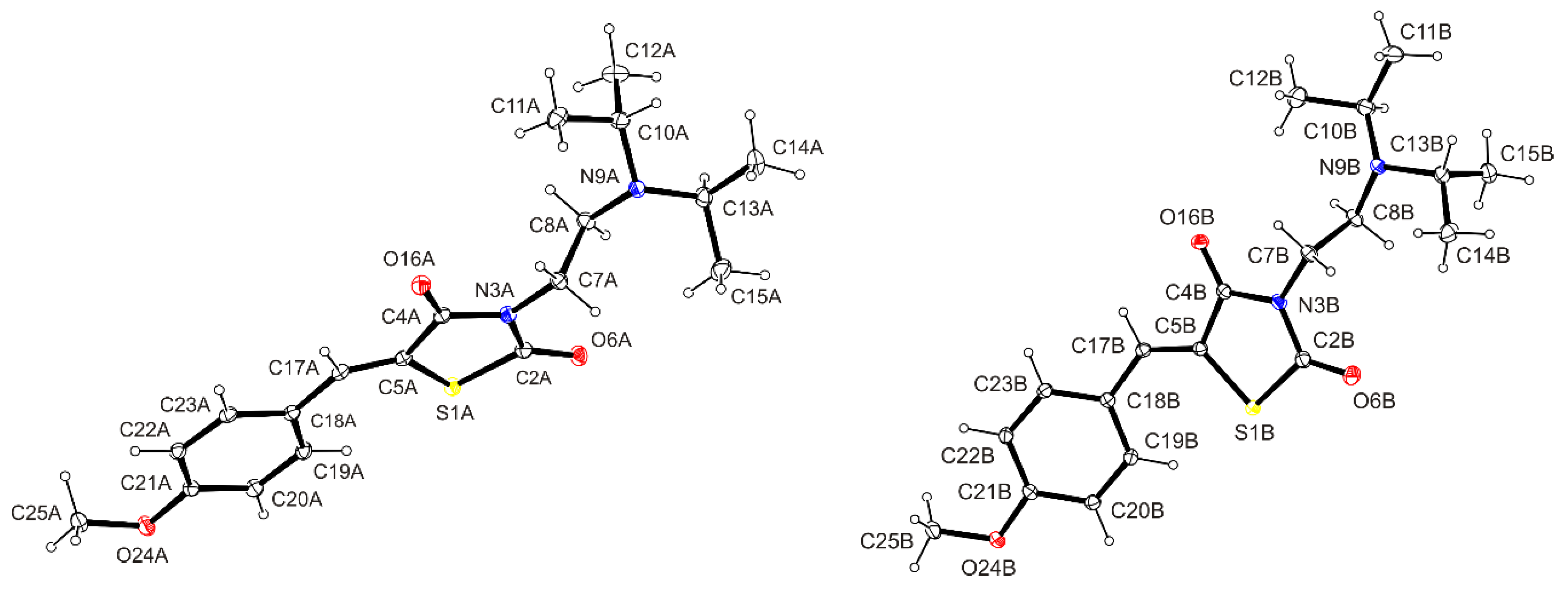
The structure of the synthesized compound 3 has been additionally confirmed by X-ray analysis (Figure 2). In the crystal structure, the asymmetric part of the unit cell was found to contain two symmetry-independent molecules, denoted as A and B, which differ to a rather moderate extent in conformation (unit weigh r.m.s.d. = 0.985 Å [15]; Figure 3). The largest conformational differences between molecules A and B correspond to the angular arrangement of the 2-diisopropylaminoethyl residue or, more precisely, its diisopropylamino fragment, although the differences were also found in the positioning of the 4-methoxyphenylmethylidene moiety, but to a much lesser extent. The spatial arrangement of diisopropylamino fragments is described with torsion angles C2–N3–C7–C8 and N3–C7–C8–N9 (molecule A: 71.04(13) and 179.92(9)°, molecule B: 87.72(13) and 173.67(9)°). The angular orientation of two isopropyl moieties in molecules A and B reveal two pairs of torsional angles—C8–N9–C10–C11/C8–N9–C10–C12 and C8–N9–C13–C14/C8–N9–C13–C15 (molecule A: −64.43(13)/62.91(12)° and −60.16(13)/179.16(10)°; molecule B: −177.54(11)/62.61(13) and −56.93(13)/69.10(13)°)—due to their rotation around the N9–C10 and N9–C13 bond, respectively. The torsional angles indicate that pairs of the bonds C8–N9/C10–C11(12) and C8–N9/C13–C14(15) in molecule An adopt a mutual synclinal (+sc, −sc) and synclinal, antiperiplanar (−sc, ap) conformation, while in molecule B, they adopt antiperiplanar, synclinal (ap, +sc) and synclinal (−sc, +sc) conformations.
The observed interatomic C5–C17 distance in molecules A and B (1.3453(16) and 1.3458(15) Å, respectively) confirms the presence of a double bond between these atoms. Simultaneously, the torsion angle S1–C5–C17–C18 (molecule A: 2.01(19)°, molecule B: −0.29(19)°) indicates a Z configuration of the 4-(methoxyphenyl)methylidene group. The dihedral angles between the mean planes of the almost flat p-methoxyphenyl and thiazolidin-2,4-dione systems are 3.63(4)° in molecule A and 19.29(4)° in molecule B due to the rotation around the C17–C18 bond.
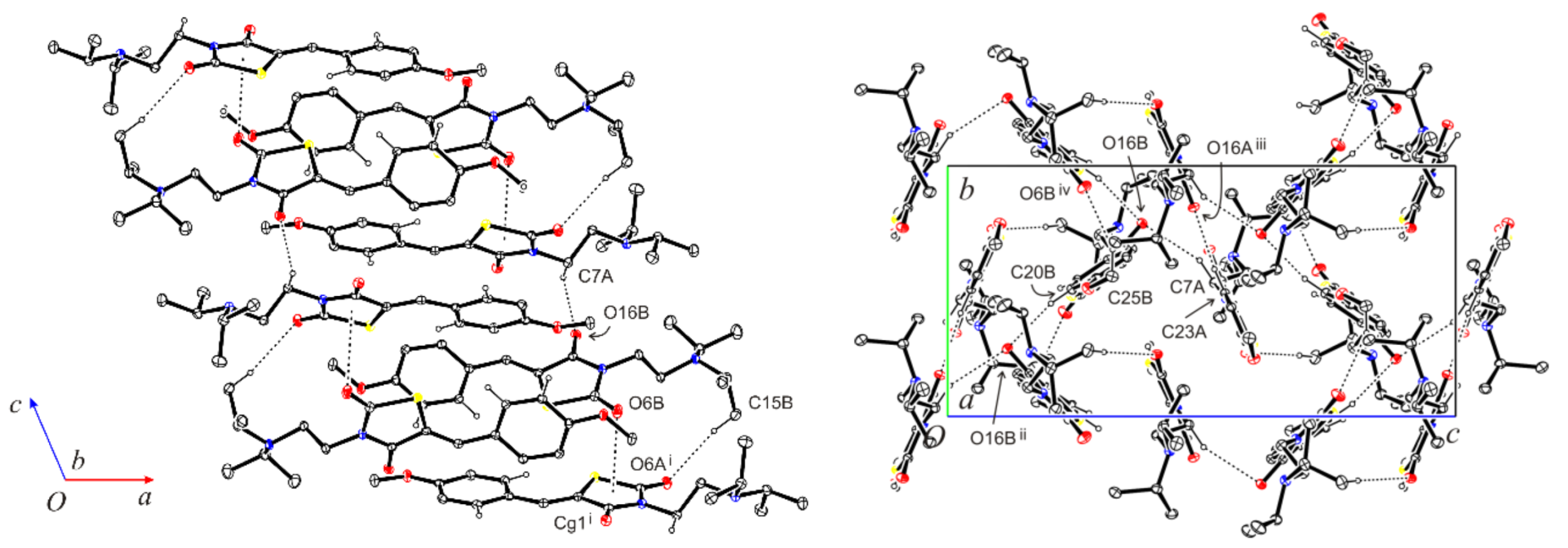
In the crystal lattice, molecules A and B are connected by the N7A–H7A∙∙∙O16B and C15B–H15D∙∙∙O6Ai hydrogen bonds and the C2B=O6B∙∙∙Cg1i intermolecular interactions into the tapes extending along the c axis. The neighboring tapes of molecules are linked through the hydrogen bonds C20B–H20B∙∙∙O16Bii, C23A–H23A∙∙∙O16Aiii and C25B–H25E∙∙∙O6Biv into layers parallel to the bc plane (Table 1, Figure 4). The layer thickness is about half of the a parameter length. Within a layer, the contacts Cg1∙∙∙Cg2v and Cg2∙∙∙Cg1v (symmetry code: (v) 1/2−x,1/2−y,1−z) between the molecules are also observed. Perpendicular distances amount to 3.3909(4) and 3.4452(5) Å, respectively (Figure 5).

3. Materials and Methods

The melting points were measured in open capillary tubes on a BÜCHI B-545 melting point apparatus (BÜCHI Labortechnik AG, Flawil, Switzerland) and were uncorrected. The elemental analyses (C, H, N) were performed using the Perkin-Elmer 2400 CHN analyzer (PerkinElmer, Waltham, MA, USA) and were within ±0.4% of the theoretical values. The 400 MHz 1H and 126 MHz 13C NMR spectra were recorded on a Varian Unity Plus 400 (400 MHz) spectrometer (Varian Inc., Paulo Alto, CA, USA). All spectra were recorded at room temperature, except where indicated otherwise, and were referenced internally to solvent reference frequencies. Chemical shifts (δ) are quoted in ppm, and coupling constants (J) are reported in Hz. The LC-MS spectra were obtained on a Finnigan MAT INCOS-50 (Thermo Finnigan LLC, San Jose, CA, USA). The reaction mixture was monitored by thin layer chromatography (TLC) using commercial glass-backed TLC plates (Merck Kieselgel 60 F254). The solvents and reagents that are commercially available were used without further purification. The starting compound 1 was prepared according to the protocol described in [16].

3.1. 3-(2-Diisopropylaminoethyl)thiazolidine-2,4-dione (2)

To a heated solution of thiazolidine-2,4-dione (1.17 g, 10 mmol) (1) and potassium hydroxide (2.76 g, 20 mmol) in acetone (20 mL), 2-diisopropylaminoethyl chloride hydrochloride (2.20 g, 11 mmol) was added slowly. The mixture was then heated to reflux for 4 h. After completion, the reaction mixture was cooled to room temperature. The organic solvent was separated from the white inorganic solid by filtration. The filtrate was evaporated to obtain the spectroscopically pure colorless liquid of 2, used in the further step without additional purification.

3.2. 3-(2-Diisopropylaminoethyl)-5-(4-methoxybenzylidene)thiazolidine-2,4-dione (3)

A mixture of compound 2 (2.7 g, 10 mmol) and 4-methoxybenzaldehyde (1.5 g, 10 mmol) in ethanol (15 mL) was heated to reflux for 5 h (monitored by TLC). After the completion of the reaction, the mixture was cooled to room temperature and the solvent was evaporated to the pure yellow liquid of 3 that was poured with distilled water. The resulting yellow solid of 3 was filtrated and recrystallized from ethanol.
Yield: 71%, yellow crystal powder, mp 83–85 °C (EtOH). 1H NMR (400 MHz, DMSO-d6, δ): 0.90 (d, J = 6.4 Hz, 12H, 4*CH3), 2.59 (t, 2H, J = 6.2 Hz, CH2), 2.95 (spt, J = 6.4 Hz, 2H, 2*CH), 3.62 (t, 2H, J = 6.2 Hz, CH2), 3.83 (s, 3H, OCH3), 7.11 (d, J = 8.3 Hz, 2H, arom.), 7.59 (d, J = 8.3 Hz, 2H, arom.), 7.88 (s, 1H = CH).
13C NMR (126 MHz, DMSO-d6, δ): 21.0, 41.7, 42.2, 48.0, 56.0, 115.5, 118.5, 125.9, 132.7, 133.1, 161.6, 166.3 (C=O), 167.8 (C=O).
LC-MS (Electrospray ionization (ESI+)): m/z 363.4 (100.0%, [M + H]+).
Anal. calc. for C19H26N2O3S: C, 62.96%; H, 7.23%; N, 7.73%. Found: C 63.10%, H 7.40%, N 8.00%.

3.3. X-ray Crystallographic Study

Crystal data. C19H26N2O3S, Mr = 362.48, monoclinic, space group C2/c, a = 44.4536(8), b = 9.1492(2), c = 20.1926(5) Å, β = 113.257(2)°, V = 7545.3(3) Å3, Z = 16 (Z’ = 2), T = 130.0(1) K.
Data collection. A pale-yellow lath crystal (EtOH) of 0.32 × 0.25 × 0.06 mm was used to record 38,844 (CuKα radiation, θmax = 76.78°) intensities on a Rigaku SuperNova Atlas diffractometer [17]. The intensity data collection employed the ω-scans mode. Accurate unit cell parameters were determined by the least-squares techniques from the θ values of 17,063 reflections, with a θ range of 4.02–76.46°. The intensity data were corrected for Lorentz, polarization and absorption effects [17]. The 7882 total unique reflections (Rint = 0.023) were used for structure determination.
Structure solution and refinement. The structure was solved by a dual-space algorithm (SHELXT) [17,18,19] and refined against F2 for all data (SHELXL) [17,18,20]. The positions of the H atoms were positioned geometrically and were refined using a riding model, with C-H: 0.98 Å (CH3), 0.99 Å (CH2), 1.00 Å (Csp3H), 0.95 Å (Csp2H) and Uiso(H) = 1.2Ueq(C) or 1.5Ueq(C) for methyl H atoms. The methyl groups were refined as rotatable rigid groups. The final refinement converged with R = 0.0319 (for 7456 data with I > 2σ(I)), wR = 0.0886 (on F2 for all data) and S = 1.077 (on F2 for all data). The largest difference peak and hole was 0.254 and −0.264 eÅ3. The molecular illustrations were drawn using ORTEP-3 for Windows [18]. The software used to prepare the material for publication were WINGX [18], OLEX [17] and PLATON [21]. The deposition number CCDC2175518 contains supplementary crystallographic data for this paper. These data can be obtained free of charge via www.ccdc.cam.ac.uk/conts/retrieving.html (or, from the Cambridge Crystallographic Data Centre, 12, Union Road, Cambridge CB2 1EZ, UK; fax: (+44)1223-336033).

4. Conclusions

In this work, a convenient and efficient two-step protocol for the synthesis of a new thiazolidine-2,4-dione-bearing molecule has been described. The compound structure was characterized and confirmed by NMR spectroscopy, LC–MS spectrometry analysis and X-ray analysis. The proposed approach allows for the avoidance of an application of a catalyst in the stage of Knoevenagel condensation and presents interest as a possible synthetic pathway for the obtention of potential biologically active small molecules with the thiazolidine-2,4-dione scaffold.

Supplementary Materials

The following are available online. Figures S1–S4: atom numbering of compound 3; 1H NMR, 13C NMR and LC–MS spectra of compound 3.

Author Contributions

Conceptualization, I.Y., S.H., A.G. and R.L.; methodology, I.Y., S.H., A.G. and R.L.; software, I.Y., S.H. and A.G.; validation, I.Y., S.H. and A.G.; investigation, I.Y., S.H. and A.G.; writing—original draft preparation, I.Y., S.H., A.G. and R.L.; writing—review and editing, I.Y., S.H., A.G. and R.L.; supervision, R.L.; project administration, R.L. All authors have read and agreed to the published version of the manuscript.

Funding

The research leading to these results has received funding from the Ministry of Healthcare of Ukraine (0121U100690) and the National Research Foundation of Ukraine (2020.02/0035).

Institutional Review Board Statement

Not applicable.

Informed Consent Statement

Not applicable.

Data Availability Statement

The data presented in this study are available in this article.

Acknowledgments

The authors would like to thank all the brave defenders of Ukraine who made the finalization of this article possible.

Conflicts of Interest

The authors declare no conflict of interest.

References

  1. Arkin, M. Protein–protein interactions and cancer: Small molecules going in for the kill. Curr. Opin. Chem. Biol. 2005, 9, 317–324. [Google Scholar] [CrossRef] [PubMed]
  2. Kue, C.S.; Kamkaew, A.; Burgess, K.; Kiew, L.V.; Chung, L.Y.; Lee, H.B. Small molecules for active targeting in cancer. Med. Res. Rev. 2016, 36, 494–575. [Google Scholar] [CrossRef] [PubMed]
  3. Kerr, W.G.; Chisholm, J.D. The next generation of immunotherapy for cancer: Small molecules could make big waves. J. Immunol. 2019, 202, 11–19. [Google Scholar] [CrossRef] [PubMed] [Green Version]
  4. Graves, D.E.; Velea, L.M. Intercalative binding of small molecules to nucleic acids. Curr. Org. Chem. 2000, 4, 915–929. [Google Scholar] [CrossRef]
  5. Holota, S.; Komykhov, S.; Sysak, S.; Gzella, A.; Cherkas, A.; Lesyk, R. Synthesis, Characterization and In Vitro Evaluation of Novel 5-Ene-thiazolo[3,2-b][1,2,4]triazole-6(5H)-ones as Possible Anticancer Agents. Molecules 2021, 26, 1162. [Google Scholar] [CrossRef]
  6. Yushyn, I.; Holota, S.; Lesyk, R. 2,2-Dichloro-N-[5-[2-[3-(4-methoxyphenyl)-5-phenyl-3,4-dihydro-2H-pyrazol-2-yl]-2-oxoethyl] sulfanyl-1,3,4-thiadiazol-2-yl]acetamide. Molbank 2022, 2022, M1328. [Google Scholar] [CrossRef]
  7. Lozynskyi, A.; Holota, S.; Yushyn, I.; Sabadakh, O.; Karpenko, O.; Novikov, V.; Lesyk, R. Synthesis and Biological Activity Evaluation of Polyfunctionalized Anthraquinonehydrazones. Lett. Drug Des. Discov. 2021, 18, 199–209. [Google Scholar] [CrossRef]
  8. Li, Q.; Al-Ayoubi, A.; Guo, T.; Zheng, H.; Sarkar, A.; Nguyen, T.; Eblen, S.T.; Grant, S.; Kellogg, G.E.; Zhang, S.H. Structure–activity relationship (SAR) studies of 3-(2-amino-ethyl)-5-(4-ethoxy-benzylidene)-thiazolidine-2,4-dione: Development of potential substrate-specific ERK1/2 inhibitors. Bioorg. Med. Chem. Lett. 2009, 19, 6042–6046. [Google Scholar] [CrossRef]
  9. Li, Q.; Wu, J.; Zheng, H.; Liu, K.; Guo, T.L.; Liu, Y.; Eblen, S.T.; Grant, S.; Zhang, S.H. Discovery of 3-(2-aminoethyl)-5-(3-phenyl-propylidene)-thiazolidine-2,4-dione as a dual inhibitor of the Raf/MEK/ERK and the PI3K/Akt signaling pathways. Bioorg. Med. Chem. Lett. 2010, 20, 4526–4530. [Google Scholar] [CrossRef]
  10. Liu, K.; Rao, W.; Parikh, H.; Li, Q.; Guo, T.L.; Grant, S.; Kellogg, G.E.; Zhang, S.H. 3,5-Disubstituted-thiazolidine-2,4-dione analogs as anticancer agents: Design, synthesis and biological characterization. Eur. J. Med.Chem. 2012, 47, 125–137. [Google Scholar] [CrossRef]
  11. Jain, V.S.; Vora, D.K.; Ramaa, C.S. Thiazolidine-2,4-diones: Progress towards multifarious applications. Bioorg. Med. Chem. 2013, 21, 1599–1620. [Google Scholar] [CrossRef] [PubMed]
  12. Asati, V.; Mahapatra, D.K.; Bharti, S.K. Thiazolidine-2,4-diones as multi-targeted scaffold in medicinal chemistry: Potential anticancer agents. Eur. J. Med. Chem. 2014, 87, 814–833. [Google Scholar] [CrossRef] [PubMed]
  13. Maccari, R.; Ottanà, R.; Curinga, C.; Vigorita, M.G.; Rakowitz, D.; Steind, T.H.; Langer, T.H. Structure–activity relationships and molecular modelling of 5-arylidene-2,4-thiazolidinediones active as aldose reductase inhibitors. Bioorg. Med. Chem. 2005, 13, 2809–2823. [Google Scholar] [CrossRef]
  14. Popov-Pergal, K.; Cekovic, Z.; Pergal, M. Condensation of 2,4-dioxotetrahydro-1,3-thiazole with aromatic aldehydes. J. Gen. Chem. USSR 1991, 61, 1958–1962. [Google Scholar]
  15. Macrae, C.F.; Sovago, I.; Cottrell, S.J.; Galek, P.T.A.; McCabe, P.; Pidcock, E.; Platings, M.; Shields, G.P.; Stevens, J.S.; Towler, M.; et al. Mercury 4.0: From visualization to analysis, design and prediction. J. Appl. Cryst. 2020, 53, 226–235. [Google Scholar] [CrossRef] [Green Version]
  16. Turkevych, N.M.; Vvedenskij, V.M.; Petlichnaya, L.P. Method of obtaining pseudothiohydantoin and thiazolidinedione-2,4. Ukr. Khim. Zh. 1961, 27, 680–681, reprinted in Chem. Abstr. 1962, 56, 73455. [Google Scholar]
  17. Dolomanov, O.V.; Bourhis, L.J.; Gildea, R.J.; Howard, J.A.K.; Puschmann, H. OLEX2: A complete structure solution, refinement and analysis program. J. Appl. Cryst. 2009, 42, 339–341. [Google Scholar] [CrossRef]
  18. Farrugia, L.J. WinGX and ORTEP for windows: An update. J. Appl. Cryst. 2012, 45, 849–854. [Google Scholar] [CrossRef]
  19. Sheldrick, G.M. SHELXT—Integrated space-group and crystal-structure determination. Acta Cryst. 2015, A71, 3–8. [Google Scholar] [CrossRef] [Green Version]
  20. Sheldrick, G.M. Crystal structure refinement with SHELXL. Acta Cryst. 2015, C71, 3–8. [Google Scholar] [CrossRef]
  21. Spek, A.L. Structure validation in chemical crystallography. Acta Cryst. 2009, 65, 148–155. [Google Scholar] [CrossRef] [PubMed]
Figure 1. Background for the present research [8,9,10].
Molbank 2022 m1394 g001
Scheme 1. Synthesis of the title compound 3. Reagents and conditions: (i)-1 (10 mmol), K2CO3 (20 mmol), 2-diisopropylaminoethyl chloride hydrochloride (11 mmol), acetone (20 mL), reflux 4 h; (ii)-2 (10 mmol), 4-methoxybenzaldehyde (10 mmol), ethanol (15 mL), reflux 5 h.
Molbank 2022 m1394 sch001
Figure 2. ORTEP view of the two symmetry-independent molecules of 3, showing the atomic labelling scheme. Non-H atoms are drawn as displacement ellipsoids at the 30% probability level. H atoms are drawn as spheres of arbitrary radii.
Molbank 2022 m1394 g002
Figure 3. Overlay of two symmetry-independent molecules—A (red) and B (blue)—by the least-squares fitting of the thiazolidin-2,4-dione systems.
Molbank 2022 m1394 g003
Figure 4. The C–H∙∙∙O hydrogen bonding and C=O∙∙∙Cg1 intermolecular interactions in the crystal structure of 3. The H atoms not involved in hydrogen bonds have been omitted for clarity; Cg1 = thiazolidin-2,4-dione system.
Molbank 2022 m1394 g004
Figure 5. π∙∙∙π Interactions between thiazolidin-2,4-dione (Cg1) and phenylene (Cg2) systems. Hydrogen atoms have been omitted for clarity. Symmetry code: (v) 1/2−x,1/2−y,1−z.
Molbank 2022 m1394 g005
Table 1. Hydrogen bonds and Y-X∙∙∙Cg interactions in the crystal structure of 3.
D—H∙∙∙AD—H (Å)H∙∙∙A (Å)D∙∙∙A (Å)D—H∙∙∙A (°)
N7A-H7A∙∙∙O16B0.992.553.4291(14)149
C15B-H15D∙∙∙O6A i0.982.603.5659(17)169
C19A-H19A∙∙∙S1A0.952.593.2963(17)132
C19B-H19B∙∙∙S1B0.952.633.3017(17)128
C20B-H20B∙∙∙O16B ii0.952.503.4307(14)166
C23A-H23A∙∙∙O16A iii0.952.433.3489(14)164
C25B-H25E∙∙∙O6B iv0.982.563.4543(16)151
Y—X∙∙∙CgY—X (Å)X∙∙∙Cg (Å)Y∙∙∙Cg (Å)Y—X∙∙∙Cg (°)
C2B=O6B∙∙∙Cg1 i1.2087(15)3.4383(10)3.7847(12)97.20(7)
Symmetry codes: (i) x,1−y,−1/2+z; (ii) 1/2−x,−1/2+y,1/2−z; (iii) 1/2−x,3/2−y,1−z; (iv) −1/2+x,1/2+y, 1/2−z; Cg1 = thiazolidin-2,4-dione system.
Publisher’s Note: MDPI stays neutral with regard to jurisdictional claims in published maps and institutional affiliations.