Short Note

2-((E)-2-((E)-4-Chloro-5-(2-((E)-5-methoxy-3,3-dimethyl-1-(3-phenylpropyl)indolin-2-ylidene) ethylidene)-1,1-dimethyl-1,2,5,6-tetrahydropyridin-1-ium-3-yl)vinyl)-5-methoxy-3,3-dimethyl-1-(3-phenylpropyl)-3H-indol-1-ium

by
Emmanuel Ramsey Buabeng
1,2 and
Maged Henary
1,2,*
1
Department of Chemistry, Georgia State University, 100 Piedmont Avenue SE, Atlanta, GA 30303, USA
2
Center for Diagnostics and Therapeutics, Georgia State University, 100 Piedmont Avenue SE, Atlanta, GA 30303, USA
*
Author to whom correspondence should be addressed.
Molbank 2021, 2021(3), M1270; https://doi.org/10.3390/M1270
Submission received: 24 May 2021 / Revised: 30 June 2021 / Accepted: 29 July 2021 / Published: 11 August 2021

Abstract

:
A heptamethine fluorophore, ERB-60, has been synthesized efficiently in four steps in a good yield. The structure of this fluorophore consists of an electron-donating group (methoxy), a hydrophobic moiety (phenylpropyl) with a rotatable bond, a quaternary ammonium fragment, and indolium rings at the terminal ends connected via polymethine chain. All these inherent chemical features fine-tuned the optical properties of the fluorophore. This compound was characterized by both 1H NMR, 13C NMR and mass spectra. The optical properties, including molar absorptivity, fluorescence, Stokes’s shift, and quantum yield, were measured in different solvents such as DMSO, DMF, MeCN, i-PrOH, MeOH, and H2O. The wavelengths of maximum absorbance of ERB-60 were found to be in the range of 745–770 nm based on the solvents used. In decreasing order, the maximum wavelength of absorbance of ERB-60 in the tested solvents was DMSO > DMF > i-PrOH > MeOH > MeCN > H2O while the decreasing order of the extinction coefficient was found to be MeCN > MeOH > DMSO > i-PrOH > H2O > DMF. ERB-60 was found to be more photostable than IR-786 iodide, a commercially available dye, and brighter than the FDA-approved dye, indocyanine green (ICG).

1. Introduction

The growing interest in the field of synthesis and optimization of bio-probes for various applications is in demand. This has prompted many organic, analytical, medicinal, computational and even physical chemists to collaborate to develop facile approaches in designing and synthesizing novel small molecules for biomedical purposes. For example, detection of DNA and proteins [1,2], fluorescence imaging of cartilage, bone, thyroid and parathyroid glands, and cancer cells [3,4,5,6], optoacoustic imaging of pancreatic cells [7], acid-base indicators, anti-tumor agents [8], and other applications in the field of biotechnology, medicine, and pharmaceuticals have been made possible through the development of small molecules as bio-probes [9,10,11,12,13].
One class of compounds that has been used in various fields for many years is cyanine dyes. Cyanine dyes consist primarily of two terminal heterocyclic rings containing nitrogen connected by a polymethine chain. These heterocyclic rings serve as electron donors and acceptors, which results in the delocalization of electrons across the polymethine chain, allowing a longer wavelength of absorption to be observed [14]. Cyanine dyes can also be categorized into mono-, tri-, penta- and heptamethine chromophores according to the number of methine chains between the two terminal heterocyclic rings [14]. The ability to easily modify the cyanine dye framework via the use of different heterocycles and N-alkylation of the ring nitrogen with various alkylating groups has permitted the tailoring of these fluorophores for various application [14]. In addition, their optical properties, solubility, targetability and toxicokinetics can effectively be fine-tuned through synthetic strategies [15].
The molecular brightness of a fluorophore is one of the most important features in fluorescence imaging [16]. In simple terms, molecular brightness is determined by the multiplication of the molar extinction coefficient and the quantum yield at a specific wavelength of the excitation light [16]. It can also be defined in broad terms as the average number of photons a fluorophore emits over a specified time [17]. Accurate determination of molecular brightness of a fluorophore is very crucial since it provides detailed information about the cells under study. Molecular brightness allows for the optimization of a fluorophore and a unique selection of such a fluorophore to obtain the highest brightness possible. It also provides a form of quality control of the fluorophore, which in turn results in achieving consistency of the imaging results [18]. In addition, molecular brightness actually defines and drastically minimizes the required dosage required for in vivo imaging. This lowers the toxicity of the imaging procedure while still maintaining a sufficient signal-to-noise ratio [18]. As such, in designing and synthesizing a fluorophore for biomedical applications, all these parameters are taken into consideration to achieve the highest brightness possible. Herein, a modified heptamethine cyanine probe with a net charge of positive two (+2) is synthesized and characterized, and the optical efficiency is analyzed. To achieve this purpose, the absorbance and fluorescence spectra were obtained in both polar protic and aprotic solvents. From these data sets, the molar extinction coefficient (ε), quantum yield (ϕ), Stoke shifts and molecular brightness values are collected. These analyzed optical properties are very beneficial for the implementation of the dye as a potential probe in the biomedical, bioanalytical or even in the pharmaceutical industry, as the dye exhibits an excellent optical property.

2. Results and Discussion

2.1. Synthesis

The synthesis of the heptamethine fluorophore with two positive charges, ERB-60, is begun by Fisher indole reaction to obtain the heterocyclic salt 1. In general, 4-methoxyphenyl hydrazine is allowed to react with 3-methyl-2-butanone in acetic acid under reflux condition while stirring vigorously for 72 h. N-alkylation of the cyclized intermediate obtained through the Fisher indole step was carried out using 3-bromophenylpropane in acetonitrile under reflux conditions for 24 h. In order to introduce the extra positive charge into the heptamethine framework, N-methyl-4-piperidone was first alkylated using methyl iodide to obtain compound 2. The final modified Vilsmeier–Haack linker 3 was obtained by a reported procedure [19]. As depicted in Scheme 1, ERB-60 was obtained by a condensation reaction consisting of heterocyclic salt 1, linker 3, and sodium acetate dissolved in acetic anhydride under reflux conditions for 5 h. The crude product obtained was purified via a coprecipitation method using a mixture of dimethyl sulfoxide and acetone to obtain ERB-60 as a green crystal in 77% yield.
As shown in Scheme 1, the chemical structure of ERB-60 consists of a rotatable bond, hydrogen bond acceptors, a quaternary ammonium moiety, a chlorine atom and a phenyl group. All these inherent features allow this compound to be useful in the field of biomedical research. For example, the introduction of the extra positive charge within the polymethine chain can improve to a greater extent the solubility of this fluorophore. Likewise, the extra positive charge of this fluorophore can permit electrostatic interaction between itself and any negative biomolecules it comes into contact with. In addition, the ability of ERB-60 to form hydrogen bonding interaction with H-bond donors via the methoxy groups can also improve its physicochemical parameters. Furthermore, various substitutions of the meso chlorine atom of ERB-60 with different nucleophiles can be achieved via the already known SNR1 mechanistic pathway [16].

2.2. Physicochemical and Optical Properties of ERB-60

The physicochemical parameters for ERB-60 were calculated using ChemAxon [4]. The logD obtained at pH = 7.4 was 6.4 while the polarizability was found to be 92.36. The molecular volume and the molecular surface area were also calculated to be 785.52 and 1214 respectively. The presence of phenylpropyl as a substituent at the ring nitrogen may account for the higher number of rotatable bonds of 13 recorded for ERB-60. In addition, the dipole moment was calculated to be 17.77 as seen in Table 1.

2.3. Optical Properties

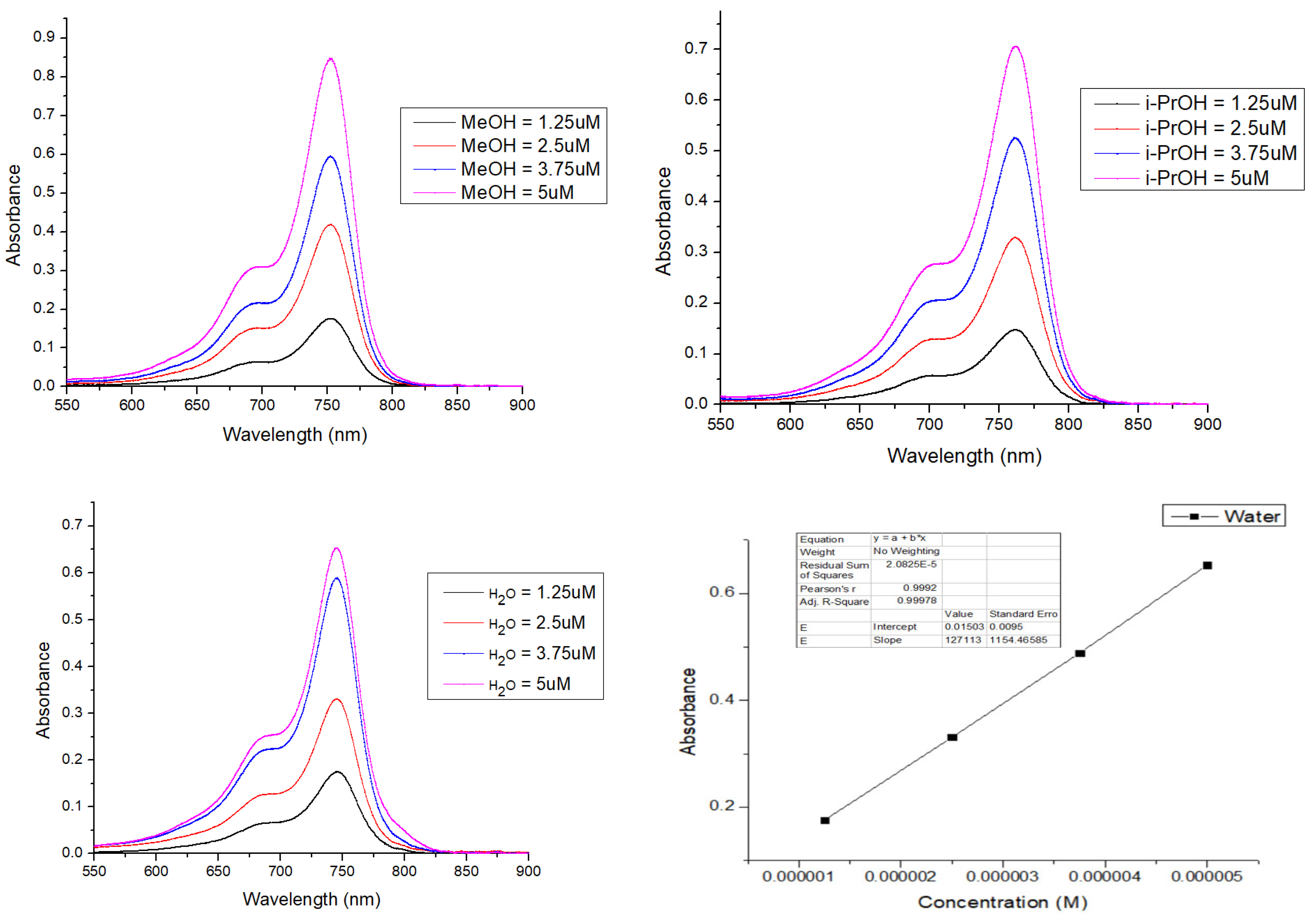
We examined the optical properties of ERB-60 in both polar protic and polar aprotic solvents. As shown in Figure 1, Figure 2 and Figure 3 and Figures S4–S6, the absorbance and emission spectra were done in methanol (MeOH), isopropanol (i-PrOH), dimethyl sulfoxides (DMSO), acetonitrile (MeCN), N,N-dimethylformamide, (DMF) and water (H2O). The absorbance profiles of ERB-60 were recorded at various concentrations (1.25 μM, 2.5 μM, 3.75 μM, 5.0 μM) and the absorbance values obtained were plotted against concentration. As observed in Figure 1, in the three polar protic solvents (water, methanol and isopropanol) tested, a linear relationship between the absorbance and the concentration was obtained as defined by the Beer–Lambert law. As shown in Table 2, the extinction coefficients calculated from the linear regressions for ERB-60 were ε = 127,113, 151,360 and 172,240 M−1 cm−1 in water, isopropanol, and methanol, respectively. From these values, it can be inferred that this fluorophore has the capacity to absorb light more in methanol followed by isopropanol. In addition, from Figure 1 and Table 2, the maximum wavelengths of absorbance for ERB-60 in water, methanol, isopropanol were found to be 745 nm, 752 nm and 761 nm respectively. ERB-60 therefore tends to exhibit a higher wavelength of absorbance in isopropanol than in a more polar solvent like water. Upon excitation at the respective wavelength of maximum absorbance, the emission wavelength of ERB-60 in water, methanol and isopropanol were found to be 761 nm, 769, and 779 nm respectively. These values correspond to a Stokes shift of 16 nm, 17 nm and 18 nm respectively. From Table 2, the fluorescence quantum yield calculated for ERB-60 was 9.8%, 24.9% and 25% and the molecular brightness was also 12, 457, 37,840, and 42, 888 and, M−1 cm−1 in water, isopropanol, and methanol, respectively. As shown in Table 2, ERB-60 therefore is brighter in methanol than in water and isopropanol but it has comparable fluorescence quantum yields as in isopropanol. We also examined the optical properties of ERB-60 in polar aprotic solvents.
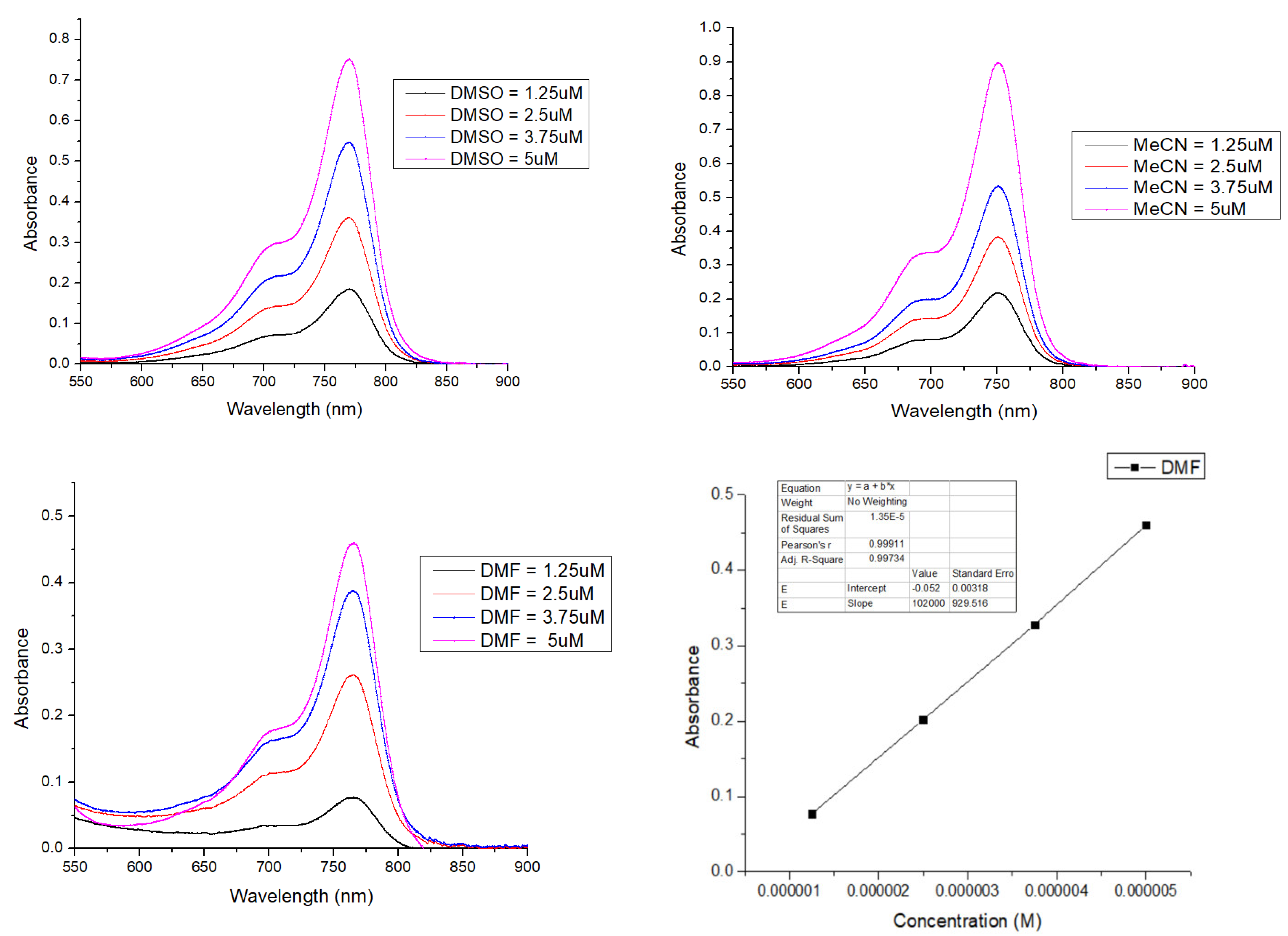
The results obtained are shown in Figure 2 and Table 2. Among the three polar aprotic solvents used, the highest wavelength of maximum absorbance was observed in dimethyl sulfoxide (DMSO). The value recorded was 770 nm. However, upon excitation at the wavelength of maximum absorbance (λmax), the emission wavelength was observed at 789 nm. The Stokes shift calculated for ERB-60 (21 nm) in acetonitrile was higher than in dimethyl sulfoxide (19 nm). Likewise, the fluorescence quantum yield (31%) and the molecular brightness (55,775 M−1 cm−1) in acetonitrile were higher than those recorded in dimethyl sulfoxide and in dimethylformamide.
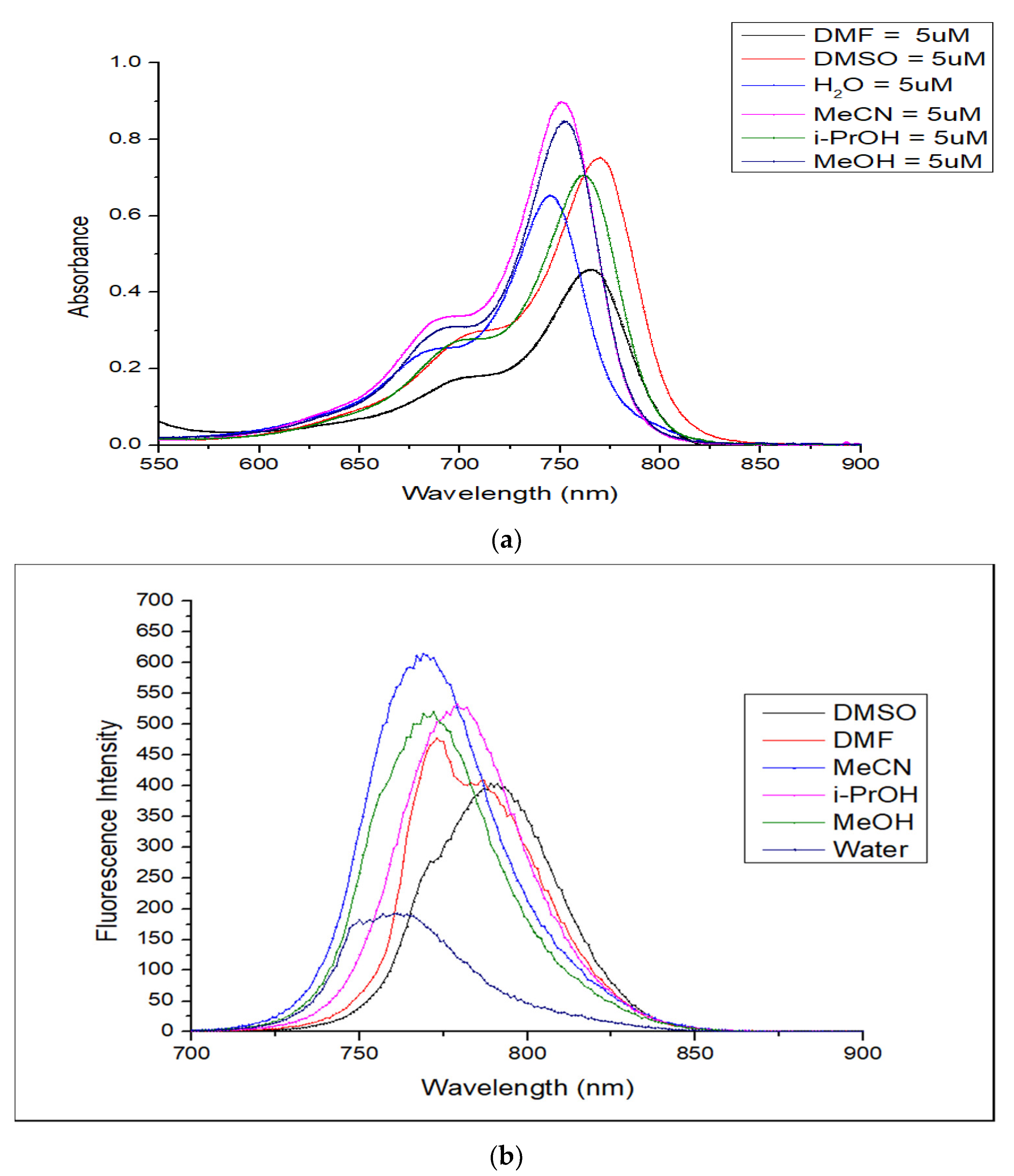
As a comparison, the absorbance and fluorescence spectra of ERB-60 were plotted, and the result is depicted in Figure 3. The decreasing order of the maximum wavelength of absorbance of ERB-60 in all tested solvents was DMSO > DMF > i-PrOH > MeOH > MeCN > H2O. The decreasing order of the extinction coefficient for ERB-60 was found to be MeCN > MeOH > DMSO > i-PrOH > H2O > DMF.
In addition, from Table 2, it can also be seen that ERB-60 exhibits higher brightness in comparison to the FDA-approved fluorophore, ICG. The molecular brightness obtained for ERB-60 in methanol was found to be 42,888 M−1 cm−1 while that of ICG was 4392 M−1 cm−1. The differences in these values might be the net two positive charge observed in ERB-60 and also the presence of the cyclohexene ring, both of which are absent in the structure of the FDA-approved heptamethine fluorophore (ICG).

2.4. Solvatochromism Properties of ERB-60

The polarity of a solvent greatly influences both the absorption and fluorescence spectra of a fluorophore. The observable shifts in the absorption maximum of most fluorophores in solvents of different polarity are partly due to the difference in the solvation energy of the ground and excited states of the solvents. In most molecular structures, the excited state of the molecule tends to be more polar than the ground state and are stabilized in more polar solvents. Such chemical compounds require only small amounts of energy for excitation, resulting in a bathochromic shift of the absorption spectra. As a result, the differences in the absorption maxima of ERB-60 observed in Figure 3 are because of the differences in the stabilization of the excited states of this molecule in the solvents tested. For example, the higher wavelength of maximum absorbance of ERB-60 in DMSO means that the excited state of this fluorophore is stabilized more in this solvent, leading to the bathochromic shift observed.

3. Photostability of ERB-60

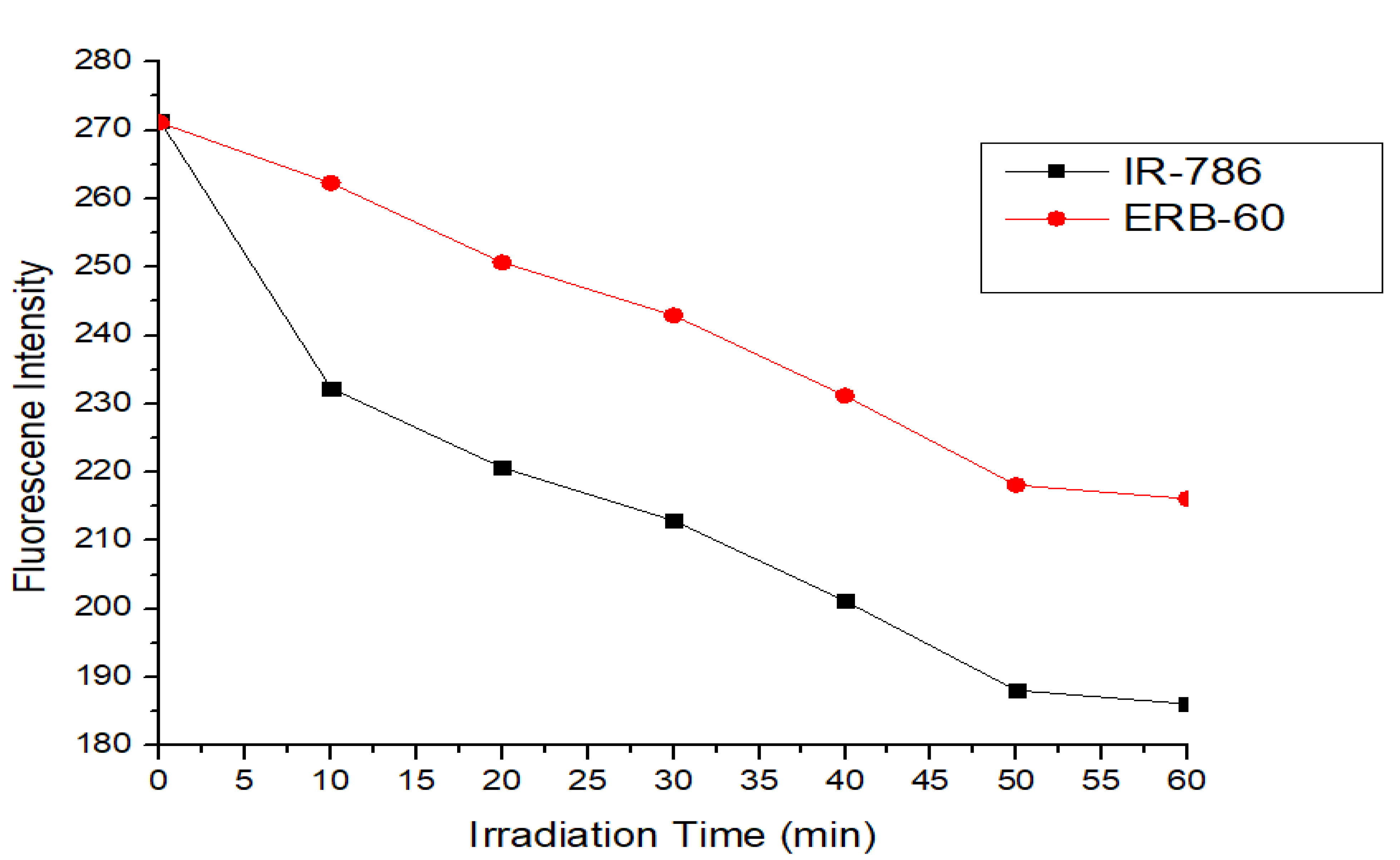
Certain classes of near-infrared dyes, such as cyanines, have been known to be much less stable especially when in solution than analogs in the visible region [20]. Such degradation has been ascribed to the presence of molecular oxygen and light [20]. This photodegradation process is usually observed with cyanines having absorption maximum at longer wavelength. Photodegradation has been an urgent problem, especially for cyanine dyes [21], and as such structural modifications are always required to improve their photostability. It has been reported by most research groups that cyanine dyes containing a rigid cyclohexenyl ring in the polymethine chain exhibit improved photostability and fluorescence quantum yield [22]. To investigate the photostability of ERB-60, a stock solution of 1 mM of this fluorophore was prepared in DMSO. An aliquot of this stock solution was diluted in methanol and the fluorescence intensity was measured at the excitation wavelength of 790 nm. The working solution was continuously irradiated with a Xenon lamp at 150 W for 1 h. The rate of photobleaching of the ERB-60 was determined based on the reduced fluorescence intensity from 270. The result obtained is depicted in Figure 4. Under the same conditions, a commercially available dye, IR-786 iodide, was used as a reference.
As shown in Figure 4, ERB-60 is more stable compared to the commercially available fluorophore, IR-786. As seen in Scheme 1, the chemical structure of ERB-60 consists of a rotatable bond, methoxy groups, a quaternary ammonium moiety, a chlorine atom and phenyl group. These inherent features, especially the increase in the net positive charge in ERB-60, might be the contributing factor explaining the improved photostability observed as compared to the commercially available IR-786 dye.
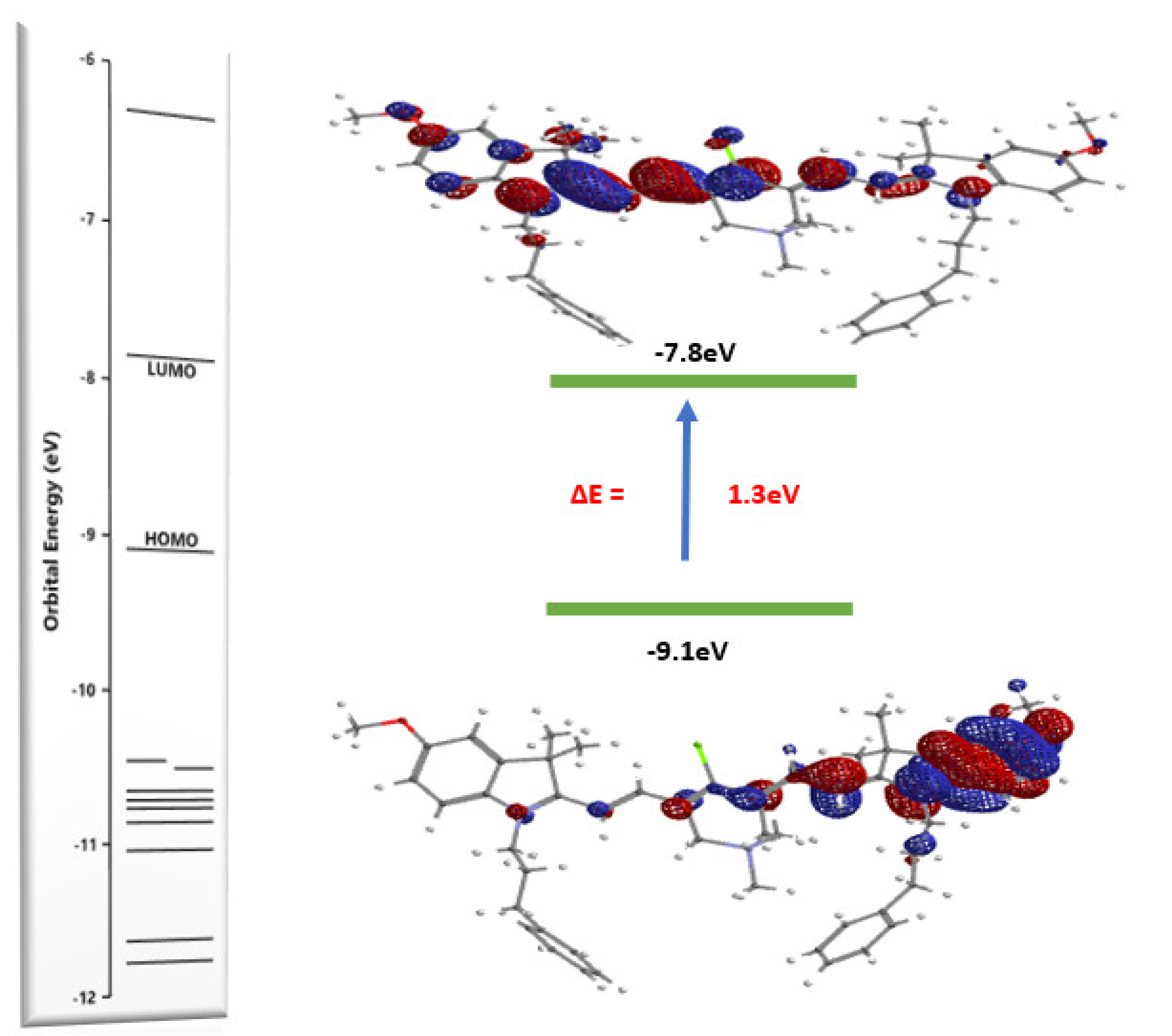
DFT Calculations of ERB-60. The molecular structure of most conjugated systems, especially ERB-60, is greatly affected upon photoexcitation. The rearrangement of electrons within the molecular orbitals requires certain quanta of energy, and with the right amount of energy, electrons in the ground state move to the excited state. Such a transition results in different photophysical processes. We were therefore interested to know the optimized geometry and electron distribution within the highest occupied molecular orbital (HOMO) and the lowest unoccupied molecular orbital (LUMO) of ERB-60. To achieve this task, we used spartan software to perform quantum chemical calculations based on the density functional theory (DFT) method with a 6–311G basis set on ERB-60. Figure 5 shows the optimized molecular structure and frontier molecular orbitals (FMO). As shown in Scheme 1, the structure of ERB-60 consists of two electron-donating groups (methoxy) at the 5th position of the indolium rings. In addition, there is phenylpropyl at the nitrogen-containing ring, and these two heterocycles are connected via polymethine chain. Within the polymethine chain, there is also a quaternary ammonium group, which creates a total net charge of positive two within the overall structure of the fluorophore. All these inherent chemical features have a significant impact on the optimized molecular structure and energy of ERB-60. As shown in Figure 5, the HOMO of ERB-60 has an energy of −9.1 eV. The methoxy group and the heterocycle at the right side of the structure contribute to the energy of the highest occupied molecular orbital (HOMO). As shown in Figure 5, upon photoexcitation, the S0–S1 excitation energy was found to be −7.8 eV. As shown in the LUMO of the compound, the electrons’ delocalization is localized on the other side of the molecule where the nitrogen atom within the terminal heterocycle bears a positive charge. The energy gap of 1.3 eV of ERB-60 corresponds to the higher wavelength of maximum absorbances observed in all tested solvents used. Interestingly, the extra positive charge on the cyclohexene ring did not affect the electron distribution within the HOMO and LUMO orbitals. However, the difference observed in the wavelengths of maximum absorbances in these solvents correlates to how these solvents stabilized the electrons distributions within the HOMO and LUMO orbital of ERB-60.

4. Materials and Methods

All chemicals and solvents were of American Chemical Society grade or HPLC purity and were used as received. All other chemicals were purchased from Fisher Scientific (Pittsburgh, PA, USA) or Acros Organics (Pittsburgh, PA, USA). The reactions were monitored using silica gel 60 F254 thin-layer chromatography plates (Merck EMD Millipore, Darmstadt, Germany). The 1H NMR and 13C NMR, spectra were obtained using high quality Kontes NMR tubes (Kimble Chase, Vineland, NJ, USA) rated to 400 MHz and were recorded on an Avance spectrometer (Bruker, Billerica, MA; 400 MHz for 1H and 100 MHz for 13C) in CDCl3. High-resolution accurate mass spectra (HRMS) were obtained at the Georgia State University Mass Spectrometry Facility using a Q-TOF micro (ESI-Q-TOF) mass spectrometer (Waters, Milford, MA, USA). All chemical shifts were recorded in parts per million (ppm). Signals are labelled as follows: s (singlet), d (doublet), t (triplet), m (multiplet) and coupling constants (J) and are measured in Hertz (Hz). All compounds tested were >95% pure. The melting points were determined with a Mel-temp melting point apparatus and are uncorrected. Energy levels and optimized geometries were calculated using Spartan20 DFT calculations at the B3LYP level.
Synthesis of 2-((E)-2-((E)-4-chloro-5-(2-((E)-5-methoxy-3,3-dimethyl-1-(3 phenylpropyl)indolin-2-ylidene)ethylidene)-1,1-dimethyl-1,2,5,6-tetrahydropyridin-1-ium-3-yl)vinyl)-5-methoxy-3,3-dimethyl-1-(3-phenylpropyl)-3H-indol-1-ium (ERB-60):
To an oven-dried 100 mL round-bottom flask containing acetic anhydride (5 mL) were added 2 equivalents of heterocyclic salt, 1, 3 equivalents of sodium acetate and 1 equivalent of modified Vilsmeier–Haack reagent 3. The reaction mixture was heated under reflux for 5 h at 80 °C until UV-Vis-NIR absorption spectroscopy indicated a total consumption of the starting material. The crude product obtained was purified via a coprecipitation method using DMSO and acetone to obtain a green metallic crystal. (Yield 77%) M.P = 242–244 °C, 1H NMR (400 MHz, CDCl3) 1.72 (s, 18H), 2.20 (t, J = 14.18 Hz, 4H), 2.84 (t, J = 6.97 Hz, 4H), 3.84 (s, 6H), 4.60 (t, J = 14.18 Hz, 4H), 5.29 (s, 4H), 6.80 (d, J = 8.62 Hz, 2H), 7.21 (d, J = 6.31 Hz, 2H), 7.28-7.33 (m, 12H), 7.35 (d, J = 17.51 Hz, 2H), 8.30 (d, J = 14.70 Hz, 2H). 13C NMR (100 MHz, CDCl3) 27.76, 28.32, 33.10,43.85, 50.42, 55.91,102.07, 108.87, 110.95, 113.55, 117.38, 126.59, 128.39, 128.83, 135.10, 139.90, 144.25, 150.50, 158.45, 174.37. HRMS (ESI) m/z calcd for C51H60ClN3O22+ 782 [M + 1] found: 753 [M + 2] – 31.
Optical and physicochemical property analyses. All optical measurements were performed in various solvents, including methanol (MeOH), dimethylformamide (DMF), acetonitrile (MeCN), water, isopropanol (i-PrOH) and dimethyl sulfoxide (DMSO). Absorbance and fluorescence emission spectra of the series of NIR fluorophores were measured using Varian Cary 50 absorbance spectrophotometer (190–1100 nm) and Shimadzu RF-1501 Spectrofluorophotometer (Shimadzu Scientific Instruments, Colombia, MD, USA) interfaced to a PC. Energy levels and optimized geometries were calculated using Spartan DFT calculations at the B3LYP level.

4.1. Preparation of Stock Solution

1 mM stock solution of ERB-60 was prepared by first weighing the corresponding amount on a 5-digit analytical balance into an amber vial. The solid was dissolved by the addition of dimethyl sulfoxide (Analytical Grade, 99.9%, Fischer Scientific, Waltham, MA, United States). The vial was capped, and the resultant solution was then sonicated for 30 min to ensure complete dissolution of solutes.

4.2. Determination of Molar Absorptivity

Working solutions with concentrations (1.25 µM, 2.5 µM, 3.75 µM, and 5 µM) in water, methanol, isopropanol, dimethylformamide, and dimethyl sulfoxide were prepared to obtained absorbance less than 1. The absorbance values at the maximum wavelength in all tested solvents were recorded using a Varian Cary-50 spectrophotometer. The absorbances measured were then plotted against concentration in mol/L using origin lab 98. The graphs obtained followed a linear correlation.

5. Conclusions

A new heptamethine cyanine dye, ERB-60, has been synthesized in four efficient steps in 77% yield. The heptamethine linker within the molecular structure of this fluorophore was modified by the introduction of a quaternary ammonium moiety to fine-tune the physical, chemical and optical properties of the synthesized fluorophore. The wavelengths of maximum absorbance of ERB-60 in different solvents were found to be in the range of 745–770 nm. The highest wavelength of absorbance was recorded in DMSO. In general, the decreasing order of the maximum wavelength of absorbance of ERB-60 in the tested solvents is DMSO > DMF > i-PrOH > MeOH > MeCN > H2O. In addition, the molar extinction coefficients of ERB-60 were found to be within 102,000–151,360 M−1 cm−1. In acetonitrile the highest molar extinction coefficient of 179,920 M−1 cm−1 was observed. As a trend, the decreasing order of the extinction coefficient for ERB-60 was found to be MeCN > MeOH > DMSO > i-PrOH > H2O > DMF. ERB-60 was found to be photostable compared to the commercially available dye IR-786 iodide and also ten times brighter than ICG in methanol.

Supplementary Materials

The following are available online. Spectroscopic data are available; NMR (Figures S1–S3), and Linear regression plots (Figures S4–S6).

Author Contributions

E.R.B. designed and carried out all the experimental and analytical work in this manuscript. E.R.B. also wrote the manuscript. M.H. supervised the research work and proofread the entire manuscript. All authors have read and agreed to the published version of the manuscript.

Funding

This research was funded by the Brains and Behavior Seed Grant, the Health Innovation Platform Grant, and the Georgia Research Alliance Venture Grant Phase 1. The APC was funded by MDPI.

Institutional Review Board Statement

Not applicable.

Informed Consent Statement

Not applicable.

Data Availability Statement

The data presented in this study are available in this article.

Acknowledgments

The authors would like to thank the Center for Diagnostics and Therapeutics at Georgia State University for EB fellowship. The authors thank the Georgia State University Brains, Behavior Seed Grant, The Atlanta Clinical and Translational Science Institute Healthcare Innova-tion Seed Grant, and The Georgia Research Alliance Ventures Phase 1 Grant for the support.

Conflicts of Interest

The authors declare no conflict of interest.

References

  1. Ahoulou, E.O.; Drinkard, K.K.; Basnet, K.; St Lorenz, A.; Taratula, O.; Henary, M.; Grant, K.B. DNA Photocleavage in the Near-Infrared Wavelength Range by 2-Quinolinium Dicarbocyanine Dyes. Molecules 2020, 25, 2926. [Google Scholar] [CrossRef]
  2. Deligeorgiev, T.G.; Vasilev, A.; Drexhage, K.H. Synthesis of novel cyanine dyes containing carbamoylethyl component—Noncovalent labels for nucleic acids detection. Dyes Pigm. 2007, 74, 320–328. [Google Scholar] [CrossRef]
  3. Hyun, H.; Park, M.H.; Owens, E.A.; Wada, H.; Henary, M.; Handgraaf, H.J.; Vahrmeijer, A.L.; Frangioni, J.V.; Choi, H.S. Structure-inherent targeting of near-infrared fluorophores for parathyroid and thyroid gland imaging. Nat. Med. 2015, 21, 192–197. [Google Scholar] [CrossRef] [PubMed] [Green Version]
  4. Hoon, H.; Eric, A.O.; Hideyuki, W.; Andrew, L.; Gwang, P.; Min Ho, P.; John, V.F.; Maged, H.; Hak, S.C. Cartilage-Specific Near-Infrared Fluorophores for Biomedical Imaging. Angew. Chem. Int. Ed. Engl. 2015, 54, 8648–8652. [Google Scholar] [CrossRef] [Green Version]
  5. Choi, H.S.; Gibbs, S.L.; Lee, J.H.; Kim, S.H.; Ashitate, Y.; Liu, F.; Hyun, H.; Park, G.; Xie, Y.; Bae, S.; et al. Targeted zwitterionic near-infrared fluorophores for improved optical imaging. Nat. Biotechnol. 2013, 31, 148–153. [Google Scholar] [CrossRef]
  6. Hoon, H.; Hideyuki, W.; Kai, B.; Julien, G.; Yogesh, Y.; Matt, L.; Maged, H.; John, V.F.; Hak, S.C. Phosphonated Near-Infrared Fluorophores for Biomedical Imaging of Bone. Angew. Chem. 2014, 126, 10844–10848. [Google Scholar] [CrossRef] [Green Version]
  7. Laramie, M.D.; Fouts, B.L.; MacCuaig, W.M.; Buabeng, R.E.; Jones, M.A.; Mukherjee, P.; Behkam, B.; McNally, L.R.; Henary, M. Improved pentamethine cyanine nanosensors for optoacoustic imaging of pancreatic cancer. Sci. Rep. 2021, 11, 4366. [Google Scholar] [CrossRef]
  8. Buabeng, R.E.; Maged, H. Developments of small molecules as inhibitors for carbonic anhydrase isoforms. Bioorg. Med. Chem. 2021, 39, 116140. [Google Scholar] [CrossRef]
  9. Levitz, A.; Marmarchi, F.; Henary, M. Introduction of various substitutions to the methine bridge of heptamethine cyanine dyes Via substituted dianil linkers. Photochem. Photobiol. Sci. 2018, 17, 1409–1416. [Google Scholar] [CrossRef] [PubMed]
  10. Owens, E.A.; Hyun, H.; Tawney, J.G.; Choi, H.S.; Henary, M. Correlating molecular character of NIR imaging agents with tissue-specific uptake. J. Med. Chem. 2015, 58, 4348–4356. [Google Scholar] [CrossRef] [PubMed]
  11. Owens, E.A.; Henary, M.; El Fakhri, G.; Choi, H.S. Tissue-specific near-infrared fluorescence imaging. Acc. Chem. Res. 2016, 49, 1731–1740. [Google Scholar] [CrossRef] [Green Version]
  12. Park, G.K.; Lee, J.H.; Levitz, A.; El Fakhri, G.; Hwang, N.S.; Henary, M.; Choi, H.S. Lysosome-Targeted Bioprobes for Sequential Cell Tracking from Macroscopic to Microscopic Scales. Adv. Mater. 2019, 31, e1806216. [Google Scholar] [CrossRef]
  13. Andrew, L.; Cory, H.; Eduardo, S.; Maged, H. “Turn on” fluorescence response of monomethine cyanines caused by noncovalent binding to ct-DNA. Dyes Pigm. 2017, 145, 202–207. [Google Scholar]
  14. Yogesh, Y.; Andrew, L.; Sanam, D.; Ritu, A.; Maged, H. Effects of heterocyclic N-alkyl chain length on cancer cell uptake of near infrared heptamethine cyanine dyes. Dyes Pigm. 2017, 145, 307–314. [Google Scholar]
  15. Levitz, A.; Marmarchi, F.; Henary, M. Synthesis and optical properties of near-infrared meso-phenyl-substituted symmetric heptamethine cyanine dyes. Molecules 2018, 23, 226. [Google Scholar] [CrossRef] [Green Version]
  16. Kiryl, D.P.; Vladislav, V.V. Guide to red fluorescent proteins and biosensors for flow cytometry. Methods Cell. Biol. 2011, 102, 431–461. [Google Scholar]
  17. Haiying, Z.; George, T.; Dennis, S.; David, M.K.; Hairong, Z.; Tommy, D.; William, C.E.; Mikhail, Y.B. Cell-free measurements of brightness of fluorescently labeled antibodies. Sci. Rep. 2017, 7, 41819. [Google Scholar]
  18. Alvaro, S.a.n.c.h.e.z.-A.; Yan, C.; Joachim, D.M. Molecular brightness determined from a generalized form of Mandel’s Q-parameter. Biophys. J. 2005, 89, 3531–3547. [Google Scholar]
  19. Thavornpradit, S.; Usama, S.M.; Park, G.K.; Shrestha, J.P.; Nomura, S.; Baek, Y.; Choi, H.S.; Burgess, K. QuatCy: A Heptamethine Cyanine Modification With Improved Characteristics. Theranostics 2019, 9, 2856–2867. [Google Scholar] [CrossRef]
  20. Henary, M.; Mojzych, M. Stability and reactivity of polymethine dyes in solution. In Heterocyclic Polymethine Dyes; Topics in Heterocyclic Chemistry; Strekowski, L., Ed.; Springer: Berlin/Heidelberg, Germany, 2008; Volume 14, pp. 221–238. [Google Scholar]
  21. Shershov, V.E.; Spitsyn, M.A.; Kuznetsova, V.E.; Timofeev, E.N.; Ivashkina, O.A.; Abramov, I.S.; Nasedkina, T.V.; Zasedatelev, A.S.; Chudinov, A.V. Near-infrared heptamethine cyanine dyes. Synthesis, spectroscopic characterization, thermal properties and photostability. Dyes Pigm. 2013, 97, 353–360. [Google Scholar] [CrossRef]
  22. Xiaozhong, M.; Matthew, L.; Maged, H. Synthesis, optical properties and cytotoxicity of meso-heteroatom substituted IR-786 analogs. Bioorg. Med. Chem. Lett. 2018, 28, 509–514. [Google Scholar]
  23. Parul, R.; Yuriy, P.B.; Ryan, D.R.; Yangchun, L.; William, S.B.; Deepti, S.; Michael, G.S.; Igor, L.; Stephen, H.F. Selective imaging and killing of cancer cells with protein-activated near-infrared fluorescing nanoparticles. Macromol. Biosci. 2011, 11, 927–937. [Google Scholar]
  24. Gala, C.; Maged, H.; Gabor, P. The effect of varying short-chain alkyl substitution on the molar absorptivity and quantum yield of cyanine dyes. Anal. Chem. Insights 2011, 6, 29–36. [Google Scholar]
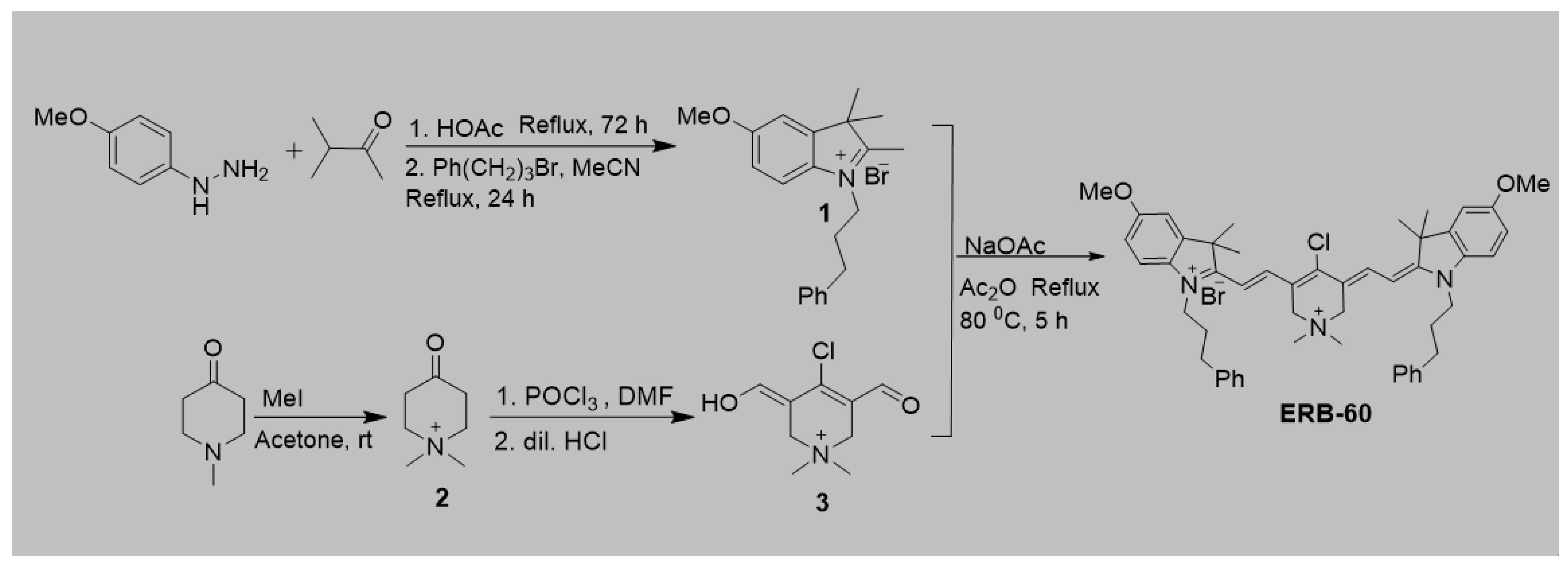
Scheme 1. Synthesis of the two positively charged heptamethine fluorophore, ERB-60.
Molbank 2021 m1270 sch001
Figure 1. Absorbance profile of ERB-60 in polar protic solvents at different concentrations (1.25 μM, 2.5 μM, 3.75 μM, 5.0 μM). The absorbance increase was linear with respect to concentration and follows the Beer–Lambert law with the extinction coefficient being calculated from the linear regression as ε = 127,113, 151,360 and 172,240 M−1 cm−1 in water, isopropanol, and methanol, respectively.
Molbank 2021 m1270 g001
Figure 2. Absorbance profiles of ERB-60 in polar aprotic solvents at different concentrations (1.25 μM, 2.5 μM, 3.75 μM, 5.0 μM). The absorbance increase was linear with respect to concentration and follows the Beer–Lambert law with the extinction coefficient being calculated from the linear regression as ε = 102,000, 151,360 and, 179,920 M−1 cm−1 in N,N-dimethylformamide, dimethyl sulfoxides, and acetonitrile respectively.
Molbank 2021 m1270 g002
Figure 3. (a) Absorbance spectra of ERB-60 (5 uM) in different solvents. (b) Fluorescence spectra of ERB-60 in both polar protic and aprotic solvents.
Molbank 2021 m1270 g003
Figure 4. Photostability studies of ERB-60 under continuous irradiation with a Xenon lamp at 150W for 1 h. The rate of photobleaching of ERB-60 was determined based on the reduced fluorescence intensity from 270 upon continuous irradiating with light (excitation wavelength, 790 nm). A commercially available dye, IR-786 iodide, was used as a reference.
Molbank 2021 m1270 g004
Figure 5. Theoretical calculations for the elucidation of the optical property of ERB-60. Optimized molecular structure and frontier molecular orbitals for ERB-60 with its energy level and, S0–S1 excitation energy based on DFT calculation at the B3LYP/6-311 G(d,p) level.
Molbank 2021 m1270 g005
Table 1. Physicochemical properties of ERB-60 calculated using ChemAxon. The data calculated (at pH 7.4) include: logD, polarizability, dipole moment (debye), number of rotatable bonds, volume (Å3), molecular surface area (Å3), and molecular weight (g/mol).
IDlogDPolarizabilityDipole Momentn rot.Molecular VolumeMolecular Surface AreaMolecular Weight
ERB-603.992.3617.7713785.521214862
Table 2. Spectral characteristics of ERB-60 in polar protic and aprotic solvents (optical properties of ICG are in methanol).
Solventsλabs
nm
λem
nm
Extinction Coefficient
ε (M−1 cm−1)
Stokes Shift (nm)Quantum Yield
(QY)
Molecular Brightness
(M−1 cm−1)
H2O745761127,113160.09812,457
MeOH752769172,240170.24942,888
i-PrOH761779149,920180.25037,840
MeCN751772179,920210.31055,775
DMF766773102,00070.26827,336
DMSO770789151,360190.16424,823
ICG [23,24]783802122,000190.0364392
Publisher’s Note: MDPI stays neutral with regard to jurisdictional claims in published maps and institutional affiliations.