Short Note

Digyaindoleacid A: 2-(1-(4-Hydroxyphenyl)-3-oxobut-1-en-2-yloxy)-3-(1H-indol-3-yl)propanoic Acid, a Novel Indole Alkaloid

1
Marine and Plant Research Laboratory of Ghana, Department of Chemistry, School of Physical and Mathematical Sciences, University of Ghana, P.O. Box LG 56 Legon-Accra, Ghana
2
Department of Bioengineering, Munzur University, 62000 Tunceli, Turkey
3
Department of Biochemistry, Cell and Molecular Biology, University of Ghana, P.O. Box LG 54 Legon-Accra, Ghana
4
Noguchi Memorial Institute for Medical Research (NMIMR), College of Health Sciences, University of Ghana, P.O. Box LG 581 Legon, Ghana
5
Marine Biodiscovery Centre, Department of Chemistry, University of Aberdeen, Old Aberdeen AB24 3UE, Scotland, UK
*
Author to whom correspondence should be addressed.
Molbank 2019, 2019(3), M1080; https://doi.org/10.3390/M1080
Submission received: 22 August 2019 / Revised: 9 September 2019 / Accepted: 12 September 2019 / Published: 14 September 2019
(This article belongs to the Section Natural Product Chemistry)

Abstract

:
Digyaindoleacid A (1) is one of the novel alkaloids produced by the Ghanaian Paenibacillus sp. DE2SH (GenBank Accession Number: MH091697) isolated from the mangrove rhizosphere soils of the Pterocarpus santalinoides tree growing in the wetlands of the Digya National Park, Brong Ahafo Region, Ghana. This compound was isolated on HPLC at tR ≈ 60 min and its structure determined by MS, 1D, and 2D-NMR data. When tested against Trypanosoma brucei subsp. brucei strain GUTat 3.1, 1 produced a half-maximal inhibitory concentration (IC50) 5.21 μM compared to the standard diminazene aceturate (IC50 = 1.86 μM). In the presence of normal mouse macrophages RAW 264.7, 1 displayed a higher selectivity towards T. brucei subsp. brucei (selectivity indices (SI) = 30.2) with low toxicity. This result is interesting given that the drug diminazene aceturate is considerably toxic and 1 is a natural product isolate. The structure of 1 incorporates the backbone of the amino acid tryptophan which is crucial in the metabolism of Trypanosoma brucei subsp. brucei strain GUTat 3.1. It is possible that 1, could interfere with the normal uptake and metabolism of tryptophan in the parasite. However, 1 (IC50 = 135.41 μM) produced weak antileishmanial activity when tested against Leishmania donovani (Laveran and Mesnil) Ross (D10).

Graphical Abstract

1. Introduction

Buruli ulcer, Chagas disease, cysticercosis, dengue fever, Guinea worm, echinococcosis, fascioliasis, human African trypanosomiasis, leishmaniasis, leprosy, lymphatic filariasis, onchocerciasis, rabies, schistosomiasis, soil transmitted helminths, trachoma and yaws represent the comprehensive and slow growing list of neglected tropical diseases (NTDs) worldwide [1]. Millions of people in the low-income countries of Africa, Asia and Americas live in areas where they are at risk of contracting at least one of these NTDs [1]. In fact, statistics are available to indicate that thousands and sometimes tens of thousands of people in low-income countries indeed contract one or more of these very terrible ailments due to geographical location, persistent poverty and repeated exposure to pathogens [2,3]. The number of deaths resulting from NTDs is also in the thousand to tens of thousands, even though, it is commendable the extent of reduction of deaths since 1960s to date as a result of World Health Organization (WHO), Centers for Disease Control and Prevention (CDC) and other organizational programs [4]. It is interesting that, with such feats, NTDs currently do not make the list of top five killers over the past decade notwithstanding the fact that millions still live with the neglected infections. The top five killers are ischemic heart disease, stroke, chronic obstructive lung disease, lower respiratory infections and diarrhea [5,6]. Coincidentally, the human anatomy affected by the current disease burden; heart, lungs, alimentary canal and central nervous system are also severely affected by NTDs such as Chagas disease, trypanosomiasis, leishmaniasis and schistosomiasis. These four NTDs also affect other organs like the liver, spleen, eye, lymph, bladder and blood vessels [7,8]. It is possible that the long lasting effects of chronic NTDs which are known to prevail even after treatment could have tremendous consequences on the impact of the world’s current top five killer diseases in low-income countries of Africa, Asia and the Americas [9].
Furthermore, drug administration has played a significant and successful role in the control, treatment and increasing reduction of deaths from NTDs. A very narrow spectrum of NTD drugs have been prescribed and administered for over three decades without replacements. However, there is current widespread development of resistance to prescription drugs for NTDs. In order to ensure that the number of deaths from NTDs is kept at their current ‘reasonably’ low percentages, scientists must intensify the research and development of new drug entities for NTDs [10,11,12].
Previously, we have isolated several microbes from soils and sediments collected from seven out of the 10 regions in Ghana [13]. Therefore, the current research in our laboratory is targeted at identifying the chemical structures of the active principles for several Ghanaian microbial extracts that have shown antitrypanosomal, antileishmanial and anticercaricidal bioactivity. The overall goal is to identify novel natural product chemical scaffolds that would provide a solid platform for the development of future drugs for the parasitic NTDs.
Large scale culture of the Ghanaian Paenibacillus sp. DE2SH (GenBank Accession Number: MH091697) in ISP2 fermentation media at pH 5.5, 28 °C, 220 rpm and 14 days incubation produced crude extracts which possessed antiparasitic activity against trypanosomes, schistosomes and leishmania. Chemical profiling of these extracts using high resolution electrospray ionization liquid chromatography mass spectrometry (HRESI-LC-MS) and nuclear magnetic resonance spectroscopy (NMR) showed the presence of several alkaloids. One of these compounds is 2-(1-(4-hydroxyphenyl)-3-oxobut-1-en-2-yloxy)-3-(1H-indol-3-yl)propanoic acid or simply Digyaindoleacid A (1). The crude extract of Paenibacillus sp. DE2SH was solvent partitioned by a modification of Kupchan’s method [14] to give four fractions hexane (FH), dichloromethane (FD), methanol/water (FM) and butanol/water (WB). Phytochemical screening on thin-layer chromatography (TLC) plates using iodine tank, Dragendorff, phosphomolybdic acid, antimony (III) chloride and ninhydrin reagents showed the WB to contain the most interesting metabolites. The fraction WB was therefore subjected to a Sephadex LH-20 size exclusion chromatography to give six fractions of which WB-SF6 was found to contain the compound of interest. Reverse phase HPLC of WB-SF6 led to the isolation of the potent antitrypanosomal agent digyaindoleacid A.

2. Results

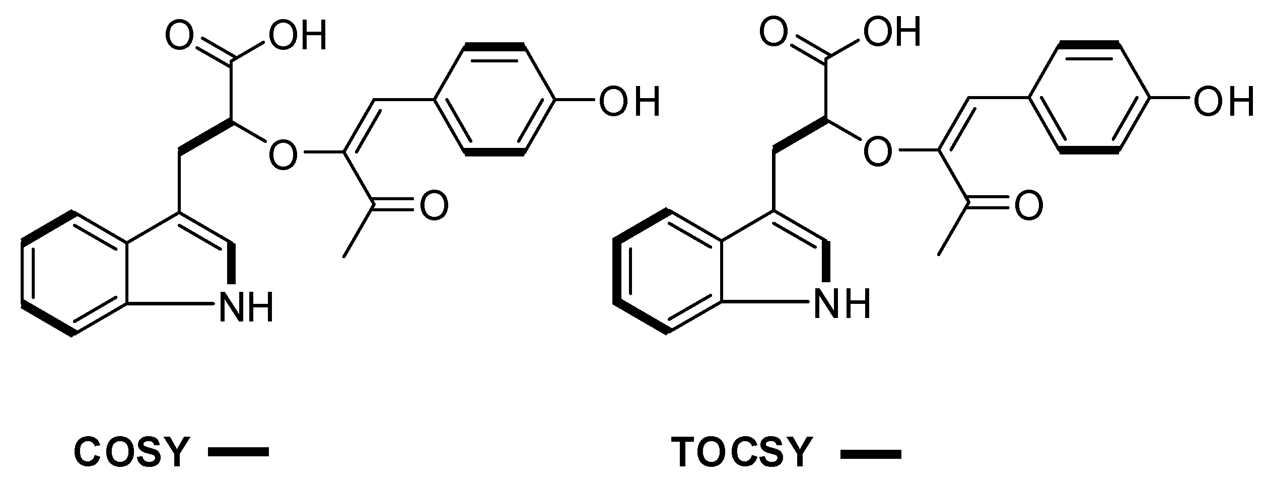
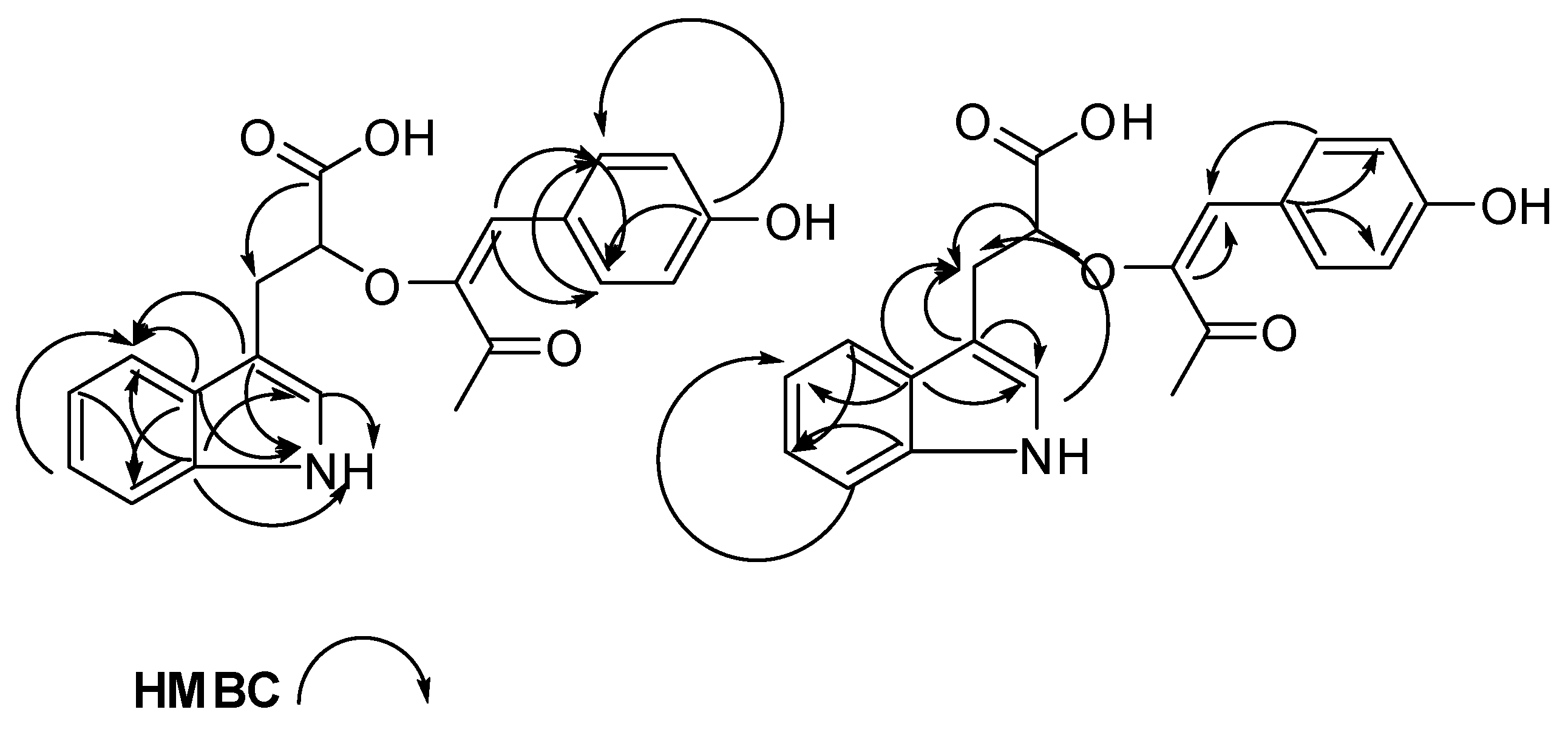
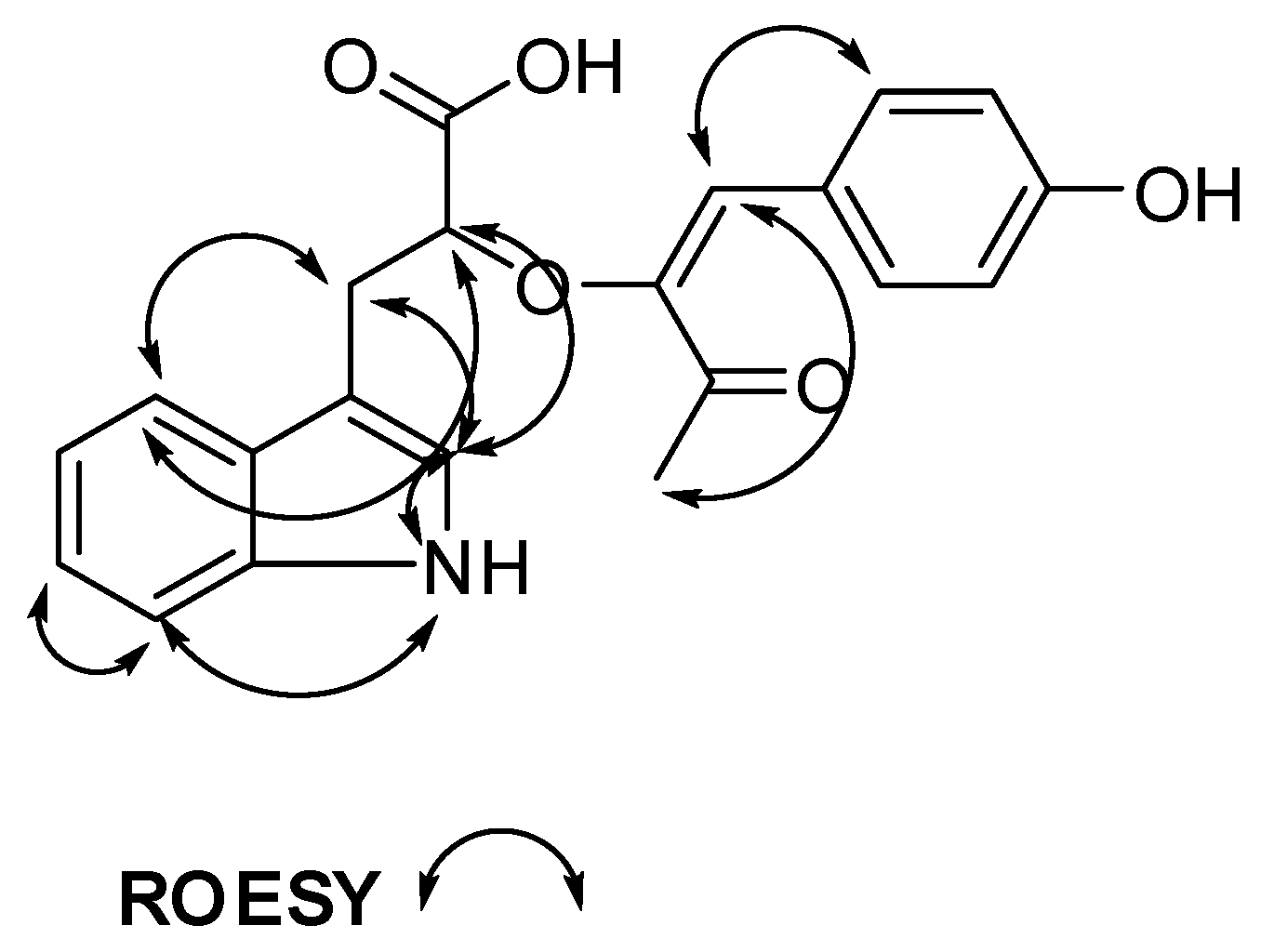
The compound 1 was obtained at tR ≈ 60 min on reverse phase HPLC (Figures S12 and S13). Compound 1 is light yellow pungent oil when completely free of solvent. Fragmentation of the molecular ion of compound 1 under the conditions for HRESI-LC-MS was very rigorous due to the four very labile functional groups, α,β-unsaturated ketone, carboxylic acid, ether and phenol present in the compound. These functional groups come together to produce fragmentation patterns that did not lend themselves to complete and comprehensive interpretation. However, facile loss of water from compound 1 produced m/z 350.1501 corresponding to molecular formula C21H20NO4+ (Δ = 0.01 ppm and 13 degrees of unsaturation). In addition, characteristic carboxylic acid and phenol inspired fragmentation pathways for compound 1 produced m/z 279.1594 corresponding to molecular formula C19H21NO.+ (Δ = 0.002 ppm and nine degrees of unsaturation). Other fragmentation pathways leading to different ions can be found in Figures S2 and S3. From this analysis, it was possible to calculate the m/z of compound 1 to be 365.1263 corresponding to molecular formula C21H19NO5 with 13 degrees of unsaturation. Analysis of the 1H, 13C, multiplicity edited pulsed field gradient heteronuclear single quantum coherence (gHSQCAD) and several pulsed field gradient 1H-13C heteronuclear multiple bond correlations (gHMBCAD) spectrum of 1, suggested the presence of eight quaternaries, eleven methines, one methylene and one methyl carbons. The 1H-NMR chemical shifts H 10.67 (1H, s, NH-1), 7.10 (1H, s, H-2), 7.53 (1H, d, J = 7.9 Hz, H-4), 6.93 (1H, t, J = 7.9 Hz, H-5), 7.02 (1H, t, J = 7.9 Hz, H-6), 7.30 (1H, d, J = 7.9 Hz, H-7), 3.10 (1H, m, H-8), 2.73 (1H, dd, J = 14.5, 8.4 Hz, H-8) and 3.92 (1H, m, H-9) together with the δC 123.1 (C-2), 111.8 (C-3), 127.7 (C-3a), 118.6 (C-4), 117.8 (C-5), 120.4 (C-6), 110.9 (C-7), 135.8 (C-7a), 30.4 (C-8), 71.4 (C-9) and 176.0 (C-10) provided direct evidence for the presence of the aromatic tryptophan hydroxyl acid residue. Detailed analysis of the 1H-1H homonuclear correlation spectroscopy (gCOSY) spectrum showed correlations H-2/NH-1, H-4/H-5, H-6/H-7 and H-8/H-9, which were further supported by similar 1H-1H total correlation spectroscopy (2D-TOCSY) correlations, including H-2/NH-1, H-4/H-5, H-6, H-7, H-5/H-4, H-6/H-4, H-7, H-5, H-7/H-4, H-5, H-6 and H-8/H-9 to further confirm the presence of a tryptophan moiety. Several gHMBCAD correlations providing connections between the isolated spin-systems of the aromatic tryptophan through quaternaries and heteroatoms were observed and included NH-1 to C-2, C-3, C-3a, C-7a, H-2 to C-3, C-3a, C-7a, H-4 to C-3, C-3a, C-6, C-7a, H-5 to C-3a, C-7, H-6 to C-4, C-7a, H-7 to C-3a, C-5 and H-8 to C-2, C-3, C-3a, C-9. In the 13C NMR spectrum, the chemical shift step-up represented by the peaks 114.3 (C-11) and 146.3 (C-12) suggested the presence of a tri-substituted double bond with one of the substituents being an oxygen atom. Such chemical shift step-ups are characteristic of substituted non-aromatic double bonds and the δH 6.53 (1H, s, H-4) confirmed the nature of the substitution. Putting together the gCOSY correlations H-16/H-17, H-19/H-20 along with the gHMBCAD correlations H-11 to C-12, C-13, C-16, C-20, H-14 to C-13, H-16 to C-11, C-18, C-20, H-17 to C-15, H-19 to C-15 and H-20 to C-11, C-16, C-18 provided proof for the existence of a 1-(4-hydroxyphenyl)-3-oxobut-1-en-2-yloxy moiety in the structure of compound 1. Detailed analysis of all the chemical shifts in comparison to the tryptophan molecules published by our group and those in the literature led to the structure of compound 1 as shown in Figure 1. Analysis of the 1H-1H rotating-frame Overhauser spectroscopy (ROESY) data gave confirmatory correlations for the structure of compound 1.
The complete NMR data for Compound 1 is given in Table 1 while the raw data can be found in the Supplementary Figures S4–S11. In Figure 2, Figure 3 and Figure 4, a visual representation of COSY, HMBC, 2D-TOCSY and ROESY correlations are shown.
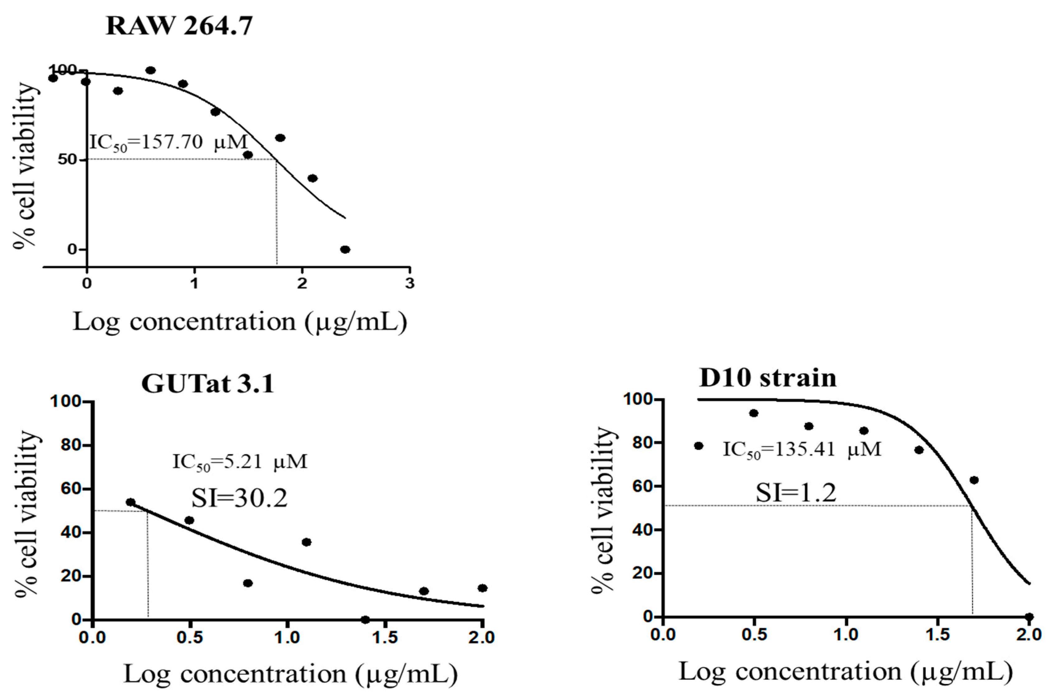
In order to determine the effects on the viability of parasites, the in vitro antitrypanosomal and antileishmanial activities of compound 1 were investigated in a 48 h cell viability assay involving the absorbance properties of resazurin. Normal cell lines (macrophages RAW 264.7) were also used to investigate potential selectivity and toxicity profiles. Compound 1 was found to exhibit significant activity against Trypanosoma brucei subsp. brucei strain GUTat 3.1 (half-maximal inhibitory concentration (IC50) 5.21 μM). The IC50 of compound 1 was approximately three times less potent than the antitrypanosomal drug diminazene aceturate (IC50 = 1.86 μM). This result is interesting, given that 1 is a natural product isolate. However, no promising antileishmanial activity against Leishmania donovani (Laveran and Mesnil) Ross (D10) was observed with IC50 = 135.41 μM which is approximately 410 times less potent than the antileishmanial drug amphotericin B (IC50 = 0.33 μM), thereby suggesting a selective activity of compound 1 against T. brucei subsp. brucei (Figure 5). Moreover, in the presence of normal mouse macrophages, 1 displayed a higher selectivity towards T. brucei subsp. brucei (SI = 30.2) as compared to Leishmania donovani (Laveran and Mesnil) Ross (D10) (SI = 1.2) (Figure 5). Therefore, compound 1 was relatively non-toxic to normal macrophages while at the same time exhibited potent antitrypanosmal activity. Compound 1 is another example of derivatized tryptophan derived alkaloids isolated from Ghanaian microbes and found to possess potent antitrypanosomal activity. We are in the process of investigating the mechanisms of action for these molecules that seem to interfere with the normal uptake and metabolism of tryptophan in the parasite.

Biosynthesis of Digyaindoleacid A (1)

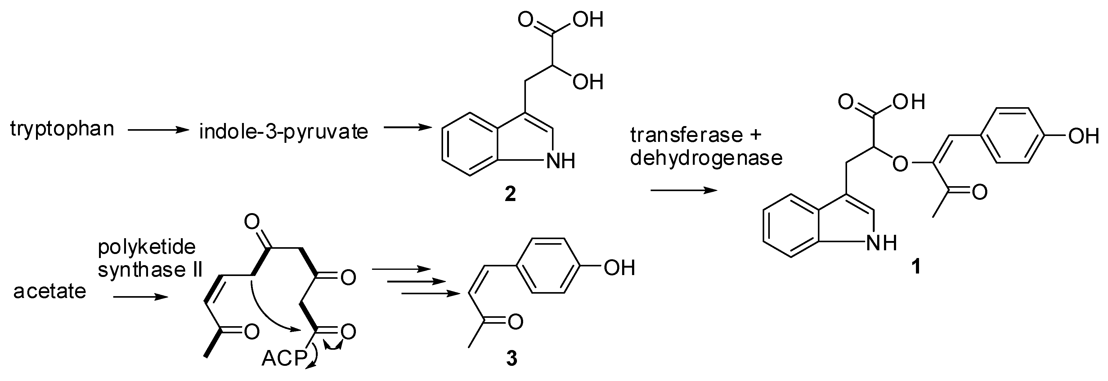
Inspection of the structure of digyaindoleacid A (1) led to the speculation that, this compound is biosynthesized through the condensation of tryptophan-derived indole-3-lactic acid (2) and 1-(4-hydroxyphenyl)-3-oxobut-1-ene (3) (Figure 6), followed by dehydrogenation. The bio-origin of 1-(4-hydroxyphenyl)-3-oxobut-1-ene (3) remains elusive although one could speculate that a type II polyketide synthase could be involved in its assembly as shown in Figure 6.

3. Experimental Section

3.1. General Experimental Procedures

1D and 2D NMR data were recorded on a Bruker AVANCE III HD Prodigy (BRUKER, Sylvenstein, Germany) at 500 and 125 MHz for 1H and 13C, respectively. This instrument was optimized for 1H observation with pulsing/decoupling of 13C and 15N, with 2H lock channels equipped with shielded z-gradients and cooled preamplifiers for 1H and 13C. The 1H and 13C chemical shifts were referenced to the solvent signals (δH 2.50 (1H, p) and δC 39.52 ppm in DMSO-d6). High-resolution mass spectrometry data were measured using a ThermoScientific LTQXL-Discovery Orbitrap (Thermo Scientific, Bremen, Germany) coupled to an Accela UPLC-DAD system. The following conditions were used for mass spectrometric analysis: Capillary voltage 45 V, capillary temperature 320 °C, auxiliary gas flow rate 10–20 arbitrary units, sheath gas flow rate 40–50 arbitrary units, spray voltage 4.5 kV, and mass range 100–2000 amu (maximum resolution 30,000). The ion source was normal electrospray ionization that acts both in positive and negative modes. Semi-preparative HPLC purifications were carried out using a Phenomenex Luna reverse-phase (C18 250 × 10 mm, L × i.d.) column connected to a Waters 1525 Binary HPLC pump Chromatograph with a 2998 photodiode array detector (PDA), column heater, and in-line degasser. Detection was achieved on-line through a scan of wavelengths from 200 to 400 nm. This system was also used to record the UV profile for the compounds. IR was measured using a PerkinElmer FT-IR (UATR Two) spectrometer. All solvents were HPLC grade. Sephadex LH-20 and HP-20 resin were obtained from Sigma Aldrich (Munich, Germany).

3.2. Sediment Sample Collection Sites

The Ghanaian Paenibacillus sp. strain DE2SH (Figure S1) was isolated from the mangrove rhizosphere soils of the Pterocarpus santalinoides tree growing in the wetland areas of the Digya National Park in the Brong Ahafo Region of Ghana (coordinates: 7°12′17.46″N and 0°05′35.01″E). This collection has been described previously in an article published by our laboratory [13].

3.3. Isolation, Purification and Taxonomy of Strain DE2SH

The isolation, purification and exact taxonomy of Paenibacillus sp. strain DE2SH (GenBank Accession Number: MH091697) has been described previously in an article published by our group (Figure S1) [13].

3.4. Fermentation

An Autoclaved Erlenmeyer flask (250 mL) plugged with non-absorbent cotton wool containing 50 mL of ISP2 (10 g of malt extract, 4 g each of yeast extract and d-glucose) fermentation media in distilled water with pH 5.5 was directly inoculated with spores of strain DE2SH and incubated at 28 °C at 220 rpm for three days. This seed culture was subsequently used to inoculate nine autoclaved 1 L Erlenmeyer flasks, each containing 200 mL ISP2 media at pH 5.5 and plugged with non-absorbent cotton wool. The 1 L flasks were incubated at 28 °C at 220 rpm for 14 days. Two days before the culture incubation period was complete, autoclaved HP-20 resin was added at 50 g/L to each of the flasks under sterile conditions and the flasks were returned to the incubator.

3.5. Extraction, Purification and Isolation

The Paenibacillus sp. strain DE2SH fermentation broth (1.8 L) was filtered through a piece of glass wool under suction in a Buchner funnel to separate the supernatant from the mycelia. The supernatant was extracted with EtOAc and the mycelia and HP-20 resin were placed in a 1 L flask and extracted sequentially and alternatively with MeOH and CH2Cl2. All extracts were combined and evaporated under reduced pressure to obtain a total crude extract (3.00 g). The total crude extract was subjected to a modification of Kupchan’s solvent partitioning process [14] that gave the four fractions; FH (0.70 g), FD (0.50 g), FM (0.30 g), and WB (1.30 g), with the compounds of interests concentrated in the polar WB fraction. The WB fraction was then subjected to Sephadex LH-20 size exclusion chromatography by gravity to obtain six fractions that were labeled WB-SF1-6. Phytochemical screening with iodine tank, Dragendorff, phosphomolybdic acid, antimony (III) chloride and Ninhydrin reagents on TLC plates followed by 1H-NMR showed the compound of interest to be concentrated in the WB-SF6 (0.10 g). Fraction WB-SF6 was therefore subjected to semi-preparative HPLC separation and purification using a Phenomenex Luna C18 column (C18 250 × 10 mm, L × i.d.). Gradients of Solvent A: H2O (0.1% HCOOH) and Solvent B: CH3CN (0.1% HCOOH) (100% A to 100% B in 30 min and hold for 30 min) were used as eluents with column flow rates set at 1.5 mL/min to afford digyaindoleacid A (1) (2.6 mg, tR ≈ 60 min).

3.6. 2-(1-(4-hydroxyphenyl)-3-oxobut-1-en-2-yloxy)-3-(1H-indol-3-yl) Propanoic Acid (1)

Light yellow pungent oil; [α]20D −0.01 (c = 0.13, Methanol); IR (neat) νmax 3367, 2947, 2835, 2263, 2133, 1740, 1647, 1449, 1368, 1213, 1112, 1022, 993 cm−1 (Table S1); UV (H2O:CH3CN) λmax 221, 280, 363 nm (Figures S12 and S13); for 1H and 13C NMR data, see Table 1; mass spectrometry data is detailed in Supplementary Figures S2 and S3.

3.7. Culture of Parasites and Mammalian Cell Lines

Blood stream forms of Trypanosoma brucei subsp. brucei strain GUTat 3.1 were cultivated in vitro to the logarithm phase using Hirum’s modified Iscove’s media (HMI9, Thermo Fisher Scientific) supplemented with 10% fetal bovine serum (Thermo Fisher Scientific) at 5% CO2 and 37 °C. Leishmania donovani (Laveran and Mesnil) Ross (D10) was cultivated in vitro to the logarithm phase using medium 199 (M199, Thermo Fisher Scientific) with 10% FBS at 5% CO2 and 37 °C. Mouse macrophages (RAW 264.7 cell lines) were cultivated in vitro to the logarithm phase using Dulbecco’s modified eagle media (DMEM, Thermo Fisher Scientific) with 10% fetal bovine serum at 5% CO2 and 37 °C [15,16].

3.8. Analysis of Cell Viability

For Trypanosoma brucei subsp. brucei strain GUTat 3.1 and Leishmania donovani (Laveran and Mesnil) Ross (D10), cells were seeded at a density of 3.0 × 10⁵ cells/mL in 96 well plates at a two-fold dilution of the compound and incubated for 24 h. Alamar blue dye (resazurin, 10% v/v) was added to wells and incubated for another 24 h. For mouse macrophages (RAW 264.7), cell lines were plated at a density of 3.0 × 10⁵ cells/mL for 48 h to allow for sufficient adherence to plates. The compound under investigation was then added to cells in a two-fold dilution and incubated for another 24 h. Alamar blue dye (10% v/v) was added to wells and incubated for another 24 h. Experiments were run in quadruplicates. Spectrophotometric absorbances were recorded at a wavelength of 570 nm. Diminazene aceturate and amphotericin B were used as a positive antitrypanosomal and antileishmanial control drugs respectively in the assays.

3.9. Statistical Analysis

Data from the cell viability assay was analyzed with Graphpad Prism version 5 (GraphPad Software, Inc., Avenida De La Playa, La Jolla, CA, USA). The half-maximal inhibitory concentration (IC50) was calculated as the concentration that caused a 50% reduction in cell viability. The IC50 values were calculated from a non-linear regression model. Graphpad Prism version 5 was employed for the analysis of unpaired t-test. p-values ≤ 0.05 were considered to be significant.

4. Conclusions

Digyaindoleacid A (1) is one of the alkaloids responsible for the antitrypanosomal activity shown by WB extracts of the Ghanaian microbial strain Paenibacillus sp. DE2SH (GenBank Accession Number: MH091697). While the antileishmanial activity of 1 against Leishmania donovani (Laveran and Mesnil) Ross (D10) is relatively weak (IC50 = 135.41) compared to the drug amphotericin B (IC50 = 0.33 μM), it was very potent against trypanosomes when compared to the drug diminazene aceturate (IC50 = 1.86 μM). Compound 1 (IC50 5.21 μM) is the latest addition to the group of tryptophan derived alkaloids in our laboratory that exhibit potent antitrypanosomal activity against Trypanosoma brucei subsp. brucei strain GUTat 3.1. These results are not surprising given the very important metabolic and physiological role of tryptophan in trypanosomes and the fact that drugs such as curaxins are based on a similar backbone [17]. It is possible that, digyaindoleacid A (1) interferes with the normal uptake and metabolism of tryptophan in Trypanosoma brucei subsp. brucei strain GUTat 3.1. Efforts to study the mechanisms of action of these tryptophan derived alkaloids are in progress in our laboratory.

Supplementary Materials

The supplementary materials are available online.

Author Contributions

K.K. and H.D. collected mangrove sediments and isolated the strain DE2SH. A.S.C. and M.C. identified the exact taxonomy of the strain. M.J. and H.D. provided access to facilities for mass spectrometry and data interpretation. K.K. performed chemical profiling to identify the major metabolite. S.K., G.M.T. and T.M. performed seed culture, large scale culture, isolation and purification of compound. K.K. measured all NMR, IR and UV, analyzed the results and integration of data to give the complete structure of the compound. A.K.D. and F.A.A. performed all the bioassay tests. K.K. wrote the article.

Funding

K.K. wishes to thank the Centre for African Wetlands (CAW), University of Ghana, for providing seed funding to enable the collection of sediments for microbe isolation and The World Academy of Sciences (TWAS) Research Grant Award_17-512 RG/CHE/AF/AC_G. K.K. is also very grateful to the Cambridge-Africa Partnership for Research Excellence (CAPREx), which is funded by the Carnegie Corporation of New York, for a postdoctoral fellowship. K.K. also appreciates the Cambridge–Africa ALBORADA Research Fund for support and the Medical Research Council (MRC)/Department for International Development (DFID) African Research Leaders MR/S00520X/1 Award. S.K. wishes to thank the Carnegie Building a New Generation of Academics (BANGA)-Africa Project Award for a PhD scholarship and M.T. is grateful for an MPhil full scholarship from the TWAS Research Grant Award_17-512 RG/CHE/AF/AC_G.

Acknowledgments

All the authors extend their gratitude to the Department of Chemistry, University of Ghana for providing the NMR facility.

Conflicts of Interest

The authors declare no conflict of interests for this article.

References

  1. Standley, C.; Boyce, M.R.; Klineberg, A.; Essix, G.; Katz, R. Organization of oversight for integrated control of neglected tropical diseases within Ministries of Health. PLoS Neglected Trop. Dis. 2018, 12, e0006929. [Google Scholar] [CrossRef] [PubMed]
  2. Simpson, H.; Quao, B.; Van Der Grinten, E.; Saunderson, P.; Ampadu, E.; Kwakye-MaClean, C.; Odoom, S.; Biritwum, N.-K.; Pullan, R.; Cano, J. Routine surveillance data as a resource for planning integration of NTD case management. Lepr. Rev. 2018, 89, 178–196. [Google Scholar]
  3. Mitra, A.K.; Mawson, A.R. Neglected Tropical Diseases: Epidemiology and Global Burden. Trop. Med. Infect. Dis. 2017, 2, 36. [Google Scholar] [CrossRef] [PubMed]
  4. Kirigia, J.M.; Mburugu, G.N. The monetary value of human lives lost due to neglected tropical diseases in Africa. Infect. Dis. Poverty 2017, 6, 165. [Google Scholar] [CrossRef] [PubMed] [Green Version]
  5. Mathers, C.; Stevens, G.; Hogan, D.; Mahanani, W.R.; Ho, J. Global and regional causes of death: Patterns and trends, 2000–2015. In Disease Control Priorities: Improving Health and Reducing Poverty, 3rd ed.; Jamison, D.T., Gelband, H., Horton, S., Jha, P., Laxminarayan, R., Mock, C.N., Nugent, R., Eds.; The International Bank for Reconstruction and Development/The World Bank: Washington, DC, USA, 2017; Volume 9, pp. 1–429. [Google Scholar]
  6. Roth, G.A.; Abate, D.; Abate, K.H.; Abay, S.M.; Abbafati, C.; Abbasi, N.; Abbastabar, H.; Abd-Allah, F.; Abdela, J.; Abdelalim, A.; et al. Global, regional, and national age-sex-specific mortality for 282 causes of death in 195 countries and territories, 1980–2017: A systematic analysis for the Global Burden of Disease Study 2017. Lancet 2018, 392, 1736–1788. [Google Scholar] [CrossRef]
  7. McDonald, M. Neglected tropical and zoonotic diseases and their impact on women’s and children’s health. In The Causes and Impacts of Neglected Tropical and Zoonotic Diseases: Opportunities for Integrated Intervention Strategies; National Academies Press: Washington, DC, USA, 2011; pp. 357–388. [Google Scholar]
  8. Mehta, P.; Hotez, P.J. NTD and NCD Co-morbidities: The Example of Dengue Fever. PLoS Negl. Trop. Dis. 2016, 10, 1–5. [Google Scholar] [CrossRef] [PubMed]
  9. Bhatnagar, T.; Kaur, P.; Kumaraswami, V. Links Between the Epidemiology and Control of Non-communicable Diseases and Neglected Tropical Diseases in Asia. In Neglected Tropical Diseases-East Asia; Utzinger, J., Yap, P., Bratschi, M., Steinmann, P., Eds.; Springer: New York, NY, USA, 2019; pp. 149–173. [Google Scholar]
  10. Weng, H.-B.; Chen, H.-X.; Wang, M.-W. Innovation in neglected tropical disease drug discovery and development. Infect. Dis. Poverty 2018, 7, 67. [Google Scholar] [CrossRef] [PubMed]
  11. Akinsolu, F.T.; Nemieboka, P.O.; Njuguna, D.W.; Ahadji, M.N.; Dezso, D.; Varga, O. Emerging Resistance of Neglected Tropical Diseases: A Scoping Review of the Literature. Int. J. Environ. Res. Public Health 2019, 16, 1925. [Google Scholar] [CrossRef] [PubMed]
  12. Kerr, K.W.; Henry, T.C.; Miller, K.L. Is the priority review voucher program stimulating new drug development for tropical diseases? PLoS Neglected Trop. Dis. 2018, 12, e0006695. [Google Scholar] [CrossRef] [PubMed]
  13. Osei, E.; Kwain, S.; Mawuli, G.; Anang, A.; Owusu, K.; Camas, M.; Camas, A.S.; Ohashi, M.; Alexandru-Crivac, C.-N.; Deng, H.; et al. Paenidigyamycin A, Potent Antiparasitic Imidazole Alkaloid from the Ghanaian Paenibacillus sp. DE2SH. Mar. Drugs 2019, 17, 9. [Google Scholar] [CrossRef] [PubMed]
  14. Dofuor, A.K.; Kwain, S.; Osei; Tetevi, G.M.; Okine, L.K.; Ohashi, M.; Gwira, T.M.; Kyeremeh, K.; Osei, E. N-(Isobutyl)-3,4-methylenedioxy Cinnamoyl Amide. Molbank 2019, 2019, M1070. [Google Scholar] [CrossRef]
  15. Azerigyik, F.A.; Amoa-Bosompem, M.; Tetteh, T.; Ayertey, F.; Antwi, A.N.; Owusu, K.B.-A.; Dadzie, K.K.; Djameh, G.I.; Tetteh-Tsifoanya, M.; Iwanaga, S.; et al. In vitro Mechanistic Assays of Tetracyclic Iridoid Compounds Isolated from Morinda lucida Benth in Leishmania species. Eur. J. Med. Plants 2018, 25, 1–14. [Google Scholar] [CrossRef]
  16. Yabu, Y.; Minagawa, N.; Kita, K.; Nagai, K.; Honma, M.; Sakajo, S.; Koide, T.; Ohta, N.; Yoshimoto, A. Oral and intraperitoneal treatment of Trypanosoma brucei brucei with a combination of ascofuranone and glycerol in mice. Parasitol. Int. 1998, 47, 131–137. [Google Scholar] [CrossRef]
  17. Gasparian, A.V.; Burkhart, C.A.; Purmal, A.A.; Brodsky, L.; Pal, M.; Saranadasa, M.; Bosykh, D.A.; Commane, M.; Guryanova, O.A.; Pal, S.; et al. Curaxins: Anticancer Compounds that Simultaneously Suppress NF-κB and Activate p53 by Targeting FACT. Sci. Transl. Med. 2011, 3, 95ra74. [Google Scholar] [CrossRef] [PubMed]
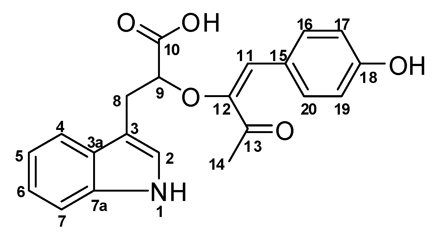
Figure 1. Structure of 2-(1-(4-hydroxyphenyl)-3-oxobut-1-en-2-yloxy)-3-(1H-indol-3-yl)propanoic acid (1).
Molbank 2019 m1080 g001
Figure 2. Key COSY and TOCSY correlations (bold lines) show all the spin systems present in compound 1.
Molbank 2019 m1080 g002
Figure 3. Key HMBC correlations (single arrows) show how the individual spin systems are connected through heteroatoms and quaternaries.
Molbank 2019 m1080 g003
Figure 4. Key ROESY correlations (double arrows) provide further confirmation of the structure of compound 1.
Molbank 2019 m1080 g004
Figure 5. Dose-response curves for compound 1: Half-maximal inhibitory concentration (IC50) values were calculated from cell viability analysis in T. brucei subsp. brucei (GUTat 3.1), Leishmania donovani (Laveran and Mesnil) Ross (D10) and mouse macrophages (RAW 264.7). Selectivity indices (SI = 30.2, 1.2) were calculated as ratios of the IC50 in the respective parasites to that in macrophages.
Molbank 2019 m1080 g005
Figure 6. Proposed biosynthesis of Digyaindoleacid A (1).
Molbank 2019 m1080 g006
Table 1. 1D and 2D-NMR spectroscopic data for compound (1) in DMSO-d6, in ppm.
#δC multδH mult (J Hz)1H-1H COSYHMBCTOCSY
1 NH 10.67, s2C-2, C-3, C-3a, C-7a
2123.1, CH7.10, s1NHC-3, C-3a, C-7a1NH
3111.8, C
3a127.7, C
4118.6, CH7.53, d (7.9)5C-3, C-3a, C-6, C-7a7, 6, 5
5117.8, CH6.93, t (7.9)4C-3a, C-74
6120.4, CH7.02, t (7.9)7C-4, C-7a4, 7, 5
7110.9, CH7.30, d (7.9)6C-3a, C-54, 6, 5
7a135.8, C
830.4, CH23.10, m
2.73, dd (14.5, 8.4)
9C-2, C-3, C-3a, C-9, C-109
971.4, CH3.92, m8 8
10176.0, C
10 OH 4.15, br
11114.3, CH6.53, s C-12, C-16, C-20, C-13
12146.3, C
13194.7, C
1423.8, CH32.38, s C-13
15125.7, C
16131.5, CH7.69, d (8.5)17C-11, C-20, C-1817
17115.3, CH6.77, d (8.4)16C-1516
18157.4, C
19115.3, CH6.77, d (8.4)20C-1520
20131.5, CH7.69, d (8.5)19C-11, C-16, C-1819