Short Note

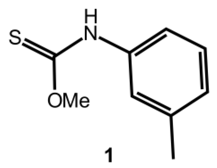
O-Methyl m-Tolylcarbamothioate

by
Chien Ing Yeo
and
Edward R. T. Tiekink
*
Research Centre for Crystalline Materials, School of Science and Technology, Sunway University, No. 5 Jalan Universiti, Bandar Sunway 47500, Selangor Darul Ehsan, Malaysia
*
Author to whom correspondence should be addressed.
Molbank 2018, 2018(3), M1020; https://doi.org/10.3390/M1020
Submission received: 4 September 2018 / Revised: 13 September 2018 / Accepted: 13 September 2018 / Published: 15 September 2018
(This article belongs to the Section Structure Determination)

Abstract

:
The synthesis, spectroscopic, and crystallographic characterisation of the title compound, O-methyl m-tolylcarbamothioate, MeOC(=S)N(H)(m-tolyl) (1), are described. The crystallographic study confirms the structure determined by spectroscopy and shows the presence of the thioamide tautomer, a syn-disposition of the thione-S and thioamide-N-H atoms and, in the crystal, thioamide-N-HS(thione) hydrogen bonding leading to an eight-membered {HNCS}2 synthon.

1. Introduction

Introducing small changes in molecules and monitoring their influence upon molecular packing is a key paradigm of crystal engineering, a discipline with the ultimate aim of controlling the manner by which molecules assemble in the crystalline state. In this context, thione molecules related to the title thiocarbamate derivative, that is, of general formula ROC(=S)N(H)R′ (for R, R′ = alkyl, aryl), have proven to readily afford crystals, making their systematic study amenable. A common feature of the molecular structure of many of these derivatives is a syn-disposition of the thione-S and thioamide-N-H atoms, enabling the formation of the eight-membered {HNCS}2 synthon in their crystals [1]. Introducing substituents in the N-bound aryl rings, for example, p-C(=O)OMe, results in an anti-disposition of the thione-S and thioamide-N-H atoms, and the formation a linear supramolecular chain mediated by thioamide-N-HO(carbonyl) hydrogen bonds in the crystal [2]. In the same way, incorporating pyridyl residues in these molecules, for example, in (p-py)CH2OC(=S)N(H)Ph, also leads to an anti-disposition within the thioamide residue and zig-zag supramolecular chains via thioamide-N-HN(pyridyl) hydrogen bonds [3]. In continuation of studies in this area, herein the synthesis and spectroscopic (1H and 13C{1H} NMR, UV and IR) characterisation of MeOC(=S)N(H)(m-tolyl) (1), Scheme 1, are described along with the determination of the crystal and molecular structures. Thiocarbamate 1 is a known compound with a CAS registry entry of 1087417-60-4. The synthesis and some characterisation data (melting point and microanalysis) of 1 has been described previously [4] and the molecule has been complexed to phosphanegold(I) in a crystallographic study, but no characterisation data for 1 were included [5]. The compound may be purchased from two vendors, namely from Ambinter [6] and from the FCH Group, Latvia [7].

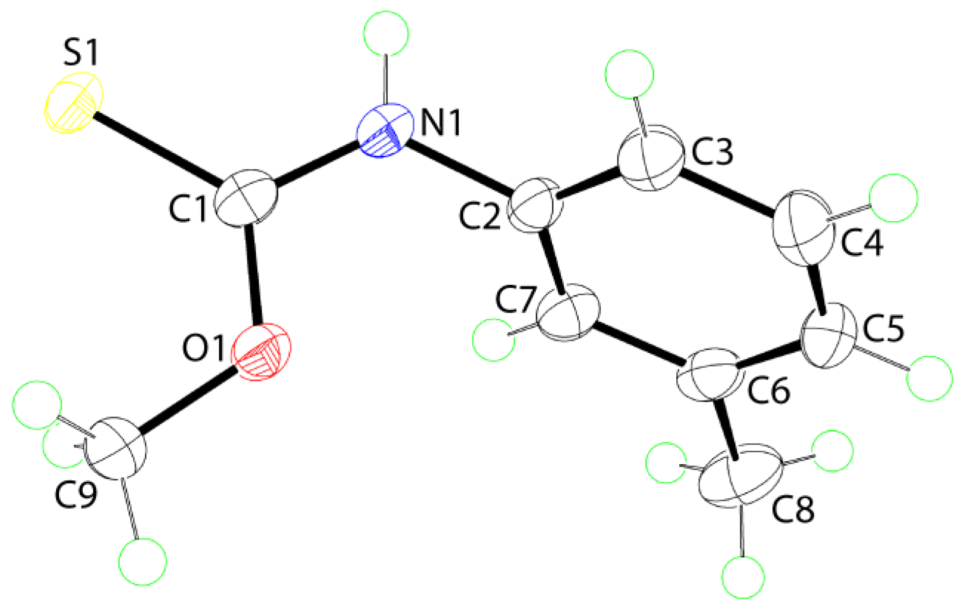
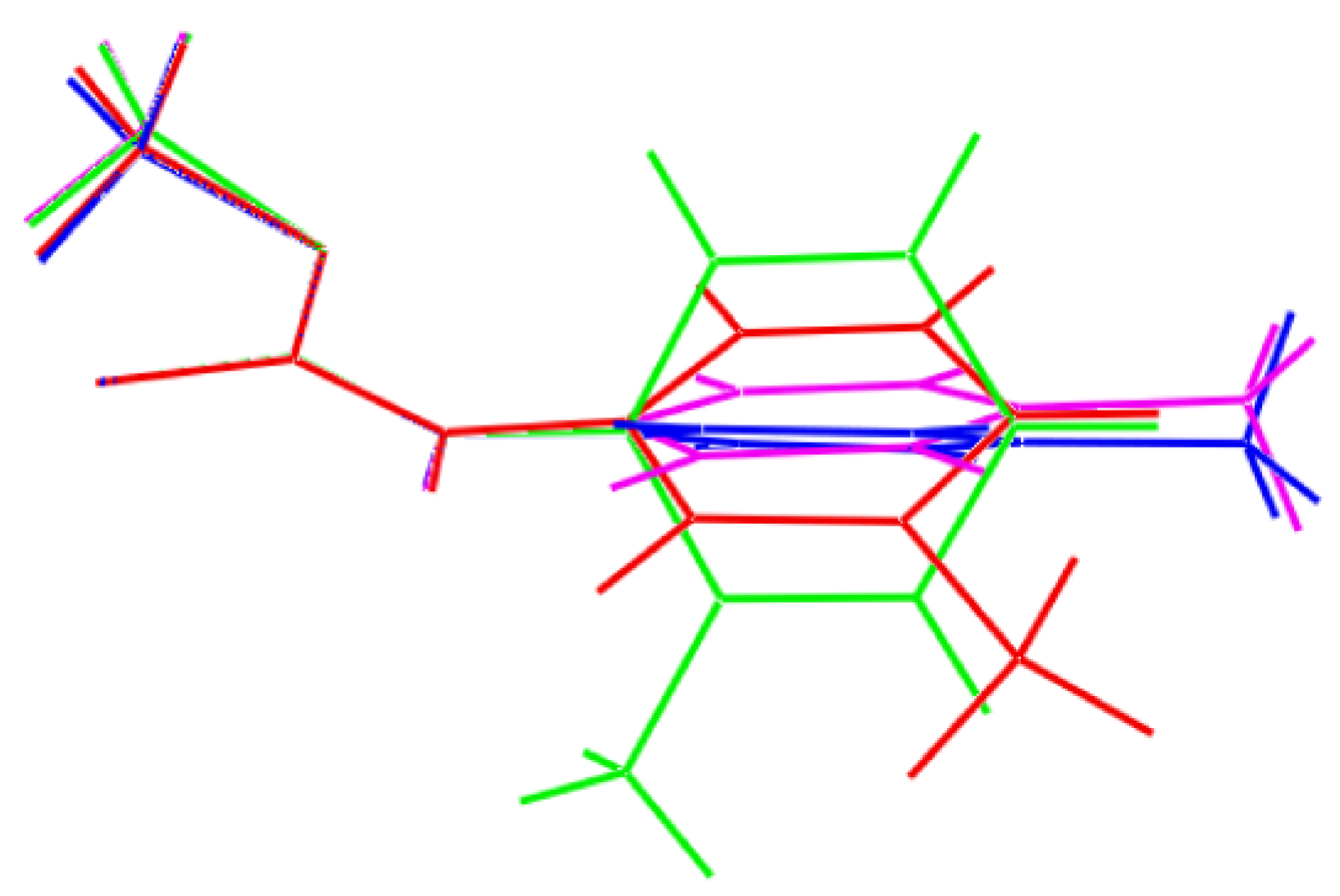
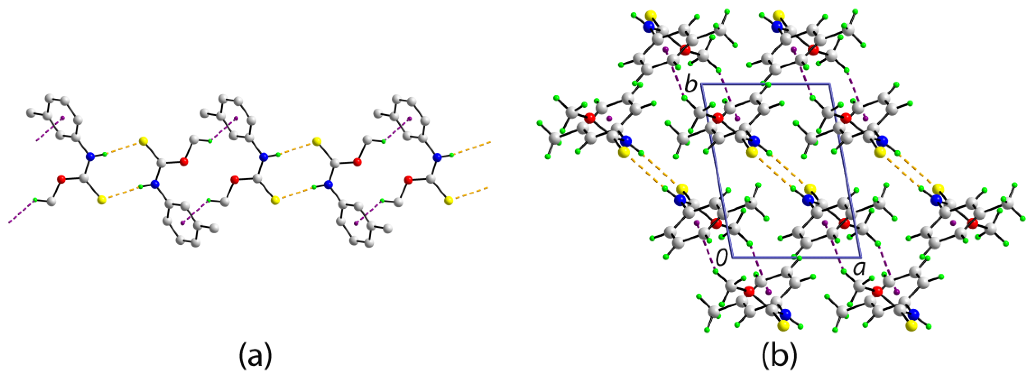
2. Results and Discussion

The title compound, 1, was prepared from the facile and stoichiometric reaction of m-tolyl isothiocyanate and NaOH conducted in methanol solution. Compound 1 is low-melting point solid that was characterised by IR, NMR, and UV spectroscopy (see Supplementary Materials for original spectra). Crystals of 1 formed in the mother liquor when kept in a refrigerator, that is, at ca. 4 °C. The isolated crystals melted almost immediately upon removal from the mother liquor under ambient conditions. This behaviour contrasts a previous literature report, where a melting point of 43 °C was reported [4]. In the present study, crystals were analysed immediately before they began to melt. This behaviour precluded the recording of a melting point and the determination of microanalytical data. In the IR, the expected bands due to ν(N-H) (3182 (br)), ν(C-N) (1444 (vs)), ν(C = S) (1203 (vs)), and ν(C-O) (1090 (m)) were observed. The 1H and 13C{1H} NMR data showed the expected resonances and integration (1H). The quaternary carbon-C1 atom exhibited a 13C{1H} resonance at δ 189.6. A single band at ca. 275 nm was observed in the UV spectrum which is ascribed to a π-π* transition [8].
The molecular structure as determined by X-ray crystallography is shown in Figure 1 and selected geometric parameters are included in the caption. The C1=S1 and C1-N1 bond lengths confirm the presence of the thioamide tautomer. The S1, O1, N1, and C1 atoms of the chromophore are strictly planar, exhibiting a r.m.s. deviation of 0.0051 Å with the appended C2 and C9 atoms, respectively, lying 0.024(2) and 0.086(3) Å out of and to one side of the plane. The variations in the bond angles about the quaternary-C1 atom follow the expected trends with those involving the doubly-bonded sulfur atom being systematically wider by ca. 12° than O1-C1-N1. There is a significant twist in the molecule as seen in the value of the C1-N1-C2-C7 torsion angle of 67.8(2)°, and this is manifested in a dihedral angle of 68.12(5)° between the thioamide and m-tolyl residues.
The most prominent feature of molecular packing in the crystal of 1 are thioamide-N-HS(thione) hydrogen bonds linking centrosymmetrically related molecules. These hydrogen bonds lead to the formation of an eight-membered {HNCS}2 synthon and a two-molecule aggregate; the geometric parameters that characterise the identified intermolecular interactions are included in the caption to Figure 2. The aggregates thus formed are linked into a supramolecular chain via methoxy-C9-Hπ(m-tolyl) interactions. The chain has a linear topology and is propagated approximately along [−1 1 0], Figure 2a. The chains pack into a three-dimensional architecture without directional interactions between them with the only contact marginally less than the sum of the van der Waals radii (3.00 Å [9]) is an inter-chain m-tolyl-C7-H7S1 contact of 2.91 Å; a view of the unit cell contents is shown in Figure 2b.
As mentioned in the Introduction, systematic structural studies underpin crystal engineering endeavours as in order to control the way molecules self-assemble in crystals, a detailed knowledge of the intermolecular interactions they form is crucial. With this in mind, the relationship between 1 and closely related literature precedents were evaluated. In the o-tolyl derivative [10], one molecule comprises the crystallographic asymmetric unit, but, in the p-tolyl crystal [11], two crystallographically independent but conformationally similar molecules comprise the asymmetric unit. As seen from the overlay diagram in Figure 3, the overlap between the central S1,O1,N1,C1 chromophore and the pendent methyl group is close between 1, the o-tolyl derivative [10] and each of the independent molecules of the p-tolyl derivative [11]. However, significant conformational differences exist between the tolyl groups. Crucially, the syn disposition is retained in all four molecules enabling the formation of the thioamide-N-HS(thione) hydrogen bonds in their respective crystals. The robust nature of the eight-membered {HNCS}2 synthon thus formed has been commented upon in a recent survey of thiocarbamate structures [1]. This is only disrupted when other, more electron-rich acceptors, for example, carbonyl-oxygen [2] and pyridyl-nitrogen [3], are present in the R and/or R′ substituents of ROC(=S)N(H)R′.
In conclusion, the X-ray crystal structure determination of 1, revealing a syn disposition of the thione-S and thioamide-H atoms in the molecular structure, and thioamide-N-HS(thione) hydrogen bonding leading to eight-membered {HNCS}2 synthons in the molecular packing, is consistent with literature expectation.

3. Materials and Methods

3.1. General Information

All chemicals and solvents were sourced from Merck and used without further purification. IR spectra were measured on a Bruker Vertex 70v FTIR spectrophotometer from 4000 to 400 cm−1; abbreviations: br, broad; m, medium; vs, very strong. 1H and 13C{1H} NMR spectra were recorded in CDCl3 solution on a Bruker Ascend 400 MHz NMR spectrometer with chemical shifts relative to tetramethylsilane; abbreviations for NMR assignments: s, singlet; m, multiplet; br, broad. The optical absorption spectra were obtained from an acetonitrile solution of 1.0 × 10⁻⁶ M in the range 200–800 nm on a Shimadzu UV-3600 plus UV/VIS/NIR spectrophotometer.

3.2. Synthesis and Characterisation

m-Tolyl isothiocyanate (2.5 mmol, 0.34 mL) was added to NaOH (2.5 mmol, 0.10 g) in MeOH (3 mL) and the mixture was stirred at room temperature for 2 h, followed by the addition of excess 5M HCl solution. The resulting mixture was stirred for another 1.5 h. The final product was extracted with chloroform (15 mL) and left for evaporation at 4 °C in a refrigerator, yielding colourless crystals after 5 weeks. Characterisation was performed on harvested crystals from the mother liquor. The title compound melts soon after removal from the mother liquor. IR (cm−1): 3182 (br) (N-H), 1444 (vs) (C-N), 1203 (vs) (C=S), 1090 (m) (C-O). 1H-NMR (CDCl3): δ 8.38 (s, br, 1H, NH), 7.26–6.99 (m, br, 4H, aryl-H), 4.13 (s, 3H, OCH3), 2.35 (s, 3H, aryl-CH3) ppm. 13C{1H} NMR (CDCl3): δ 189.6 (Cq), 139.1 (aryl-C1), 136.9 (aryl-C3), 128.9 (aryl-C5), 126.5 (aryl-C2), 122.4 (aryl-C4), 119.0 (aryl-C6), 58.9 (OCH3), 21.4 (aryl-CH3) ppm. UV (acetonitrile): λmax = 274.50, ε = 210,000 L·cm−1·mol−1.

3.3. Crystallography

Intensity data for 1 were measured at T = 100(2) K on an Agilent Technologies SuperNova Dual diffractometer fitted with Mo Kα radiation so that θmax was 27.5°. Data reduction, including absorption correction, was accomplished with CrysAlis Pro [12]. Of the 3380 reflections measured, 3380 were unique (Rint = 0.022), and of these, 1756 data satisfied the I ≥ 2σ(I) criterion of observability. The structure was solved by direct methods [13] and refined (anisotropic displacement parameters and C-bound H atoms in the riding model approximation; the N-bound H atom was refined without restraint) on F2 [14]. A weighting scheme of the form w = 1/(σ2(Fo2) + (0.043P)2 + 0.163P) was introduced, where P = (Fo2 + 2Fc2)/3. Based on the refinement of 115 parameters, the final values of R and wR (all data) were 0.036 and 0.095, respectively. The molecular structure diagram was generated with ORTEP for Windows [15] and the packing diagram using DIAMOND [16].
Crystal data for C9H11NOS (1): M = 181.25, triclinic, P¯1, a = 5.9025(5), b = 8.2726(6), c = 10.0552(7) Å, α = 105.957(6), β = 101.970(6), γ = 96.271(6)°, V = 454.53(6) Å3, Z = 2, Dx = 1.324 g cm−3 and μ = 0.306 mm−1. CCDC deposition number: 1864622.

Supplementary Materials

The following are available online. 1H and 13C{1H} NMR, UV, and IR spectra, and crystallographic data for 1 in Crystallographic Information File (CIF) format. CCDC 1864622 also contains the supplementary crystallographic data for this paper. These data can be obtained free of charge via http://www.ccdc.cam.ac.uk/conts/retrieving.html.

Author Contributions

C.I.Y. was the only experimentalist, who obtained and analysed all data, apart from the X-ray crystallography, which was performed by E.R.T.T.

Funding

This research received no external funding. The APC was funded by Sunway University.

Acknowledgments

The X-ray crystallography laboratory at the University of Malaya is thanked for providing the X-ray intensity data.

Conflicts of Interest

The authors declare no conflict of interest.

References

  1. Jotani, M.M.; Yeo, C.I.; Tiekink, E.R.T. A new monoclinic polymorph of N-(3-methylphenyl)ethoxycarbothioamide: Crystal structure and Hirshfeld surface analysis. Acta Crystallogr. E 2017, 73, 1889–1897. [Google Scholar] [CrossRef] [PubMed]
  2. Ho, S.Y.; Bettens, R.P.A.; Dakternieks, D.; Duthie, A.; Tiekink, E.R.T. Prevalence of the thioamide {H‒N‒C=S}2 synthon—Solid-state (X-ray crystallography), solution (NMR) and gas-phase (theoretical) structures of O-methyl-N-aryl-thiocarbamides. CrystEngComm 2005, 7, 682–689. [Google Scholar] [CrossRef]
  3. Xiao, H.-L.; Wang, K.-F.; Jian, F.-F. (4-Pyridyl)methyl N-phenylthiocarbamate. Acta Crystallogr. E 2006, 62, o2852–o2853. [Google Scholar] [CrossRef]
  4. Burrows, A.A.; Hunter, L. The associating effect of the hydrogen atom. Part XV*. The S-H-N bond. Esters of thion- and dithio-carbamic acids. J. Chem. Soc. 1952, 4118–4122. [Google Scholar] [CrossRef]
  5. Kuan, F.S.; Ho, S.Y.; Tadbuppa, P.P.; Tiekink, E.R.T. Electronic and steric control over AuAu, C-HO and C-Hinteractions in the crystal structures of mononuclear triarylphosphinegold(I) carbonimidothioates: R3PAu[SC(OMe)=NR′] for R = Ph, o-tol, m-tol or p-tol, and R′ = Ph, o-tol, m-tol, p-tol or C6H4NO2-4. CrystEngComm 2008, 10, 548–564. [Google Scholar] [CrossRef]
  6. Ambinter. Available online: http://www.ambinter.com/ (accessed on 13 September 2018).
  7. FCH Group, Latvia. Available online: http://fchgroup.net/ (accessed on 13 September 2018).
  8. Ho, S.Y.; Cheng, E.C.C.; Tiekink, E.R.T.; Yam, V.W.W. Luminescent phosphine gold(I) thiolates: Correlation between crystal structure and photoluminescent properties in [R3PAu{SC(OMe)=NC6H4NO2-4}] (R = Et, Cy, Ph) and [(Ph2P-R-PPh2){AuSC(OMe)=NC6H4NO2-4}2] (R = CH2, (CH2)2, (CH2)3, (CH2)4, Fc). Inorg. Chem. 2006, 45, 8165–8174. [Google Scholar] [CrossRef] [PubMed]
  9. Spek, A.L. Structure validation in chemical crystallography. Acta Crystallogr. D 2009, 65, 148–155. [Google Scholar] [CrossRef] [PubMed] [Green Version]
  10. Kuan, F.S.; Tadbuppa, P.; Tiekink, E.R.T. Crystal structure of o-methyl N-(o-tolyl)thiocarbamate, SC(OCH3)NH(C6H4CH3). Z. Kristallogr. New Cryst. Struct. 2005, 220, 393–396. [Google Scholar]
  11. Ho, S.Y.; Kuan, F.S.; Tiekink, E.R.T. (E)-O-Methyl N-(4-methylphenyl)thiocarbamate. Acta Crystallogr. E 2007, 63, o1723–o1724. [Google Scholar] [CrossRef]
  12. Rigaku Oxford Diffraction, CrysAlis PRO; Agilent Technologies Inc.: Santa Clara, CA, USA, 2011.
  13. Sheldrick, G.M. A short history of SHELX. Acta Crystallogr. A 2008, 64, 112–122. [Google Scholar] [CrossRef] [PubMed]
  14. Sheldrick, G.M. Crystal structure refinement with SHELXL. Acta Crystallogr. C 2015, 71, 3–8. [Google Scholar] [CrossRef] [PubMed]
  15. Farrugia, L.J. WinGX and ORTEP for Windows: An update. J. Appl. Crystallogr. 2012, 45, 849–854. [Google Scholar] [CrossRef]
  16. Brandenburg, K. DIAMOND; Crystal Impact GbR: Bonn, Germany, 2006. [Google Scholar]
Scheme 1. Chemical structure diagram for 1.
Molbank 2018 m1020 sch001
Figure 1. The molecular structures of 1 showing atom labelling and displacement ellipsoids at the 70% probability level. Selected geometric parameters: C1=S1 = 1.6721(16), C1-O1 = 1.3342(19), and C1-N1 = 1.331(2) Å; S1-C1-O1 = 124.84(12), S1-C1-N1 = 123.32(12), and O1-C1-N1 = 111.81(14)°.
Molbank 2018 m1020 g001
Figure 2. The molecular packing in the crystal of 1: (a) a view of the supramolecular chain sustained by N-HS hydrogen bonds and methoxy-C-Hπ(m-tolyl) interactions shown as orange and purple dashed lines, respectively. Non-participating hydrogen atoms have been omitted. (b) A view of the unit cell contents in projection down the c-axis. Selected geometric parameters for the specified intermolecular interactions: N1-H1nS1i = 2.52(2) Å, N1S1i = 3.3757(18) Å with angle at H1n = 169.5(17)°. C9-H9aCg(C2-C7)ii = 2.80 Å, C9Cg(C2-C7)ii = 3.4892(19) Å with angle at H9a = 128°. Symmetry operations: (i) 1-x, 1-y, 1-z and (ii) 2-x, -y, 1-z.
Molbank 2018 m1020 g002
Figure 3. Superimposition of MeOC(=S)N(H)(tolyl) molecules for m-tolyl (1; red image), o-tolyl (green), p-tolyl (first independent molecule; blue) and p-tolyl (second molecule; pink). The molecules have been overlapped so the central NOS chromophores are coincident.
Molbank 2018 m1020 g003

