Article

Vasorelaxant and Antioxidant Activities of Spilanthes acmella Murr.

1
Department of Pharmacology, Faculty of Medicine, Srinakharinwirot University, Bangkok 10110, Thailand
2
Department of Chemistry, Faculty of Science, Srinakharinwirot University, Bangkok 10110, Thailand
3
Department of Clinical Microbiology, Faculty of Medical Technology, Mahidol University, Bangkok 10700, Thailand
4
Laboratory of Pharmacology, Chulabhorn Research Institute, Bangkok 10210, Thailand
5
Laboratory of Medicinal Chemistry, Chulabhorn Research Institute , Bangkok 10210, Thailand
*
Authors to whom correspondence should be addressed.
Int. J. Mol. Sci. 2008, 9(12), 2724-2744; https://doi.org/10.3390/ijms9122724
Submission received: 12 June 2008 / Revised: 14 December 2008 / Accepted: 14 December 2008 / Published: 18 December 2008
(This article belongs to the Section Biochemistry)

Abstract

:
This study reports the effect of Spilanthes acmella Murr. extracts on phenylephrine-induced contraction of rat thoracic aorta as well as their antioxidant activity. Results show that the extracts exert maximal vasorelaxations in a dose-dependent manner, but their effects are less than acetylcholine-induced nitric oxide (NO) vasorelaxation. Significant reduction of vasorelaxations is observed in both NG-nitro-l-arginine methyl ester (l-NAME) and indomethacin (INDO). In the presence of l-NAME plus INDO, synergistic effects are observed, leading to loss of vasorelaxation of both acetylcholine and the extracts. Similarly, the vasorelaxations of the extracts are completely abolished upon the removal of endothelial cells. This demonstrates that the extracts exhibit vasorelaxation via partially endothelium-induced NO and prostacyclin in a dose-dependent manner. Significantly, the ethyl acetate extract exerts immediate vasorelaxation (ED50 76.1 ng/mL) and is the most potent antioxidant (DPPH assay). The chloroform extract shows the highest vasorelaxation and antioxidation (SOD assay). These reveal a potential source of vasodilators and antioxidants.

1. Introduction

Spilanthes acmella Murr. (para-cress or toothache plant) is a medicinal plant of the Compositae family [1]. It is known in Thai as Phak-Kratt Huawaen and has long been used as a traditional medicine for toothache, headache and treatment of asthma, rheumatism, fever, sore throat and haemorrhoids [13]. Its root decoction has been used as a laxative and diuretic drug [2, 4]. It is an annual or short-lived herb, spreads in low open places and requires moist soil [5]. Its flowers and leaves have a pungent taste, accompanied with tingling and numbness [2].
Constituents found in Spilanthes acmella Murr. were spilanthol, isobutylamide derivatives [6, 7], α-and β-amyrin esters, stigmasterol, myricyl alcohol including sitosterol glucosides [8] and triterpenoidal saponins [9]. Spilanthol showed interesting bioactivities, e.g. strong local anesthetic [10], analgesic [11, 12] and insecticidal activities [3]. Crude flower head extracts of the plant exhibited potent ovicidal, marked larvicidal (LC50 of 61.43 ppm) and pupicidal activities [13, 14]. The extracts were also shown to exhibit pancreatic lipase inhibition which has potential as candidates for weight loss and obesity control [15]. In addition, the crude flower head extracts displayed strong diuretic action which caused marked increase in urine Na+, K+ and a reduction in the osmolarity of urine [16]. Moreover, it has been used as anti-toothache formulation for pain relieving within 2–10 min and also useful in treating the swelling and infection of gums [17]. The Spilanthes acmella is one of the active ingredients in compositions for acute- or long-term treatment of microbial infections, particularly, oral pathogenic microorganisms, dental caries, periodontosis, gum disease, gum bleeding and/or plaque reduction [18]. In addition, its root powder extract is very effective in treating HIV/AID infection [19]. Presently, cardiovascular disease is one of the most important public health concerns, accountable for morbidity and mortality. Thus new vasoactive ingredients that influence the endothelial cells, which are key regulating cells in the vessel wall, are the striking areas. So far, the effect of Spilanthes acmella Murr. on vascular function has not been reported. In this regard, it was of great interest to explore its medicinal value with regards to cardiovascular functions such as regulation of vascular tone. In the present study, we have investigated the effects of Spilanthes acmella Murr. extracts on rat thoracic aorta and its mechanism of action as well as its antioxidant activity.

2. Results

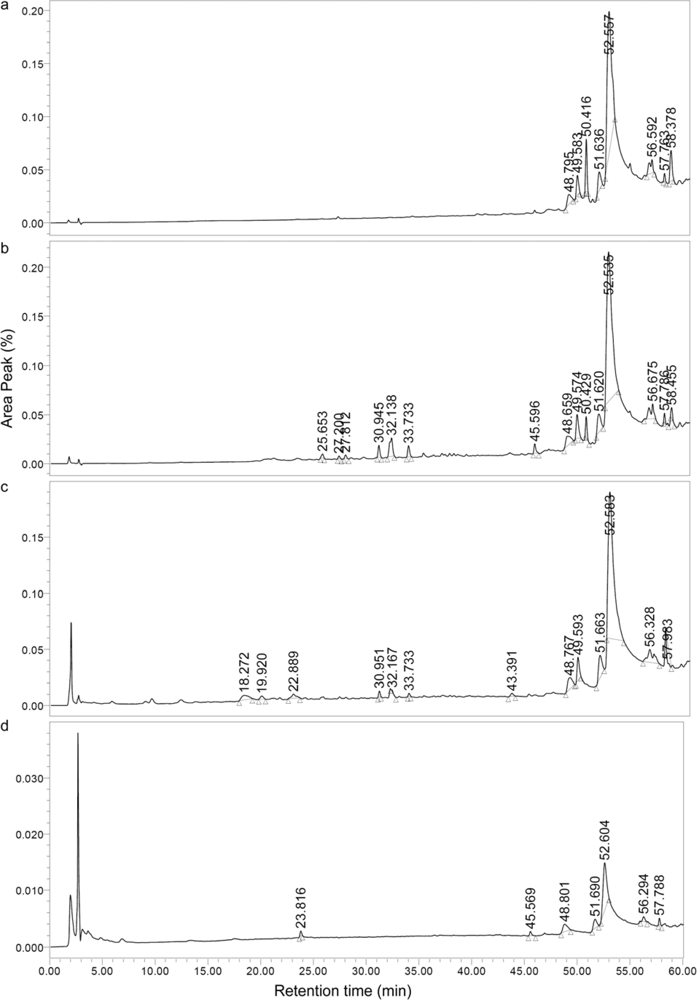
2.1. Components and HPLC (high performance liquid chromatography) profiles of the extracts

Preliminary screening of components in the Spilanthes acmella Murr. extracts was performed using thin layer chromatography (TLC) chromatograms, IR and 1H-NMR spectra. It was found that hexane, chloroform, ethyl acetate and methanol extracts showed UV absorption on TLC. The ethyl acetate and methanol extracts exhibited very strong absorptions, compared to the chloroform extract. The IR spectra of the extracts showed absorptions (cm−1) corresponding to OH (3360–3385), CH (2850–2942), CO (1710–1736), 1454–1574 and C-O (1047–1385) functions. The ethyl acetate and methanol extracts exhibited very strong OH and CO (ester or acid) and strong C-O absorptions. The chloroform extract displayed strong OH, medium CO (ester or acid) and C-O absorptions. The hexane extract showed strong CO (ester or acid), but medium OH and weak C-O absorptions. All the extracts showed strong CH absorptions, the the hexane extract displaying the strongest ones. The 1H-NMR spectra of the hexane and chloroform extracts exhibited the presence of triterpenes (δ 0.9–2.4, 3.4–3.7, 4.1–4.3 and 5.3–5.4). The ethyl acetate extract showed the signals of triterpenes and aromatics (δ 0.84–2.4, 3.5–4.3, 5.3–5.5 and 6.8–7.8). The methanol extract displayed the presence of aromatic protons (δ 6.8–8.4).
Based on the IR and 1H-NMR data, it may be concluded that the hexane and chloroform extracts contain triterpenes and long chain hydrocarbon esters or alcohols. The ethyl acetate extract contains triterpene esters and phenolic compounds, while the components of the methanol extract are presumably phenolic compounds and esters.
Profiles of the extracts were obtained by reversed phase HPLC running from a more polar to less polar mobile phase (Figure 1). Major nonpolar components of the extracts appeared in the 48.80–58.38 min retention time (RT) range. In the hexane extract the major component (66.06%) was found at a RT of 52.56 min, while th major components of the chloroform (65.72%), ethyl acetate (67.34%) and methanol (64.18%) extracts were observed at RTs of 52.54, 52.58 and 52.60 min, respectively. It was noted that minor contents (0.22–4.72%) of polar components in chloroform and ethyl acetate extracts appeared at RTs of 18.27–33.73 min. However, the methanol extract showed a minor polar component (2.64%) at a RT of 23.82 min (Table 1).

2.2. Vasorelaxant activity

Effects of Spilanthes acmella Murr. extracts (hexane, chloroform, ethyl acetate and methanol) on vascular function of rat thoracic aorta precontracted with phenylephrine (PE) were investigated under various conditions; in the presence or absence of inhibitors, namely, NG-nitro-l-arginine methyl ester (l-NAME) and indomethacin (INDO) and under removal of functional endothelial cells. In addition, the effects of acetylcholine (ACh) as a positive control, sodium nitroprusside (SNP) a negative control and vehicle such as polyethyleneglycol (PEG) were studied. Results confirmed that the vasorelaxation of ACh was related to nitric oxide (NO).

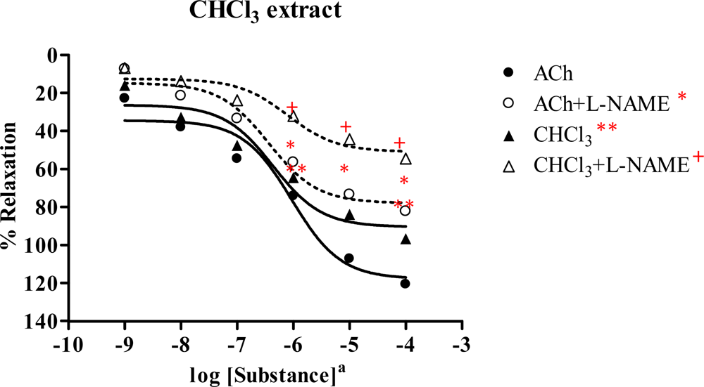
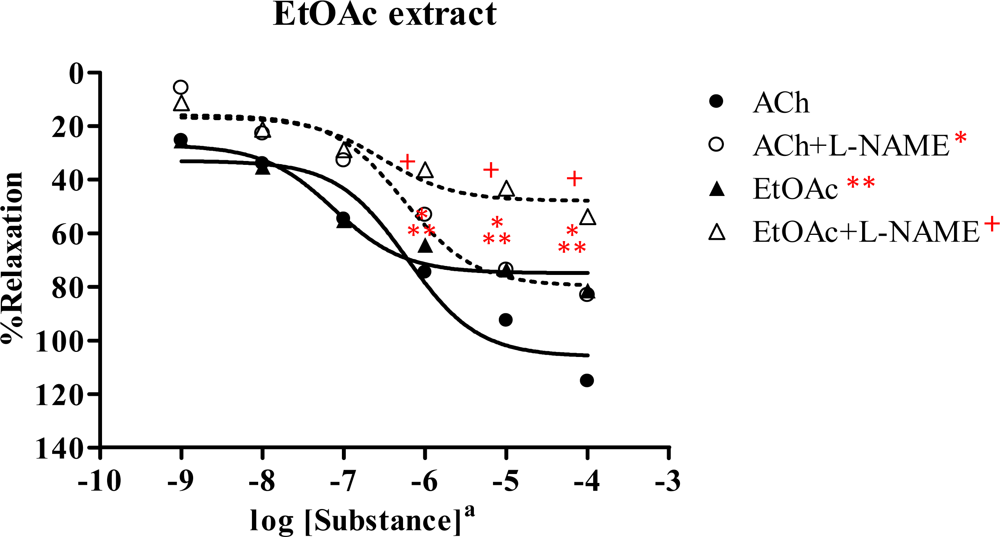
2.3. Effect of Spilanthes acmella Murr. extracts on the vascular function of rat thoracic aorta in the absence or presence of a NOS inhibitor (L-NAME, 1 mM)

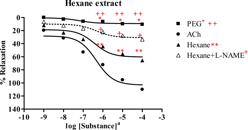
2.3.1. Hexane extract

In the rat aortic ring preparation, the hexane extract exerted vasorelaxation in the dose-dependent manner, but PEG showed no effect on the vessels (Figure 2 and Table 2). The activity of the hexane extract reached up to 65.7% of the Rmax induced by ACh (110.1%) showing ED50 3.60×10⁻⁷ mg/mL and 4.66×10⁻⁷ M, respectively. This indicated that the hexane extract likely acted as a partial agonist. In the presence of a NOS inhibitor (l-NAME), the dose-response curve of hexane extract was shifted to the right, with Rmax 33.5% and ED50 4.80×10⁻⁷ mg/mL. This suggested that the hexane extract caused vasorelaxation by partially producing NO from the endothelial cells, while PEG had no effect on induction of vasorelaxation.

2.3.2. Chloroform extract

Similarly, the chloroform extract showed dose-dependent vasorelaxation (Figure 3 and Table 2). The Rmax of ACh and the chloroform extract were 120.6 and 96.6%, with ED50 9.43×10⁻⁷ M and 4.28×10⁻⁷mg/mL, respectively. In the presence of l-NAME, the dose-response curve of chloroform extract was shifted to the right. The extract showed Rmax 54.4% and ED50 1.00×10⁻⁶ mg/mL, whereas Rmax and ED50 of ACh were 82.1% and 3.61×10⁻⁷ M, respectively. This demonstrated that the chloroform extract exhibited vasorelaxation by partly production of NO from the endothelial cells.

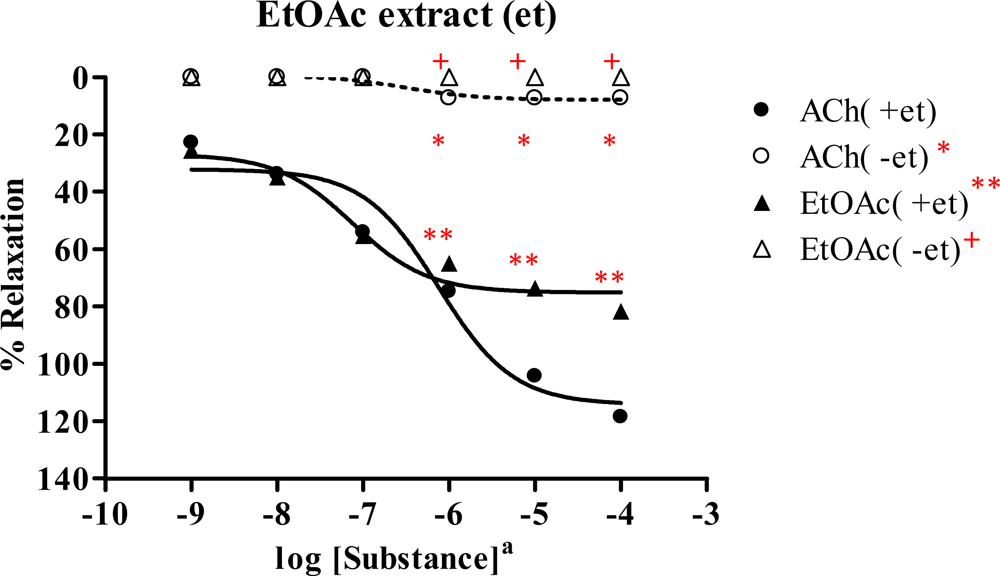
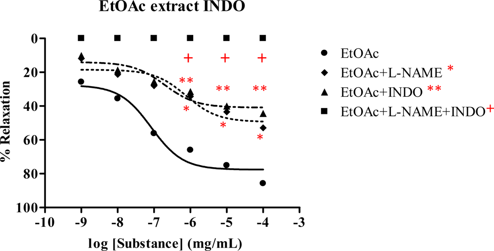
2.3.3. Ethyl acetate extract

A similar result was obtained from ethyl acetate extract, showing vasorelaxation in the dose-dependent manner (Figure 4 and Table 2). The ACh and plant extract exerted Rmax 115.5 and 81.6%, with ED50 of 5.91×10⁻⁷ M and 7.61×10⁻⁸ mg/mL, respectively. This showed that the ethyl acetate extract possibly acted as a partial agonist. In the presence of l-NAME, the dose-response curve of the extract was shifted to the right with Rmax 54.0%, ED50 3.88×10⁻⁷ mg/mL, whereas the Rmax and ED50 of ACh were 83.4% and 5.65×10⁻⁷ M, respectively. This suggested that the ethyl acetate extract caused vasorelaxation by partly synthesis of NO from the endothelial cells.

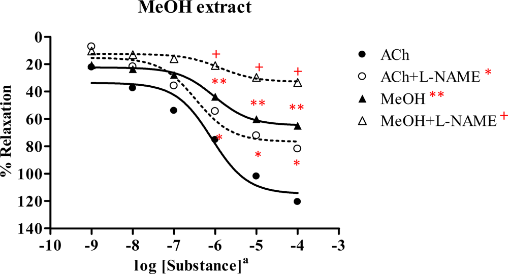
2.3.4. Methanol extract

The methanol extract displayed vasorelaxant activity in a dose-dependent manner (Figure 5 and Table 2). The Rmax of ACh and extract were 120.7 and 65.1% with ED50 8.18×10⁻⁷ M and 9.55×10⁻⁷ mg/mL, respectively. This indicated that the extract possibly acted as partial agonist. In the presence of l-NAME, the dose-response curve of extract was shifted to the right with Rmax 33.4% and ED50 1.38×10⁻⁶ mg/mL, whereas the Rmax and ED50 of ACh were 82.0% and 3.43×10⁻⁷M, respectively. This illustrated that the methanol extract exerted vasorelaxation by partial NO production from the endothelial cells.

2.4. Effect of Spilanthes acmella Murr. on the vascular function of rat thoracic aorta in the absence of endothelial cells

The vasorelaxation of Spilanthes acmella Murr. extracts was examined with and without intact endothelial cells, comparing with ACh. The results (Table 3) illustrated that the vasorelaxation of all the tested extracts was abolished when endothelial cells were removed. An example, the dose-response curve of the ethyl acetate extract is shown in Figure 6. Similar results were observed for the control, ACh. This confirmed that the vasorelaxant activity of Spilanthes acmella Murr. extracts was mediated via endothelial cells producing NO.

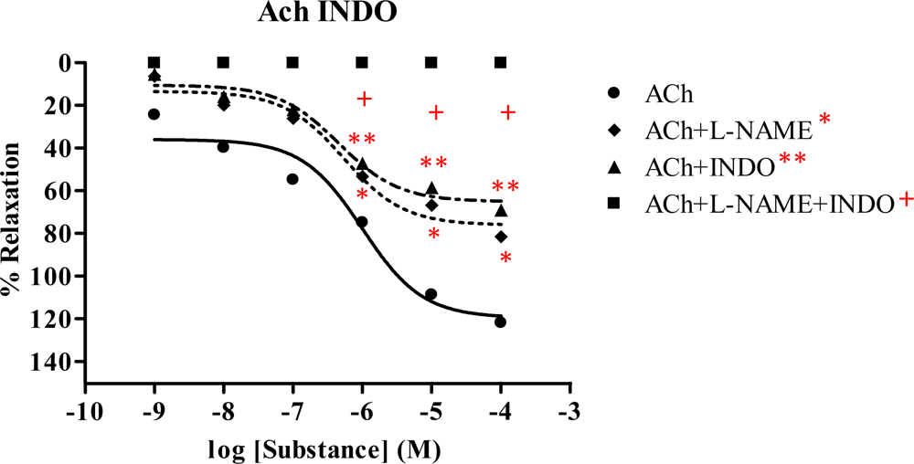
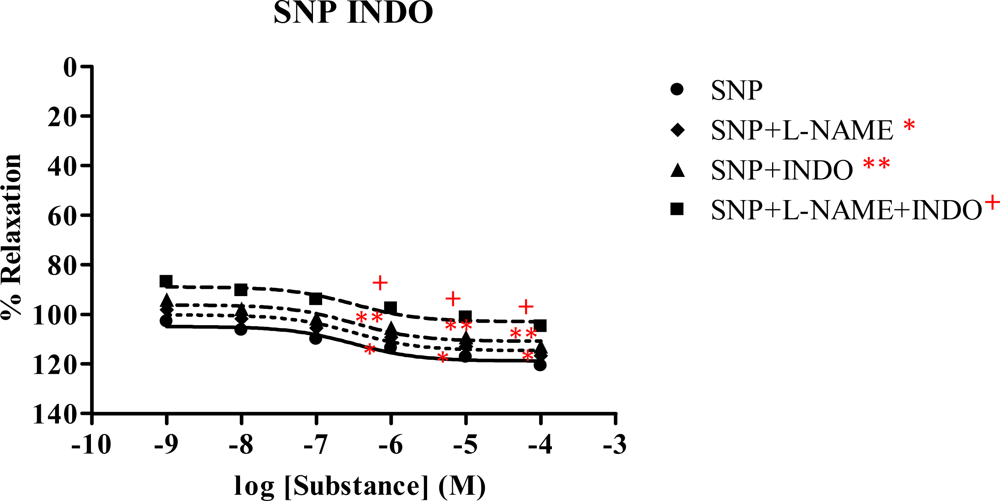
2.5. Effect of Spilanthes acmella Murr. on the vascular function of rat thoracic aorta in the presence of a cyclooxygenase inhibitor (INDO)

The vasorelaxation of Spilanthes acmella Murr. extracts was studied in the presence of INDO (1 mM), comparing with l-NAME (1 mM) and l-NAME plus INDO. The results are summarized in Table 4 and some dose-response curves are shown in Figures 79. It was observed that in each experiment with l-NAME (1 mM) or INDO (1 mM) the vasorelaxation was reduced in a dose-dependent manner. The inhibition effect of the INDO was stronger than that of l-NAME. However, the antagonist effects of l-NAME or INDO were more pronounced in the case of l-NAME plus INDO. Such effects were not observed in the SNP dose-response curves. Interestingly, the vasorelaxations were abolished by all the tested extracts and by the ACh in the presence of l-NAME (1 mM) plus INDO (1 mM), but no significant change was noted in the case of SNP. The results confirmed that the extracts caused partial vasorelaxation through endothelial cells producing NO and prostacyclin (PGI2).

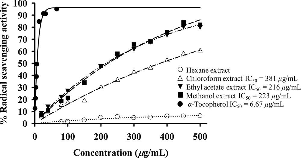
2.6. Antioxidant activity

The antioxidant activity of Spilanthes acmella Murr. extracts was measured using DPPH and SOD assays. The results (Table 5) showed that all the tested extracts exhibited antioxidative activity. In DPPH assay (Figure 10) at 200 μg/mL, the ethyl acetate and methanol extracts displayed comparable activity and the highest radical scavenging activity (47.90 and 47.76%) with IC50 216 and 223 μg/mL, while α-tocopherol (a positive control) showed antioxidant activity with IC50 6.67 μg/mL. The chloroform extract exhibited 29.82% radical scavenging activity. The hexane extract produced some activity (4.90% radical scavenging).
The SOD assay at 200 μg/mL (Figure 11), the chloroform extract showed the highest antioxidant activity (57.92% NBT inhibition). The ethyl acetate and methanol extracts exhibited weak to moderate SOD activity.

3. Discussion

We have disclosed the effect of Spilanthes acmella Murr.extracts on phenylephrine-induced contraction of rat thoracic aorta. All the tested plant extracts elicit maximal vasorelaxations in a dose-related manner, although such vasorelaxations are less than those produced by ACh. The vasorelaxations are exerted by partial production of NO from functional endothelial cells, which is demonstrated by a significant reduction of the activity in the presence l-NAME (Table 2).
The chloroform extract exhibited the highest Rmax of 96.6%, compared to the other extracts. The ethyl acetate extract shows strong vasorelaxation (Rmax 81.6%), while the nonpolar hexane and polar methanol extracts exert comparable activity, with Rmax 65.67 and 65.09%, respectively. Significantly, the ethyl acetate extract exhibits immediate vasorelaxation with ED50 7.61×10⁻⁸ mg/mL (Figure 4) when compared to the chloroform extract showing ED50 4.28×10⁻⁷ mg/mL (Figure 3). This perhaps due to the fact that the ethyl acetate extract has higher affinity for the receptor than the chloroform extract and thus the ethyl acetate extract is the most potent vasorelaxant. Furthermore, the vasorelaxant activities of the tested extracts were all abolished by the removal of functional endothelial cells (Table 3 and Figure 6). This again confirms that the vasorelaxation of the extracts is modulated via NO production by endothelial cells, a fact which is known for Ach involvement in mediating vasorelaxation by NO, PGI2 and endothelium-derived hyperpolarizing factor [2022]. Therefore, a set of experiments was studied in the presence of INDO (1 mM) compared with l-NAME (1mM). The results (Table 4) show that the vasorelaxation of the tested extracts (Figure 8) and ACh (Figure 7) is significantly reduced in a dose-dependent manner when compared to that of in the presence of l-NAME. Particularly, the INDO exhibits stronger reduction of such vasorelaxation than the l-NAME, but none was not observed for the SNP (Figure 9). Additionally, in the presence of INDO (1 mM) plus l-NAME (1 mM), synergistic effects are observed, leading to complete loss of the activity of the tested extracts and ACh. However, such an effect is not markedly noticeable for the SNP. These data confirm that the vasorelaxant activity of Spilanthes acmella Murr. is exerted by functional endothelial cells producing partial synthesis of NO and PGI2 which are inhibited by l-NAME and INDO, respectively. NO is an important signaling molecule implicated in cardiovascular functions such as vascular tone. PGI2, endothelium-derived relaxing factor (EDRF), is a powerful vasorelaxant and antioxidant. It exerts antifibrotic properties preventing the development of fibrosis and cirrhosis in liver diseases including antiplatelet aggregation. PGI2 is clinically used for treatment of pulmonary hypertension and portopulmonary hypertension [23].
Many studies have reported the role of NO related to superoxide radical (O2.−) [24]. Thus, the antioxidative activity of the extracts was evaluated. It was found that the plant extracts exhibited antioxidant activity at 200 μg/mL. In a DPPH assay, the polar ethyl acetate and methanol extracts exerted the highest radical scavenging activity, while the chloroform extract shows the highest SOD activity. The nonpolar hexane extract produces some activity in both the DPPH and SOD assays.
It is noted that the ethyl acetate extract is the most potent, with immediate vasorelaxation and with the highest radical scavenging activity. This is presumably due to the presence of bioactive polar phenolic and triterpenoid ester compounds in the extract. Moreover, the chloroform extract possesses the highest Rmax accompanied by the highest SOD activity. This may result from the presence of triterpenoids and fatty alcohols or esters in this extract. Moderate vasorelaxations are observed for the nonpolar hexane and polar methanol extracts. This suggests that both hydrophobic and hydrophilic constituents show comparable vasorelaxant activity (Table 2). At this point, the ethyl acetate extract is the most promising vasorelaxant, with a significant ED50 of 76.1 ng/mL and it is also the most active antioxidant (DPPH assay). It has been reported that flavonoid, phenolic, polyphenol and triterpenoid compounds exhibit both vasorelaxant and antioxidant activities, in some cases behaving as either vasorelaxants or antioxidants [2426]. Therefore, our results imply a good correlation between vasorelaxant and antioxidant activities deriving from a diverse group of compounds.
It is well recognized that endothelium NO is diminished by reacting with O2.− to form peroxynitrite as a potent oxidant, which is inhibited by antioxidant, thus in turn improving NO induced vasorelaxation [24, 2729]. This was evidenced by phenolic antioxidants such as prinsepiol isolated from roots of Valeriana prionophylla [24]. A number of flavonoids exerted vasorelaxation through endothelium produced NO, e.g. chrysin (5, 7-dihydroxyflavone) presented in honey, propolis, fruits, vegetables and beverages [3032], isoliquiritigenin isolated from various plants such as Dahlia variabilis [33] and recently from Hydnophytum formicarum Jack. [34]. Triterpenoids are commonly found in plant species, for instances, oleanolic acid and erythrodiol elicit vasorelaxation via endothelium induced NO [26]. In addition, root extracts of Caesalpinia benthamiana, which are rich in phenolic compounds, e.g. gallic acid, resveratrol and tannin, had significant vasorelaxant and powerful scavenging activity versus O2. [25]. Some plant extracts of Casimiroa edulis and Mammea africana (Guttiferae) caused vasorelaxation through endothelium dependent producing NO [35, 36]. The vasorelaxation was also mediated by both endothelium dependent and endothelium independent pathways [37]. Moreover, extracts of ethnomedical plant used as antihypertensive and anti-stroke exhibited their vasorelaxants via endothelium dependent producing NO or via direct stimulation of NO release. Examples are Diospyros kaki Thunb., Polygonum aviculare L., Magnolia liliflora Desr., Sorbus commixta Hedl, Selaginella tamariscina Spr. and Guazuma ulmifolia [38, 39].
In this study, Spilanthes acmella Murr. extracts produce vasorelaxation via partial release of NO and PGI2 from endothelium as well as showing antioxidant activities. Some countries in Asia including Thailand use this plant as fresh vegetable and cooking ingredient [40]. This demonstrates beneficial effects and applications of the Spilanthes acmella Murr. to medical uses and as a health food.

4. Experimental Section

4.1. Materials

4.1.1. Chemicals

L-Phenylephrine hydrochloride, sodium nitroprusside, NG-nitro-L-arginine methyl ester, acetylcholine, ketamine hydrochloride and indomethacin were obtained from Sigma Chemical Co. (St. Louis, MO, USA). Polyethyleneglycol was purchased from Fluka. Plant extracts were dissolved in PEG, while methanol extract was dissolved in 0.9% normal saline. Then the solutions were further diluted by normal saline.

4.1.2. Plant materials

Aerial parts of Spilanthes acmella Murr. were collected from Nakornsrithammarat Province, Thailand. It has been identified (BKF 112361) by The Forest Herbarium, Royal Forestry Department. A voucher specimen has been deposited at Department of Chemistry, Faculty of Science, Srinakharinwirot University, Thailand.

4.2. Methods

4.2.1. Extraction

The air dried Spilanthes acmella Murr. (1,050 g) was ground and extracted with hexane (3×5 days), followed by filtration. The filtrates were combined and evaporated in vacuo to give a crude hexane extract (11 g). Similarly, the extraction was performed using chloroform, ethyl acetate and methanol to obtain the corresponding chloroform (10 g), ethyl acetate (18 g) and methanol (31 g) extracts, respectively.

4.2.2. Components and HPLC profiles of the extracts

TLC chromatograms of the extracts were run on pre-coated silica gel 60F254 (Merck). Phenolic groups were detected by UV fluorescence. Their IR (neat, Perkin Elmer Spectrum One FT–IR spectrometer) and 1H-NMR (CDCl3 or DMSO-d6, Bruker Avance 300 MHz) spectra were recorded. HPLC profiles were performed with flow rate 1 mL/min using a Hichrom Exsil 100-5ODS column (25 cm length with 4.6 mm diameter) and a Waters 2996 Photodiode Array Detector. The tested extracts were prepared by dissolving 1 mg in 1 mL methanol. Aliquots of 20 μL were injected using gradient elution with decreasing polarity from H2O: CH3OH (9:1) to CH3OH and running for 100 min. Peaks of components were detected at 228.0 nm (hexane, chloroform and ethyl acetate extracts) and at 280.0 nm (methanol extract).

4.2.3. Biological activity

The study was performed using the hexane, chloroform, ethyl acetate and methanol extracts of Spilanthes acmella Murr., obtained as described above.

4.2.3.1. Vasorelaxant assay

4.2.3.1.1. Isometric tension measurements

The protocols for handling animals were approved by the Animal Care Committee at the Srinakharinwirot University and done at the National Laboratory Animal Centre, Mahidol University. Male Sprague-Dawley rats (170–250 g) were anesthetized with intraperitoneal ketamine hydrochloride (0.05 mL/kg). The thoracic aorta was quickly removed to cold Kreb-Henseleit buffer containing (mM): 118 NaCl; 4.7 KCl; 1.2 KH2PO4; 1.2 MgSO4·7H2O; 11.0(+)-glucose; 25.0 NaHCO3; and 2.5 CaCl2·2H2O, pH 7.4, aerated with 95% O2, 5% CO2. After removed debris tissue, the vessel was cut into rings, each 2–3 mm-long and hanged in the organ bath containing Kreb-Henseleit solution at 37 °C, aerated with 95% O2, 5% CO2 and also connected to a force-displacement transducer (Model MLTO50 Force transducer Range: 50, P.R. China) and equilibrated for 50–60 min under a 1 g resting tension. During an incubation period, the Kreb-Henseleit solution was changed every 20 min. After the incubation period, the maximal contraction of the rings was determined with high dose of PE (10−5 M) and then washed 5 times until resting tension was recovered. Isometric tension [41] was recorded by Macintosh MacLab 4E AD Instrument connected to computer hard drive. The endothelial intact was examined using high dose of ACh (10−5 M) at a level of submaximal tension. If the relaxation response to ACh was less than 80%, the ring would be discarded. Then, the ring was washed 5 times to remove the residue of ACh. The vessel was again equilibrated for 50–60 min and the responses of vessel were performed by the following protocols. Submaximal contraction was induced using PE (10−7 M), then cumulative dose-response curves to the agonists (10−9–10−4 M or mg/mL). Finally, the dose-response curve of SNP was performed in order to test the functional vessel. With inhibitors (L-NAME or INDO) or vehicle, the vessels were pretreated with such compounds prior to submaximal contraction with PE then examine the endothelial response to the tested compounds. After each cumulative dose-response curve, the thoracic aorta preparation was washed and equilibrated 50–60 min before working on the next dose-response curve of tested compounds.

4.2.3.1.2. Statistical analyses

The unpaired two-tailed Student’s t test and one-way ANOVA were used in the statistical analysis when appropriate. Post-hoc comparisons of individual groups were performed using the Tukey-Kramer test. The ED50 values for the vasorelaxants were calculated using nonlinear regression analyses (GraphPad Prism 4, GraphPad Software Inc., Sandiego, CA, USA). A p-value less than 0.05 was considered significant. The data were expressed as mean ± s.e.m. for the number of animals.

4.2.3.2. Antioxidative assay

Two assay methods were used: 2,2-diphenyl-1-picrylhydrazyl (DPPH) and superoxide dismutase (SOD). The antioxidative activity of the crude extracts was elucidated by the DPPH radical scavenging assay [42]. When DPPH (a stable purple color) reacts with an antioxidant, it is reduced to yield a light-yellow colored diphenylpicrylhydrazine. Color changes can be spectrophotometrically measured. In this study, experiment was initiated by preparing 0.2 mM solution of DPPH in methanol. One mL of this solution was added sample solution (1 mg/mL dissolved in methanol, 0.5 mL). After 30 min, absorbance was measured at 517 nm and the percentage of radical scavenging activity was calculated from the following equation:
% Radical scavenging = ( 1 - Abs .sample / Abs .cont) × 100
where Abs.cont is the absorbance of the control reaction and Abs.sample is the absorbance in the presence of sample.
The SOD activity was assayed by measuring inhibition of the photoreduction of nitro blue tetrazolium (NBT) [43]. The indirect assay is comprised of several reactions: the photochemically excited riboflavin was first reduced by methionine into a semiquinone, which donated an electron to oxygen to form the superoxide source. The superoxide readily converted NBT into a purple formazan product. In this regard, the SOD activity was inversely related to the amount of formazan formation.

5. Conclusions

Our findings report for the first time that Spilanthes acmella Murr. extracts possess vasorelaxant and antioxidant activities. The plant extracts elicit vasorelaxations via partially endothelium induced NO and PGI2 in a dose-dependent manner. However, other underlying mechanisms may participate. Significantly, the ethyl acetate extract exhibits immediate vasorelaxation in nanogram levels and is the most potent antioxidant in the DPPH assay. The chloroform extract displays the highest vasorelaxation with the highest antioxidant (SOD assay). Furthermore, the nonpolar hexane and polar methanol extracts show moderate vasorelaxant activity. This demonstrates an important role for Spilanthes acmella Murr. as a new natural source of vasodilators and antioxidants. Furthermore, the results provide a guideline to further isolate bioactive ingredient, essentially, from chloroform and ethyl acetate extracts. However, the methanol and hexane extracts are also interesting to explore. Hopefully, new constituents will be isolated and investigated for bioactivities. Particularly, mechanism of vasorelaxation in more details involved EDHF via different potassium channels will be investigated.

Acknowledgments

This project was in part supported by the budget grant of Srinakharinwirot University (B.E. 2549) and the annual research grant of Mahidol University (B.E. 2551–2555). We thank Dr. Vilailak Prachyawarakorn, Chulabhorn Research Institute, for the technical support on the HPLC profiles determination.

References

  1. Chopra, RN; Nayara, SL; Chopra, IC. Glossary of Indian medicinal plants; Council of Scientific & Industrial Research: New Delhi, 1956; pp. 168–169. [Google Scholar]
  2. Bunyapraphatsara, N; Chokechareunporn, O. Tradition medicinal plants; Prachachon: Bangkok, 1999. [Google Scholar]
  3. Farnsworth, NR; Bunyapraphatsara, N. Thai medicinal plants recommended for primary health care system; Prachachon: Bangkok, 1992. [Google Scholar]
  4. Perry, LM; Metzger, J. Medicinal plants of East and South-East Asia; MIT Press: Cambridge, 1980. [Google Scholar]
  5. Wilkinson, J. Herbs and flowers of the cottage garden; Book Builders: Melbourne, 1989. [Google Scholar]
  6. Gokhale, VG; Bhide, BV. Chemical investigation of Spilanthes acmella. J. Ind. Chem. Soc 1945, 22, 250–252. [Google Scholar]
  7. Ramsewak, RS; Erickson, AJ; Nair, MG. Bioactive N-isobutylamides from the flower buds of Spilanthes acmella. Phytochemistry 1999, 51, 729–732. [Google Scholar]
  8. Krishnaswamy, NR; Prasanna, S. α and β-Amyrin esters and sitosterol glucoside from Spilanthes acmella. Phytochemistry 1975, 14, 1666–1667. [Google Scholar]
  9. Mukharya, DK; Ansari, AH. Olean-12-en-3-O-beta-D-galactopyranosyl (1→4)-O-alpha-L-rhamnopyranoside: A new triterpenoidal saponin from the roots of Spilanthes acmella (Murr.). Indian J. Chem. B 1987, 26, 86. [Google Scholar]
  10. Saengsirinavin, C; Vanveerakul, B; Chalermsanyakorn, P. Local anesthetic action of Spilanthes acmella. Murr Siriraj Hospital Gazazette 1990, 42, 123–131. [Google Scholar]
  11. Ansari, AH; Mukharya, DK; Saxena, VK. Analgesic study of N-isobutyl-4,5-decadienamide isolated from the flowers of Spilanthes acmella (Murr). Indian J. Pharm. Sci 1988, 50, 106. [Google Scholar]
  12. Ratnasooriya, WD; Pieris, KPP. Attenuation of persistent pain and hyperalgesia by Spilanthus acmella flowers in rats. Pharm. Biol 2005, 43, 614–619. [Google Scholar]
  13. Saraf, DK; Dixit, VK. Spilanthes acmella Murr.: Study on its extract spilanthol as larvicidal compound. Asian J. Exp. Sci 2002, 16, 9–19. [Google Scholar]
  14. Pitasawat, B; Choochote, W; Kanjanapothi, D; Panthong, A; Jitpakdi, A; Chaithong, U. Screening for larvicidal activity of ten carminative plants. Southeast Asian J. Trop. Med. Public Health 1998, 29, 660–662. [Google Scholar]
  15. Ekanem, AP; Wang, M; Simon, JE; Moreno, DA. Antiobesity properties of two African plants (Afromomum meleguetta and Spilanthes acmella) by pancreatic lipase inhibition. Phytother. Res 2007, 21, 1253–1255. [Google Scholar]
  16. Ratnasooriya, WD; Pieris, KPP; Samaratunga, U; Jayakody, JRAC. Diuretic activity of Spilanthes acmella flowers in rats. J. Ethnopharmacol 2004, 91, 317–320. [Google Scholar]
  17. Pandey, HK; Rawut, PS; Kumar, N; Gauri, S. A herbal formulation for toothache and related disorders and a process for preparation thereof. IN Patent 2004DE00260. Chem. Abstr 2007, 147, 350526. [Google Scholar]
  18. Adler, RJ. Compositions for the acute and/or long term treatment of periodontal diseases using herb extracts. Eur Patent WO 2006059196. Chem. Abstr 2006, 145, 14791. [Google Scholar]
  19. Bajarang, BB. Invention of herbal drug against HIV infection/AIDS. IN Patent 2005MU01512. Chem. Abstr 2007, 147, 413250. [Google Scholar]
  20. Quignard, JF; Félétou, M; Thollon, C; Vilaine, JP; Duhault, J; Vanhoutte, PM. Potassium ions and endothelium-derived hyperpolarizing factor in guinea-pig carotid and porcine coronary arteries. Br. J. Phrmacol 1999, 127, 27–34. [Google Scholar]
  21. Ferrer, M; Marin, J; Encabo, A; Alonso, MJ; Balfagón, G. Role of K+ channels and sodium pump in the vasodilatation induced by acetylcholine, nitric oxide and cyclic GMP in the rabbit aorta. Gen. Pharmacol 1999, 33, 35–41. [Google Scholar]
  22. Edwards, G; Dora, KA; Gardener, MJ; Garland, CJ; Weston, AH. K+ is an endothelium-derived hyperpolarizing factor in rat arteries. Nature 1998, 396, 269–272. [Google Scholar]
  23. Zardi, EM; Dobrina, A; Amoroso, A; Afeltra, A. Prostacyclin in liver disease: A potential therapeutic option. Expert Opin. Biol .Ther 2007, 7, 785–790. [Google Scholar]
  24. Piccinelli, AL; Arana, S; Caceres, A; Bianca, REV; Sorrentino, R; Rastrelli, L. New lignans from the roots of Valeriana prionophylla with antioxidative and vasorelaxant activities. J. Nat. Prod 2004, 67, 1135–1140. [Google Scholar]
  25. Zamblé, A; Martin-Nizard, F; Sahpaz, S; Hennebelle, T; Staels, B; Bordet, R; Duriez, P; Brunet, C; Bailleul, F. Vasoactivity, antioxidant and aphrodisiac properties of Caesalpinia benthamiana roots. J. Ethnopharmacol 2008, 116, 112–119. [Google Scholar]
  26. Rodríguez-Rodríguez, R; Herrera, MD; Perona, JS; Ruiz-Gutiérrez, V. Potential vasorelaxant effects of oleanolic acid and erythrodiol, two triterpenoids contained in ‘orujo’ olive oil, on rat aorta. Br. J. Nutr 2004, 92, 635–642. [Google Scholar] [Green Version]
  27. Demirci, B; McKeown, PP; Bayraktutan, DVMU. The bimodal regulation of vascular function by superoxide anion: Role of endothelium. BMB rep 2008, 41, 223–229. [Google Scholar]
  28. Shin, JI; Lee, YK; Kim, YM; Hwang, JT; Park, OJ. Possible link between NO concentrations and COX-2 expression in systems treated with soy-isoflavones. Ann. N. Y. Acad. Sci 2007, 1095, 564–573. [Google Scholar]
  29. Zenebe, W; Pechánová, O; Andriantsitohaina, R. Red wine polyphenols induce vasorelaxation by increased nitric oxide bioactivity. Physiol. Res 2003, 52, 425–432. [Google Scholar]
  30. Villa, IC; Vera, R; Galisteo, M; O’Valle, F; Romero, M; Zarzuelo, A; Duarte, J. Endothelial nitric oxide production stimulated by bioflavonoid chrysin in rat isolated arota. Planta. Med 2005, 71, 829–834. [Google Scholar]
  31. Norton, C; Kalea, AZ; Harris, PD; Klimis-Zacas, DJ. Wild blueberry-rich diets affect the contractile machinery of the vascular smooth muscle in the Sprague-Dawley rat. J. Med. Food 2005, 8, 8–13. [Google Scholar]
  32. DalBó, S; Moreira, EG; Brandão, FC; Horst, H; Pizzolatti, MG; Micke, GA; Ribeiro-do-Valle, RM. Mechanisms underlying the vasorelaxant effect induced by proanthocyanidin-rich fraction from Croton celtidifolius in rat small resistance arteries. J. Pharmacol. Sci 2008, 106, 234–241. [Google Scholar]
  33. Bate-Smith, EC; Swain, T. The isolation of 2,4,4′-trihydroxychalcone from yellow varieties of Dahlia variabilis. J. Chem. Soc 1953, 2185–2187. [Google Scholar]
  34. Prachayasittikul, S; Buraparuangsang, P; Worachartcheewan, A; Isarankura-Na-Ayudhya, C; Ruchirawat, S; Prachayasittikul, V. Antimicrobial and antioxidative activities of bioactive constituents from Hydnophytum formicarum Jack. Molecules 2008, 13, 904–921. [Google Scholar]
  35. Baisch, ALM; Urban, H; Ruiz, AN. Endothelium-dependent vasorelaxing activity of aqueous extracts of lyophilized seeds of Casimiroa edulis (AECe) on rat mesenteric arterial bed. J. Ethnopharmacol 2004, 95, 163–167. [Google Scholar]
  36. Dongmo, AB; Azebaze, AGB; Nguelefack, TB; Ouahouo, BM; Sontia, B; Meyer, M; Nkengfack, AE; Kamanyi, A; Vierling, W. Vasodilator effect of the extracts and some coumarins from the stem bark of Mammea africana (Guttiferae). J. Ethnopharmacol 2007, 111, 329–334. [Google Scholar]
  37. Jiang, HD; Cai, J; Xu, J-H; Zhou, X-M; Xia, Q. Endothelium-dependent and direct relaxation induced by ethyl acetate extract from Flos Chrysanthemi in rat thoracic aorta. J. Ethnopharmacol 2005, 101, 221–226. [Google Scholar]
  38. Yin, HH; Kang, DG; Choi, DH; Kwon, TO; Lee, HS. Screening of vasorelaxant activity of some medicinal plants used in Oriental medicines. J. Ethnopharmacol 2005, 99, 113–117. [Google Scholar]
  39. Magos, GA; Mateos, JC; Páez, E; Fernández, G; Lobato, C; Márquez, C; Enríquez, RG. Hypotensive and vasorelaxant effects of the procyanidin fraction from Guazuma ulmifolia bark in normotensive and hypertensive rats. J. Ethnopharmacol 2008, 117, 58–68. [Google Scholar]
  40. Siemonsma, JS; Piluck, K. Plant resources of South East Asia No 8 Vegetable; Pudoc Scientific Publishers: Wageningen, The Netherlands, 1993. [Google Scholar]
  41. Woodman, OL; Wongsawatkul, O; Sobey, CG. Contribution of nitric oxide, cyclic GMP and K+ channel to acetylcholine induced dilation of rat conduit and resistance arteries. Clin. Exp. Pharmacol. Toxicol 2000, 27, 32–34. [Google Scholar]
  42. Prachayasittikul, S; Suksrichavalit, T; Isarankura-Na-Ayudhya, C; Ruchirawat, S; Prachayasittikul, V. Antimicrobial and antioxidative activities of 1-Adamantylthio derivatives of 3-substitued pyridines. EXCLI J 2008, 7, 63–70. [Google Scholar]
  43. Piacham, T; Isarankura-Na-Ayudhya, C; Nantasenamat, C; Yainoy, S; Ye, L; Prachayasittikul, V. Metalloantibiotic Mn(II)-bacitracin complex mimicking manganese superoxide dismutase. Biochem. Biophys. Res. Comm 2006, 341, 925–930. [Google Scholar]
Figure 1. HPLC profiles of plant extracts in hexane (a, 228.0 nm), chloroform (b, 228.0 nm), ethyl acetate (c, 228.0 nm), and methanol (d, 280.0 nm).
Ijms 09 02724f1
Figure 2. Effect of hexane extract on the vascular function of rat thoracic aorta in the absence or presence of NOS inhibitor (l-NAME, 1 mM) compared with those of ACh and PEG.Data represent as means ± s.e.m. of 6 experiments, each performed in duplicate.★ p < 0.05, ACh versus PEG,★★ p < 0.05, ACh versus hexane extract,+ p < 0.05, hexane extract versus hexane extract +l-NAME,++ p < 0.05, PEG versus hexane extracta: hexane extract as mg/mL, ACh as molar
Ijms 09 02724f2
Figure 3. Effect of CHCl3 extract and ACh on the vascular function of rat thoracic aorta in the absence or presence of 1 mM l-NAME.Data represent as means ± s.e.m. of 5–6 experiments, each performed in duplicate.★ p < 0.05, ACh versus ACh+l-NAME,★ ★ p < 0.05, ACh versus CHCl3 extract,+ p < 0.05, CHCl3 extract versus CHCl3 extract+l-NAMEa: CHCl3 extract as mg/mL, ACh as molar
Ijms 09 02724f3
Figure 4. Effect of EtOAc extract and ACh on the vascular function of rat thoracic aorta in the absence or presence of 1 mM l-NAME.Data represent as means ± s.e.m. of 5 experiments, each performed in duplicate.★ p < 0.05, ACh versus ACh+l-NAME,★ ★ p < 0.05, ACh versus EtOAc extract,+ p < 0.05, EtOAc extract versus EtOAc extract+l-NAMEa: EtOAc extract as mg/mL, ACh as Molar
Ijms 09 02724f4
Figure 5. Effect of MeOH extract and ACh on the vascular function of rat thoracic aorta in the absence and presence of 1 mM L-NAME.Data represent as means ± s.e.m. of 6 experiments, each performed in duplicate.★ p < 0.05, ACh versus ACh+l-NAME,★ ★ p < 0.05, ACh versus MeOH extract,+ p < 0.05, MeOH extract versus MeOH extract+l-NAMEa: MeOH extract as mg/mL, ACh as molar
Ijms 09 02724f5
Figure 6. Effect of EtOAc extract and ACh on the vascular function of rat thoracic aorta under removal of endothelium (−et) compared with intact endothelium (+et).Data represent as means ± s.e.m. of 4 experiments, each performed in duplicate.★ p < 0.05, ACh(+et) versus ACh(−et),★★ p < 0.05, ACh(+et) versus EtOAc extract (+et),+ p < 0.05, EtOAc extract (+et) versus EtOAc extract (−et)a: EtOAc extract as mg/mL, ACh as Molar
Ijms 09 02724f6
Figure 7. Effect of ACh in the presence of l-NAME plus INDO on the vascular function of rat thoracic aorta precontracted with PE.Data represent as means ± s.e.m. of 5 experiments, each performed in duplicate.★ p < 0.05, ACh versus ACh+l-NAME,★★ p < 0.05, ACh versus ACh+INDO,+ p < 0.05, ACh versus ACh+l-NAME+INDO
Ijms 09 02724f7
Figure 8. Effect of EtOAc extract on the vascular function of rat thoracic aorta in the presence of INDO compared with l-NAME and with l-NAME plus INDO.Data represent as means ± s.e.m. of 5 experiments, each performed in duplicate.★ p < 0.05, EtOAc extract versus EtOAc extract+l-NAME, ★★p < 0.05, EtOAc extract versus EtOAc extract+INDO,+ p < 0.05, EtOAc extract versus EtOAc extract+l-NAME+INDO
Ijms 09 02724f8
Figure 9. Effect of SNP in the presence of l-NAME, INDO and l-NAME plus INDO on the vascular function of rat thoracic aorta.Data represent as means ± s.e.m. of 6 experiments, each performed in duplicate.★★ p < 0.05, SNP versus SNP+l-NAME,★★ p < 0.05, SNP versus SNP+INDO,+ p < 0.05, SNP versus SNP+l-NAME+INDO
Ijms 09 02724f9
Figure 10. Radical scavenging activity of α-tocopherol and extracts.
Ijms 09 02724f10
Figure 11. Superoxide dismutase activity of crude extracts.
Ijms 09 02724f11
Table 1. Retention times and area peaks of plant extracts.
ExtractRT (min)Area (%)ExtractRT (min)Area (%)
Hexane48.803.86Chloroformb25.650.61
49.584.2027.200.37
50.427.7927.810.55
51.645.3830.951.69
52.5666.0632.144.72
56.595.09
57.760.97Ethyl acetateb18.272.55
58.386.6419.920.84
22.891.57
Chloroforma52.5465.7230.950.72
32.170.22
Ethyl acetatea52.5867.3433.730.38
Methanola52.6064.18Methanolb23.822.64
a Minor contents at RT range 48–58 min are similar to hexane extract, but data are not shown.
b Minor contents of polar components at RT range 18.27–33.73 min.
Table 2. Vasorelaxant activity of Spilanthes acmella Murr. extracts on rat thoracic aorta.
CompoundVasorelaxant activity
Without l-NAMEWith l-NAME (1 mM)
Rmax (%)ED50 (mg/mL)Rmax (%)ED50 (mg/mL)
Hexane extracta65.67 ± 0.9843.601×10−733.49 ± 1.1224.805×10−7
ACha110.08 ± 1.8014.657×10−7NANA
Chloroform extractb96.64 ± 1.1124.279×10−754.38 ± 0.5751.003×10−6
ACha120.63 ± 1.6719.428×10−782.15 ± 1.1583.611×10−7
Ethyl acetate extractb81.64 ± 0.5307.614×10−853.99 ± 1.5173.885×10−7
AChb115.47 ± 0.9515.912×10−783.36 ± 1.1695.650×10−7
Methanol extracta65.09 ± 0.4099.550×10−733.38 ± 0.4031.375×10−6
ACha120.69 ± 0.2208.183×10−782.01 ± 0.5823.427×10−7
AChb119.20 ± 0.3447.878×10−780.86 ± 0.3684.163×10−7
SNPb120.53 ± 2.2701.727×10−7114.84 ± 0.7163.171×10−7
a: data obtained from 6 experiments
b: data obtained from 5 experiments ED50 of ACh and SNP are expressed as molar
NA: not tested, p < 0.05
Table 3. Effect of endothelial cells on vasorelaxant activity of Spilanthes acmella Murr. extracts.
CompoundaVasorelaxant activity
+Et−Et
Rmax (%)ED50 (mg/mL)Rmax (%)ED50 (mg/mL)
Hexane extract64.82 ± 0.8702.556×10−70-
ACh104.31 ± 1.5952.440×10−77.16 ± 2.0103.128×10−7
Chloroform extract98.00 ± 0.6944.422×10−75.64 ± 0.0803.128×10−7
ACh120.36 ± 0.8237.534×10−77.42 ± 0.1593.128×10−7
Ethyl acetate extract81.68 ± 0.6827.638×10−80-
ACh118.58 ± 0.5507.477×10−77.42 ± 0.1593.128×10−7
Methanol extract65.62 ± 0.6511.002×10−60-
ACh119.93 ± 0.4957.967×10−77.28 ± 0.1383.128×10−7
a: data obtained from 4 experiments, ED50 of ACh is expressed as molar, p < 0.05
+Et: in the presence of endothelial cells,
−Et: in the absence of endothelial cells
Table 4. Effect of inhibitors on vasorelaxant activity of Spilanthes acmella Murr. extracts.
CompoundVasorelaxant activity
−Inhibitor+l-NAME (1 mM)+INDO (1 mM)+l-NAME (1 mM) + INDO (1 mM)
Rmax (%)ED50 (mg/mL)Rmax (%)ED50 (mg/mL)Rmax(%)ED50 (mg/mL)Rmax (%)ED50 (mg/mL)
ACha121.74 ± 1.4409.990×10−781.34 ± 0.7705.455×10−768.78 ± 0.9194.575×10−70-
Hexane extracta69.05 ± 0.6935.016×10−731.29 ± 0.6193.163×10−726.64 ± 0.7683.162×10−70-
Chloroform extracta94.25 ± 0.8735.278×10−751.62 ± 0.7061.598×10−646.70 ± 0.5116.661×10−70-
Ethyl acetate extracta85.84 ± 1.1968.552×10−852.95 ± 0.9767.804×10−744.48 ± 0.3503.133×10−70-
Methanol extractb65.56 ± 0.5359.444×10−733.24 ± 0.6081.404×10−632.19 ± 0.4201.072×10−60-
SNPb120.84 ± 1.1763.164×10−7116.70 ± 1.2903.167×10−7112.93 ± 0.6133.155×10−7104.98 ± 1.4073.165×10−7
a: data obtained from 5 experiments,
b: data obtained from 6 experiments
ED50 of ACh and SNP are expressed as molar, p < 0.05, −Inhibitor: in the absence of l-NAME or INDO
Table 5. Radical scavenging activity and NBT inhibition of extracts.
Extract% Radical scavenging activitya (DPPH assay); 200 μg/mL% NBT inhibitionb (SOD assay); 200 μg/mL
Hexane4.900.41
Chloroform29.8257.92
Ethyl acetate47.9033.05
Methanol47.7647.02
a: α-Tocopherol was used as a positive control.
b: Native SOD (8.1 U) from bovine erythrocytes was used as a positive control.

