Review

Role of PARP Inhibitors: A New Hope for Breast Cancer Therapy

by
Kamalendu De
1,
Malabendu Jana
2,
Bhabadeb Chowdhury
3,
Gloria M. Calaf
4,* and
Debasish Roy
5
1
Department of Biological Sciences (Botany), Midnapore City College, Midnapore 721129, West Bengal, India
2
Department of Neurological Science, Rush University School of Medicine, Chicago, IL 773, USA
3
HIV Dynamics and Replication Program, National Institute of Health, Frederick, MD 21702, USA
4
Instituto de Alta Investigación, Universidad de Tarapacá, Arica 1000000, Chile
5
Department of Natural Sciences, Hostos College of The City University of New York, Bronx, NY 718, USA
*
Author to whom correspondence should be addressed.
Int. J. Mol. Sci. 2025, 26(6), 2773; https://doi.org/10.3390/ijms26062773
Submission received: 2 January 2025 / Revised: 5 March 2025 / Accepted: 14 March 2025 / Published: 19 March 2025
(This article belongs to the Special Issue Hormone Receptors and Signaling in Breast Cancer)

Abstract

Tumors formed by the unchecked growth of breast cells are known as breast cancer. The second most frequent cancer in the world is breast cancer. It is the most common cancer among females. In 2022, 2,296,840 women were diagnosed with breast cancer. The therapy of breast cancer is evolving through the development of Poly (ADP-ribose) polymerase (PARP) inhibitors, which are offering people with specific genetic profiles new hope as research into the disease continues. It focuses on patients with BRCA1 and BRCA2 mutations. This review summarizes the most recent research on the mechanisms of action of PARP inhibitors and their implications for breast cancer therapy. We review how therapeutic applications are developing and highlight recent studies showing the effectiveness of these medicines whether used alone or in combination. Furthermore, the significance of customized therapy is highlighted in enhancing patient outcomes as we address the function of genetic testing in identifying candidates for PARP inhibition. Recommendations for future research areas to maximize the therapeutic potential of PARP inhibitors are also included, along with challenges and limits in their clinical usage. The objective of this review is to improve our comprehension of the complex interaction between breast cancer biology and PARP inhibition. This knowledge will help to guide screening approaches, improve clinical practice, and support preventive initiatives for people at risk.

Graphical Abstract

1. Introduction

Breast cancer is a major worldwide health concern. Globally, breast cancer is the most often diagnosed cancer. The four types of breast cancer are identified by distinct histological and pathological characteristics: luminal A, luminal B, HER2 overexpression, and triple-negative breast cancer (TNBC) [1]. In 2022, there were 2,296,840 new cases reported, and it was the fourth leading cause of mortality, with 666.103 deaths [2]. The majority of women with breast cancer are diagnosed between the ages of 50 and 69 [3]. Higher risks are associated with factors including alcohol intake, obesity, and physical inactivity [4].
Breast cancer rates are greater in industrialized nations including the United States, Canada, and some Western European countries [5]. Numerous variables, including lifestyle and screening habits, are sometimes blamed for this [6]. Urbanization and changes in lifestyle have contributed to a notable increase in breast cancer in emerging nations [7]. It has been demonstrated that routine screening lowers death rates by facilitating early breast cancer identification [8]. There are significant differences in access to screening; industrialized countries usually have greater screening rates and better access than underdeveloped countries [9]. Better survival rates result from major therapy advancements, such as immunotherapies and targeted medicines. In high-income nations, the 5-year survival rate for breast cancer is around 90%; however, in low- and middle-income nations, this number may be lower owing to late-stage detection and fewer treatment choices [10]. Global campaigns are raising awareness of breast cancer and providing financing for research, which will improve detection methods and treatment choices. Mutations in the PI3K/Akt/mTOR pathway, which is also crucial in cell proliferation and survival, can accelerate the development of breast cancer [11].
Erb-b2 receptor tyrosine kinase 2 (ERBB2), progesterone receptor (PGR), and estrogen receptor (ER) are three key receptors frequently used to classify breast cancers. TNBC is a subtype of breast cancer characterized by the absence of these receptors [12]. Cancer cells grow in response to estrogens when the ER is positive. Nevertheless, TNBC lacks this receptor. PGRs, which are linked to the hormone progesterone, are likewise absent from TNBC. When compared to other breast cancer subtypes, TNBC is more aggressive and is linked to a worse prognosis and increased chance of recurrence [13]. Younger women and women of African heritage are more likely to experience it [14]. Treatment options for TNBC are more restricted since the disease does not respond to hormone treatments or medications that target HER2 [15]. Chemotherapy is usually part of standard treatment, and researchers are also investigating targeted treatments and immunotherapies. The molecular features of TNBC are being investigated by researchers to find possible targeted medicines and biomarkers that may predict the course of therapy. Novel approaches to immunotherapy and clinical trials hold great promise for improving the prognosis of TNBC patients [16].
A new taxonomy of breast cancer based on genetic characteristics was proposed by several studies using microarray analysis. At least five molecular breast cancer subtypes have been identified as a result of gene expression microarray-based class discovery studies: basal-like, HER2, normal breast-like, luminal A, and luminal B [17,18,19,20,21,22]. Microarray research reveals the strongest difference between the transcriptomes of ER-negative (ER) and estrogen receptor-positive (ERþ) breast cancers. Among the molecular subtypes of breast cancer determined by gene expression profiling studies, the basal-like group is the focus of controversy. The differences between triple-negative and basal-like breast cancers are important to pathologists, and the characteristics of basal-like breast cancer have been studied [21,23,24,25]. There are few direct comparisons between the molecular subtypes described by microarrays and immunohistochemistry markers [26,27]. There is no widely recognized definition for basal-like breast cancers. Various groups have attempted to define basal-like breast cancers using panels of immunohistochemical markers and microarray-based expression profiling [28], including (1) the triple-negative immunophenotype (absence of ER, PGR, and HER2 expression); (2) the expression of one or more high-molecular-weight/basal cytokeratins (CK5/6, CK14, and CK17); (3) the absence of ER and HER2 expression combined with CK5/6 and/or EGFR expression [26], and (4) the absence of ER, PGR, and HER2 expression in combination with CK5/6 and/or EGFR expression. These cancers exhibit unique clinical manifestations [29], including histological features [27,30] and chemotherapeutic response [18,20,31,32,33,34,35,36,37].
Approximately 15% of all breast cancers are basal-like tumors, which primarily affect African-American women and young patients [14]. Most basal-like breast cancers lack or show low levels of ER and PGR, and lack HER2 protein overexpression and HER2 gene amplification, while expressing genes and proteins typically found in “basal” or myoepithelial cells of the normal breast, such as high-molecular-weight cytokeratins (5/6, 14 and 17) [26,28,30,38], P-cadherin [39], caveolins [40,41], and EGFR [26], and, in a minority of cases, harboring EGFR gene amplification [42] or aneusomy [43].
Up to 85% of patients have TP53 gene mutations or p53 immunohistochemistry expression [44,45], and these malignancies commonly involve changes to the pRB and p16 G1/S cell cycle checkpoint [46,47]. In contrast to tumors of other molecular subtypes, a study showed that about 30% of basal-like breast cancers concurrently exhibit overexpression of p16 and p53 immunoreactivity (pRB/p16þ/p53þ) and a lack of pRB expression [46]. Contrary to “basal” or myoepithelial cells of a normal breast, basal-like breast cancers almost uniformly express cytokeratins 8 and/or 18, raising doubts about the initial histogenetic implications of the microarray-based taxonomy of breast cancers from basal/myoepithelial cells [23,48]. Basal-like cancers also exhibit remarkably high proliferation indices, as determined by mitotic counting or by the Ki67 labeling index [33,46,49].
Nonetheless, there is broad consensus that triple-negative cancers are tumors that do not express ER, PGR, or HER2. Depending on the criteria used to define ER and PGR positivity and the techniques employed for HER2 assessment, these tumors make up 10–17% of all breast carcinomas [32,34,50,51,52,53,54,55,56,57]. The ASCO/CAP guidelines have changed the classification of HER2 and hormone receptor positivity, which may somewhat alter prevalence rates for triple-negative breast tumors in future research [58,59]. When compared to patients with non-basal-like/non-triple-negative controls, patients with triple-negative tumors [51,55], like those with basal-like cancers [38], have a noticeably lower survival rate after the initial metastatic event.
Although there are many parallels between triple-negative and basal-like breast cancers, the labels are not interchangeable, despite people using them interchangeably in the past [21,24,44,60,61]. Most tumors expressing “basal” markers are triple-negative [25,26,28,44,52,62], and most triple-negative malignancies do have a basal-like phenotype [31,52,60]. However, not all triple-negative tumors exhibit a basal-like phenotype by expression array analysis, and not all basal-like cancers, identified by gene expression profiling, lack ER, PGR, and HER2 [21,24,25,60,62,63,64,65,66].
By using gene expression profiling, the authors of [62] demonstrated that only 71% of triple-negative malignancies were of the basal-like subtype and that only 77% of molecular basal-like tumors were triple-negative. Similar findings were noted by de Ronde et al. and Parker et al. [22,66], who found that 18–40% of basal-like breast tumors identified by gene expression profiling did not exhibit a triple-negative phenotype, while 8–29% of triple-negative cancers did not exhibit a basal-like subtype by expression array analysis. The triple-negative phenotype is not a good surrogate marker for basal-like breast tumors [19,52,62].
Regarding the connection between the BRCA1 DNA repair associated (BRCA1) germ-line mutations and basal-like breast cancer, there is mounting evidence that basal-like breast tumors and the BRCA1 pathway are related [67,68]. Most tumors of BRCA1 germ-line mutation carriers, especially those identified before the age of 50, share morphological characteristics with basal-like cancers [69,70] and exhibit a basal-like phenotype as determined by immunohistochemistry [71,72] or expression arrays [19]. When compared to sporadic breast carcinomas and the BRCA2 DNA repair associated (BRCA2) mutant tumors, basal-like breast cancers and tumors arising in carriers of the BRCA1 germ-line mutation exhibit a peculiar pattern of cell cycle protein expression [45,69,70,73]. They both rarely harbor CCND1 gene amplification [45,70], but they express lower levels of p27 and higher levels of Skp2, cyclin E, and caspase-3 [69,73]. The molecular genetic profiles of sporadic basal-like malignancies are comparable to those of tumors that arise in carriers of BRCA1 mutations, although they do not have BRCA1 somatic mutations [74,75,76,77,78]. This is partly because these tumors have a malfunctioning BRCA1 pathway [67,68,79].
In breast cancer, the Wnt/β-catenin signaling pathway is important [80]. Numerous biological functions, such as cell division, proliferation, and survival, depend on this route. A critical component of the signaling cascade of Wnt signaling, which plays a major role in cellular processes, is the β-catenin pathway. The cytoplasmic accumulation and stabilization of β-catenin occur as a result of Wnt proteins binding to their receptors on cell surfaces. Upon translocating to the nucleus, this β-catenin engages in transcription factor-mediated gene expression regulation affecting cellular processes like survival, differentiation, and proliferation. The development and spread of breast cancer are linked to the dysregulation of Wnt/β-catenin signaling [81].
Uncontrolled cell survival and proliferation may result from abnormal activation of this mechanism. The process of epithelial–mesenchymal transition (EMT), which is facilitated by metastasis, is mediated by Wnt/β-catenin signaling [81]. In TNBC, the Wnt/β-catenin signaling may be especially important, as its activation is frequently linked to aggressive tumor activity. Owing to its involvement in cancer, the Wnt/β-catenin pathway is investigated as a possible target for therapy. To stop tumor development and metastasis, researchers are looking at ways to block this route or its consequences. Because of its intricacy, and because Wnt signaling is essential for regular physiological activities, targeting the Wnt/β-catenin pathway is difficult. Overall, the Wnt/β-catenin signaling pathway affects the growth and progression of tumors, making it a crucial component of the biology of breast cancer [82].
The BRCA1 and BRCA2 genes, which stand for breast cancer gene 1 and gene 2, produce proteins that aid in the repair of damaged DNA in normal conditions [83]. Each gene is found in two copies in every person: one copy from each parent. Individuals who inherit a deleterious alteration (also known as a mutation or pathogenic variation) in one gene are more likely to develop several malignancies, including ovarian and breast cancers, among others [84]. Mutations in BRCA1 and BRCA2 are mainly inherited, so parents pass them on to their offspring. A genetic predisposition to breast cancer is linked to the BRCA1 and BRCA2 genes, with an ongoing danger of 40–60% in BRCA1 mutation carriers and 13–30% in BRCA2 alterations carriers [85,86].
There is a greater risk associated with BRCA1 than BRCA2 [87]. The majority of people with a detrimental mutation in either the BRCA1 or BRCA2 gene from one parent also have a healthy copy of the gene from the other parent. They will have one healthy copy of each gene to prevent cells from developing into cancer. However, during a person’s lifespan, the typical copy may alter or disappear. This modification refers to somatic alteration. When a somatic modification occurs in the single normal copy of one gene, the capacity of the cell to repair DNA is compromised, and it may develop into cancer [88]. Autosomal dominant inheritance is frequently observed in cases of BRCA1 and BRCA2 mutations [89]. This implies that the chance of acquiring malignancies, particularly breast and ovarian cancers, can be raised by receiving even one mutant copy of the gene from either parent. BRCA mutations may be indicated by a strong family history of pancreatic, prostate, ovarian, or breast malignancies [90].
There may be an inherited mutation in families where several relatives have been afflicted by these malignancies. Certain BRCA1 and BRCA2 mutations are more common in some groups, such as Ashkenazi Jews [91]. This is because of the founder effect, which is the genetic features of a small population amplified in their offspring. Although the majority of BRCA mutations are inherited, they can also develop on their own during the lifetime of a person because of environmental influences or mistakes in DNA replication; however, this is less common. Growing older can increase mutations because aging causes more cellular divisions, which raises the possibility of mistakes [92]. Knowing these variables can assist in estimating cancer risk and deciding if genetic testing is suitable for certain people or families. Most BRCA1 and BRCA2 mutations are inherited; that is, they are not brought on by alterations in lifestyle or exposure to toxins in the environment. Those with these genetic changes already have an increased risk of cancer due to environmental factors and lifestyle choices. Environmental contaminants and lifestyle variables (such as food, exercise, and smoking) might raise one’s overall chance of acquiring cancer [93].
These variables may further increase the risk of breast, ovarian, or other cancers in people with BRCA mutations. DNA damage can be caused by radiation, certain compounds, and environmental contaminants [94]. They may not directly cause BRCA mutations, but they can accelerate the growth of cancer in cells that already have genetic predispositions or mutations. In those with BRCA mutations, leading a healthy lifestyle may help lower their chance of developing cancer in general. This includes minimizing alcohol intake, abstaining from tobacco, eating a balanced diet, and exercising often. Although studies on the impact of environmental and lifestyle variables on cancer are ongoing, there is little proof to support that they might produce BRCA1 or BRCA2 mutations on their own. Poly (ADP-ribose) polymerase (PARP) inhibitors have emerged as a promising therapeutic strategy in the treatment of breast cancer, particularly for patients with hereditary BRCA1 and BRCA2 mutations [95]. These inhibitors exploit the concept of “synthetic lethality”, targeting cancer cells’ reliance on PARP for DNA repair [96].
PARP1/2 inhibitors are small-molecule compounds that block PARP1 activity by competing with NAD+ for the active site. Interest in PARP inhibitors has increased since blocking PARP1 results in notable cell death in cancer cells with gene mutations, including BRCA1 and BRCA2. A genetic mutation that results in cell death and the combined effects of a drug cause synthetic lethality. By inhibiting PARP, these drugs hinder the ability of cancer cells to repair DNA damage, leading to increased cell death and offering a novel approach to managing this aggressive disease. PARP inhibitors are changing the face of breast cancer treatment as research advances, giving individuals with certain genetic profiles fresh hope [97]. Research is focused on understanding the mechanisms underlying PARP inhibitors and identifying effective treatment strategies that target this system. It seeks to deepen our understanding of the complex features of breast cancer and suggests further investigation, screening guidelines, and preventive measures.

2. The BRCA1 Gene

The BRCA1 gene, found on chromosome 17, is important for preserving genomic stability [98]. Through a process known as homologous recombination, it encodes a protein that aids in the repair of DNA breaks [99]. The prevention of the build-up of genetic mutations that can increase cancer is made possible by this process. Additionally, BRCA1 is engaged in several cellular functions, such as apoptosis and cell cycle regulation [1]. In addition to regulating the cell cycle and apoptosis, BRCA1 is essential for maintaining genomic stability and ensuring that DNA damage is properly repaired. BRCA1 participates in the DNA damage response in cell cycle regulation, particularly at the G1/S and G2/M checkpoints [100]. Before allowing the cell to continue through the cell cycle, it aids in determining whether the DNA is intact. When damage is identified, BRCA1 can assist in initiating signaling pathways that cause the cell cycle to stop, giving the body time to heal [101]. Cyclins and cyclin-dependent kinases (CDKs), which are essential for the progression of the cell cycle, interact with BRCA1 [102].
By preventing cells from proliferating while their DNA is damaged, this interaction can help control how the cell cycle transitions between different phases. BRCA1 guarantees that the cell only advances through the cycle once the DNA is intact by aiding in the repair of DNA double-strand breaks through homologous recombination [103]. When DNA damage is irreversible, BRCA1 can induce apoptosis to get rid of potentially cancerous cells [104]. It aids in the activation of pathways that result in programmed cell death, stopping the spread of mutations. In response to severe DNA damage, BRCA1 can enhance the ability of a cell to undergo apoptosis by regulating the expression and activity of several pro-apoptotic proteins [105].
BRCA1 functions as a tumor suppressor, lowering the risk of tumorigenesis in tissues where it is normally expressed, by promoting apoptosis in damaged cells. The function of BRCA1 in controlling the cell cycle and apoptosis highlights how important it is for preserving cellular integrity and halting the growth of cancer. The risk of developing ovarian and breast cancers is markedly elevated in individuals with mutations in the BRCA1 gene. BRCA1 mutation carriers are more likely than the general population to develop these cancers [106]. People with a family history of breast or ovarian cancer are often advised to undergo genetic testing for BRCA1 mutations so preventive measures can be implemented and early detection occurs [107]. The wider effects of BRCA1 in other cancers and its potential as a therapeutic target are still being investigated.

3. The BRCA2 Gene

Similar to BRCA1, the BRCA2 gene is found on chromosome 13 and is essential for preserving genomic stability [108]. It encodes a protein necessary for homologous recombination, a crucial step in precise DNA repair, to repair double-strand breaks in DNA. By supporting and directing the RAD51 protein (RAD51 homology1), which is necessary for the homologous recombination repair pathway, BRCA2 aids in the repair of DNA damage [109]. By ensuring that cells can precisely repair damaged DNA, this process stops mutations that may eventually result in cancer. During the S and G2 phases, BRCA2 plays a role in cell cycle checkpoint control, preventing cells from dividing until DNA damage is repaired [110].
The BRCA2 gene is essential for preserving DNA integrity and preventing the development of cancer. The risk of developing breast, ovarian, and other cancers is greatly increased by mutations in BRCA2 [106]. Like those with BRCA1 mutations, people with BRCA2 mutations are more likely to develop these cancers than the general population. Studies reveal a correlation between BRCA2 mutations and various other cancer types, such as pancreatic and prostate cancers [111]. The detection of these mutations can direct prophylactic surgeries and enhanced surveillance treatment options such as targeted therapies that take advantage of the vulnerabilities of the cancer cells in DNA repair. This has important ramifications for genetic counseling and cancer treatment approaches. For those with a family history of breast or ovarian cancer, genetic testing for BRCA2 mutations is frequently necessary.

4. The Interaction Between BRCA1 and BRCA2 Genes

In the biological mechanisms that preserve genomic stability, BRCA1 and BRCA2 collaborate closely, especially with DNA repair [112]. Effective homologous recombination, a critical mechanism for mending DNA double-strand breaks, depends on their interaction. The pathway of homologous recombination repair requires both BRCA1 and BRCA2. While BRCA2 is mainly in charge of the later stages of the DNA damage response, particularly stabilizing RAD51, a protein essential to the repair process, BRCA1 is involved in the early stages of the response [1]. Signal transducer BRCA1 is attracted to DNA damage sites and performs its function [101]. Once there, it can aid in the recruitment of RAD51 and BRCA2 to the damaged site, facilitating the effective repair of DNA [109].
Both proteins are involved in the control of cell cycle checkpoints. While BRCA2 ensures that the repair mechanisms are working properly before the cell divides, BRCA1 can stop the cell cycle to make time for repair. With other elements involved in DNA repair, BRCA1 and BRCA2 can form protein complexes that coordinate their actions. To improve their cooperative function in repair pathways, they can interact with other repair proteins such as PALB2 (partner and localizer of BRACA2) and XRCC1 (X-ray repair cross-complementing Protein1), which serves as a bridge between BRCA1 and BRCA2 [113]. BRCA1 and BRCA2 work together as tumor suppressors. The ability to properly repair DNA is impaired when either gene is mutated, which increases the risk of genomic instability and, especially, ovarian and breast cancers. To preserve genomic stability and guarantee successful DNA repair, BRCA1 and BRCA2 must interact. Together, these genes play a crucial role in maintaining cellular health and protecting from cancer by preventing the accumulation of mutations that can cause the disease.

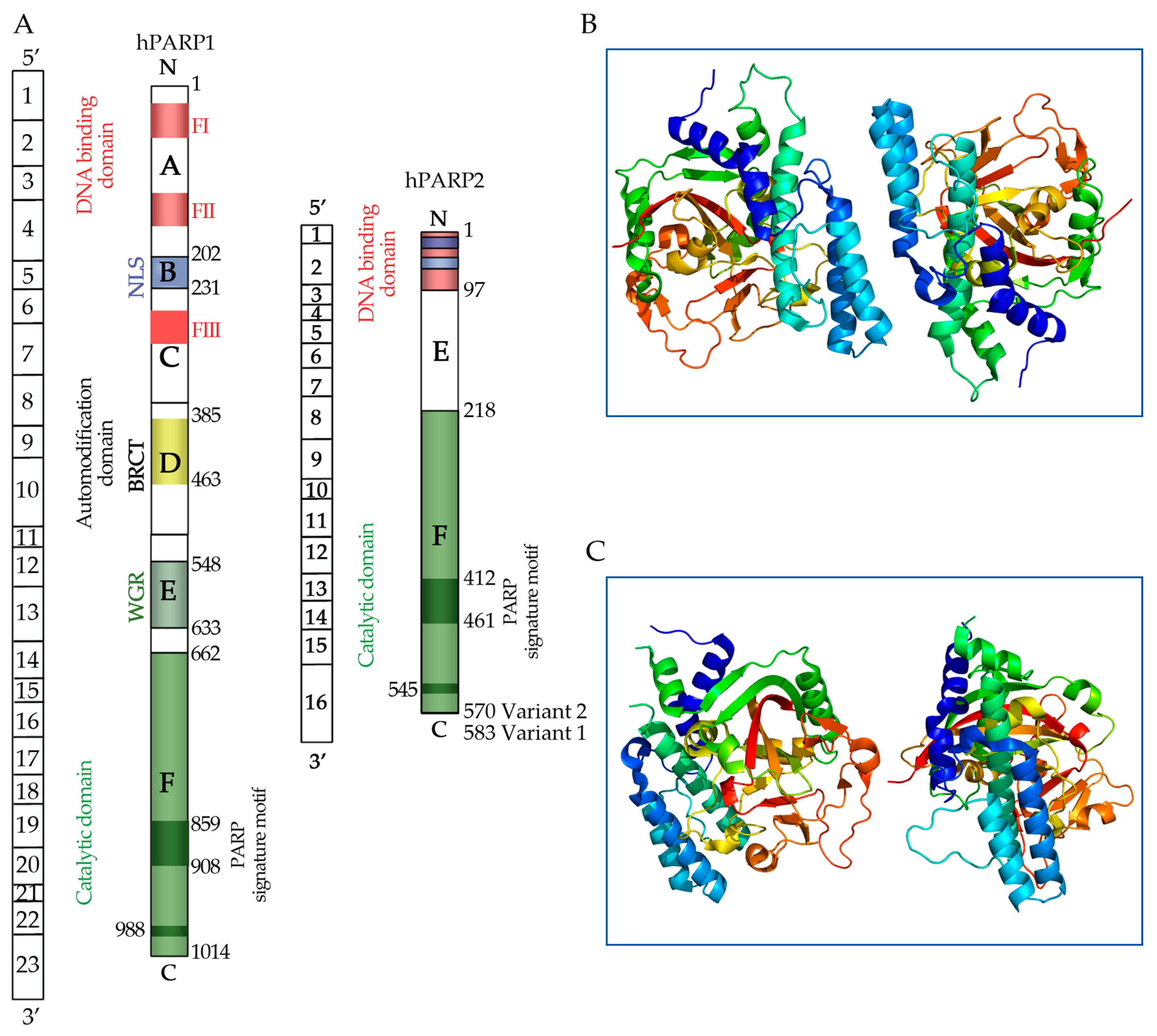
5. Poly (ADP-Ribose) Polymerase (PARP) Molecules

The enzyme family known as PARP molecules is involved in some cellular functions associated with DNA repair, cell signaling, and programmed cell death (apoptosis) [114]. PARP is a complex enzyme primarily composed of ADP-ribose units, which are linked to phospholipid groups and ribose sugars, forming a branched polymer (Figure 1).
Its structure includes multiple functional domains that facilitate its role in detecting and repairing DNA damage. The C-terminus is essential for auto-modification and interactions with other proteins, while the N-terminus is critical for DNA binding [118]. PARP consists of two main regions: the NAD+ binding site and a protein domain, which includes three unique subdomains: zinc finger, catalytic, and auto modification domains. The zinc finger domain is unnecessary for DNA binding, but is important for the catalytic activity of PARP [119]. The catalytic domain transfers ADP-ribose units from NAD+ to target proteins, and the auto-modification domain allows PARP to modify itself by forming poly (ADP-ribose) chains, influencing protein interactions and signaling pathways [120]. The C-terminus can generate large poly (ADP-ribose) chains, often enriched with tryptophan, glycine, and arginine residues, which can modulate enzyme activity and cellular responses to DNA damage. The N-terminal domain aids in the localization of DNA damage sites, ensuring efficient repair processes [114]. The most well-known member of this family, PARP1, is essential to the healing of DNA single-strand breaks [121]. DNA damage, especially single-strand breaks, is detected by PARP enzymes, which then start the repair process. By adding poly (ADP-ribose) chains to target proteins, they catalyze the recruitment of extra repair factors to the damage site. In addition to repairing DNA, PARP enzymes are involved in several signaling pathways that control how cells react to damage and stress [122].
This includes modulating protein interactions and influencing the activity of other enzymes. The primary location of PARP molecules is the cell nucleus, where they are essential for DNA repair processes [123]. However, cellular conditions can affect their distribution. Near sites of DNA damage, the majority of PARP proteins, particularly PARP1, are in the nucleus. Here, they interact with other proteins involved in the DNA damage response and aid in the repair of single-strand breaks. PARP proteins are predominantly nuclear, but they are also present in the cytoplasm. Additionally, their cytoplasmic activity might be connected to different signaling cascades and reactions to intracellular stress [124]. According to some studies, certain members of the PARP family may localize to mitochondria, where they may play roles in apoptosis and mitochondrial function [125]. PARP activity can trigger cell death pathways in cases of severe and irreversible DNA damage, aiding in the removal of damaged cells. The synthesis of poly (ADP-ribose) by PARP enzymes requires NAD+ as a substrate; this process can deplete cellular NAD+ stores, which can affect overall cell metabolism and energy balance [126].
PARP inhibitors have emerged as a major field of cancer therapy because of their function in DNA repair, especially for tumors containing BRCA1 and BRCA2 mutations.

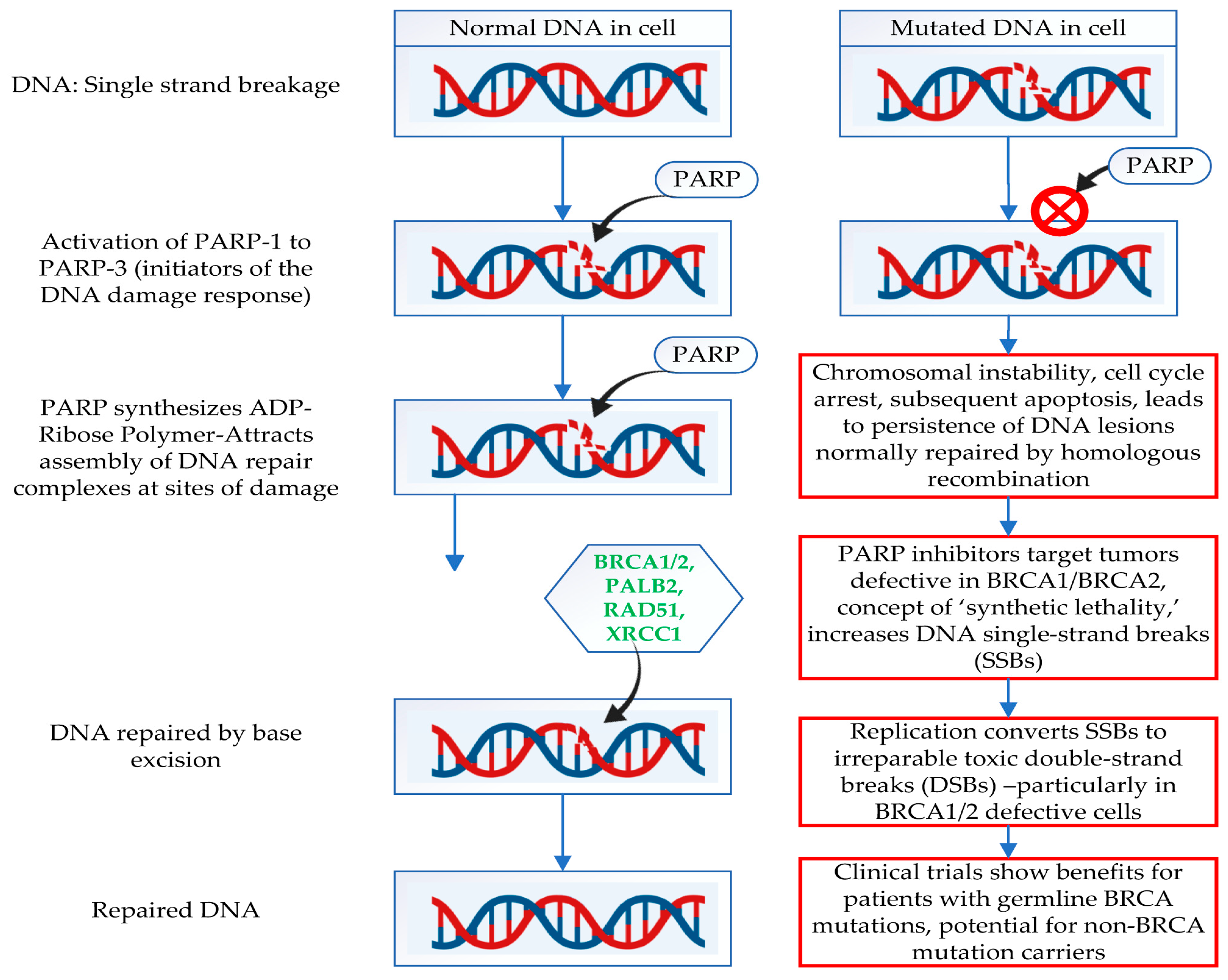
6. Mechanism of Action of PARP Inhibitors

A class of medications known as PARP inhibitors (PARPis) is mainly used to treat breast cancers, especially those with BRCA1 and BRCA2 mutations. An enzyme called PARP helps to mend DNA strand breaks when they occur. When DNA damage occurs, PARP recognizes the break and uses the base excision repair (BER) pathway to help repair it. The process of homologous recombination to repair double-strand breaks in DNA depends on the BRCA1 and BRCA2 proteins [127].
Figure 2 corresponds to a comparison between normal and BRCA1/2 mutated cell DNA repair mechanisms and the role of PARP molecules.
The usual mode of action of PARP inhibitors is the inhibition of poly (ADP-ribose) polymerase (PARP) enzymes, which are important in DNA repair. The novel PARP inhibitors, such as veliparib and rucaparib, can differ in their potency, selectivity, and ability to trap PARP-DNA complexes.
It is possible to compare the modes of action of medications with the authorized PARP inhibitors (olaparib, talazoparib, and niraparib). By inhibiting the PARP1 and PARP2 enzymes, olaparib stops DNA single-strand breaks from being repaired. Olaparib prevents the repair of single-strand breaks by inhibiting these enzymes. In cancer cells with defective BRCA1/2 or other defects in homologous recombination repair (HRR), DNA replication results in irreversible double-strand breaks [128].
Other novel PARP inhibitors have been investigated and tried. Several PARP inhibitors that target both PARP1 and PARP2 are now being developed, albeit they may also concentrate on blocking other PARP family members, such as PARP3. Some improve the effects of PARP trapping to increase the effectiveness of treatment, or to demonstrate specific action targeting PARP1. To improve PARP trapping and DNA damage, new inhibitors frequently aim to increase potency or selectivity. To further boost anti-tumor benefits, some are being studied in combination tactics, especially when taken with immune checkpoint inhibitors, chemotherapy, or targeted treatments. Most of the research on these drugs is being conducted in clinical studies that look at different patient groups, combination treatments, and dosage schedules. Their ability to balance potency, toxicity, and selectivity will determine their exact effect.
The ubiquitous post-translational modification of proteins known as poly(ADP-ribosyl)ation, which is catalyzed by PARP, is essential for controlling several cellular functions, including transcription, cell division, proliferation, DNA methylation, and apoptosis [129]. This alteration affects cellular function by modifying the activities of enzymes and the interactions of proteins, DNA, and RNA [130]. For life to continue and for species to remain stable, DNA repair is a fundamental mechanism for maintaining the stability and integrity of the DNA structure [131]. Complex systems for identifying, detecting, and fixing DNA damage are present in cells. Single-strand breaks (SSBs) and double-strand breaks (DSBs) are the two general categories into which DNA damage can be divided [132]. With a heavy reliance on PARP, SSBs are mainly treated by the nucleotide excision repair (NER), base excision repair (BER), and mismatch repair (MMR) pathways [133,134].

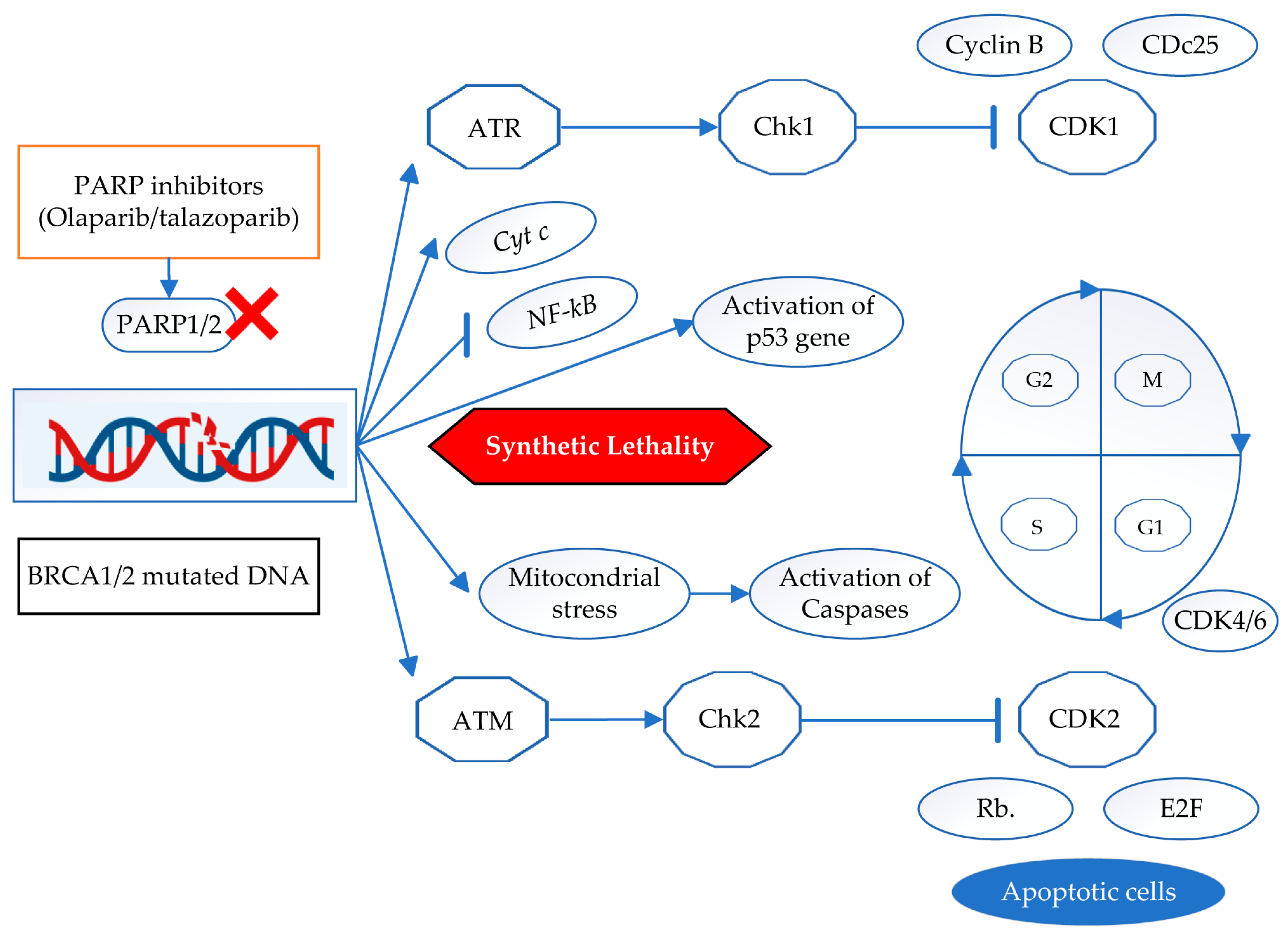
7. Cell Signaling Pathways of PARP Inhibitors

Inhibitors of PARP mainly target pathways involved in DNA repair, but they also have other effects. An outline of the main cell signaling pathways that PARP inhibitors affect is a sequential event. The Ataxia Telangiectasia Mutated (ATM) and Ataxia Telangiectasia and Rad3-related (ATR) kinases are activated because of increased DNA damage caused by PARP inhibitors [135]. These kinases play a vital role in cellular response regulation and DNA damage recognition. Checkpoint proteins (such as CHK1 and CHK2) become phosphorylated because of ATM/ATR activation, stopping the cell cycle and facilitating repair processes. When there is significant and irreversible damage to DNA, p53 is triggered, which results in cell cycle arrest or apoptosis. In BRCA-deficient cells, PARP inhibitors can increase p53 activation, which triggers apoptotic signaling [136]. Inhibition of PARP can affect cytochrome c release and mitochondrial stress, which can activate caspases and cause apoptosis [137].
PARP inhibitors may affect the ratio of pro- and anti-apoptotic (BCL-2) and pro-apoptotic (BAX, BAK) proteins, inducing apoptosis in cancer cells [138]. The NF-κB pathway, which is involved in inflammatory responses and cell survival, can be modulated by PARP inhibitors [139]. Because the inhibition interferes with NF-κB-mediated survival signals, it may accelerate cell death. PARP inhibitors cause double-strand breaks in BRCA-deficient cells that are irreversible because there is no functioning HRR. This causes DNA damage to accumulate and eventually results in cell death. In certain situations, PARP inhibitors may also cause cellular senescence, a permanent cell cycle arrest state that can be brought on by significant DNA damage. There is growing evidence that PARP inhibitors can influence autophagy, a process that, depending on the situation, can either induce cell survival or cause cell death. Complex and interconnected signaling pathways are triggered by PARP inhibitors, and in cancer cells, especially those with compromised DNA repair mechanisms, this ultimately results in increased DNA damage, cell cycle arrest, and apoptosis. Knowing these pathways can help develop treatment plans and find biomarkers to help choose patients for PARP inhibitor therapies and other cancers because of their capacity to cause synthetic lethality in BRCA-deficient cells. Figure 3 depicts the BRCA1/2-mutated and PARP-inhibited cancer cell signaling mechanisms.
The licensed PARP inhibitors (olaparib, talazoparib, and niraparib) and the ones now being studied (veliparib, rucaparib, and others) have different pharmacokinetics, side-effect profiles, clinical indications, and mechanisms of action. Olaparib primarily inhibits the PARP1 and PARP2 enzymes, which stops DNA repair. Single-strand DNA breaks cannot be fixed when these enzymes are suppressed, leading to double-strand breaks and ultimately cell death, particularly in cancer cells that rely on this repair mechanism. Prostate, pancreatic, breast, and ovarian cancers linked to BRCA1/2 mutations have all been authorized for it [128]. Olaparib’s half-life is moderate, and it is recommended to take the drug twice a day, although olaparib can cause common side effects such as fatigue, nausea, anemia, and neutropenia [140]. Both PARP1 and PARP2 are inhibited by talazoparib. Trapping the PARP-DNA complex increases the potency of the complex and enhances its therapeutic advantages, making it more effective at inhibiting PARP1 and causing DNA damage [141]. Therefore, it might be proposed that BRCA mutations in breast cancer can be treated with it. Like olaparib, talazoparib can produce fatigue, anemia, and thrombocytopenia; but, due to its stronger strength, side effects could be more severe [142].
Niraparib inhibits both PARP1 and PARP2, but what makes it unique is how long it remains in cells. It can continue to block PARP even after the drug has been taken out of the system because of its long half-life. Regardless of BRCA status, it is approved for ovarian cancer; nevertheless, it is most effective when there are BRCA mutations or homologous recombination deficits (HRDs) [118,143]. Furthermore, it typically needs to be taken once a day and has a long half-life, raising the question of cumulative toxicity from extended use. Additionally, it may cause thrombocytopenia, anemia, and exhaustion [144].
Inhibiting PARP1 and PARP2, rucaparib works similarly to olaparib and niraparib. However, it can trap PARP more efficiently at the DNA site, which, in certain circumstances, may lead to stronger anti-cancer effects [145]. It is used to treat ovarian and prostate cancers with BRCA mutations. Additionally, it is being researched for several cancers, particularly with other therapies. It can be taken twice daily due to its light half-life. Rucaparib, like the other PARP inhibitors, can cause anemia, tiredness, and gastrointestinal problems [146]. It has significant hematologic side effects, but a toxicity profile simpler to manage than talazoparib’s. Olaparib, talazoparib, and niraparib have been approved based on strong evidence from clinical trials in specific cancer types. Their efficacy is especially apparent in cancers with BRCA mutations and limitations in homologous recombination repair [147]. Clinical trials are being conducted to see whether rucaparib can treat various cancers, especially when used with other treatments, since it has shown promise in treating prostate and ovarian cancers [148].

8. Clinical Implications

Olaparib, talazoparib, and niraparib are examples of PARP inhibitors that work especially well for patients with hereditary breast and ovarian cancer syndromes caused by BRCA mutations [149]. To increase the effectiveness of therapy, they are frequently used with other medical procedures like immunotherapy and chemotherapy [150]. Patients who may benefit from PARP inhibition can be identified by the presence of BRCA mutations or other deficiencies in DNA repair pathways (e.g., mutations in other homologous recombination-related genes). PARP inhibitors offer a novel therapeutic option for cancers whose DNA repair mechanisms are compromised [151].
They have greatly advanced the treatment of breast cancer. In addition to olaparib, talazoparib, and niraparib, more PARP inhibitors are being researched for the treatment of breast cancer. Rucaparib was first created to treat ovarian cancer, but it is now being investigated for its potential to treat breast cancer, especially in those with BRCA mutations. Although veliparib has been studied mainly with chemotherapy, it is also being considered for the treatment of breast cancer. Research is continuing, and as our knowledge of cancer treatment advances, new compounds might appear. Oncology research on BRCA1 and BRCA2 mutations and their association with PARP inhibitors is still very active, especially in cases of breast and ovarian cancer. Recent research has revealed that people with BRCA1/2 mutations are not the only ones being investigated for PARP inhibitors like olaparib, niraparib, and rucaparib. The first approved PARP inhibitor, olaparib, is used in the post-chemotherapy phase of advanced breast cancer treatment and shows promising clinical outcomes. Patients with HER2-negative metastatic breast cancer (mBC) that has spread and BRCA mutations benefit most from it [152].
The strong affinity of talazoparib for PARP makes it a more potent PARP inhibitor than olaparib, more effective against BRCA-mutant breast cancer, and longer-acting [153,154]. Niraparib is a potent inhibitor that can be used regardless of a patient’s BRCA status because it may also affect other DNA repair pathways [155]. Similar to olaparib and talazoparib, rucaparib is effective against cancers with BRCA mutations. It has proven effective in treating both ovarian and breast cancers and is used after patients have had previous treatments [156].
A list of PARP inhibitors, along with their therapeutic names and their application fields, is provided in Table 1.
Additionally, they are being studied in tumors with homologous recombination deficiency (HRD), which might benefit from comparable therapeutic modalities. To increase efficacy and overcome resistance, there is growing interest in combining PARP inhibitors with other treatments, such as immunotherapies and targeted agents. Current research focuses on controlling PARP inhibitor side effects and enhancing the quality of life of patients while they are receiving treatment. Trials are being conducted to investigate the efficacy of PARP inhibitors in nascent stages and diverse forms of cancer. A specific area of interest is their application in neoadjuvant environments. The field of genetic testing is changing, and recommendations are being made for more comprehensive testing for HRD in different types of cancer. This could help identify more patients who could benefit from PARP inhibitors. Patients are receiving more and more education about their genetic status, available treatments, and the consequences of having a BRCA mutation.

9. Bioinformatic Analysis of Clinical Datasets

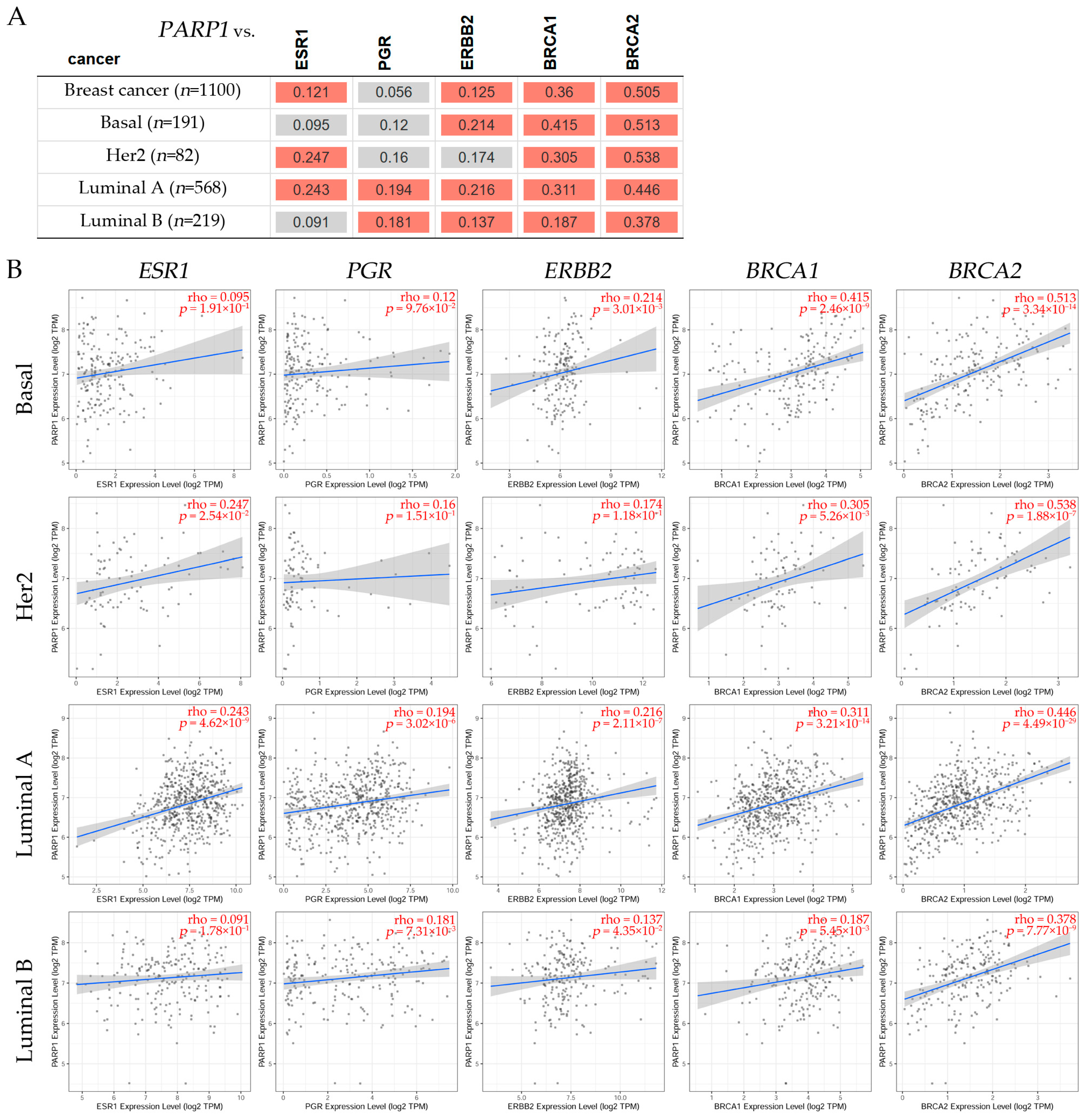
9.1. Correlation Analysis Between PARP1/2 and Genes Such as ESR1, PGR, ERBB2, BRCA1, and BRCA2 Expression Levels in Breast Cancer Subtypes

To investigate the association between PARP1/2 and the estrogen receptor alpha (ESR1), PGR, ERBB2, BRCA1, and BRCA2 genes, a correlation assessment was conducted using the Tumor Immune Estimation Resource v2.0 (TIMER2.0) dataset (Figure 4 and Figure 5). The Gene Correlation module of TIMER2.0 was used to explore such correlations across several breast cancer subtypes, and the statistical analysis was facilitated by Spearman’s test.
It can be observed in Figure 4A a positive correlation between PARP1 and BRCA1, BRCA2 in basal, HER2, luminal A, and luminal B patients. A positive correlation between PARP1 and ERBB2 was found in basal, luminal A, and luminal B, but was non-significant in HER2 patients. On the other hand, there was no significant correlation between PARP1 and ESR1 in basal or LumB, but there was a significant correlation between PARP1 and HER2 and luminal A breast cancer patients. There was also a significant correlation between PARP1 in PGR gene expression in luminal A and luminal B patients. It is concluded from these studies that PARP1 gene expression is correlated with subtypes in breast cancer patients such as the basal subtype, and it can be positive for BRCA1/2 and ERBB2 gene expression, but is non-significant for ESR1 and PGR, given the opportunity to give specific drugs for such genes; the HER2 subtype can be positive for BRCA1/2 and ESR1; the luminal A subtype can be positive for all genes mentioned above, including BRCA1/2, ESR1, PGR, and ERBB2; and the luminal B subtype can be positive for BRCA1/2, PGR, and ERBB2, but not for ESR1 gene expression.
In Figure 4B, representative scatter plots show the correlations (Spearman’s, p < 0.05) between PARP1 and ESR1, PGR, ERBB2, BRCA1, and BRCA2 expression levels in breast cancer subtypes such as basal, HER2, luminal A, and luminal B. Results indicated that the basal subtype had non-significance between PARP1 and ESR1, PGR, but was positive for ERBB2 and BRCA1/2. The HER2 subtype showed non-significance between PARP1 and PGR and ERBB2, but was positive for luminal A and luminal B. The luminal A subtype showed positive significance between PARP1 and ESR1, PGR, ERBB2, BRCA1, and BRCA2 expression levels, but negative significance in luminal B subtypes. A positive correlation can be seen between PARP1 and BRCA1, BRCA2 in basal, HER2, luminal A, and luminal B patients. A positive correlation between PARP1 and ERBB2 was found in basal, luminal A, and luminal B, but was negative in HER2 patients. However, there was no significant correlation between PARP1 and ESR1 in basal or luminal B, but there was a significant correlation in HER2 and luminal A breast cancer patients. There was also a significant correlation between PARP1 and PGR gene expression in luminal A and luminal B patients.
Figure 5A shows the correlation between PARP2 and genes such as ESR1, PGR, ERBB2, BRCA1, and BRCA2. The red color indicates a significant positive (Spearman’s, p < 0.05), and the gray has a non-significant correlation. It can be observed in Figure 5A a positive correlation between PARP2 and BRCA1, BRCA2 and with ERBB2 in the basal subtype; a positive correlation between PARP2 and BRCA1, BRCA2 and with ESR1, but a negative correlation with ERBB2 in the HER2 subtype; and a positive correlation between PARP2 and BRCA1/2, ERBB2, PGR, and ESR1 in the luminal A subtype. However, there was no significant correlation between PARP2 and ESR, PGR, and ERBB2, being positive with BRACA1/2 in luminal B breast cancer patients. In Figure 5B, representative scatterplots show the correlations (Spearman’s, p < 0.05) between PARP2 and ESR1, PGR, ERBB2, BRCA1, and BRCA2 expression levels in breast cancer subtypes such as basal, HER2, luminal A, and luminal B. Results indicated that the basal breast cancer subtype had non-significance between PARP2 and ESR1, PGR, but was positive for ERBB2, BRCA1/2. The HER2 subtype showed non-significance between PARP2 and PGR, a negative association with ERBB2, and a positive association with BRCA1/2. The luminal A subtype showed positive significance between PARP2 and ESR1, PGR, ERBB2, BRCA1, and BRCA2 expression levels, whereas the luminal B subtypes showed a positive association with BRCA1/2, but was non-significant with ESR1, PRG, and ERBB2 breast cancer patients.

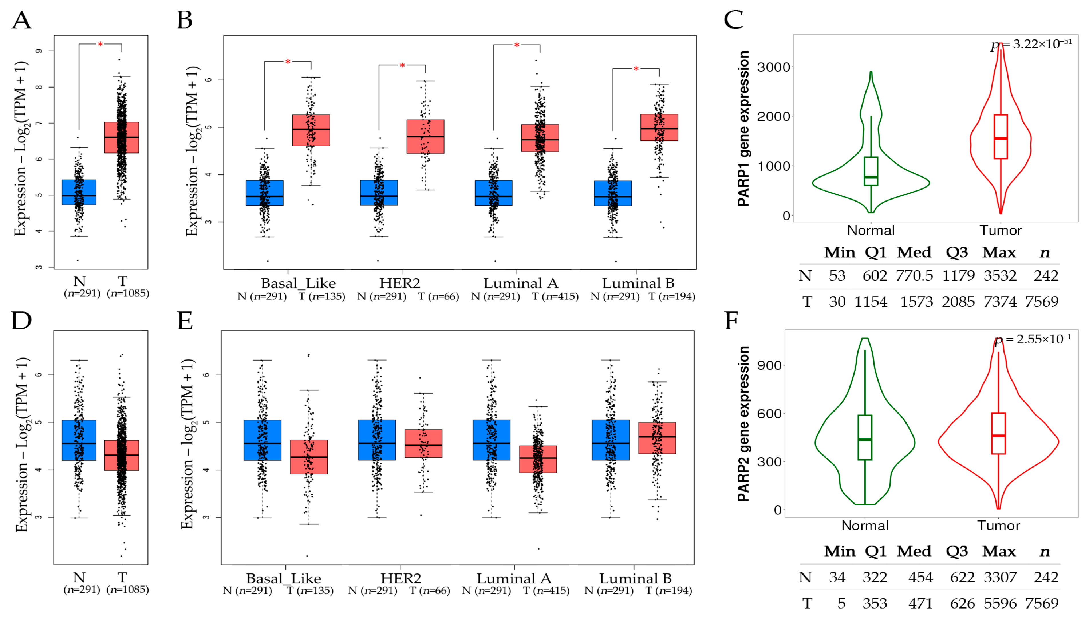
9.2. Comparative Assessment of PARP1 and PARP2 Expression Levels in Tumor and Normal Tissues in Breast Cancer Subtypes

An analysis of PARP1 and PARP2 gene expression levels across tumor grades in breast cancer can be seen in Figure 6. The Gene Expression Profiling Interactive Analysis (GEPIA2) dataset provided the comparison of gene expression between tumor and normal tissues, with the distribution of gene expression levels represented through box plots. The Student’s t-test was used to compare the expression of PARP1/2 between tumor tissues and corresponding normal tissues.
A comparison of PARP1 and PARP2 mRNA expression levels between (A) normal and tumor breast tissues, as well as between (B) several breast cancer subtypes such as basal-like, HER2, luminal A, and luminal B, including normal tissues, is provided in Figure 6. GEPIA2 and TNMplot datasets were used for this analysis. A statistically significant PARP1 overexpression was found in breast tumors in comparison with normal tissue (Figure 6A) and across its several subtypes (Figure 6B). A significant upregulation of PARP1 mRNA was observed (Figure 6C) in tumors compared to controls (p = 3.22 × 10−51). The median expression was higher in tumors than in the normal tissues. This overexpression may play a part in the aggressiveness and progression of tumors, as evidenced by its correlation with advanced tumor grades and stages. These findings highlight the diagnostic and prognostic usefulness of PARP1 in breast cancer by demonstrating that it can be investigated as a trustworthy biomarker with excellent specificity at raised cut-off levels. PARP2 expression levels were non-significant (Figure 6D–F).

9.3. PARP1 and PARP2 as Potential Markers in Chemotherapy-Treated Breast Cancer Patients

Figure 7 shows an overall survival (OS) analysis of PARP1 and PARP2 expression levels in breast cancer patients. GEPIA2 was used to explore the OS of breast cancer patients (Figure 7A,D) by analyzing data from the TCGA, GTEx, and GEO databases; the threshold values for separating the groups with low and high expression were the cut-off low (50%) and cut-off high (50%). The ROC Plotter web platform was used to assess the expression levels of PARP1/2 (Figure 7B,C,E,F, respectively) in breast cancer patients with a pathological complete response (n = 1775), treated with chemotherapy and a positive ER status.
Patients were grouped into two categories depending on their PARP expression level: the high expression and low expression groups. OS assessment indicated that patients with high PARP1 (Figure 7A) and PARP2 (Figure 7D) expression levels (red line) presented worse survival rates than low PARP expression groups (black lines). Additionally, the receiver operating characteristic (ROC) Plotter dataset was used to assess PARP1 and PARP2 expression levels in breast cancer patients whose ER status was positive and who were treated with chemotherapy. Results indicated that responders showed higher mean (598) and median (559) PARP2 expression levels than non-responders, with a mean value of 454 and median of 406 (Figure 7E). The ROC Plotter showed that PARP2 had an area under the curve (AUC) of 0.649 presenting a predictive power with a significant p-value of 2.7 × 10⁻¹³ (Figure 7F); the Mann–Whitney test p-value (1.8 × 10⁻¹²) showed a significant difference between both groups, and the fold change was 1.3, implying that responders had increased PARP2 expression levels. However, PARP1 was non-significant. Therefore, OS results suggested that PARP2 expression could be a potential biomarker to predict chemotherapy response in ER-positive patients.

10. Conclusions

Since the bioinformatics showed that there was a correlation between PARP1 and BRCA1/2, and at the same time, a correlation with ERBB2 in basal; such results indicate that those breast cancer patients can receive not only PARP inhibitors, but also Anti-HER2 Monoclonal Antibodies as therapeutic drugs. Such analysis by bioinformatics showed that there was a correlation between PARP1 and BRCA1/2, and at the same time, with ESR1 in the HER2 subgroup; those breast cancer patients can receive not only PARP inhibitors, but also tamoxifen or other drugs for positive ESR1 patients as therapeutic drugs. Furthermore, there was a correlation between PARP1 and BRCA1/2, and at the same time, with ERBB2, PGR, and ESR1 in the luminal A subgroup; such results can indicate that those breast cancer patients classified as subgroup luminal A can receive not only PARP inhibitors, but Anti-HER2 Monoclonal Antibodies, tamoxifen, or other drugs for positive ESR1 patients and antiPGR as therapeutic drugs. A correlation was also reported between PARP1 and BRCA1/2, and at the same time, with ERBB2 and PGR in the luminal B subgroup; such results can indicate that those breast cancer patients can receive Anti-HER2 Monoclonal Antibodies and anti-PGR drugs available in the clinic at present.
The bioinformatics study revealed a correlation between PARP2 and BRCA1/2 and ERBB2 in the basal subgroup of breast cancer patients. These patients can receive PARP inhibitors and anti-HER2 Monoclonal Antibodies as therapeutic drugs. There was also a correlation between PARP2 and BRCA1/2 and ESR1 in the HER2 subgroup of breast cancer patients. Those patients can receive not only PARP inhibitors, but also tamoxifen or other therapeutic drugs for positive ESR1 patients. Since bioinformatics showed that there was a correlation between PARP2 and BRCA1/2, ERBB2, PGR, and ESR1 in the luminal A subgroup, patients can receive PARP inhibitors, anti-HER2 Monoclonal Antibodies, tamoxifen, antiPGR as therapeutic drugs, or other drugs for positive ESR1 patients. It was also shown that there was a correlation between PARP2 and BRCA1/2 in the luminal B subgroup, indicating that those breast cancer patients can receive Anti-HER2 Monoclonal Antibodies as therapeutic drugs available in the clinic at present.
On the other hand, through bioinformatics, it was observed that there was a higher overall expression of PARP1 in tumors than in normal tissues and across subtypes, as well as more upregulation of mRNA in tumors than in normal tissues; such results indicate that those breast cancer patients can receive only PARP1 inhibitors. A non-significant difference was found when PARP2 was studied. Concerning survival, results showed that those patients with increased levels of PARP1 and PARP2 expression had worse survival rates than those with low levels. A critical focus is based on finding more selective novel PARP inhibitors that lower toxicity and enhance patients’ overall therapeutic experience. PARP inhibitors frequently work with other treatments, such as immunotherapy and chemotherapy.
Using PARP inhibitors for cancers unrelated to BRCA is a crucial objective. The greatest impact of PARP inhibitors has been demonstrated in BRCA-mutant malignancies, such as ovarian, breast, and prostate cancers, although BRCA mutations only affect a tiny fraction of patients. To increase the number of patients who can benefit from PARP inhibition, clinical investigations are looking into its usage in malignancies with various types of homologous recombination deficit (HRD) and with other treatments. It is yet unknown how to determine which individuals will benefit the most from PARP inhibitors, and not all patients respond to them.
To be constructive in our conclusions, we consider that PARP inhibition has minimal impact on some tumor cells because malignancies are heterogeneous, including cells that may respond differentially to treatment. Research into personalized medicine, where medication is based on individual genetic profiles, would be very beneficial to optimize these combinations and minimize negative effects. Targeted therapy has advanced significantly with PARP inhibitors in the treatment of breast cancer, especially for patients with BRCA1/2 mutations. As long as research on combination therapies and treatment protocols is conducted, the future of PARP inhibitors as a mainstay of breast cancer care looks bright. PARPis will play a more significant role in the clinical treatment of cancer. Further research is expected to expand treatment that benefits more patients. Overall, developing PARPis brings new hope to cancer treatment, especially for BRCA mutation-positive patients, as improving the selectivity of PARP-1, enhancing the inhibitory capability of PARPis, and adopting strategies to reduce drug toxicity and side effects are all ideas and directions for developing PARPis.
Future research is needed for the creation of novel PARP inhibitors or other combinations of drugs with enhanced safety and efficacy profiles. Clinical trials and ongoing research will play a critical role in determining this future. Such trials have shown encouraging efficacy, frequently leading to increased response rates and progression-free survival rates in patients with triple-negative breast cancer and other subtypes. There are compelling reasons to keep creating new and enhanced versions. By focusing on distinct pathways or successfully treating malignancies when there is no response to treatments, novel inhibitors may help to overcome this resistance. Today, the most common tumors treated with PARP inhibitors are those associated with BRCA mutations, including breast. More selective novel PARP inhibitors must lower toxicity and enhance patients’ overall therapeutic experience. PARP inhibitors frequently work with other treatments, such as immunotherapy and chemotherapy.

Author Contributions

Writing—original draft preparation, K.D., M.J., B.C. and D.R.; writing—review and editing, K.D., M.J., B.C., G.M.C. and D.R.; Funding acquisition, G.M.C. All authors have read and agreed to the published version of the manuscript.

Funding

This research was funded by Fondo Nacional de Desarrollo Científico y Tecnológico (FONDECYT/ANID), grant number FONDECYT 1231537 (G.M.C.).

Institutional Review Board Statement

Not applicable.

Informed Consent Statement

Not applicable.

Data Availability Statement

TIMER2.0 is freely available at http://timer.cistrome.org/ (accessed on 7 November 2024). GEPIA2, Gene Exploration Profiling Interactive Analysis, is freely available at http://gepia2.cancer-pku.cn/ (accessed on 7 November 2024). TNMplot: differential gene expression analysis in Tumor, Normal, and Metastatic tissues, is freely available at https://tnmplot.com/ (accessed on 7 November 2024). Kaplan-Meier Plotter is freely available at https://www.kmplot.com/ (accessed on 7 November 2024). ROC Plotter is freely available at https://rocplot.org/ (accessed on 8 November 2024). The data generated in the present study may be requested from the corresponding author.

Acknowledgments

The authors want to thank Leodan A. Crispin for the bioinformatic analysis and for revising the manuscript (Instituto de Alta Investigación, Universidad de Tarapacá).

Conflicts of Interest

The authors declare no conflicts of interest. The funders had no role in the design of the study; in the collection, analyses, or interpretation of data; in the writing of the manuscript; or in the decision to publish the results.

References

  1. Ismail, T.; Alzneika, S.; Riguene, E.; Al-Maraghi, S.; Alabdulrazzak, A.; Al-Khal, N.; Fetais, S.; Thanassoulas, A.; AlFarsi, H.; Nomikos, M. BRCA1 and Its Vulnerable C-Terminal BRCT Domain: Structure, Function, Genetic Mutations and Links to Diagnosis and Treatment of Breast and Ovarian Cancer. Pharmaceuticals 2024, 17, 333. [Google Scholar] [CrossRef] [PubMed]
  2. WHO. Breast Cancer. World Health Organization. Available online: https://www.who.int/news-room/fact-sheets/detail/breast-cancer (accessed on 12 November 2024).
  3. Obeagu, E.I.; Obeagu, G.U. Breast cancer: A review of risk factors and diagnosis. Medicine 2024, 103, e36905. [Google Scholar] [CrossRef] [PubMed]
  4. Marino, P.; Mininni, M.; Deiana, G.; Marino, G.; Divella, R.; Bochicchio, I.; Giuliano, A.; Lapadula, S.; Lettini, A.R.; Sanseverino, F. Healthy Lifestyle and Cancer Risk: Modifiable Risk Factors to Prevent Cancer. Nutrients 2024, 16, 800. [Google Scholar] [CrossRef] [PubMed]
  5. Yuan, M.; Zhu, Y.; Ren, Y.; Chen, L.; Dai, X.; Wang, Y.; Huang, Y.; Wang, H. Global burden and attributable risk factors of breast cancer in young women: Historical trends from 1990 to 2019 and forecasts to 2030 by sociodemographic index regions and countries. J. Glob. Health 2024, 14, 04142. [Google Scholar] [CrossRef]
  6. Grauman, A.; Sundell, E.; Johansson, J.V.; Cavalli-Bjorkman, N.; Fahlquist, J.N.; Hedstrom, M. Perceptions of lifestyle-related risk communication in patients with breast and colorectal cancer: A qualitative interview study in Sweden. Arch. Public Health 2024, 82, 154. [Google Scholar] [CrossRef]
  7. Gao, M.; Wik, S.L.; Yu, Q.; Xue, F.; Chan, S.C.; Chow, S.H.; Adebisi, Y.A.; Zhong, C.C.; Lucero-Prisno, D.E., III; Wong, M.C.; et al. Disease Burden, Risk Factors, and Temporal Trends in Breast Cancer in Low- and Middle-Income Countries: A Global Study. Public Health Chall. 2024, 3, e223. [Google Scholar] [CrossRef]
  8. Conte, L.; Lupo, R.; Lezzi, A.; Sciolti, S.; Rubbi, I.; Carvello, M.; Calabro, A.; Botti, S.; Fanizzi, A.; Massafra, R.; et al. Breast Cancer Prevention Practices and Knowledge in Italian and Chinese Women in Italy: Clinical Checkups, Free NHS Screening Adherence, and Breast Self-Examination (BSE). J. Cancer Educ. 2024, 40, 30–43. [Google Scholar] [CrossRef]
  9. Chou, C.Y.; Shen, T.T.; Wang, W.C.; Wu, M.P. Favorable breast cancer mortality-to-incidence ratios of countries with good human development index rankings and high health expenditures. Taiwan J. Obstet. Gynecol. 2024, 63, 527–531. [Google Scholar] [CrossRef]
  10. Goh, S.P.; Ong, S.C.; Chan, J.E. Economic evaluation of germline genetic testing for breast cancer in low- and middle-income countries: A systematic review. BMC Cancer 2024, 24, 316. [Google Scholar] [CrossRef]
  11. Khorasani, A.B.S.; Hafezi, N.; Sanaei, M.J.; Jafari-Raddani, F.; Pourbagheri-Sigaroodi, A.; Bashash, D. The PI3K/AKT/mTOR signaling pathway in breast cancer: Review of clinical trials and latest advances. Cell Biochem. Funct. 2024, 42, e3998. [Google Scholar] [CrossRef]
  12. Mustafa, M.; Abbas, K.; Alam, M.; Ahmad, W.; Moinuddin; Usmani, N.; Siddiqui, S.A.; Habib, S. Molecular pathways and therapeutic targets linked to triple-negative breast cancer (TNBC). Mol. Cell Biochem. 2024, 479, 895–913. [Google Scholar] [CrossRef]
  13. Varzaru, V.B.; Vlad, T.; Popescu, R.; Vlad, C.S.; Moatar, A.E.; Cobec, I.M. Triple-Negative Breast Cancer: Molecular Particularities Still a Challenge. Diagnostics 2024, 14, 1875. [Google Scholar] [CrossRef]
  14. Hernandez, A.E.; Westrick, A.C.; Stoler, J.; Kesmodel, S.B.; Pinheiro, P.S.; Figueroa, M.; Kobetz, E.N.; Rebbeck, T.; Goel, N. Associations Between Neighborhood-Level Income and Triple-Negative Breast Cancer in a Majority-Minority Population. Ann. Surg. Oncol. 2024, 31, 988–996. [Google Scholar] [CrossRef]
  15. Lyons, T.G. Targeted Therapies for Triple-Negative Breast Cancer. Curr. Treat. Options Oncol. 2019, 20, 82. [Google Scholar] [CrossRef]
  16. Wu, S.; Ge, A.; Deng, X.; Liu, L.; Wang, Y. Evolving immunotherapeutic solutions for triple-negative breast carcinoma. Cancer Treat. Rev. 2024, 130, 102817. [Google Scholar] [CrossRef]
  17. Perou, C.M.; Sorlie, T.; Eisen, M.B.; van de Rijn, M.; Jeffrey, S.S.; Rees, C.A.; Pollack, J.R.; Ross, D.T.; Johnsen, H.; Akslen, L.A.; et al. Molecular portraits of human breast tumours. Nature 2000, 406, 747–752. [Google Scholar] [CrossRef]
  18. Sorlie, T.; Perou, C.M.; Tibshirani, R.; Aas, T.; Geisler, S.; Johnsen, H.; Hastie, T.; Eisen, M.B.; van de Rijn, M.; Jeffrey, S.S.; et al. Gene expression patterns of breast carcinomas distinguish tumor subclasses with clinical implications. Proc. Natl. Acad. Sci. USA 2001, 98, 10869–10874. [Google Scholar] [CrossRef]
  19. Sorlie, T.; Tibshirani, R.; Parker, J.; Hastie, T.; Marron, J.S.; Nobel, A.; Deng, S.; Johnsen, H.; Pesich, R.; Geisler, S.; et al. Repeated observation of breast tumor subtypes in independent gene expression data sets. Proc. Natl. Acad. Sci. USA 2003, 100, 8418–8423. [Google Scholar] [CrossRef]
  20. Hu, Z.; Fan, C.; Oh, D.S.; Marron, J.S.; He, X.; Qaqish, B.F.; Livasy, C.; Carey, L.A.; Reynolds, E.; Dressler, L.; et al. The molecular portraits of breast tumors are conserved across microarray platforms. BMC Genom. 2006, 7, 96. [Google Scholar] [CrossRef]
  21. Rakha, E.A.; Reis-Filho, J.S.; Ellis, I.O. Basal-like breast cancer: A critical review. J. Clin. Oncol. 2008, 26, 2568–2581. [Google Scholar] [CrossRef]
  22. Parker, J.S.; Mullins, M.; Cheang, M.C.; Leung, S.; Voduc, D.; Vickery, T.; Davies, S.; Fauron, C.; He, X.; Hu, Z.; et al. Supervised risk predictor of breast cancer based on intrinsic subtypes. J. Clin. Oncol. 2009, 27, 1160–1167. [Google Scholar] [CrossRef] [PubMed]
  23. Gusterson, B. Do ‘basal-like’ breast cancers really exist? Nat. Rev. Cancer 2009, 9, 128–134. [Google Scholar] [CrossRef] [PubMed]
  24. Rakha, E.A.; Reis-Filho, J.S.; Ellis, I.O. Impact of basal-like breast carcinoma determination for a more specific therapy. Pathobiology 2008, 75, 95–103. [Google Scholar] [CrossRef]
  25. Rakha, E.A.; El-Sayed, M.E.; Reis-Filho, J.; Ellis, I.O. Patho-biological aspects of basal-like breast cancer. Breast Cancer Res. Treat. 2009, 113, 411–422. [Google Scholar] [CrossRef]
  26. Nielsen, T.O.; Hsu, F.D.; Jensen, K.; Cheang, M.; Karaca, G.; Hu, Z.; Hernandez-Boussard, T.; Livasy, C.; Cowan, D.; Dressler, L.; et al. Immunohistochemical and clinical characterization of the basal-like subtype of invasive breast carcinoma. Clin. Cancer Res. 2004, 10, 5367–5374. [Google Scholar] [CrossRef]
  27. Livasy, C.A.; Karaca, G.; Nanda, R.; Tretiakova, M.S.; Olopade, O.I.; Moore, D.T.; Perou, C.M. Phenotypic evaluation of the basal-like subtype of invasive breast carcinoma. Mod. Pathol. 2006, 19, 264–271. [Google Scholar] [CrossRef]
  28. Cheang, M.C.; Voduc, D.; Bajdik, C.; Leung, S.; McKinney, S.; Chia, S.K.; Perou, C.M.; Nielsen, T.O. Basal-like breast cancer defined by five biomarkers has superior prognostic value than triple-negative phenotype. Clin. Cancer Res. 2008, 14, 1368–1376. [Google Scholar] [CrossRef]
  29. Carey, L.A.; Perou, C.M.; Livasy, C.A.; Dressler, L.G.; Cowan, D.; Conway, K.; Karaca, G.; Troester, M.A.; Tse, C.K.; Edmiston, S.; et al. Race, breast cancer subtypes, and survival in the Carolina Breast Cancer Study. JAMA 2006, 295, 2492–2502. [Google Scholar] [CrossRef]
  30. Fulford, L.G.; Easton, D.F.; Reis-Filho, J.S.; Sofronis, A.; Gillett, C.E.; Lakhani, S.R.; Hanby, A. Specific morphological features predictive for the basal phenotype in grade 3 invasive ductal carcinoma of breast. Histopathology 2006, 49, 22–34. [Google Scholar] [CrossRef]
  31. Banerjee, S.; Reis-Filho, J.S.; Ashley, S.; Steele, D.; Ashworth, A.; Lakhani, S.R.; Smith, I.E. Basal-like breast carcinomas: Clinical outcome and response to chemotherapy. J. Clin. Pathol. 2006, 59, 729–735. [Google Scholar] [CrossRef]
  32. Haffty, B.G.; Yang, Q.; Reiss, M.; Kearney, T.; Higgins, S.A.; Weidhaas, J.; Harris, L.; Hait, W.; Toppmeyer, D. Locoregional relapse and distant metastasis in conservatively managed triple negative early-stage breast cancer. J. Clin. Oncol. 2006, 24, 5652–5657. [Google Scholar] [CrossRef]
  33. Rakha, E.A.; Ellis, I.O.; Reis-Filho, J.S. Immunohistochemical heterogeneity of breast carcinomas negative for estrogen receptors, progesterone receptors and Her2/neu (basal-like breast carcinomas). Mod. Pathol. 2008, 21, 1060–1061; author reply 1061–1062. [Google Scholar] [CrossRef]
  34. Carey, L.A.; Dees, E.C.; Sawyer, L.; Gatti, L.; Moore, D.T.; Collichio, F.; Ollila, D.W.; Sartor, C.I.; Graham, M.L.; Perou, C.M. The triple negative paradox: Primary tumor chemosensitivity of breast cancer subtypes. Clin. Cancer Res. 2007, 13, 2329–2334. [Google Scholar] [CrossRef]
  35. Kennecke, H.; Yerushalmi, R.; Woods, R.; Cheang, M.C.; Voduc, D.; Speers, C.H.; Nielsen, T.O.; Gelmon, K. Metastatic behavior of breast cancer subtypes. J. Clin. Oncol. 2010, 28, 3271–3277. [Google Scholar] [CrossRef]
  36. Thike, A.A.; Cheok, P.Y.; Jara-Lazaro, A.R.; Tan, B.; Tan, P.; Tan, P.H. Triple-negative breast cancer: Clinicopathological characteristics and relationship with basal-like breast cancer. Mod. Pathol. 2010, 23, 123–133. [Google Scholar] [CrossRef]
  37. Thike, A.A.; Iqbal, J.; Cheok, P.Y.; Chong, A.P.; Tse, G.M.; Tan, B.; Tan, P.; Wong, N.S.; Tan, P.H. Triple negative breast cancer: Outcome correlation with immunohistochemical detection of basal markers. Am. J. Surg. Pathol. 2010, 34, 956–964. [Google Scholar] [CrossRef]
  38. Fulford, L.G.; Reis-Filho, J.S.; Ryder, K.; Jones, C.; Gillett, C.E.; Hanby, A.; Easton, D.; Lakhani, S.R. Basal-like grade III invasive ductal carcinoma of the breast: Patterns of metastasis and long-term survival. Breast Cancer Res. 2007, 9, R4. [Google Scholar] [CrossRef]
  39. Arnes, J.B.; Brunet, J.S.; Stefansson, I.; Begin, L.R.; Wong, N.; Chappuis, P.O.; Akslen, L.A.; Foulkes, W.D. Placental cadherin and the basal epithelial phenotype of BRCA1-related breast cancer. Clin. Cancer Res. 2005, 11, 4003–4011. [Google Scholar] [CrossRef]
  40. Pinilla, S.M.; Honrado, E.; Hardisson, D.; Benitez, J.; Palacios, J. Caveolin-1 expression is associated with a basal-like phenotype in sporadic and hereditary breast cancer. Breast Cancer Res. Treat. 2006, 99, 85–90. [Google Scholar] [CrossRef]
  41. Savage, K.; Leung, S.; Todd, S.K.; Brown, L.A.; Jones, R.L.; Robertson, D.; James, M.; Parry, S.; Rodrigues Pinilla, S.M.; Huntsman, D.; et al. Distribution and significance of caveolin 2 expression in normal breast and invasive breast cancer: An immunofluorescence and immunohistochemical analysis. Breast Cancer Res. Treat. 2008, 110, 245–256. [Google Scholar] [CrossRef]
  42. Reis-Filho, J.S.; Pinheiro, C.; Lambros, M.B.; Milanezi, F.; Carvalho, S.; Savage, K.; Simpson, P.T.; Jones, C.; Swift, S.; Mackay, A.; et al. EGFR amplification and lack of activating mutations in metaplastic breast carcinomas. J. Pathol. 2006, 209, 445–453. [Google Scholar] [CrossRef] [PubMed]
  43. Gilbert, J.A.; Goetz, M.P.; Reynolds, C.A.; Ingle, J.N.; Giordano, K.F.; Suman, V.J.; Blair, H.E.; Jenkins, R.B.; Lingle, W.L.; Reinholz, M.M.; et al. Molecular analysis of metaplastic breast carcinoma: High EGFR copy number via aneusomy. Mol. Cancer Ther. 2008, 7, 944–951. [Google Scholar] [CrossRef] [PubMed]
  44. Rakha, E.; Ellis, I.; Reis-Filho, J. Are triple-negative and basal-like breast cancer synonymous? Clin. Cancer Res. 2008, 14, 618. [Google Scholar] [CrossRef]
  45. Reis-Filho, J.S.; Savage, K.; Lambros, M.B.; James, M.; Steele, D.; Jones, R.L.; Dowsett, M. Cyclin D1 protein overexpression and CCND1 amplification in breast carcinomas: An immunohistochemical and chromogenic in situ hybridisation analysis. Mod. Pathol. 2006, 19, 999–1009. [Google Scholar] [CrossRef]
  46. Subhawong, A.P.; Subhawong, T.; Nassar, H.; Kouprina, N.; Begum, S.; Vang, R.; Westra, W.H.; Argani, P. Most basal-like breast carcinomas demonstrate the same Rb-/p16+ immunophenotype as the HPV-related poorly differentiated squamous cell carcinomas which they resemble morphologically. Am. J. Surg. Pathol. 2009, 33, 163–175. [Google Scholar] [CrossRef]
  47. Katz, E.; Dubois-Marshall, S.; Sims, A.H.; Gautier, P.; Caldwell, H.; Meehan, R.R.; Harrison, D.J. An in vitro model that recapitulates the epithelial to mesenchymal transition (EMT) in human breast cancer. PLoS ONE 2011, 6, e17083. [Google Scholar] [CrossRef]
  48. Weigelt, B.; Reis-Filho, J.S. Histological and molecular types of breast cancer: Is there a unifying taxonomy? Nat. Rev. Clin. Oncol. 2009, 6, 718–730. [Google Scholar] [CrossRef]
  49. Reis-Filho, J.S.; Tutt, A.N. Triple negative tumours: A critical review. Histopathology 2008, 52, 108–118. [Google Scholar] [CrossRef]
  50. Reis-Filho, J.S.; Lakhani, S.R. Breast cancer special types: Why bother? J. Pathol. 2008, 216, 394–398. [Google Scholar] [CrossRef]
  51. Dent, R.; Trudeau, M.; Pritchard, K.I.; Hanna, W.M.; Kahn, H.K.; Sawka, C.A.; Lickley, L.A.; Rawlinson, E.; Sun, P.; Narod, S.A. Triple-negative breast cancer: Clinical features and patterns of recurrence. Clin. Cancer Res. 2007, 13, 4429–4434. [Google Scholar] [CrossRef]
  52. Rakha, E.A.; El-Sayed, M.E.; Green, A.R.; Lee, A.H.; Robertson, J.F.; Ellis, I.O. Prognostic markers in triple-negative breast cancer. Cancer 2007, 109, 25–32. [Google Scholar] [CrossRef] [PubMed]
  53. Tischkowitz, M.; Brunet, J.S.; Begin, L.R.; Huntsman, D.G.; Cheang, M.C.; Akslen, L.A.; Nielsen, T.O.; Foulkes, W.D. Use of immunohistochemical markers can refine prognosis in triple negative breast cancer. BMC Cancer 2007, 7, 134. [Google Scholar] [CrossRef] [PubMed]
  54. Bauer, K.R.; Brown, M.; Cress, R.D.; Parise, C.A.; Caggiano, V. Descriptive analysis of estrogen receptor (ER)-negative, progesterone receptor (PR)-negative, and HER2-negative invasive breast cancer, the so-called triple-negative phenotype: A population-based study from the California cancer Registry. Cancer 2007, 109, 1721–1728. [Google Scholar] [CrossRef] [PubMed]
  55. Harris, L.N.; Broadwater, G.; Lin, N.U.; Miron, A.; Schnitt, S.J.; Cowan, D.; Lara, J.; Bleiweiss, I.; Berry, D.; Ellis, M.; et al. Molecular subtypes of breast cancer in relation to paclitaxel response and outcomes in women with metastatic disease: Results from CALGB 9342. Breast Cancer Res. 2006, 8, R66. [Google Scholar] [CrossRef]
  56. Morris, G.J.; Naidu, S.; Topham, A.K.; Guiles, F.; Xu, Y.; McCue, P.; Schwartz, G.F.; Park, P.K.; Rosenberg, A.L.; Brill, K.; et al. Differences in breast carcinoma characteristics in newly diagnosed African-American and Caucasian patients: A single-institution compilation compared with the National Cancer Institute’s Surveillance, Epidemiology, and End Results database. Cancer 2007, 110, 876–884. [Google Scholar] [CrossRef]
  57. Schneider, B.P.; Winer, E.P.; Foulkes, W.D.; Garber, J.; Perou, C.M.; Richardson, A.; Sledge, G.W.; Carey, L.A. Triple-negative breast cancer: Risk factors to potential targets. Clin. Cancer Res. 2008, 14, 8010–8018. [Google Scholar] [CrossRef]
  58. Wolff, A.C.; Hammond, M.E.; Schwartz, J.N.; Hagerty, K.L.; Allred, D.C.; Cote, R.J.; Dowsett, M.; Fitzgibbons, P.L.; Hanna, W.M.; Langer, A.; et al. American Society of Clinical Oncology/College of American Pathologists guideline recommendations for human epidermal growth factor receptor 2 testing in breast cancer. Arch. Pathol. Lab. Med. 2007, 131, 18–43. [Google Scholar] [CrossRef]
  59. Hammond, M.E.; Hayes, D.F.; Wolff, A.C.; Mangu, P.B.; Temin, S. American society of clinical oncology/college of american pathologists guideline recommendations for immunohistochemical testing of estrogen and progesterone receptors in breast cancer. J. Oncol. Pract. 2010, 6, 195–197. [Google Scholar] [CrossRef]
  60. Rakha, E.A.; Tan, D.S.; Foulkes, W.D.; Ellis, I.O.; Tutt, A.; Nielsen, T.O.; Reis-Filho, J.S. Are triple-negative tumours and basal-like breast cancer synonymous? Breast Cancer Res. 2007, 9, 404. [Google Scholar] [CrossRef]
  61. Kreike, B.; van Kouwenhove, M.; Horlings, H.; Weigelt, B.; Peterse, H.; Bartelink, H.; van de Vijver, M.J. Gene expression profiling and histopathological characterization of triple-negative/basal-like breast carcinomas. Breast Cancer Res. 2007, 9, R65. [Google Scholar] [CrossRef]
  62. Bertucci, F.; Finetti, P.; Cervera, N.; Esterni, B.; Hermitte, F.; Viens, P.; Birnbaum, D. How basal are triple-negative breast cancers? Int. J. Cancer 2008, 123, 236–240. [Google Scholar] [CrossRef] [PubMed]
  63. Calza, S.; Hall, P.; Auer, G.; Bjohle, J.; Klaar, S.; Kronenwett, U.; Liu, E.T.; Miller, L.; Ploner, A.; Smeds, J.; et al. Intrinsic molecular signature of breast cancer in a population-based cohort of 412 patients. Breast Cancer Res. 2006, 8, R34. [Google Scholar] [CrossRef] [PubMed]
  64. Jumppanen, M.; Gruvberger-Saal, S.; Kauraniemi, P.; Tanner, M.; Bendahl, P.O.; Lundin, M.; Krogh, M.; Kataja, P.; Borg, A.; Ferno, M.; et al. Basal-like phenotype is not associated with patient survival in estrogen-receptor-negative breast cancers. Breast Cancer Res. 2007, 9, R16. [Google Scholar] [CrossRef] [PubMed]
  65. Rouzier, R.; Perou, C.M.; Symmans, W.F.; Ibrahim, N.; Cristofanilli, M.; Anderson, K.; Hess, K.R.; Stec, J.; Ayers, M.; Wagner, P.; et al. Breast cancer molecular subtypes respond differently to preoperative chemotherapy. Clin. Cancer Res. 2005, 11, 5678–5685. [Google Scholar] [CrossRef]
  66. de Ronde, J.J.; Hannemann, J.; Halfwerk, H.; Mulder, L.; Straver, M.E.; Vrancken Peeters, M.J.; Wesseling, J.; van de Vijver, M.; Wessels, L.F.; Rodenhuis, S. Concordance of clinical and molecular breast cancer subtyping in the context of preoperative chemotherapy response. Breast Cancer Res. Treat. 2010, 119, 119–126. [Google Scholar] [CrossRef]
  67. Turner, N.; Tutt, A.; Ashworth, A. Hallmarks of ‘BRCAness’ in sporadic cancers. Nat. Rev. Cancer 2004, 4, 814–819. [Google Scholar] [CrossRef]
  68. Turner, N.C.; Reis-Filho, J.S. Basal-like breast cancer and the BRCA1 phenotype. Oncogene 2006, 25, 5846–5853. [Google Scholar] [CrossRef]
  69. Foulkes, W.D.; Brunet, J.S.; Stefansson, I.M.; Straume, O.; Chappuis, P.O.; Begin, L.R.; Hamel, N.; Goffin, J.R.; Wong, N.; Trudel, M.; et al. The prognostic implication of the basal-like (cyclin E high/p27 low/p53+/glomeruloid-microvascular-proliferation+) phenotype of BRCA1-related breast cancer. Cancer Res. 2004, 64, 830–835. [Google Scholar] [CrossRef]
  70. Vaziri, S.A.; Krumroy, L.M.; Elson, P.; Budd, G.T.; Darlington, G.; Myles, J.; Tubbs, R.R.; Casey, G. Breast tumor immunophenotype of BRCA1-mutation carriers is influenced by age at diagnosis. Clin. Cancer Res. 2001, 7, 1937–1945. [Google Scholar]
  71. Foulkes, W.D.; Stefansson, I.M.; Chappuis, P.O.; Begin, L.R.; Goffin, J.R.; Wong, N.; Trudel, M.; Akslen, L.A. Germline BRCA1 mutations and a basal epithelial phenotype in breast cancer. J. Natl. Cancer Inst. 2003, 95, 1482–1485. [Google Scholar] [CrossRef]
  72. Lakhani, S.R.; Reis-Filho, J.S.; Fulford, L.; Penault-Llorca, F.; van der Vijver, M.; Parry, S.; Bishop, T.; Benitez, J.; Rivas, C.; Bignon, Y.J.; et al. Prediction of BRCA1 status in patients with breast cancer using estrogen receptor and basal phenotype. Clin. Cancer Res. 2005, 11, 5175–5180. [Google Scholar] [CrossRef] [PubMed]
  73. Palacios, J.; Honrado, E.; Osorio, A.; Cazorla, A.; Sarrio, D.; Barroso, A.; Rodriguez, S.; Cigudosa, J.C.; Diez, O.; Alonso, C.; et al. Phenotypic characterization of BRCA1 and BRCA2 tumors based in a tissue microarray study with 37 immunohistochemical markers. Breast Cancer Res. Treat. 2005, 90, 5–14. [Google Scholar] [CrossRef] [PubMed]
  74. Jones, C.; Ford, E.; Gillett, C.; Ryder, K.; Merrett, S.; Reis-Filho, J.S.; Fulford, L.G.; Hanby, A.; Lakhani, S.R. Molecular cytogenetic identification of subgroups of grade III invasive ductal breast carcinomas with different clinical outcomes. Clin. Cancer Res. 2004, 10, 5988–5997. [Google Scholar] [CrossRef] [PubMed]
  75. van Beers, E.H.; van Welsem, T.; Wessels, L.F.; Li, Y.; Oldenburg, R.A.; Devilee, P.; Cornelisse, C.J.; Verhoef, S.; Hogervorst, F.B.; van’t Veer, L.J.; et al. Comparative genomic hybridization profiles in human BRCA1 and BRCA2 breast tumors highlight differential sets of genomic aberrations. Cancer Res. 2005, 65, 822–827. [Google Scholar] [CrossRef]
  76. Wessels, L.F.; van Welsem, T.; Hart, A.A.; van’t Veer, L.J.; Reinders, M.J.; Nederlof, P.M. Molecular classification of breast carcinomas by comparative genomic hybridization: A specific somatic genetic profile for BRCA1 tumors. Cancer Res. 2002, 62, 7110–7117. [Google Scholar]
  77. Melchor, L.; Honrado, E.; Garcia, M.J.; Alvarez, S.; Palacios, J.; Osorio, A.; Nathanson, K.L.; Benitez, J. Distinct genomic aberration patterns are found in familial breast cancer associated with different immunohistochemical subtypes. Oncogene 2008, 27, 3165–3175. [Google Scholar] [CrossRef]
  78. Valenza, C.; Marsicano, R.M.; Trapani, D.; Curigliano, G. PARP inhibitor resistant BRCA-mutated advanced breast cancer: Current landscape and emerging treatments. Curr. Opin. Oncol. 2024, 36, 474–479. [Google Scholar] [CrossRef]
  79. Turner, N.C.; Reis-Filho, J.S.; Russell, A.M.; Springall, R.J.; Ryder, K.; Steele, D.; Savage, K.; Gillett, C.E.; Schmitt, F.C.; Ashworth, A.; et al. BRCA1 dysfunction in sporadic basal-like breast cancer. Oncogene 2007, 26, 2126–2132. [Google Scholar] [CrossRef]
  80. Zhu, S.; Meng, L.; Wei, P.; Gu, G.; Duan, K. Sinensetin suppresses breast cancer cell progression via Wnt/beta-catenin pathway inhibition. Transl. Cancer Res. 2024, 13, 348–362. [Google Scholar] [CrossRef]
  81. Xue, W.; Yang, L.; Chen, C.; Ashrafizadeh, M.; Tian, Y.; Sun, R. Wnt/beta-catenin-driven EMT regulation in human cancers. Cell Mol. Life Sci. 2024, 81, 79. [Google Scholar] [CrossRef]
  82. Xu, X.; Zhang, M.; Xu, F.; Jiang, S. Wnt signaling in breast cancer: Biological mechanisms, challenges and opportunities. Mol. Cancer 2020, 19, 165. [Google Scholar] [CrossRef] [PubMed]
  83. Awaji, A.A.; Maigoro, A.Y.; Aborode, A.T.; Akintola, A.A.; Fatoba, D.O.; Idris, E.B.; Idris, A.B.; Jafri, S.; Shoaib, E.; Onifade, I.A.; et al. Identification of key molecular pathways and genes in BRCA1 and BRCA2-mutant ovarian cancer: Evidence from bioinformatics analysis. Genome Instab. Dis. 2024, 5, 164–182. [Google Scholar] [CrossRef]
  84. Young, A.L.; Butow, P.N.; Rhodes, P.; Tucker, K.M.; Williams, R.; Healey, E.; Wakefield, C.E. Talking across generations: Family communication about BRCA1 and BRCA2 genetic cancer risk. J. Genet. Couns. 2019, 28, 516–532. [Google Scholar] [CrossRef]
  85. Daniele, A.; Divella, R.; Pilato, B.; Tommasi, S.; Pasanisi, P.; Patruno, M.; Digennaro, M.; Minoia, C.; Dellino, M.; Pisconti, S.; et al. Can harmful lifestyle, obesity and weight changes increase the risk of breast cancer in BRCA 1 and BRCA 2 mutation carriers? A Mini review. Hered. Cancer Clin. Pract. 2021, 19, 45. [Google Scholar] [CrossRef]
  86. Kufel-Grabowska, J.; Wasag, B. Diagnosis and treatment of patients with breast cancer and mutation in the BRCA1/2 genes. Oncol. Clin. Pract. 2024, 20, 222–228. [Google Scholar] [CrossRef]
  87. Yu, S.; Qiu, X.; Wang, Z.; Xiao, J.; Ji, H.; Shan, H.; Shao, Q.; Xia, H.; Cao, F.; Li, J.; et al. Breast cancer risk associated with BRCA1 and BRCA2 pathogenic variants in the Eastern Chinese population. Cancer Pathog. Ther. 2024, 2, e01–e026. [Google Scholar] [CrossRef]
  88. Minello, A.; Carreira, A. BRCA1/2 Haploinsufficiency: Exploring the Impact of Losing one Allele. J. Mol. Biol. 2024, 436, 168277. [Google Scholar] [CrossRef]
  89. Barili, V.; Ambrosini, E.; Bortesi, B.; Minari, R.; De Sensi, E.; Cannizzaro, I.R.; Taiani, A.; Michiara, M.; Sikokis, A.; Boggiani, D.; et al. Genetic Basis of Breast and Ovarian Cancer: Approaches and Lessons Learnt from Three Decades of Inherited Predisposition Testing. Genes 2024, 15, 219. [Google Scholar] [CrossRef]
  90. Szentmartoni, G.; Muhl, D.; Csanda, R.; Szasz, A.M.; Herold, Z.; Dank, M. Predictive Value and Therapeutic Significance of Somatic BRCA Mutation in Solid Tumors. Biomedicines 2024, 12, 593. [Google Scholar] [CrossRef]
  91. Yi, H.; Trivedi, M.S.; Crew, K.D.; Schechter, I.; Appelbaum, P.; Chung, W.K.; Allegrante, J.P.; Kukafka, R. Understanding Social, Cultural, and Religious Factors Influencing Medical Decision-Making on BRCA1/2 Genetic Testing in the Orthodox Jewish Community. Public Health Genom. 2024, 27, 57–67. [Google Scholar] [CrossRef]
  92. Ren, P.; Zhang, J.; Vijg, J. Somatic mutations in aging and disease. Geroscience 2024, 46, 5171–5189. [Google Scholar] [CrossRef] [PubMed]
  93. Sandhu, A.P.S.; Tanvir; Singh, K.; Singh, S.; Antaal, H.; Luthra, S.; Singla, A.; Nijjar, G.S.; Aulakh, S.K.; Kaur, Y. Decoding Cancer Risk: Understanding Gene-Environment Interactions in Cancer Development. Cureus 2024, 16, e64936. [Google Scholar] [CrossRef] [PubMed]
  94. Agboola, O.O.; Emmanuel, A.O.; Sabina, C.E.; Anejo-Okopi, J.; Naomi, A.I.; Ene, U.I.; Olowoyo, J.O. Occurrence of Carcinogens and their Potential Effects on Human Health—A Review. UMYU Sci. 2024, 3, 129–143. [Google Scholar] [CrossRef]
  95. Yuan, P.; Ma, N.; Xu, B. Poly (adenosine diphosphate-ribose) polymerase inhibitors in the treatment of triple-negative breast cancer with homologous repair deficiency. Med. Res. Rev. 2024, 44, 2774–2792. [Google Scholar] [CrossRef]
  96. Previtali, V.; Bagnolini, G.; Ciamarone, A.; Ferrandi, G.; Rinaldi, F.; Myers, S.H.; Roberti, M.; Cavalli, A. New Horizons of Synthetic Lethality in Cancer: Current Development and Future Perspectives. J. Med. Chem. 2024, 67, 11488–11521. [Google Scholar] [CrossRef]
  97. Subhan, M.A.; Parveen, F.; Shah, H.; Yalamarty, S.S.K.; Ataide, J.A.; Torchilin, V.P. Recent Advances with Precision Medicine Treatment for Breast Cancer including Triple-Negative Sub-Type. Cancers 2023, 15, 2204. [Google Scholar] [CrossRef]
  98. Miresmaeili, M.; Nabi-Afjadi, M.; Lagzian, A.; Fathi, Z.; Yazdanpour, M.; Zalpoor, H.; Yaghoubzad-Maleki, M.; Moeini, A.M.; Arman, I. Identification of Novel Functional Single Nucleotide Polymorphisms in the BRCA1 Gene of Breast Cancer Patients. Iran. J. Sci. 2024, 48, 821–833. [Google Scholar] [CrossRef]
  99. Aubuchon, L.N.; Verma, P. Endogenous base damage as a driver of genomic instability in homologous recombination-deficient cancers. DNA Repair. 2024, 141, 103736. [Google Scholar] [CrossRef]
  100. Khamidullina, A.I.; Abramenko, Y.E.; Bruter, A.V.; Tatarskiy, V.V. Key Proteins of Replication Stress Response and Cell Cycle Control as Cancer Therapy Targets. Int. J. Mol. Sci. 2024, 25, 1263. [Google Scholar] [CrossRef]
  101. Mubarak, M.H.; Kharisma, M. Impact of Estrogen Therapy on BRCA1-Associated Breast Cancer Progression in Transgender Women. Int. J. Health Med. 2024, 1, 134–155. [Google Scholar] [CrossRef]
  102. Ding, L.; Cao, J.; Lin, W.; Chen, H.; Xiong, X.; Ao, H.; Yu, M.; Lin, J.; Cui, Q. The Roles of Cyclin-Dependent Kinases in Cell-Cycle Progression and Therapeutic Strategies in Human Breast Cancer. Int. J. Mol. Sci. 2020, 21, 1960. [Google Scholar] [CrossRef] [PubMed]
  103. Xie, Y.; Xiao, D.; Li, D.; Peng, M.; Peng, W.; Duan, H.; Yang, X. Combined strategies with PARP inhibitors for the treatment of BRCA wide type cancer. Front. Oncol. 2024, 14, 1441222. [Google Scholar] [CrossRef] [PubMed]
  104. Raimundo, L.; Calheiros, J.; Saraiva, L. Exploiting DNA Damage Repair in Precision Cancer Therapy: BRCA1 as a Prime Therapeutic Target. Cancers 2021, 13, 3438. [Google Scholar] [CrossRef] [PubMed]
  105. Singh, A.; Ravendranathan, N.; Frisbee, J.C.; Singh, K.K. Complex Interplay between DNA Damage and Autophagy in Disease and Therapy. Biomolecules 2024, 14, 922. [Google Scholar] [CrossRef]
  106. Pourmasoumi, P.; Moradi, A.; Bayat, M. BRCA1/2 Mutations and Breast/Ovarian Cancer Risk: A New Insights Review. Reprod. Sci. 2024, 31, 3624–3634. [Google Scholar] [CrossRef]
  107. Bedrosian, I.; Somerfield, M.R.; Achatz, M.I.; Boughey, J.C.; Curigliano, G.; Friedman, S.; Kohlmann, W.K.; Kurian, A.W.; Laronga, C.; Lynce, F.; et al. Germline Testing in Patients With Breast Cancer: ASCO-Society of Surgical Oncology Guideline. J. Clin. Oncol. 2024, 42, 584–604. [Google Scholar] [CrossRef]
  108. Andrian, S.M.; Benny, A.; Hemalatha, N. Oncogenomic analysis to find the role of BRCA1 and BRCA2 in male. In Information Technology & Bioinformatics International Conference On Advance It, Engineering and Management SACAIM 2023; Hemalatha, N., Shetty, K.A., Rakesh Kumar, B., Eds.; Redshine Publication: Mumbai, Maharashtra, India, 2023; Volume 2, p. 26. [Google Scholar]
  109. Ramirez-Otero, M.A.; Costanzo, V. “Bridging the DNA divide”: Understanding the interplay between replication- gaps and homologous recombination proteins RAD51 and BRCA1/2. DNA Repair. 2024, 141, 103738. [Google Scholar] [CrossRef]
  110. Patel, V.; Casimiro, S.; Abreu, C.; Barroso, T.; de Sousa, R.T.; Torres, S.; Ribeiro, L.A.; Nogueira-Costa, G.; Pais, H.L.; Pinto, C.; et al. DNA damage targeted therapy for advanced breast cancer. Explor. Target. Antitumor Ther. 2024, 5, 678–698. [Google Scholar] [CrossRef]
  111. Foulkes, W.D.; Polak, P. Probing the relevance of BRCA1 and BRCA2 germline pathogenic variants beyond breast and ovarian cancer. J. Natl. Cancer Inst. 2024, 116, 1871–1874. [Google Scholar] [CrossRef]
  112. Chahat; Nainwal, N.; Murti, Y.; Yadav, S.; Rawat, P.; Dhiman, S.; Kumar, B. Chahat; Nainwal, N.; Murti, Y.; Yadav, S.; Rawat, P.; Dhiman, S.; Kumar, B. Advancements in targeting tumor suppressor genes (p53 and BRCA 1/2) in breast cancer therapy. Mol. Divers. 2024. [Google Scholar] [CrossRef]
  113. Reddy, P.P.; Phale, A.; Das, R. Structural analysis of genetic variants of the human tumor suppressor Palb2 coiled-coil domain. BioRxvid 2024, 3, 1–24. [Google Scholar] [CrossRef] [PubMed]
  114. Munyembaraga, V.; Cyuzuzo, D.; Dao, T.N.P.; Oyinloye, B.E.; Onikanni, S.A.; Chang, H.H. Modulatory Potential of Poly (ADP-Ribose) Polymerase 1 (PARP1) in BRCA-Mutated Tumors. Eur. J. Cancer Care 2024, 2024, e1–e12. [Google Scholar] [CrossRef]
  115. Yelamos, J.; Farres, J.; Llacuna, L.; Ampurdanes, C.; Martin-Caballero, J. PARP-1 and PARP-2: New players in tumour development. Am. J. Cancer Res. 2011, 1, 328–346. [Google Scholar]
  116. Wikipedia. PARP1. Available online: https://en.wikipedia.org/wiki/PARP1 (accessed on 3 February 2025).
  117. Wikipedia. PARP2. Available online: https://en.wikipedia.org/wiki/PARP2 (accessed on 3 February 2025).
  118. Min, A.; Im, S.A. PARP Inhibitors as Therapeutics: Beyond Modulation of PARylation. Cancers 2020, 12, 394. [Google Scholar] [CrossRef]
  119. Deeksha, W.; Rajakumara, E. Regulatory apoptotic fragment of PARP1 complements catalytic fragment for PAR and DNA-dependent activity but inhibits DNA-induced catalytic stimulation of PARP2. DNA Repair. 2024, 133, 103593. [Google Scholar] [CrossRef]
  120. Bastos, I.M.; Rebelo, S.; Silva, V.L.M. A review of poly(ADP-ribose)polymerase-1 (PARP1) role and its inhibitors bearing pyrazole or indazole core for cancer therapy. Biochem. Pharmacol. 2024, 221, 116045. [Google Scholar] [CrossRef]
  121. van Beek, L.; McClay, E.; Patel, S.; Schimpl, M.; Spagnolo, L.; Maia de Oliveira, T. PARP Power: A Structural Perspective on PARP1, PARP2, and PARP3 in DNA Damage Repair and Nucleosome Remodelling. Int. J. Mol. Sci. 2021, 22, 5112. [Google Scholar] [CrossRef]
  122. Al-Rahahleh, R.Q.; Sobol, R.W. Poly-ADP-ribosylation dynamics, signaling, and analysis. Environ. Mol. Mutagen. 2024, 65, 315–337. [Google Scholar] [CrossRef]
  123. Nosella, M.L.; Kim, T.H.; Huang, S.K.; Harkness, R.W.; Goncalves, M.; Pan, A.; Tereshchenko, M.; Vahidi, S.; Rubinstein, J.L.; Lee, H.O.; et al. Poly(ADP-ribosyl)ation enhances nucleosome dynamics and organizes DNA damage repair components within biomolecular condensates. Mol. Cell 2024, 84, 429–446.e17. [Google Scholar] [CrossRef]
  124. Marti, J.M.; Fernandez-Cortes, M.; Serrano-Saenz, S.; Zamudio-Martinez, E.; Delgado-Bellido, D.; Garcia-Diaz, A.; Oliver, F.J. The Multifactorial Role of PARP-1 in Tumor Microenvironment. Cancers 2020, 12, 739. [Google Scholar] [CrossRef]
  125. Ramesh, S.; Almeida, S.D.; Hammigi, S.; Radhakrishna, G.K.; Sireesha, G.; Panneerselvam, T.; Vellingiri, S.; Kunjiappan, S.; Ammunje, D.N.; Pavadai, P. A Review of PARP-1 Inhibitors: Assessing Emerging Prospects and Tailoring Therapeutic Strategies. Drug Res. 2023, 73, 491–505. [Google Scholar] [CrossRef] [PubMed]
  126. Li, L.; Zhou, X.; Liu, W.; Chen, Z.; Xiao, X.; Deng, G. Supplementation with NAD+ and its precursors: A rescue of female reproductive diseases. Biochem. Biophys. Rep. 2024, 38, 101715. [Google Scholar] [CrossRef] [PubMed]
  127. Voutsadakis, I.A.; Stravodimou, A. Homologous Recombination Defects and Mutations in DNA Damage Response (DDR) Genes Besides BRCA1 and BRCA2 as Breast Cancer Biomarkers for PARP Inhibitors and Other DDR Targeting Therapies. Anticancer. Res. 2023, 43, 967–981. [Google Scholar] [CrossRef]
  128. Che, H.; Wang, L.W.; Ye, X.Y.; He, X. Olaparib research update: Mechanism, structure and clinical trials. Med. Chem. Res. 2025, 34, 535–548. [Google Scholar] [CrossRef]
  129. Dibitetto, D.; Widmer, C.A.; Rottenberg, S. PARPi, BRCA, and gaps: Controversies and future research. Trends Cancer 2024, 10, 857–869. [Google Scholar] [CrossRef]
  130. Li, X.; Zou, L. BRCAness, DNA gaps, and gain and loss of PARP inhibitor-induced synthetic lethality. J. Clin. Investig. 2024, 134, e181062. [Google Scholar] [CrossRef]
  131. Malamos, P.; Papanikolaou, C.; Gavriatopoulou, M.; Dimopoulos, M.A.; Terpos, E.; Souliotis, V.L. The Interplay between the DNA Damage Response (DDR) Network and the Mitogen-Activated Protein Kinase (MAPK) Signaling Pathway in Multiple Myeloma. Int. J. Mol. Sci. 2024, 25, 6991. [Google Scholar] [CrossRef]
  132. Tan, J.; Sun, X.; Zhao, H.; Guan, H.; Gao, S.; Zhou, P.K. Double-strand DNA break repair: Molecular mechanisms and therapeutic targets. MedComm (2020) 2023, 4, e388. [Google Scholar] [CrossRef]
  133. Ndlovu, H.; Lawal, I.O.; Mdanda, S.; Kgatle, M.M.; Mokoala, K.M.G.; Al-Ibraheem, A.; Sathekge, M.M. [(18)F]F-Poly(ADP-Ribose) Polymerase Inhibitor Radiotracers for Imaging PARP Expression and Their Potential Clinical Applications in Oncology. J. Clin. Med. 2024, 13, 3426. [Google Scholar] [CrossRef]
  134. Xu, L.; Xu, P.; Wang, J.; Ji, H.; Zhang, L.; Tang, Z. Advancements in clinical research and emerging therapies for triple-negative breast cancer treatment. Eur. J. Pharmacol. 2025, 988, 177202. [Google Scholar] [CrossRef]
  135. Huang, L.; Shao, J.; Lai, W.; Gu, H.; Yang, J.; Shi, S.; Wufoyrwoth, S.; Song, Z.; Zou, Y.; Xu, Y.; et al. Discovery of the first ataxia telangiectasia and Rad3-related (ATR) degraders for cancer treatment. Eur. J. Med. Chem. 2024, 267, 116159. [Google Scholar] [CrossRef] [PubMed]
  136. De, P.; Sun, Y.; Carlson, J.H.; Friedman, L.S.; Leyland-Jones, B.R.; Dey, N. Doubling down on the PI3K-AKT-mTOR pathway enhances the antitumor efficacy of PARP inhibitor in triple negative breast cancer model beyond BRCA-ness. Neoplasia 2014, 16, 43–72. [Google Scholar] [CrossRef] [PubMed]
  137. Gallyas, F., Jr.; Sumegi, B. Mitochondrial Protection by PARP Inhibition. Int. J. Mol. Sci. 2020, 21, 2767. [Google Scholar] [CrossRef] [PubMed]
  138. Campbell, K.J.; Leung, H.Y. Evasion of cell death: A contributory factor in prostate cancer development and treatment resistance. Cancer Lett. 2021, 520, 213–221. [Google Scholar] [CrossRef]
  139. Klapp, V.; Alvarez-Abril, B.; Leuzzi, G.; Kroemer, G.; Ciccia, A.; Galluzzi, L. The DNA Damage Response and Inflammation in Cancer. Cancer Discov. 2023, 13, 1521–1545. [Google Scholar] [CrossRef]
  140. Jin, W.; Yang, Q.; Zhang, Z.; Li, J. Olaparib-associated toxicity in cancer patients: A systematic review and meta-analysis. Eur. J. Clin. Pharmacol. 2025, 81, 65–81. [Google Scholar] [CrossRef]
  141. Pacaud, R.; Thomas, S.; Chaudhuri, S.; Lazar, A.; Timmerman, L.A.; Munster, P.N. Low dose DNA methyltransferase inhibitors potentiate PARP inhibitors in homologous recombination repair deficient tumors. Breast Cancer Res. 2025, 27, 8. [Google Scholar] [CrossRef]
  142. Narang, A.; Hage Chehade, C.; Ozay, Z.I.; Nordblad, B.; Swami, U.; Agarwal, N. Talazoparib for the treatment of prostate cancer. Expert. Opin. Pharmacother. 2024, 25, 1717–1727. [Google Scholar] [CrossRef]
  143. Sun, K.; Mikule, K.; Wang, Z.; Poon, G.; Vaidyanathan, A.; Smith, G.; Zhang, Z.Y.; Hanke, J.; Ramaswamy, S.; Wang, J. A comparative pharmacokinetic study of PARP inhibitors demonstrates favorable properties for niraparib efficacy in preclinical tumor models. Oncotarget 2018, 9, 37080–37096. [Google Scholar] [CrossRef]
  144. Longoria, T.C.; Tewari, K.S. Pharmacokinetic drug evaluation of niraparib for the treatment of ovarian cancer. Expert. Opin. Drug Metab. Toxicol. 2018, 14, 543–550. [Google Scholar] [CrossRef]
  145. Kim, D.; Nam, H.J. PARP Inhibitors: Clinical Limitations and Recent Attempts to Overcome Them. Int. J. Mol. Sci. 2022, 23, 8412. [Google Scholar] [CrossRef] [PubMed]
  146. Valabrega, G.; Scotto, G.; Tuninetti, V.; Pani, A.; Scaglione, F. Differences in PARP Inhibitors for the Treatment of Ovarian Cancer: Mechanisms of Action, Pharmacology, Safety, and Efficacy. Int. J. Mol. Sci. 2021, 22, 4203. [Google Scholar] [CrossRef] [PubMed]
  147. Lin, Z.; Wang, L.; Xing, Z.; Wang, F.; Cheng, X. Update on Combination Strategies of PARP Inhibitors. Cancer Control 2024, 31, 10732748241298329. [Google Scholar] [CrossRef] [PubMed]
  148. Pearre, D.C.; Tewari, K.S. Targeted treatment of advanced ovarian cancer: Spotlight on rucaparib. Ther. Clin. Risk Manag. 2018, 14, 2189–2201. [Google Scholar] [CrossRef]
  149. Mateo, J.; Lord, C.J.; Serra, V.; Tutt, A.; Balmana, J.; Castroviejo-Bermejo, M.; Cruz, C.; Oaknin, A.; Kaye, S.B.; de Bono, J.S. A decade of clinical development of PARP inhibitors in perspective. Ann. Oncol. 2019, 30, 1437–1447. [Google Scholar] [CrossRef]
  150. Boussios, S.; Abson, C.; Moschetta, M.; Rassy, E.; Karathanasi, A.; Bhat, T.; Ghumman, F.; Sheriff, M.; Pavlidis, N. Poly (ADP-Ribose) Polymerase Inhibitors: Talazoparib in Ovarian Cancer and Beyond. Drugs R. D 2020, 20, 55–73. [Google Scholar] [CrossRef]
  151. Klinakis, A.; Karagiannis, D.; Rampias, T. Targeting DNA repair in cancer: Current state and novel approaches. Cell Mol. Life Sci. 2020, 77, 677–703. [Google Scholar] [CrossRef]
  152. Tutt, A.N.J.; Garber, J.E.; Kaufman, B.; Viale, G.; Fumagalli, D.; Rastogi, P.; Gelber, R.D.; de Azambuja, E.; Fielding, A.; Balmana, J.; et al. Adjuvant Olaparib for Patients with BRCA1- or BRCA2-Mutated Breast Cancer. N. Engl. J. Med. 2021, 384, 2394–2405. [Google Scholar] [CrossRef]
  153. McCann, K.E. Advances in the use of PARP inhibitors for BRCA1/2-associated breast cancer: Talazoparib. Future Oncol. 2019, 15, 1707–1715. [Google Scholar] [CrossRef]
  154. Kowalik, A.; Chudecka-Głaz, A.; Kufel-Grabowska, J.; Skoneczna, I.; Kubiatowski, T. Diagnostics and treatment of BRCA-associated cancers with olaparib—expert position statement. Oncol. Clin. Pract. 2024, 20, 229–244. [Google Scholar] [CrossRef]
  155. Jones, P.; Wilcoxen, K.; Rowley, M.; Toniatti, C. Niraparib: A Poly(ADP-ribose) Polymerase (PARP) Inhibitor for the Treatment of Tumors with Defective Homologous Recombination. J. Med. Chem. 2015, 58, 3302–3314. [Google Scholar] [CrossRef] [PubMed]
  156. Musella, A.; Bardhi, E.; Marchetti, C.; Vertechy, L.; Santangelo, G.; Sassu, C.; Tomao, F.; Rech, F.; D’Amelio, R.; Monti, M.; et al. Rucaparib: An emerging parp inhibitor for treatment of recurrent ovarian cancer. Cancer Treat. Rev. 2018, 66, 7–14. [Google Scholar] [CrossRef] [PubMed]
  157. Li, T.; Fu, J.; Zeng, Z.; Cohen, D.; Li, J.; Chen, Q.; Li, B.; Liu, X.S. TIMER2.0 for analysis of tumor-infiltrating immune cells. Nucleic Acids Res. 2020, 48, W509–W514. [Google Scholar] [CrossRef]
  158. Tang, Z.; Kang, B.; Li, C.; Chen, T.; Zhang, Z. GEPIA2: An enhanced web server for large-scale expression profiling and interactive analysis. Nucleic Acids Res. 2019, 47, W556–W560. [Google Scholar] [CrossRef] [PubMed]
  159. Bartha, A.; Gyorffy, B. TNMplot.com: A Web Tool for the Comparison of Gene Expression in Normal, Tumor and Met-astatic Tissues. Int. J. Mol. Sci. 2021, 22, 2622. [Google Scholar] [CrossRef]
  160. Fekete, J.T.; Gyorffy, B. ROCplot.org: Validating predictive biomarkers of chemotherapy/hormonal therapy/anti-HER2 therapy using transcriptomic data of 3,104 breast cancer patients. Int. J. Cancer 2019, 145, 3140–3151. [Google Scholar] [CrossRef]
Figure 1. (A) A diagrammatic depiction of the protein domains and gene organization of human PARP1 and PARP2. The darkened green box inside the catalytic domain represents the region that is homologous to the PARP signature (residues 859–908 of PARP1 and 412–461 of PARP2 in variant 2) and the essential residue for polymerase activity (glutamic acid 988 of PARP1 and glutamic acid 545 of PARP2 in variant 2). Image modified from Yelamos et al. [115]. (B) Poly (ADP-ribose) polymerase 1 (PARP1) and (C) poly (ADP-ribose) polymerase 2 (PARP2) structures. Courtesy of Wikipedia [116,117]. Abbreviations: BRCT: BRCA1 C-terminus motif; WGR: domain with unknown function; FI, FII: zinc fingers motifs; FIII: zinc ribbon domain; NLS: nuclear localization signal; NoLS: nucleolar localization signal. FI and FII red-wine color denotes the zinc finger motifs; the FIII red color indicated the zin ribbon domain; the yellow color represents the BRCA1 carboxy-terminal (BRCT) motif via which PARP-1 participates in protein-protein interactions within the auto-modification domain; the green color denotes the catalytic domain, and the dark green color indicates the PARP signature motif.
Ijms 26 02773 g001
Figure 2. Comparison between normal cell and BRCA1/2 mutated cell DNA repair mechanism and the role of PARP molecules. Abbreviations: BRCA1 = breast cancer gene 1, BRCA2 = breast cancer gene 2, PALB2 = partner and localizer of BRCA2, XRCC1 = X-ray repair cross-complementing protein 1, RAD51 = RAD51 homology1. Red X in circle: Inhibition of PARP.
Ijms 26 02773 g002
Figure 3. The BRCA1 and BRCA2 genes are essential for homologous recombination (HR), which repairs double-strand DNA breaks. However, this repair pathway is compromised and results in genomic instability when BRCA1 or BRCA2 is mutated. Single-strand DNA breaks are repaired by the enzymes known as poly (ADP-ribose) polymerase (PARP1/2). Talazoparib and olaparib are examples of PARP inhibitors that block PARP action, preventing single-strand break repair. The cells that have a mutation in BRCA1/2 are unable to repair the double-strand breaks caused by these lesions. The buildup of unrepaired DNA damage causes synthetic lethality, which in turn, causes cell death. During this phase, several stress response pathways are activated. Both the Ataxia Telangiectasia Mutated (ATM) and Ataxia Telangiectasia and Rad3-related (ATR) pathways detect DNA damage and initiate the DNA damage response (DDR). Activation of Chk1 and Chk2 inhibits cyclin-dependent kinases (CDK1, CDK2, and CDK4/6), leading to cell cycle arrest. The transcription factors E2F and Rb (retinoblastoma) play a role in facilitating cell cycle arrest and cell death. Apoptosis induction including p53 activation enhances apoptosis (programmed cell death), mitochondrial stress leads to cytochrome c release, which activates caspases, triggering apoptosis, and PARP inhibition disrupts NF-κB-mediated survival signaling, further promoting cell death. Ultimately, these events lead to cell death via apoptosis due to the inability of BRCA1/2-mutated cells to repair accumulated DNA damage. Abbreviations: ATM–Ataxia Telangiectasia Mutated; ATR–Ataxia Telangiectasia and Rad3-related kinases; BRCA1/2, breast cancer gene 1/ breast cancer gene 2; Chk1–checkpoint kinase 1; Chk2–checkpoint kinase 2; CDK1–cyclin-dependent kinase 1; CDK2–cyclin-dependent kinase 2; CDK4–cyclin-dependent kinase 4; CDK6–cyclin-dependent kinase 6; Cyt c–cytochrome c; NF-κB–nuclear factor kappa B; Rb–retinoblastoma; E2F–a family of heterodimeric transcription factor. Red X: Inhibition of PARP.
Ijms 26 02773 g003
Figure 4. (A) The table shows the correlation between PARP1 and genes such as the estrogen receptor alpha (ESR1), the progesterone receptor (PGR), the erb-b2 receptor tyrosine kinase 2 (ERBB2), the BRCA1 DNA repair associated (BRCA1), and the BRCA2 DNA repair associated (BRCA2). The red color indicates a significant positive (Spearman’s, p < 0.05), and the gray has a non-significant correlation. (B) Representative scatter plots show the correlations (Spearman’s, p < 0.05) between PARP1 and ESR1, PGR, ERBB2, BRCA1, and BRCA2 expression levels in breast cancer subtypes such as basal, HER2, luminal A, and luminal B. These plots display linear regression lines and correlation coefficients (ρ) in red in the upper right corner. Each plot shows a blue line indicating the linear regression fit that marks the association between PARP1 and ESR1, PGR, ERBB2, BRCA1, and BRCA2 expression levels, respectively. The shaded area in grey denotes the confidence interval. Data obtained from TIMER2.0, accessed on 7 November 2024 [157].
Ijms 26 02773 g004
Figure 5. (A) The table shows the correlation between PARP2 and genes such as the estrogen receptor alpha (ESR1), the progesterone receptor (PGR), the erb-b2 receptor tyrosine kinase 2 (ERBB2), the BRCA1 DNA repair associated (BRCA1), and the BRCA2 DNA repair associated (BRCA2). The red color designates a significant positive correlation (Spearman’s, p < 0.05), blue indicated a significant negative correlation (Spearman’s, p < 0.05), and gray stands for a non-significant result. (B) Representative scatter plots show the correlations (Spearman’s, p < 0.05) between PARP2 and ESR1, PGR, ERBB2, BRCA1, and BRCA2 expression levels in breast cancer subtypes such as basal, HER2, luminal A, and luminal B. These plots display linear regression lines and correlation coefficients (ρ) in red in the upper right corner. Each plot shows a blue line indicating the linear regression fit that marks the association between PARP2 and ESR1, PGR, ERBB2, BRCA1, and BRCA2 expression levels, respectively. The shaded area in grey denotes the confidence interval. Data obtained from TIMER2.0, accessed on 7 November 2024 [157].
Ijms 26 02773 g005
Figure 6. (A) Comparison of PARP1 expression in normal (n = 291) and tumor (n = 1085) tissues. (B) Expression level of PARP1 in several breast cancer subtypes such as basal-like, HER2, luminal A, and luminal B, including normal tissues. (C) Violin plot presenting PARP1 expression level with normal (n = 242) and tumor (n = 7569) tissues. (D) Comparison of PARP2 expression in normal (n = 291) and tumor (n = 1085) tissues. (E) Expression level of PARP2 in several breast cancer subtypes such as basal-like, HER2, luminal A, and luminal B, including normal tissues. (F) Violin plot presenting PARP2 expression level with normal (n = 242) and tumor (n = 7569) tissues. Data (A,B,D,E) obtained from the GEPIA2 dataset [158], accessed on 7 November 2024; violin data (C,F) retrieved from the TNMplot dataset [159], accessed on 8 November 2024. Abbreviations: N: normal, T: tumor * p < 0.05. The red color denotes tumor tissue and blue or green indicates normal tissue. Abbreviations, N: Normal tissue, T: Tumor tissue.
Ijms 26 02773 g006
Figure 7. Analysis of PARP1 and PARP2 expression levels in breast cancer prognosis and treatment response. Kaplan–Meier (KM) curves presenting the overall survival (OS) of (A) PARP1 expression levels grouping patients into high and low gene expressions. (B) Box plots analyzing PARP1 expression levels between non-responders (n = 713) and responders (n = 253) to chemotherapy in breast cancer patients. (C) Analysis of the ROC curve assessing the predictive power of PARP1 expression in patients’ chemotherapy responses. Similarly, (D) KM curves showing the OS of PARP2 expression levels grouping patients into high and low gene expressions. (E) Box plots analyzing PARP2 expression levels between non-responders (n = 713) and responders (n = 253) to chemotherapy in breast cancer patients. (F) Analysis of the ROC curve assessing the predictive power of PARP2 expressions in patients’ chemotherapy responses. Black lines in KM curves indicate low gene expression, and red ones denote high gene expression. KM data (A,D) obtained from GEPIA2 [158], accessed on 7 November 2024. Responder data (B,C,E,F) retrieved from ROC Plotter [160], accessed on 8 November 2024. The black dot in the red line (C,F) indicates the strongest cutoff, whereas the black dotted line represents no predictive value.
Ijms 26 02773 g007
Table 1. List of PARP inhibitors, along with their therapeutic names and their application fields.
SL. NoName of PARP
Inhibitors
Stage of ApplicationReferences
1OlaparibOlaparib, the first PARP inhibitor to be approved, is used in advanced breast cancer treatment after chemotherapy and provides clinically effective results. It is especially useful in patients with BRCA mutations and HER2-negative metastatic breast cancer (mBC).(Tutt et al., 2021) [152]
2TalazoparibTalazoparib is a stronger PARP inhibitor than olaparib, more effective against BRCA-mutant breast cancer, and longer-acting because of its strong affinity for PARP.(McCann, 2019; Kowalik et al., 2024) [153,154]
3NiraparibDeveloped originally for ovarian cancer, it is being studied for breast cancer. Regardless of a patient’s BRCA status, niraparib is a strong inhibitor that can be used because it may affect other DNA repair pathways.(Jones et al., 2015) [155]
4RucaparibLike olaparib and talazoparib, rucaparib works well in cancers with BRCA mutations. It is used after patients have undergone prior treatment and has demonstrated efficacy in treating both breast and ovarian cancers.(Musella et al., 2018) [156]
Disclaimer/Publisher’s Note: The statements, opinions and data contained in all publications are solely those of the individual author(s) and contributor(s) and not of MDPI and/or the editor(s). MDPI and/or the editor(s) disclaim responsibility for any injury to people or property resulting from any ideas, methods, instructions or products referred to in the content.

