Communication

Nor1 and Mitophagy: An Insight into Sertoli Cell Function Regulating Spermatogenesis Using a Transgenic Rat Model

by
Bhola Shankar Pradhan
1,2,*,
Deepyaman Das
3,4,
Hironmoy Sarkar
3,
Indrashis Bhattacharya
5,
Neerja Wadhwa
6 and
Subeer S. Majumdar
1,7
1
Cellular Endocrinology Laboratory, National Institute of Immunology, Aruna Asaf Ali Marg, JNU Complex, New Delhi 110067, India
2
Łukasiewicz Research Network, PORT Polish Center for Technology Development, 147 Stabłowicka Street, 54-066 Wrocław, Poland
3
Cell Biology and Bacteriology Laboratory, Department of Microbiology, Raiganj University, Raiganj 7337342, India
4
Department of Zoology, Raiganj University, Raiganj 733134, India
5
Department of Zoology, Central University of Kerala, Periye Campus, Kasaragod 671316, India
6
Embryo Biotechnology Lab, National Institute of Immunology, New Delhi 110067, India
7
Gujarat Biotechnology University, GIFT City Campus, Gandhinagar 382355, India
*
Author to whom correspondence should be addressed.
Int. J. Mol. Sci. 2025, 26(18), 9209; https://doi.org/10.3390/ijms26189209
Submission received: 20 August 2025 / Revised: 16 September 2025 / Accepted: 18 September 2025 / Published: 20 September 2025

Abstract

Male infertility is a global health concern, and many cases are idiopathic in nature. The development and differentiation of germ cells (Gcs) are supported by Sertoli cells (Scs). Differentiated Scs support the development of Gcs into sperm, and hence, male fertility. We previously reported on a developmental switch in Scs around 12 days of age onwards in rats. During the process of the differentiation of Scs, the differential expression of mitophagy-related genes and its role in male fertility are poorly understood. To address this gap, we evaluated the microarray dataset GSE48795 to identify 12 mitophagy-related hub genes, including B-Cell Leukemia/Lymphoma 2 (Bcl2) and FBJ murine osteosarcoma viral oncogene homolog (Fos). We identify Neuron-derived orphan receptor 1 (Nor1) as a potential mitophagy-related gene of interest due to its strong regulatory association with two hub genes, Bcl2 and Fos, which were differentially expressed during Sc maturation. To validate this finding, we generated a transgenic rat model with the Sc-specific knockdown of Nor1 during puberty. A functional analysis showed impaired spermatogenesis with reduced fertility in these transgenic rats. Our findings suggest that Nor1 may be an important mitophagy-related gene regulating the function of Scs and thereby regulating male fertility.

1. Introduction

The increase in cases of male infertility worldwide has become a global concern [1,2,3]. Recent reports suggest the prevalence of infertility is around 17.5% [4,5,6]. The causes of male infertility are diverse, and most of these cases are idiopathic in nature [7,8,9]. Moreover, most of these cases do not respond to the conventional modes of therapy, including hormonal supplementation [10,11,12,13]. Therefore, there is a need to explore the cellular and molecular mechanism underlying male infertility.
The development and function of Scs are important for the process of spermatogenesis, which regulates the division, differentiation, and survival of male Gcs, which develop into sperm [14,15]. The proper function of Scs is critical for male fertility [16]. Upon maturation, Scs support the robust process of spermatogenesis [17,18]. However, the maturation of Scs is impaired in many patients with idiopathic male infertility [19]. Thus, a better understanding of the molecular mechanisms regulating the process of Sc maturation may provide key insights into the etiology of male infertility.
One such pathway involves mitochondrial homeostasis and its dysfunction, which contribute significantly to the rising number of cases of male infertility [20,21,22]. Mitochondria are essential for energy production in all testicular cell types, including Scs [23,24]. The proper function of mitochondria is regulated by several quality control mechanisms such as mitochondrial fusion and fission, as well as mitophagy [25,26]. Mitophagy is the removal of excessive mitochondria or damaged mitochondria that are beyond repair [27,28,29,30]. During the process of mitophagy, the microtubule-associated protein 1A/1B light chain 3 (LC3) is recruited to the autophagosomal membrane, further binding to the damaged mitochondria expressing mitophagy receptors. Later on, mitophagosomes fuse with lysosomes for their degradation [31,32,33]. This process of the outer membrane fusion of mitochondria is mediated by mitofusins: mitofusin 1 (Mfn1) and mitofusin 2 (Mfn2), and the fusion of the inner membrane is mediated by Optic Atrophy 1, mitochondrial dynamin-like GTPase (Opa1). The dysfunction of Mfn1, Mfn2, or Opa1 leads to various diseases due to compromised OXPHOS activity [34,35,36,37]. Moreover, the cascade of events involving phosphatase, tensin homolog (PTEN)-induced kinase I (PINK1), a serine/threonine kinase, and E3 ubiquitin ligase Parkin activate the ubiquitin proteasome system (UPS) [38,39] and recruit autophagosomes for mitophagy [40,41,42]. These pathways suggest that mitochondrial quality control may have an important role in the process of spermatogenesis.
Indeed, in many infertile male patients, various mutations in mitochondrial DNA (mtDNA) have been reported [43,44,45]. Mitochondrial dysfunction has been shown to be correlated with increased oxidative stress and decreased respiratory capacity in sperm, reflecting impaired energy production, which is important for sperm motility [46]. These mutations also regulate mitochondrial metabolism and human fertility [47]. A mutation in the mitochondrial polymerase gamma (POLG) also contributes to male infertility by impairing mtDNA replication [45,48]. A mouse model carrying mutations in the proofreading subunit of the mtDNA polymerase gamma (PolGAD257A) exhibited male infertility due to mitochondrial dysfunction [49,50].
Similarly, the inhibition of mitochondrial fusion by the deletion of Mfn1Mfn2 in the Gcs in a mouse model leads to azoospermia [51]. Moreover, the disruption of mitochondrial fission through mitochondrial fission factor (Mff) deficiency in a mouse model showed a reduced sperm count and subfertility [52]. These results suggest the importance of the mitochondrial fusion and fission processes in spermatogenesis. Recent reports suggest that autophagy also plays an important role in post-meiotic spermatids [53,54]. The deletion of autophagy related 7 (Atg7), an important autophagy gene in primordial germ cells, inhibits acrosome biogenesis [54]. This evidence further extends to Scs. Excess mitochondria agglomerate into residual bodies for their phagocytic degradation by Scs just before spermiation [55]. Some data indirectly suggests higher mitophagic activity in immature, proliferative Scs as compared to aged Scs [56]. A similar pattern is observed in neural stem cells, in which active mitophagy clears away defective mitochondria during the differentiation process [57]. Ethanol exposure has been shown to enhance mitophagy in rat Scs as a response to stress conditions, which is important for the survival of Scs [58]. Scs utilize autophagy and mitophagy for degrading ingested apoptotic substrates and maintaining the testicular microenvironment [59]. An important transcription factor GATA binding protein 4 (GATA4), which is required for the survival of Scs, is also regulated by mitophagy [60]. All these pieces of evidence suggest the dynamic nature of mitophagy may regulate the function of Scs. They also support the notion that there is a change in mitophagy-related activity during the differentiation of Scs. However, the specific roles of mitophagy-related genes (MRGs) in the development of Scs and male fertility are not fully understood. This gap in the literature forms the basis of our investigation. Therefore, we plan to identify and characterize the role of MRGs in the function of Scs and male fertility in this study.
Our previous work provides the foundation for this study. We reported that Scs remain in an immature, proliferative state in 5-day-old rats [61]. We also characterize the differential gene expression profiles of Scs across different developmental stages, i.e., immature (5-day-old rats), maturing (12-day-old rats), and mature (60-day-old rats) using the GEO dataset GSE48795 [62]. In the current study, we evaluated the microarray dataset GSE48795 to identify 12 mitophagy-related hub genes, including Bcl2 and Fos. We identify Nor1, also known as Nuclear Receptor 4A3 (NR4A3), as a potential MRG gene of interest due to its strong regulatory association with two hub genes, Bcl2 and Fos, which were differentially expressed during Sc maturation. Importantly, Nor1 was upregulated in maturing Scs (12-day-old rats).
Nor1, a pro-apoptotic factor, is a member of the NR4A subfamily [63]. Previous in vitro studies show the association of Nor1 with mitophagy. For instance, NOR1 translocates to the mitochondria under proinflammatory conditions and promotes mitochondrial fragmentation via mitophagy in human beta cells [64]. Further, the knockdown of Nor1 in C2C12 myotubes reduces peroxisome proliferator-activated receptor gamma coactivator 1α (Pgc1α), Transcription factor A, and mitochondrial (Tfam) and Mfn2 levels, whereas the upregulation of Nor1 in HeLa cells promotes apoptosis through a mitochondria-dependent pathway [65]. The loss of Nor1 is embryonically lethal, and even a partial loss leads to lethality in some cases in mice [66]. On the other hand, a mutation of the Nor1 gene locus leads to the generation of viable mice with only minor defects in inner ear development [67]. In mice, the Sc-specific knockdown of Nor1 leads to compromised spermatogenesis due to enhanced levels of beta-catenin and SMAD family member 3 (Smad3) [68]. Based on this evidence, we generated a transgenic rat model with Sc-specific Nor1 knockdown to test the importance of the MRG gene(s) associated with male fertility. Our findings revealed that Nor1-deficient rats were infertile, providing strong evidence suggesting the involvement of MRGs during Sc development and male fertility.

2. Results

2.1. Identification of Mitophagy-Related Genes (MRGs) During the Development of Scs

To investigate the role of MRGs in Sc development, we analyzed microarray data from the GSE48795 dataset to identify differentially expressed genes (DEGs). An outline of the bioinformatic workflow for screening hub genes is shown in Figure 1.
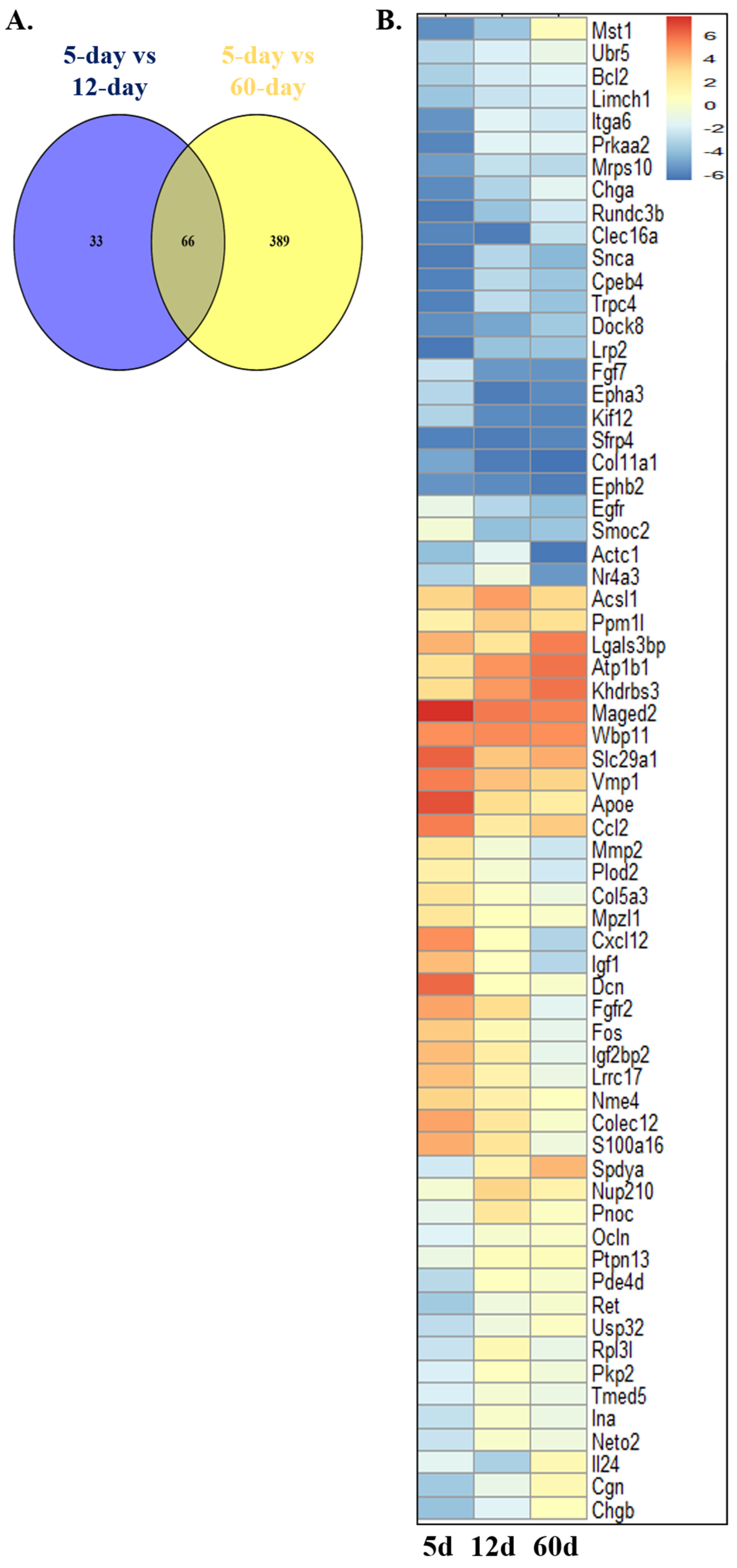
We used an adjusted p value of <0.05 and a cutoff of |log2(FC)| > 1 to identify significant DEGs. We compared the transcriptomic profile of a 5-day-old Sc with a 12-day-old Sc and identified a total of 1402 DEGs. Similarly, by comparing the RNA-seq data of a 5-day-old Sc with that of a 60-day-old Sc, we identified a total of 4612 DEGs. Then, we referred to a list of the mitophagy-related differentially expressed genes (MRDEGs) of humans from the PubMed website using the term “mitophagy-related genes” [69,70] and GeneCards database [71]. The GeneCards database provides comprehensive information about human genes. A total of 3073 MRGs were obtained after combining the results and removing duplicates. Further, we identified MRDEGs in rats. For this, we initially identified the orthologs for these MRDEGs of humans in rats from the NCBI gene database. After the successful identification of the orthologs, we screened 99 and 455 MRDEGs for rats in Sc development from 1402 (5- vs. 12-day-old Scs) and 4612 (5- vs. 60-day-old Scs) DEGs, respectively (Figure 2 and Supplementary File S1).
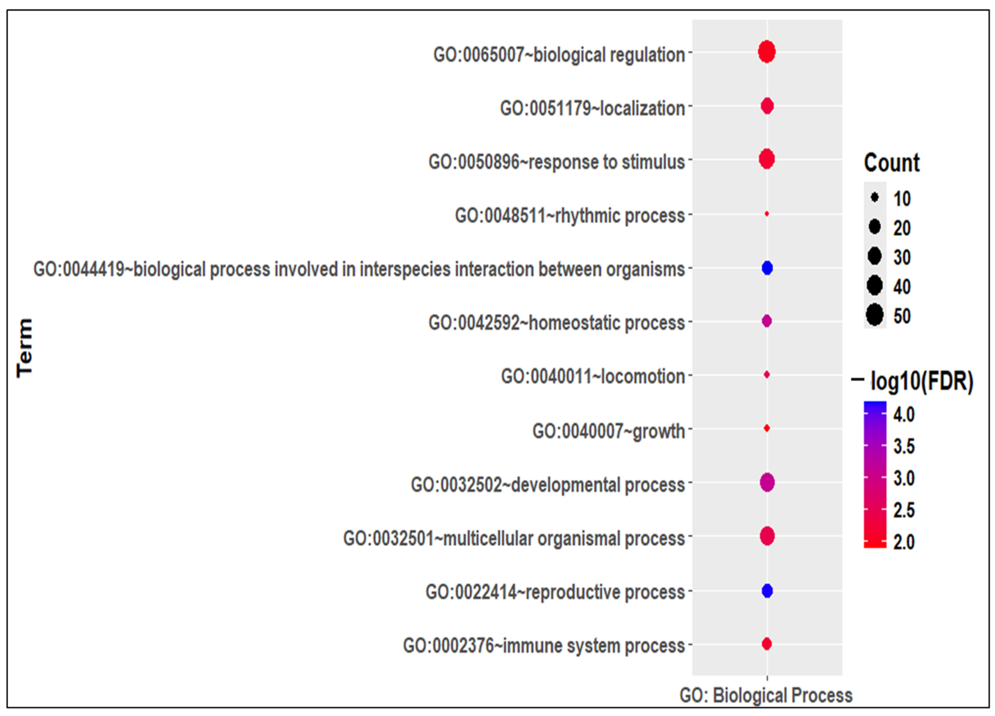
Thus, there were a total of 488 mitophagy-related genes in Sc development (MRGSCD) considering both the phases. A total of 66 MRGSCDs were identified to be common between the two experimental conditions, i.e., 5d vs. 12d and 5d vs. 60d (Figure 2). Thus, these 66 genes can be considered to be crucial mitophagy-related genes. A functional enrichment analysis of the common 66 MRGSCDs showed that GO biological processes like GO:0022414—“reproductive process” and GO:0032502—“developmental process” were enriched (Figure 3). Similarly, GO molecular functions like GO:0005488—“binding” were enriched (Supplementary Figure S1).
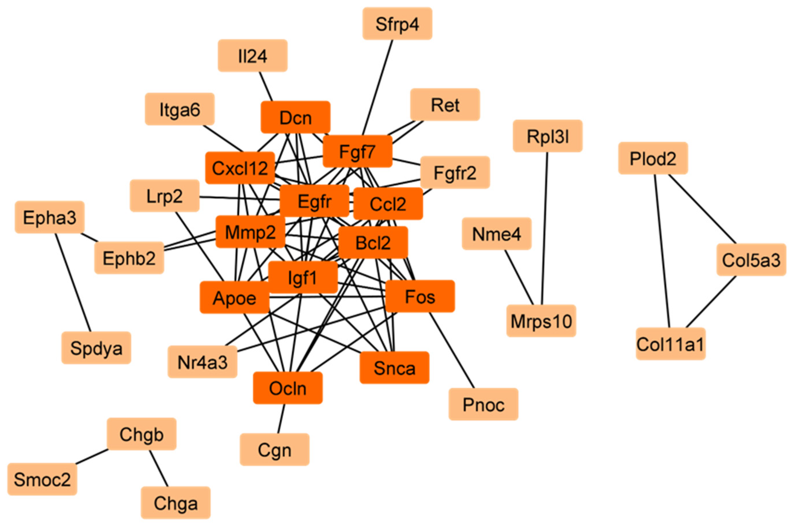
The reconstruction of a protein interaction network for 66 common MRGSCDs would unravel the vital signaling cascade of mitophagy-related genes in Sc development common to both stages. So next, with these 66 MRDEGs, we reconstructed a protein–protein interaction network (PPIN) using STRING v12 (with a confidence score > 0.4) (Figure 4).
This PPIN consists of 33 nodes and 76 edges. In a PPIN, hub genes are the nodes with the greatest number of edges or the most connections. Subsequently, using a network analyzer in Cytoscape 3.10.3., we identified 12 hub genes in the PPIN, viz. epidermal growth factor receptor (Egfr), Bcl2, C-C motif chemokine ligand 2 (Ccl2), matrix metallopeptidase 2 (Mmp2), insulin-like growth factor 1 (Igf1), fibroblast growth factor 7 (Fgf7), apolipoprotein E (Apoe), Fos, C-X-C motif chemokine ligand 12 (Cxcl12), Occludin (Ocln), decorin (Dcn), and synuclein alpha (Snca) (Figure 4). Notably, among these 66 common genes, Nor1 was highlighted as a key gene of interest due to its significant upregulation in both 12-day- and 60-day-old Scs (Figure 2B and Supplementary File S1). Moreover, it was found that Nor1 was involved in the interactions with only two important hub genes, i.e., Bcl2 and Fos in the PPIN. So, we performed further studies on Nor1 to validate the findings of this analysis.

2.2. Validation of the Differential Expression of Nor1 in Scs

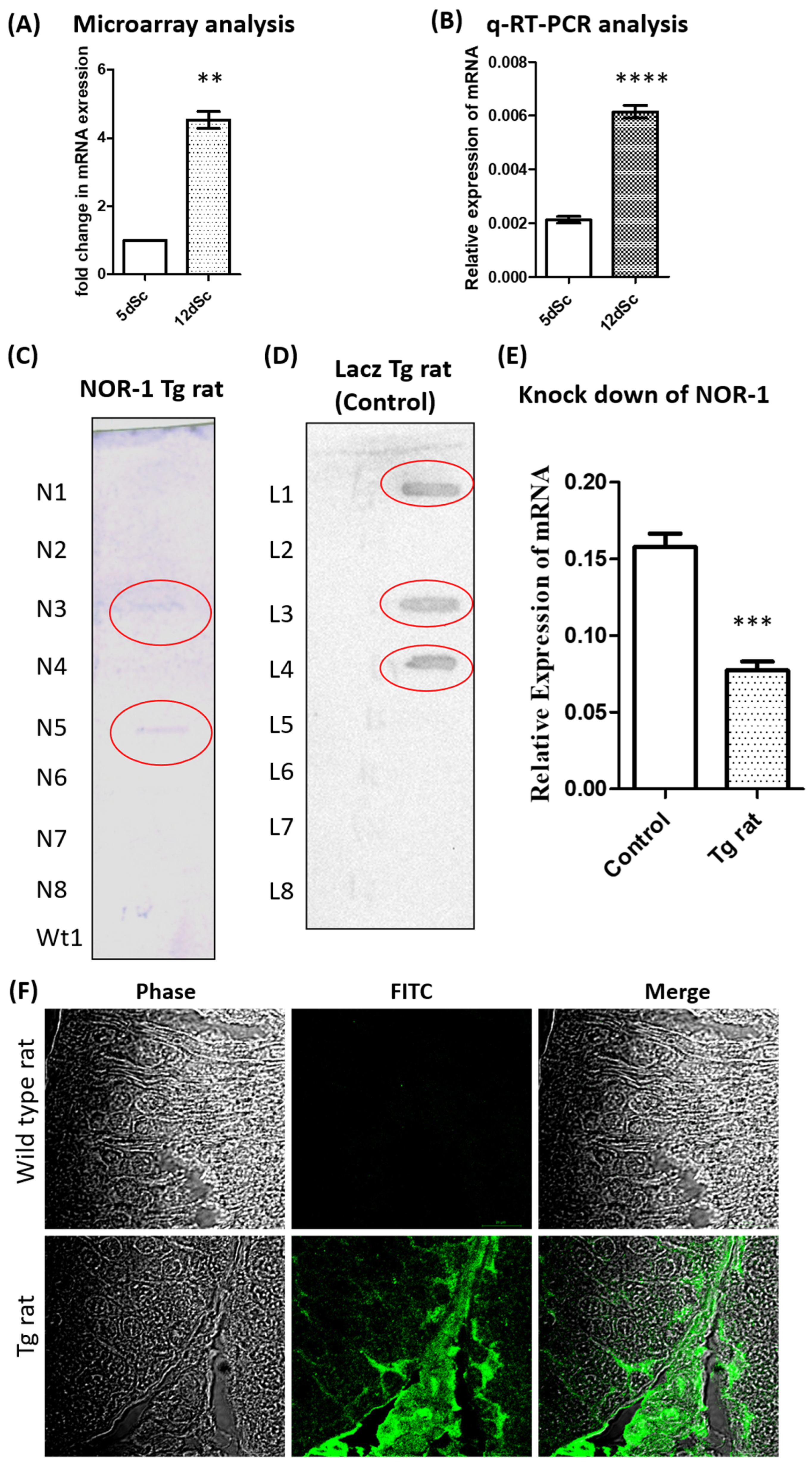
Since Nor1 was identified as an important gene related to mitophagy in our analysis, we validated the expression of Nor1 in the 5-day-old Scs and 12-day-old Scs, as it was upregulated in the microarray analysis (Figure 5A). These cells were treated with follicle-stimulating hormone (FSH) and testosterone (T), which were identical to those of the microarray samples. The q-RT-PCR data showed that Nor1 was significantly (p ≤ 0.05) upregulated in the Sc isolated from the 12-day-old Scs as compared to that of the 5-day-old Scs (n = 4) (Figure 5B).

2.3. Generation of Transgenic Rat with Sc-Specific NOR1 Knockdown

Since we identified Nor1 to be upregulated in a 12-day-old Sc from the rat microarray data, we generated a transgenic rat with the Sc cell-specific knockdown of Nor1 (Tg rat) using the Pem promoter to determine the functional role of Nor1 in spermatogenesis and mitophagy. As a control, we generated the transgenic rat with the Sc cell-specific knockdown of LacZ (LacZ control rat). The integration of the transgene was confirmed by slot blot analysis using the GFP probe (Figure 5C,D). We performed a q-RT-PCR analysis to determine the knockdown efficiency of Nor1 in the testis of the Tg rat. We observed that the expression levels of Nor1 in the Tg rat were reduced by 47% as compared to the LacZ control rat (Figure 5E). Since the Pem promoter drives the expression of shRNA against Nor1 and gfp, we analyzed the testicular section of the Tg rats and age-matched wild-type (wt) control rats for the expression of GFP using anti-GFP antibodies to determine the transgene expression. We observed the expression of GFP in the testis of the Tg rat but not in that of the control (Figure 5F), suggesting that the Pem promoter was active in the Tg rats and able to drive the expression of shRNA.

2.4. Attenuation of Spermatogenesis in Testes of Sc-Specific Nor1 Knockdown Tg Rats

The Tg rat line could not propagate beyond the F1 generation as the positive male pups born from the mating of F1-generation siblings were infertile (Figure 6A). On the other hand, the LacZ control rats were maintained beyond the F1 generation. All of our studies were conducted using the F1 generation of shRNA knockdown rats, as the shRNA transgene behaves like dominant-negative alleles of the gene of interest; hence, Tg rats can be analyzed in the F1 generation itself without waiting for homozygosity.
The F1-generation male rats from the Tg rats and the LacZ control male rats were analyzed for defects in reproductive functions. The testis weight of the Tg rats (0.79 ± 0.11 g) decreased significantly (p ≤ 0.05) compared to that of the LacZ control rats (1.497 ± 0.02 g) (Figure 6B). Further, we analyzed the sperm count from the epididymis of the Tg rats and control. We observed a significant (p ≤ 0.05) reduction in sperm counts in the Tg rats (4 ± 1.52 × 10⁶/mL/epididymis) as compared to the age-matched LacZ control rat (144.7 ± 4.667 × 10⁶/mL/epididymis) (Figure 6C). Although the total sperm count was found to be drastically reduced, more than 80% of the sperm was found to be motile in the Tg rats.
Qualitative histopathological evaluations of the testes of the Tg rats (2 months old and 10 months old) and LacZ control rat (10 months old) were performed to determine the effect of NOR1 knockdown. We observed that that of the LacZ control rat showed a normal tubular structure, whereas those of the Tg rats showed the sloughing of spermatogenic cells in the majority of the tubules (Figure 6D). Moreover, multiple vacuoles were observed in the seminiferous epithelium of the Tg rat, suggesting that spermatogenesis was impaired due to the decline in Nor1.

3. Discussion

Our study provides novel insights into the role of MRGs during the development of Scs and their effect on spermatogenesis. We identified 12 hub MRGs (Fos, Bcl2, Egfr, Ccl2, Mmp2, Igf1, Fgf7, Apoe, Cxcl12, Ocln, Dcn, and Snca) that are differentially expressed during Sc differentiation. To assess their importance, we performed a functional genomics study and identified Nor1 as a key MRG that interacts with two major hub genes, Bcl2 and Fos (Figure 2, Figure 3 and Figure 4). Nor1 expression was significantly upregulated in 12-day-old Scs compared to 5-day-old Scs, and we validated its differential expression (Figure 5). To investigate the role of Nor1 in mitophagy and spermatogenesis, we generated transgenic (Tg) rats with reduced Nor1 expression specifically in Scs during puberty (Figure 5). Male Tg rats were infertile due to a low sperm count and displayed abnormalities in testicular architecture (Figure 6). Overall, our findings suggest that Nor1 is an important MRG and plays a critical role in spermatogenesis in adult rats.
Several of the hub genes we identified show dynamic regulation during the development of Scs. Bcl2, a key member of the Bcl2 family of proteins, has an anti-apoptotic role [72]. Previous studies have shown that a reduction in levels of Bcl2 leads to apoptosis in various cell types [73]. Interestingly, Bcl2-deficient mice show normal spermatogenesis [74]. In infertile males, there are higher Bcl2 mRNA levels, which indicate an attempt to prevent germ cell death [75]. It was upregulated in the 12 d old Scs in our analysis, suggesting that a higher level of Bcl2 has a protective mechanism against cell death. Moreover, Bcl2 is a known inhibitor of Parkin-mediated mitophagy, suggesting that it may regulate mitochondrial quality control in Scs [76].
Fos, primarily in the form of cFos, is a pivotal transcription factor. cFos upregulates Matrix metallo protease8 (Mmp8), which degrades gap junction proteins that play an important role in the development of Scs [77]. We observed a downregulation of cFos in 12 d old Scs onwards, which coincides with the upregulation of gap junction proteins during puberty. Further, elevated reactive oxygen species (ROS) levels can upregulate the level of cFos [78,79]. The stress-induced upregulation of cFos may be a protective mechanism for the cells [80].
During the metastasis of a cancer cell, it translocates to the mitochondria and regulates mitochondrial dynamics [81]. Interestingly, knockout mice models lacking EGFR ligands show normal spermatogenesis [82]. However, EGFR-overexpressing mice show infertility [83]. Egfr was observed to be downregulated during Sc differentiation, suggesting that its downregulation may be required for proper Sc maturation.
The chemokine Ccl2 promotes autophagy and mitochondrial biogenesis [84,85]. Ccl2 overexpression leads to mitochondrial accumulation [86]. We observed that its level was reduced in 12 d old Scs, suggesting that the downregulation of Ccl2 may facilitate mitochondrial turnover during Sc maturation. Similarly, Mmp2 modulates mitochondrial proteins [87] and its expression is regulated by FSH in Scs [88]. Its level was reduced in the 12 d old Scs, indicating that mitochondrial remodeling in Scs may be developmentally regulated.
Igf1 is shown to activate mitophagy through nuclear respiratory factor 2/ Sirtuin 3 (Nrf2/Sirt3) signaling [89] and also regulates mitochondrial dynamics [90]. However, Igf1 knockout mice are fertile [91]. Our data show that the Igf1 level was reduced during Sc maturation, suggesting that the downregulation may be a developmental shift in energy metabolism. We observed a similar pattern (downregulation in 12 d old Scs) for Fgf7, Apoe, Fos, Cxcl12, and Dcn, all of which are associated with mitochondrial or autophagy-related functions [92,93,94,95,96].
On the other hand, Ocln and Snca were upregulated in the differentiated 12 d old Scs. Importantly, Ocln knockout male mice are infertile [97]. It has a role in the structural integrity and mitochondrial stability of Scs [98]. The elevated level of Ocln during the development of Scs supports its crucial role in the function of Scs. The elevated level of Snca is associated with mitochondrial dysfunction [99] and excessive mitophagy [100]. These reports suggest that the regulation of Scna may have a role in mitochondrial homeostasis during Sc maturation.
The identification of these differentially expressed hub genes related to mitophagy suggests that developmental changes in Scs are associated with mitophagy. To further validate this, we selected Nor1 for a detailed investigation. Nor1 is also associated with two hub genes, Bcl2 and Fos. Based on these findings, we generated a transgenic (Tg) rat with an Sc-specific reduction in Nor1 to validate our analysis of mitophagy-related genes in Scs. We observed that Tg rats with Sc-specific NOR1 decline were infertile due to a reduced sperm count and abnormal testicular architecture (Figure 6). These findings are consistent with previous reports on Nor1 [68], supporting the reliability of our in vivo functional analysis of Nor1 and the identification of mitophagy-related hub genes.
Rat models were chosen because our microarray data are from rats, and rats are physiologically closer to humans than mice [101,102]. Mice are widely used for genetic studies due to the advancement in embryo transfer techniques. However, rats have distinct advantages for studying spermatogenesis. In particular, spermatogenesis requires approximately 55 days to complete a full cycle in a rat, which is closer to that of a human (64 days) [103,104,105]. Also, the rat seminiferous epithelium has 14 defined stages compared to 12 in mice [103,104].
The proximal Rhox-5 promoter (Pem promoter) drives the expression of target genes only in Scs from 14 days postnatally onwards [106]. We selected this promoter, as Nor1 is upregulated from 12 days onwards in rats. On the other hand, the anti Mullerian hormone (Amh) promoter is active from birth to the neonatal stage. For instance, the expression of Amh in mouse Scs starts at embryonic day 12 dpc, which is gradually reduced during Sc differentiation and in adult testes [107]. The proximal promoter of Amh (180 bp) is sufficient for driving its expression specifically in Scs [108]. Therefore, we selected proximal Rhox-5 promoter (Pem promoter) to knockdown Nor1 in Scs during puberty.
The limitation of this study is that we did not perform any studies on the functional aspect of mitophagy in these Tg rats due to the lower number of Tg rats generated by these lines. Further studies will be needed to determine the effect of Nor1 on mitochondrial structure and function in these Tg rats.
In conclusion, our integrative genomic and functional analyses identified 12 key hub genes related to mitophagy that are differentially expressed during the development of Scs. Furthermore, we demonstrated that Nor1 plays an important role in spermatogenesis in rat testes. These findings provide new insights into the roles of other MRGs during Sc development and further highlight the critical contribution of mitochondrial dysfunction to male infertility.

4. Materials and Methods

4.1. Animals

Wistar rats (Rattus norvegicus) were obtained from the Small Animal Facility of the National Institute of Immunology (New Delhi, India). The rats were housed in a climate-controlled facility under standard light conditions (14 h light/10 h dark cycle), temperature (23 °C), and humidity (50 ± 5%). The rats were housed in individually ventilated cages with (IVC) ad libitum access to acidified autoclaved water and were handled by trained personnel. Rats were used as per the guidelines laid down by the CPCSEA (Committee for the Purpose of Control and Supervision of the Experiments on Animals). Protocols for experiments were approved by the Institutional Animal Ethics Committee (IAEC) of National Institute of Immunology, India, constituted by CPCSEA (IAEC number: IAEC 220/09). Rats were euthanized by cervical dislocation as approved by the Institutional Animal Ethics Committee of National Institute of Immunology. For the primary Sc culture, about 100 (5-day-old male) rats and 60 (12-days-old male) rats were used. For the study involving transgenic rats, about 220 rats (including non-transgenic littermates) were used.

4.2. Screening of Hub Genes for Sertoli Cell Development in Rats

We identified differentially expressed genes (DEGs) by analyzing microarray data from the GSE48795 dataset [62] in order to examine the function of mitophagy-related genes in Sertoli cell development. The Bioconductor project’s basic packages in the NCBI Gene Expression Omnibus (GEO) database, which were based on limmav3.26.8, were utilized for data retrieval, logarithmic transformation, and quantile normalization of the data [109]. Benjamini–Hochberg (false discovery rate (FDR)) method was used for identifying significant DEGs. Considering this, an adjusted p value < 0.05 along with |log2(FC)| > 1 was considered to identify significant DEGs. A cut-off of adjusted p value < 0.05 and |log2(FC)| > 1 was considered to identify significant DEGs. The orthologs for humans in rats were identified from NCBI’s gene database [110]. Then, to reconstruct the PPIN for Sertoli cell development, we considered both physical and functional interactions from STRING v12 [111]. A default cut-off, i.e., confidence score > 0.4, was considered for the reconstruction of the PPIN. To identify hub genes, we used a network analyzer in Cytoscape 3.10.3 [112] to calculate network degree centrality. A network degree centrality > average of total degree count of the nodes, i.e., > 4.5, was considered the cut-off for identifying hub genes.

4.3. Functional Enrichment Analysis

We used DAVID v2023q4 for GO biological process and GO molecular function enrichment analyses of the genes [113].

4.4. Differential Expression Analysis of Nor1 in 5-Day-Old and 12-Day-Old Rat Sc Cultures

We identified Nor1 as a candidate gene from the analysis. The differential expression of Nor1 from this analysis was validated by q-RT-PCR analysis in the four separate sets of Sc cultures (n = 4 biological replicates) of 5-day- and 12-day-old rat testes. Testes from about 20–30 and 10–20 male rats were pooled for 5-day- and 12-day-old rat Sc cultures, respectively [61]. The Scs were isolated, cultured, and treated with FSH and testosterone in combination on day 4 of culture, as we reported previously [61,62]. Further, the total RNAs were extracted from these groups of cells as described previously [114,115]. Whole RNA was extracted using TRI reagent (Sigma Aldrich, St. Louis, MO, USA) as per the manufacturer’s instructions. The quantity and quality (260/280) of RNA were determined using NanoDrop 2000c spectrophotometer (Thermo Scientific, Waltham, MA, USA). The 260/280 and 260/230 absorbance ratios of all the RNA preparations used in this study were within the range of 1.8–2.0. This suggested that all the RNA preparations were of sufficiently good quality for gene expression analysis using real-time PCR. One microgram of RNA was treated with 0.5 U DNaseI (Thermo Scientific, Waltham, MA, USA) to remove any contaminating genomic DNA fragments. This was followed by single-strand c-DNA synthesis using M-MLV reverse transcriptase (Promega, Madison, WI, USA) as per the manufacturer’s protocol. qRT-PCR amplifications were performed using RealplexS (Eppendorf, Hamburg, Germany) in a total volume of 10 μL (1 μL of cDNA), with 0.5 μM of each primer and 5 μL of Power SYBR Green Master Mix (Applied Biosystems, Foster City, CA, USA).

4.5. Plasmids and Cloning

The knockdown of Nor1 using shRNA in the Scs of the prepubertal rats was performed as described previously [68,116,117]. shRNA was designed by using online tools available at Dharmacon (https://rnaidesigner.thermofisher.com/rnaiexpress/setOption.do?designOption=shrna&pid=-38224801…; accessed on 17 May 2011) and Clontech (https://www.takarabio.com/assets/a/112504?srsltid=AfmBOopKDxPbPeL8erHa66yZk3C3LfUdHdyNvmriqXyHMvG-1…; accessed on 17 May 2011). shRNA sequences were verified for target specificity by BLAT (http://genome.ucsc.edu/cgi-bin/hgBlat?command=start) and BLAST 2.2.26 analysis (http://www.ebi.ac.uk/Tools/sss/ncbiblast/). The shRNAs against Nor1 and bacterial LacZ (as a control) were designed and cloned under the Pem promoter. Pem promoter is reported to drive shRNA expression specifically in pubertal Sertoli cells [106]. The sequence for shRNA for Nor1 was TCGAGAGACAAGAGACGTCGAAATTTCAAGAGAATTTCGACGTCTCTTGTCTTTTTTTACCGGTCCGC, and the sequence for shRNA for LacZ was TCGAGGCATCGAGCTGGATAATAATTCAAGAGATTATTATCCAGCTCGATGCTTTTTTACCGGTCCGC.

4.6. Generation of Transgenic (Tg) Rats with Reduced NOR1 in Scs

The construct was linearized with Stu I and was used to generate a transgenic rat (Tg) as we reported previously [101,114,118]. Briefly, the linearized construct (a total of 30 µg DNA with a concentration of 1 µg/µL) was injected into the testis of a 38-day-old Wistar rat and electroporated using 8 square 90 V electric pulses in alternating directions with a time constant of 0.05 second and an inter-pulse interval of ~1 second via an electric pulse generator. The electroporated male rats were mated with wild-type female rats 60 days post-electroporation. Pups that were born were screened by PCR using genomic DNA obtained from their tail biopsies, and those positive for the transgene were regarded as F1 generation of transgenic (Tg) rats.
The integration of transgene in the transgenic rats was confirmed by slot blot analysis as described previously [115,116]. For slot blot analysis, around 2 µg of gDNA isolated from the tail biopsies was denatured at 95 °C for 10 min and blotted onto Hybond N+ (Amersham Pharmacia Biotech, Buckinghamshire, UK) in a slot blot apparatus (Cleaver Scientific Co., Rugby, UK) under vacuum. The membrane was UV cross-linked at energy of 12 × 104 µJ/cm2 in a CL-1000 Ultraviolet crosslinker (UVP, Upland, CA, USA). The non-radioactive DIG probe was used to detect the transgene. The probe was prepared by amplifying a fragment that contained part of the gfp (630 bp) using the following primers: GFPF: GACGTAAACGGCCACAAGTT and GFPR: GGCGGTCACGAACTCCAG. During PCR, the probe was labeled with DIG. The membranes were pre-hybridized for 2 h at 37 °C in hybridization solution without labeled probe and then hybridized separately at 37 °C with probe specific for GFP for 16 h. The membranes were washed twice, for 5 min each, at room temperature in 2× saline sodium citrate buffer and 0.1% SDS. This was followed by another washing for 15 min at 60 °C (in 0.1X saline sodium citrate buffer and 0.1% SDS). Detection was performed by using the DIG labeling and BCIP/NBT substrate according to the manufacturer’s recommendation (DIG system user’s guide for filter hybridization, Roche, Mannheim, Germany).
For this study, we generated two female founders for Nor1 knockdown rats and two founders for LacZ knockdown rats. The efficiencies of generation of transgenic rats for both the lines were close to 33%, which is in agreement with our previous study, which showed that 33.08% of the progeny generated from single cohabitation of an electroporated fore founder (generated by us) with a wild-type female was transgenic [101].

4.7. Validation of Knockdown of NOR1 by q-RT-PCR

Total RNA was extracted from the whole testicular extract of control and 10-month-old Tg rats (n = 4 biological replicates) as we described previously [114,115]. q-RT-PCR amplifications were performed to detect expression of Nor1 and Ppia (loading control) using the RealplexS (Eppendorf) as described previously [114,115]. Primers for Nor1 were as follows: NF: GCTTGTCTCTGCACCATTCA; and NR: TGAGCTAGGCCTCGAAGGTA. Primers for Ppia were as follows: PF: ATGGTCAACCCCACCGTGT; and PR: TCTGCTGTCTTTGGAACTTTGTCT. The expressions of mRNA of the target genes were evaluated by the efficiency-corrected 2−ΔΔCT method.

4.8. Immunofluorescence Microscopy

The testes of Tg rats (10 months old) (n = 3) and age-matched WT rats (n = 3) were collected and fixed in Bouins solution, and IF was performed as described previously [114,115]. To stain for transgene GFP, a primary rabbit polyclonal anti-GFP antibody was used at a dilution of 1:250 and incubated for 2 h at room temperature. A goat anti-rabbit IgG conjugated with Alexa 488 (Thermoscientific, Waltham, MA, USA) at a dilution of 1:1000 was used as the secondary antibody.

4.9. Tissue Histology

Tissue histology was performed as described previously [114,115]. Briefly, the testicular tissue samples of rats were fixed in Bouins solution at room temperature for 24 h. Dehydration of tissues was performed in a series of ascending concentrations of ethanol for 1 h at each grade of ethanol. The tissues were embedded in paraffin, and 4 µm sections were cut. Sections were stained with hematoxylin and eosin and were examined to evaluate the status of spermatogenesis in the control (10 months old) and Tg rats (2 months and 10 months old). Around 50 to 60 tubules per section were analyzed per rat.

4.10. Fertility Analyses of Nor1 Knockdown Rats

Testes weights of Nor1 knockdown transgenic rats (10 months old) (n = 3) and LacZ shRNA knockdown control rats (n = 3) were recorded. The numbers of epididymal spermatozoa were analyzed as described previously [114,115]. Total numbers of sperm present in each epididymis were counted after releasing the sperm in 1 mL of 1× PBS by mincing the epididymis, and the average was determined. Motility of at least 200 epididymal spermatozoa per field was assessed under light microscope.

4.11. Statistical Analysis

For the validation of differential expression of Nor1 by q-RT-PCR analysis, we used Mann–Whitney tests. For fertility studies, at least three or more sets of observations were analyzed by Mann–Whitney tests. A value of p  ≤  0.05 was considered significant. All statistical analyses were performed using GraphPad Prism v. 5.01 (GraphPad Software, La Jolla, CA, USA).

Supplementary Materials

The following supporting information can be downloaded at: https://www.mdpi.com/article/10.3390/ijms26189209/s1.

Author Contributions

Experiments were conceived and designed by B.S.P. and S.S.M. Experiments were performed by B.S.P. and D.D. Data of the manuscript were analyzed by B.S.P., H.S. and I.B. Primary draft of the manuscript was written by B.S.P. and reviewed by B.S.P., D.D., H.S., N.W. and I.B. All authors have read and agreed to the published version of the manuscript.

Funding

This study was funded by the Department of Biotechnology, Govt. of India, under grants BT/PR11313/AAQ/01/376/2008, BT/HRD/35/01/01/2010 (TATA Innovation Award), and BT/PR10805/AAQ/1/576/2013; the Polish National Science Centre under grant (2020/39/D/NZ5/02004); and the Department of Biotechnology, Govt. of India, under grant (BT/PR46488/AAQ/1/886/2022).

Institutional Review Board Statement

The rats were housed and used as per the guidelines laid down by the CPCSEA (Committee for the Purpose of Control and Supervision of the Experiments on Animals). The protocols for the experiments were approved by the Institutional Animal Ethics Committee (IAEC) of the National Institute of Immunology, India, constituted by CPCSEA (approval code: IAEC 220/09; approval date: 1 June 2009; extended every four years).

Informed Consent Statement

Not applicable.

Data Availability Statement

The original contributions presented in this study are included in the article/Supplementary Materials. Further inquiries can be directed to the corresponding author.

Acknowledgments

We are thankful to all the staff at the Small Animal Facility. Thanks are due to Ram Singh, Dharamvir Singh, and Birendar Roy for their technical assistance. We are grateful to the Director of the NII for their valuable support.

Conflicts of Interest

The authors declare no conflicts of interest.

Abbreviations

The following abbreviations are used in this manuscript:
GcGerm cell
ScSertoli cell
Bcl2B-Cell Leukemia/Lymphoma 2
FosFBJ murine osteosarcoma viral oncogene homolog
LC3Microtubule-associated protein 1A/1B light chain 3
Mfn1mitofusin 1
Mfn2mitofusin 2
Opa1Optic Atrophy 1, mitochondrial dynamin-like GTPase
PTENphosphatase and tensin homolog
PINK1phosphatase and tensin homolog induced kinase I
UPSubiquitin proteasome system
mtDNAmitochondrial DNA
POLGpolymerase gamma
Mffmitochondrial fission factor
Atg7autophagy related 7
GATA4GATA binding protein 4
MRGmitophagy-related genes
Tg ratSertoli-cell-specific Nor1 knockdown transgenic rat
Pgc1αperoxisome proliferator-activated receptor gamma coactivator 1 α
TfamTranscription factor A, mitochondrial
Smad3SMAD family member 3
DEGsdifferentially expressed genes
MRDEGsmitophagy-related differentially expressed genes
PPINprotein–protein interaction network
Egfrepidermal growth factor receptor
Ccl2C-C motif chemokine ligand 2
Mmp2matrix metallopeptidase 2
Igf1insulin-like growth factor 1
Fgf7fibroblast growth factor 7
Apoeapolipoprotein E
Cxcl12C-X-C motif chemokine ligand 12
Oclnoccludin
Dcndecorin
Scnasynuclein alpha
GFPgreen fluorescent protein
Mmp8Matrix metallo protease8
ROSreactive oxygen species
Amhanti mullerian hormone
GEOGene Expression Omnibus
FDRFalse discovery rate
MRGSCDmitophagy-related genes in Sc development

References

  1. Agarwal, A.; Baskaran, S.; Parekh, N.; Cho, C.L.; Henkel, R.; Vij, S.; Arafa, M.; Panner Selvam, M.K.; Shah, R. Male infertility. Lancet 2021, 397, 319–333. [Google Scholar] [CrossRef] [PubMed]
  2. Potiris, A.; Moustakli, E.; Trismpioti, E.; Drakaki, E.; Mavrogianni, D.; Matsas, A.; Zikopoulos, A.; Sfakianakis, A.; Tsakiridis, I.; Dagklis, T.; et al. From Inflammation to Infertility: How Oxidative Stress and Infections Disrupt Male Reproductive Health. Metabolites 2025, 11, 267. [Google Scholar] [CrossRef] [PubMed]
  3. Yan, Q.; Wang, Q. Exploring the Characters of Non-Coding RNAs in Spermatogenesis and Mal e Infertility. Int. J. Mol. Sci. 2025, 26, 1128. [Google Scholar] [CrossRef]
  4. Qu, N. Pharmacological Effects and Immune Mechanisms of Oriental Medicines in Restoring Male Infertility. Int. J. Mol. Sci. 2025, 26, 4642. [Google Scholar] [CrossRef]
  5. Sharpe, R.M. Sperm counts and fertility in men: A rocky road ahead. Science & Socie ty Series on Sex and Science. EMBO Rep. 2012, 13, 398–403. [Google Scholar]
  6. Vander Borght, M.; Wyns, C. Fertility and infertility: Definition and epidemiology. Clin. Biochem. 2018, 62, 2–10. [Google Scholar] [CrossRef] [PubMed]
  7. Olesen, I.A.; Andersson, A.M.; Aksglaede, L.; Skakkebaek, N.E.; Rajpert-de Meyts, E.; Joergensen, N.; Juul, A. Clinical, genetic, biochemical, and testicular biopsy findings among 1,213 men evaluated for infertility. Fertil. Steril. 2017, 107, 74–82.e7. [Google Scholar] [CrossRef]
  8. Tournaye, H.; Krausz, C.; Oates, R.D. Novel concepts in the aetiology of male reproductive impairment. Lancet Diabetes Endocrinol. 2017, 5, 544–553. [Google Scholar] [CrossRef]
  9. Zecevic, N.; Veselinovic, A.; Perovic, M.; Stojsavljevic, A. Association Between Zinc Levels and the Impact of Its Deficiency on Idiopathic Male Infertility: An Up-to-Date Review. Antioxidants 2025, 14, 165. [Google Scholar] [CrossRef]
  10. Bouvattier, C. Neonatal gonadotropin therapy in male congenital hypogonadotropic hypo gonadism. Nat. Rev. Endocrinol. 2011, 8, 172–182. [Google Scholar] [CrossRef]
  11. Irvine, D.S. Epidemiology and aetiology of male infertility. Hum. Reprod. 1998, 13 (Suppl. 1), 33–44. [Google Scholar] [CrossRef]
  12. Maroto, M.; Torvisco, S.N.; García-Merino, C.; Fernández-González, R.; Pericuesta, E. Mechanisms of Hormonal, Genetic, and Temperature Regulation of Germ Ce ll Proliferation, Differentiation, and Death During Spermatogenesis. Biomolecules 2025, 15, 500. [Google Scholar] [CrossRef]
  13. Santi, D.; Corona, G.; Salonia, A.; Ferlin, A. Current drawbacks and future perspectives in the diagnosis and treatme nt of male factor infertility, with a focus on FSH treatment: An exper t opinion. J. Endocrinol. Investig. 2025, 48, 1085–1100. [Google Scholar] [CrossRef] [PubMed]
  14. Du, L.; Chen, W.; Cheng, Z.; Wu, S.; He, J.; Han, L.; He, Z.; Qin, W. Novel Gene Regulation in Normal and Abnormal Spermatogenesis. Cells 2021, 10, 666. [Google Scholar] [CrossRef] [PubMed]
  15. Griswold, M.D. The central role of Sertoli cells in spermatogenesis. Semin. Cell Dev. Biol. 1998, 9, 411–416. [Google Scholar] [CrossRef] [PubMed]
  16. Chen, Z.F.; Shen, Y.F.; Gao, D.W.; Lin, D.F.; Ma, W.Z.; Chang, D.G. Metabolic pathways and male fertility: Exploring the role of Sertoli c ells in energy homeostasis and spermatogenesis. Am. J. Physiol. Endocrinol. Metab. 2025, 329, E160–E178. [Google Scholar] [CrossRef]
  17. Shah, W.; Khan, R.; Shah, B.; Khan, A.; Dil, S.; Liu, W.; Wen, J.; Jiang, X. The Molecular Mechanism of Sex Hormones on Sertoli Cell Development an d Proliferation. Front. Endocrinol. 2021, 12, 648141. [Google Scholar] [CrossRef]
  18. Wang, L.; Bu, T.; Wu, X.; Gao, S.; Li, X.; Jesus, A.B.; Wong, C.K.C.; Chen, H.; Chung, N.P.Y.; Sun, F.; et al. Cell-Cell Interaction-Mediated Signaling in the Testis Induces Reprodu ctive Dysfunction-Lesson from the Toxicant/Pharmaceutical Models. Cells 2022, 11, 591. [Google Scholar] [CrossRef]
  19. Hai, Y. The roles and regulation of Sertoli cells in fate determinations of sp ermatogonial stem cells and spermatogenesis. Semin. Cell Dev. Biol. 2014, 29, 66–75. [Google Scholar] [CrossRef]
  20. Boguenet, M.; Bouet, P.E.; Spiers, A.; Reynier, P.; May-Panloup, P. Mitochondria: Their role in spermatozoa and in male infertility. Hum. Reprod. Update 2021, 27, 697–719. [Google Scholar] [CrossRef]
  21. Chang, Z.; Miao, L.; Wang, P. Mitochondrial Ribosome Regulation Drives Spermatogenesis and Male Fert ility. Biol. Cell 2025, 117, e12007. [Google Scholar] [CrossRef]
  22. Tesarik, J.; Mendoza-Tesarik, R. Mitochondria in Human Fertility and Infertility. Int. J. Mol. Sci. 2023, 24, 8950. [Google Scholar] [CrossRef]
  23. Costa, J.; Braga, P.C.; Rebelo, I.; Oliveira, P.F.; Alves, M.G. Mitochondria Quality Control and Male Fertility. Biology 2023, 12, 827. [Google Scholar] [CrossRef]
  24. Park, Y.J.; Pang, M.G. Mitochondrial Functionality in Male Fertility: From Spermatogenesis to Fertilization. Antioxidants 2021, 10, 98. [Google Scholar] [CrossRef]
  25. Wang, X.; Yin, L.; Wen, Y.; Yuan, S. Mitochondrial regulation during male germ cell development. Cell. Mol. Life Sci. 2022, 79, 91. [Google Scholar] [CrossRef]
  26. Youle, R.J.; Narendra, D.P. Mechanisms of mitophagy. Nat. Rev. Mol. Cell Biol. 2011, 12, 9–14. [Google Scholar] [CrossRef]
  27. Mizushima, N. Autophagy: Process and function. Genes Dev. 2007, 21, 2861–2873. [Google Scholar] [CrossRef]
  28. Mizushima, N.; Sugita, H.; Yoshimori, T.; Ohsumi, Y. A New Protein Conjugation System in Human the counterpart of the yeast Apg12p conjugation system essential for autophagy. J. Biol. Chem. 1998, 273, 33889–33892. [Google Scholar] [CrossRef]
  29. Picca, A.; Faitg, J.; Auwerx, J.; Ferrucci, L.; D’Amico, D. Mitophagy in human health, ageing and disease. Nat. Metab. 2023, 5, 2047–2061. [Google Scholar] [CrossRef] [PubMed]
  30. Tsukada, M.; Ohsumi, Y. Isolation and characterization of autophagy-defective mutants of Sacch aromyces cerevisiae. FEBS Lett. 1993, 333, 169–174. [Google Scholar] [CrossRef] [PubMed]
  31. Kuma, A.; Komatsu, M.; Mizushima, N. Autophagy-monitoring and autophagydeficient mice. Autophagy 2017, 13, 1619–1628. [Google Scholar] [CrossRef] [PubMed]
  32. McWilliams, T.G.; Prescott, A.R.; Allen, G.F.G.; Tamjar, J.; Munson, M.J.; Thomson, C.; Muqit, M.M.K.; Ganley, I.G. mito-QC illuminates mitophagy and mitochondrial architecture in vivo. J. Cell Biol. 2016, 214, 333–345. [Google Scholar] [CrossRef] [PubMed]
  33. Sun, N.; Yun, J.; Liu, J.; Malide, D.; Liu, C.; Rovira, I.I.; Holmström, K.M.; Fergusson, M.M.; Yoo, Y.H.; Combs, C.A.; et al. Measuring in vivo Mitophagy. Mol. Cell 2015, 60, 685–696. [Google Scholar] [CrossRef]
  34. Chan, D.C. Mitochondrial dynamics and its involvement in disease. Annu. Rev. Pathol. Mech. Dis. 2020, 15, 235–259. [Google Scholar] [CrossRef]
  35. Tábara, L.C.; Segawa, M.; Prudent, J. Molecular mechanisms of mitochondrial dynamics. Nat. Rev. Mol. Cell Biol. 2025, 26, 123–146. [Google Scholar] [CrossRef]
  36. Zaman, M.; Shutt, T.E. The Role of Impaired Mitochondrial Dynamics in MFN2-Mediated Pathology. Front. Cell Dev. Biol. 2022, 10, 858286. [Google Scholar] [CrossRef]
  37. Zhao, S.; Heng, N.; Wang, H.; Wang, H.; Zhang, H.; Gong, J.; Hu, Z.; Zhu, H. Mitofusins: From mitochondria to fertility. Cell. Mol. Life Sci. 2022, 79, 370. [Google Scholar] [CrossRef]
  38. Chan, N.C.; Chan, D.C. Parkin uses the UPS to ship off dysfunctional mitochondria. Autophagy 2011, 7, 771–772. [Google Scholar] [CrossRef]
  39. Rakovic, A.; Ziegler, J.; Mårtensson, C.U.; Prasuhn, J.; Shurkewitsch, K.; König, P.; Paulson, H.L.; Klein, C. PINK1-dependent mitophagy is driven by the UPS and can occur independe ntly of LC3 conversion. Cell Death Differ. 2019, 26, 1428–1441. [Google Scholar] [CrossRef] [PubMed]
  40. Clague, M.J.; Urbé, S. Diverse routes to mitophagy governed by ubiquitylation and mitochondri al import. Trends Cell Biol. 2025, 35, 527–538. [Google Scholar] [CrossRef] [PubMed]
  41. Narendra, D.P.; Youle, R.J. The role of PINK1-Parkin in mitochondrial quality control. Nat. Cell Biol. 2024, 26, 1639–1651. [Google Scholar] [CrossRef]
  42. Sekine, S.; Youle, R.J. PINK1 import regulation; a fine system to convey mitochondrial stress to the cytosol. BMC Biol. 2018, 16, 2. [Google Scholar] [CrossRef]
  43. Baklouti-Gargouri, S.; Ghorbel, M.; Ben Mahmoud, A.; Mkaouar-Rebai, E.; Cherif, M.; Chakroun, N.; Sellami, A.; Fakhfakh, F.; Ammar-Keskes, L. Identification of a novel m.9588G > a missense mutation in the mitocho ndrial COIII gene in asthenozoospermic Tunisian infertile men. J. Assist. Reprod. Genet. 2014, 31, 595–600. [Google Scholar] [CrossRef]
  44. Carra, E.; Sangiorgi, D.; Gattuccio, F.; Rinaldi, A.M. Male infertility and mitochondrial DNA. Biochem. Biophys. Res. Commun. 2004, 322, 333–339. [Google Scholar] [CrossRef]
  45. Demain, L.A.; Conway, G.S.; Newman, W.G. Genetics of mitochondrial dysfunction and infertility. Clin. Genet. 2017, 91, 199–207. [Google Scholar] [CrossRef]
  46. Zhang, X.; Xiong, Y.; Tan, Y.; Wang, L.; Li, R.; Zhang, Y.; Huang, H. Melatonin rescues impaired penetration ability of human spermatozoa in duced by mitochondrial dysfunction. Reproduction 2019, 158, 465–475. [Google Scholar] [CrossRef] [PubMed]
  47. Irigoyen, P.; Pintos-Polasky, P.; Rosa-Villagrán, L.; Skowronek, M.; Cassina, A.; Sapiro, R. Mitochondrial metabolism determines the functional status of human spe rm and correlates with semen parameters. Front. Cell Dev. Biol. 2022, 10, 926684. [Google Scholar] [CrossRef] [PubMed]
  48. Luoma, P.; Melberg, A.; Rinne, J.O.; Kaukonen, J.A.; Nupponen, N.N.; Chalmers, R.M.; Oldfors, A.; Rautakorpi, I.; Peltonen, L.; Majamaa, K.; et al. Parkinsonism, premature menopause, and mitochondrial DNA polymerase γ mutations: Clinical and molecular genetic study. Lancet 2004, 364, 875–882. [Google Scholar] [CrossRef] [PubMed]
  49. Kujoth, G.C.; Hiona, A.; Pugh, T.D.; Someya, S.; Panzer, K.; Wohlgemuth, S.E.; Hofer, T.; Seo, A.Y.; Sullivan, R.; Jobling, W.A.; et al. Mitochondrial DNA mutations, oxidative stress, and apoptosis in mammalian aging. Science 2005, 309, 481–484. [Google Scholar] [CrossRef]
  50. Trifunovic, A.; Wredenberg, A.; Falkenberg, M.; Spelbrink, J.N.; Rovio, A.T.; Bruder, C.E.; Bohlooly, Y.M.; Gidlof, S.; Oldfors, A.; Wibom, R.; et al. Premature ageing in mice expressing defective mitochondrial DNA polyme rase. Nature 2004, 429, 417–423. [Google Scholar] [CrossRef]
  51. Varuzhanyan, G.; Rojansky, R.; Sweredoski, M.J.; Graham, R.L.; Hess, S.; Ladinsky, M.S.; Chan, D.C. Mitochondrial fusion is required for spermatogonial differentiation an d meiosis. eLife 2019, 8, 51601. [Google Scholar] [CrossRef]
  52. Chen, H.; Ren, S.; Clish, C.; Jain, M.; Mootha, V.; McCaffery, J.M.; Chan, D.C. Titration of mitochondrial fusion rescues Mff-deficient cardiomyopathy. J. Cell Biol. 2015, 211, 795–805. [Google Scholar] [CrossRef]
  53. Shang, Y.; Wang, H.; Jia, P.; Zhao, H.; Liu, C.; Liu, W.; Song, Z.; Xu, Z.; Yang, L.; Wang, Y. Autophagy regulates spermatid differentiation via degradation of PDLIM 1. Autophagy 2016, 12, 1575–1592. [Google Scholar] [CrossRef]
  54. Wang, H.; Wan, H.; Li, X.; Liu, W.; Chen, Q.; Wang, Y.; Yang, L.; Tang, H.; Zhang, X.; Duan, E. Atg7 is required for acrosome biogenesis during spermatogenesis in mice. Cell Res. 2014, 24, 852. [Google Scholar] [CrossRef]
  55. Dietert, S.E. Fine structure of the formation and fate of the residual bodies of mou se spermatozoa with evidence for the participation of lysosomes. J. Morphol. 1966, 120, 317–346. [Google Scholar] [CrossRef]
  56. Eid, N.; Ito, Y.; Horibe, A.; Hamaoka, H.; Kondo, Y. A Method for In Vivo Induction and Ultrastructural Detection of Mitoph agy in Sertoli Cells. Methods Mol. Biol. 2018, 1748, 103–112. [Google Scholar]
  57. Beckervordersandforth, R.; Ebert, B.; Schäffner, I.; Moss, J.; Fiebig, C.; Shin, J.; Moore, D.L.; Ghosh, L.; Trinchero, M.F.; Stockburger, C.; et al. Role of mitochondrial metabolism in the control of early lineage progr ession and aging phenotypes in adult hippocampal neurogenesis. Neuron 2017, 93, 560–573.e6. [Google Scholar] [CrossRef]
  58. Eid, N.; Ito, Y.; Otsuki, Y. Enhanced mitophagy in sertoli cells of ethanol-treated rats: Morpholog ical evidence and clinical relevance. J. Mol. Histol. 2011, 43, 71–80. [Google Scholar] [CrossRef]
  59. Yéfimova, M.; Messaddeq, N.; Harnois, T.; Meunier, A.; Clarhaut, J.; Noblanc, A.; Benzakour, O. A chimerical phagocytosis model reveals the recruitment by sertoli cel ls of autophagy for the degradation of ingested illegitimate substrate s. Autophagy 2013, 9, 653–666. [Google Scholar] [CrossRef] [PubMed]
  60. Yamamuro, T.; Nakamura, S.; Yamano, Y.; Endo, T.; Yanagawa, K.; Tokumura, A.; Yoshimori, T. Rubicon prevents autophagic degradation of gata4 to promote sertoli ce ll function. PLoS Genet. 2021, 17, 1009688. [Google Scholar] [CrossRef] [PubMed]
  61. Bhattacharya, I. A switch in Sertoli cell responsiveness to FSH may be responsible for robust onset of germ cell differentiation during prepubartal testicula r maturation in rats. Am. J. Physiol. Endocrinol. Metab. 2012, 303, E886–E898. [Google Scholar] [CrossRef]
  62. Gautam, M.; Bhattacharya, I.; Rai, U.; Majumdar, S.S. Hormone induced differential transcriptome analysis of Sertoli cells d uring postnatal maturation of rat testes. PLoS ONE 2018, 13, e0191201. [Google Scholar] [CrossRef]
  63. Close, A.F.; Dadheech, N.; Villela, B.S.; Rouillard, C.; Buteau, J. The orphan nuclear receptor Nor1/Nr4a3 is a negative regulator of β-ce ll mass. J. Biol. Chem. 2019, 294, 4889–4897. [Google Scholar] [CrossRef]
  64. Close, A.F.; Dadheech, N.; Lemieux, H.; Wang, Q.; Buteau, J. Disruption of Beta-Cell Mitochondrial Networks by the Orphan Nuclear R eceptor Nor1/Nr4a3. Cells 2020, 9, 168. [Google Scholar] [CrossRef]
  65. Paez, H.G.; Ferrandi, P.J.; Pitzer, C.R.; Mohamed, J.S.; Alway, S.E. Loss of NOR1 represses muscle metabolism through mTORC1-mediated signa ling and mitochondrial gene expression in C2C12 myotubes. FASEB J. 2023, 37, e23050. [Google Scholar] [CrossRef] [PubMed]
  66. DeYoung, R.A.; Baker, J.C.; Cado, D.; Winoto, A. The orphan steroid receptor Nur77 family member Nor1 is essential for early mouse embryogenesis. J. Biol. Chem. 2003, 278, 47104–47109. [Google Scholar] [CrossRef]
  67. Ponnio, T.; Burton, Q.; Pereira, F.A.; Wu, D.K.; Conneely, O.M. The nuclear receptor Nor1 is essential for proliferation of the semici rcular canals of the mouse inner ear. Mol. Cell. Biol. 2002, 22, 935–945. [Google Scholar] [CrossRef] [PubMed]
  68. Shukla, M.; Ganguli, N.; Sen Sharma, S.; Majumdar, S.S. Sertoli cell specific decline in NOR1 leads to germ cell apoptosis and reduced fertility. J. Cell. Biochem. 2018, 119, 6514–6526. [Google Scholar] [CrossRef]
  69. Sheng, Z.; Zhang, R.; Ji, Z.; Liu, Z.; Zhou, Y. Identification of mitophagy-related key genes and their correlation wi th immune cell infiltration in acute myocardial infarction via bioinfo rmatics analysis. Front. Cardiovasc. Med. 2025, 11, 1501608. [Google Scholar] [CrossRef] [PubMed]
  70. Zhao, K.; Wu, Y.; Zhao, D.; Zhang, H.; Lin, J.; Wang, Y. Six mitophagy-related hub genes as peripheral blood biomarkers of Alzh eimer’s disease and their immune cell infiltration correlation. Front. Neurosci. 2023, 17, 1125281. [Google Scholar] [CrossRef]
  71. Stelzer, G.; Rosen, N.; Plaschkes, I.; Zimmerman, S.; Twik, M.; Fishilevich, S.; Stein, T.I.; Nudel, R.; Lieder, I.; Mazor, Y.; et al. The GeneCards Suite: From Gene Data Mining to Disease Genome Sequence Analyses. Curr. Protoc. Bioinform. 2016, 54, 1.30.1–1.30.33. [Google Scholar] [CrossRef] [PubMed]
  72. Luo, H.; Peng, F.; Weng, B.; Tang, X.; Chen, Y.; Yang, A.; Chen, B.; Ran, M. miR-222 Suppresses Immature Porcine Sertoli Cell Growth by Targeting t he GRB10 Gene Through Inactivating the PI3K/AKT Signaling Pathway. Front. Genet. 2020, 11, 581593. [Google Scholar] [CrossRef] [PubMed]
  73. Gao, Y.; Wu, H.; He, D.; Hu, X.; Li, Y. Downregulation of BCL11A by siRNA induces apoptosis in B lymphoma cell lines. Biomed. Rep. 2013, 1, 47–52. [Google Scholar] [CrossRef] [PubMed]
  74. Russell, L.D.; Chiarini-Garcia, H.; Korsmeyer, S.J.; Knudson, C.M. Bax-dependent spermatogonia apoptosis is required for testicular devel opment and spermatogenesis. Biol. Reprod. 2002, 66, 950–958. [Google Scholar] [CrossRef] [PubMed]
  75. Steger, K.; Wilhelm, J.; Konrad, L.; Stalf, T.; Greb, R.; Diemer, T.; Kliesch, S.; Bergmann, M.; Weidner, W. Both protamine-1 to protamine-2 mRNA ratio and Bcl2 mRNA content in te sticular spermatids and ejaculated spermatozoa discriminate between fe rtile and infertile men. Hum. Reprod. 2008, 23, 11–16. [Google Scholar] [CrossRef]
  76. Hollville, E.; Carroll, R.G.; Cullen, S.P.; Martin, S.J. Bcl-2 family proteins participate in mitochondrial quality control by regulating Parkin/PINK1-dependent mitophagy. Mol. Cell 2014, 55, 451–466. [Google Scholar] [CrossRef]
  77. Chen, Y.; Wang, J.; Pan, C.; Li, D.; Han, X. Microcystin-leucine-arginine causes blood-testis barrier disruption an d degradation of occludin mediated by matrix metalloproteinase-8. Cell. Mol. Life Sci. 2018, 75, 1117–1132. [Google Scholar] [CrossRef]
  78. Lv, D.; Ji, Y.; Zhang, Q.; Shi, Z.; Chen, T.; Zhang, C.; Wang, X.; Ren, T.; Gao, Z.; Zhong, C. Mailuoshutong pill for varicocele-associated male infertility-Phytoche mical characterisation and multitarget mechanism. Front. Pharmacol. 2022, 13, 961011. [Google Scholar] [CrossRef]
  79. Nakamura, B.N.; Lawson, G.; Chan, J.Y.; Banuelos, J.; Cortés, M.M.; Hoang, Y.D.; Ortiz, L.; Rau, B.A.; Luderer, U. Knockout of the transcription factor NRF2 disrupts spermatogenesis in an age-dependent manner. Free Radic. Biol. Med. 2010, 49, 1368–1379. [Google Scholar] [CrossRef]
  80. Kyrgiafini, M.A.; Sarafidou, T.; Mamuris, Z. The Role of Long Noncoding RNAs on Male Infertility: A Systematic Review and In Silico Analysis. Biology 2022, 11, 1510. [Google Scholar] [CrossRef]
  81. Che, T.F.; Lin, C.W.; Wu, Y.Y.; Chen, Y.J.; Han, C.L.; Chang, Y.L.; Wu, C.T.; Hsiao, T.H.; Hong, T.M.; Yang, P.C. Mitochondrial translocation of EGFR regulates mitochondria dynamics an d promotes metastasis in NSCLC. Oncotarget 2015, 6, 37349–37366. [Google Scholar] [CrossRef]
  82. Schneider, M.R.; Gratao, A.A.; Dahlhoff, M.; Boersma, A.; Angelis, M.; Hoang-Vu, C.; Wolf, E.; Klonisch, T. EGFR ligands exert diverging effects on male reproductive organs. Exp. Mol. Pathol. 2010, 88, 216–218. [Google Scholar] [CrossRef] [PubMed]
  83. Wong, R.W.; Kwan, R.W.; Mak, P.H.; Mak, K.K.; Sham, M.H.; Chan, S.Y. Overexpression of epidermal growth factor induced hypospermatogenesis in transgenic mice. J. Biol. Chem. 2000, 275, 18297–18301. [Google Scholar] [CrossRef]
  84. Murphy, M.P.; Hartley, R.C. Mitochondria as a therapeutic target for common pathologies. Nat. Rev. Drug Discov. 2018, 17, 865–886. [Google Scholar] [CrossRef]
  85. O’Connor, T.; Borsig, L.; Heikenwalder, M. CCL2-CCR2 Signaling in Disease Pathogenesis. Endocr. Metab. Immune Disord. Drug Targets 2015, 15, 105–118. [Google Scholar] [CrossRef]
  86. Luciano-Mateo, F.; Cabré, N.; Fernández-Arroyo, S.; Baiges-Gayà, G.; Hernández-Aguilera, A.; Rodríguez-Tomàs, E.; Joven, J. Chemokine c–c motif ligand 2 overexpression drives tissue-specific met abolic responses in the liver and muscle of mice. Sci. Rep. 2020, 10, 11954. [Google Scholar] [CrossRef]
  87. Bassiouni, W.; Valencia, R.; Mahmud, Z.; Seubert, J.M.; Schulz, R. Matrix metalloproteinase-2 proteolyzes mitofusin-2 and impairs mitocho ndrial function during myocardial ischemia-reperfusion injury. Basic Res. Cardiol. 2023, 118, 29. [Google Scholar] [CrossRef]
  88. Longin, J.; Le Magueresse-Battistoni, B. Evidence that MMP-2 and TIMP-2 are at play in the FSH-induced changes in Sertoli cells. Mol. Cell. Endocrinol. 2002, 189, 25–35. [Google Scholar] [CrossRef]
  89. Hou, X.; Li, Z.; Higashi, Y.; Delafontaine, P.; Sukhanov, S. Insulin-Like Growth Factor I Prevents Cellular Aging via Activation of Mitophagy. J. Aging Res. 2020, 2020, 4939310. [Google Scholar] [CrossRef]
  90. Riis, S.; Murray, J.B.; O’Connor, R. IGF-1 Signalling Regulates Mitochondria Dynamics and Turnover through a Conserved GSK-3β-Nrf2-BNIP3 Pathway. Cells 2020, 9, 147. [Google Scholar] [CrossRef] [PubMed]
  91. Yakar, S.; Liu, J.L.; Fernandez, A.M.; Wu, Y.; Schally, A.V.; Frystyk, J.; Chernausek, S.D.; Mejia, W.; Le Roith, D. Liver-specific igf-1 gene deletion leads to muscle insulin insensitivi ty. Diabetes 2001, 50, 1110–1118. [Google Scholar] [CrossRef]
  92. Giallongo, C.; Dulcamare, I.; Tibullo, D.; Del Fabro, V.; Vicario, N.; Parrinello, N.; Romano, A.; Scandura, G.; Lazzarino, G.; Conticello, C.; et al. CXCL12/CXCR4 axis supports mitochondrial trafficking in tumor myeloma microenvironment. Oncogenesis 2022, 11, 6. [Google Scholar] [CrossRef]
  93. Hu, E.; Mueller, E.; Oliviero, S.; Papaioannou, V.E.; Johnson, R.; Spiegelman, B.M. Targeted disruption of the c-fos gene demonstrates c-fos-dependent and -independent pathways for gene expression stimulated by growth factor s or oncogenes. EMBO J. 1994, 13, 3094–3103. [Google Scholar] [CrossRef] [PubMed]
  94. Liu, X.; Hu, X.; Niu, C.; Yang, Y.; Huang, Z.; Xie, J. Fibroblast growth factor 7 protects osteoblasts against oxidative dama ge through targeting mitochondria. FASEB J. 2024, 38, e23524. [Google Scholar] [CrossRef]
  95. Neill, T.; Torres, A.; Buraschi, S.; Owens, R.T.; Hoek, J.B.; Baffa, R.; Iozzo, R.V. Decorin induces mitophagy in breast carcinoma cells via peroxisome pro liferator-activated receptor γ coactivator-1α (PGC-1α) and mitostatin. J. Biol. Chem. 2014, 289, 4952–4968. [Google Scholar] [CrossRef] [PubMed]
  96. Wynne, M.E.; Ogunbona, O.; Lane, A.R.; Gokhale, A.; Zlatic, S.A.; Xu, C.; Wen, Z.; Duong, D.M.; Rayaprolu, S.; Ivanova, A.; et al. APOE expression and secretion are modulated by mitochondrial dysfuncti on. eLife 2023, 12, e85779. [Google Scholar] [CrossRef]
  97. Saitou, M.; Furuse, M.; Sasaki, H.; Schulzke, J.D.; Fromm, M.; Takano, H.; Noda, T.; Tsukita, S. Complex phenotype of mice lacking occludin, a component of tight junct ion strands. Mol. Biol. Cell 2000, 11, 4131–4142. [Google Scholar] [CrossRef]
  98. Beeman, N.E.; Baumgartner, H.K.; Webb, P.G.; Schaack, J.B.; Neville, M.C. Disruption of occludin function in polarized epithelial cells activate s the extrinsic pathway of apoptosis leading to cell extrusion without loss of transepithelial resistance. BMC Cell Biol. 2009, 10, 85. [Google Scholar] [CrossRef] [PubMed]
  99. Pathak, D.; Berthet, A.; Bendor, J.T.; Yu, K.; Sellnow, R.C.; Orr, A.L.; Nguyen, M.K.; Edwards, R.H.; Manfredsson, F.P.; Nakamura, K. Loss of alpha-Synuclein Does Not Affect Mitochondrial Bioenergetics in Rodent Neurons. eNeuro 2017, 4, ENEURO.0216-16.2017. [Google Scholar] [CrossRef]
  100. Choubey, V.; Safiulina, D.; Vaarmann, A.; Cagalinec, M.; Wareski, P.; Kuum, M.; Zharkovsky, A.; Kaasik, A. Mutant A53T alpha-synuclein induces neuronal death by increasing mitoc hondrial autophagy. J. Biol. Chem. 2011, 286, 10814–10824. [Google Scholar] [CrossRef]
  101. Pradhan, B.S.; Majumdar, S.S. An Efficient Method for Generation of Transgenic Rats Avoiding Embryo Manipulation. Mol. Ther. Nucleic Acids 2016, 5, e293. [Google Scholar] [CrossRef] [PubMed][Green Version]
  102. Zhao, S.; Shetty, J.; Hou, L.; Delcher, A.; Zhu, B.; Osoegawa, K.; Jong, P.; Nierman, W.C.; Strausberg, R.L.; Fraser, C.M. Human, mouse, and rat genome large-scale rearrangements: Stability ver sus speciation. Genome Res. 2004, 14, 1851–1860. [Google Scholar] [CrossRef]
  103. Ahmed, E.A.; de Rooij, D.G. Staging of mouse seminiferous tubule cross-sections. Methods Mol. Biol. 2009, 558, 263–277. [Google Scholar]
  104. Clouthier, D.E.; Avarbock, M.R.; Maika, S.D.; Hammer, R.E.; Brinster, R.L. Rat spermatogenesis in mouse testis. Nature 1996, 381, 418–421. [Google Scholar] [CrossRef]
  105. El-Domyati, M.; Al-Din, A.; Barakat, M.; El-Fakahany, H.; Xu, J.; Sakkas, D. Deoxyribonucleic acid repair and apoptosis in testicular germ cells of aging fertile men: The role of the poly(adenosine diphosphate-ribosyl)ation pathway. Fertil. Steril. 2009, 91, 2221–2229. [Google Scholar] [CrossRef] [PubMed]
  106. Rao, M.K.; Wayne, C.M.; Meistrich, M.L.; Wilkinson, M.F. Pem homeobox gene promoter sequences that direct transcription in a Se rtoli cell-specific, stage-specific, and androgen-dependent manner in the testis in vivo. Mol. Endocrinol. 2003, 17, 223–233. [Google Scholar] [CrossRef][Green Version]
  107. Münsterberg, A.; Lovell-Badge, R. Expression of the mouse anti-müllerian hormone gene suggests a role in both male and female sexual differentiation. Development 1991, 113, 613–624. [Google Scholar] [CrossRef] [PubMed]
  108. Giuili, G.; Shen, W.H.; Ingraham, H.A. The nuclear receptor SF-1 mediates sexually dimorphic expression of Mu llerian Inhibiting Substance, in vivo. Development 1997, 124, 1799–1807. [Google Scholar] [CrossRef]
  109. Ritchie, M.E.; Phipson, B.; Wu, D.; Hu, Y.; Law, C.W.; Shi, W.; Smyth, G.K. limma powers differential expression analyses for RNA-sequencing and m icroarray studies. Nucleic Acids Res. 2015, 43, e47. [Google Scholar] [CrossRef]
  110. Sayers, E.W.; Bolton, E.E.; Brister, J.R.; Canese, K.; Chan, J.; Comeau, D.C.; Connor, R.; Funk, K.; Kelly, C.; Kim, S.; et al. Database resources of the national center for biotechnology informatio n. Nucleic Acids Res. 2022, 50, D20–D26. [Google Scholar] [CrossRef]
  111. Szklarczyk, D.; Kirsch, R.; Koutrouli, M.; Nastou, K.; Mehryary, F.; Hachilif, R.; Gable, A.L.; Fang, T.; Doncheva, N.T.; Pyysalo, S.; et al. The STRING database in 2023: Protein-protein association networks and functional enrichment analyses for any sequenced genome of interest. Nucleic Acids Res. 2023, 51, D638–D646. [Google Scholar] [CrossRef] [PubMed]
  112. Shannon, P.; Markiel, A.; Ozier, O.; Baliga, N.S.; Wang, J.T.; Ramage, D.; Amin, N.; Schwikowski, B.; Ideker, T. Cytoscape: A software environment for integrated models of biomolecula r interaction networks. Genome Res. 2003, 13, 2498–2504. [Google Scholar] [CrossRef]
  113. Dennis, G., Jr.; Sherman, B.T.; Hosack, D.A.; Yang, J.; Gao, W.; Lane, H.C.; Lempicki, R.A. DAVID: Database for Annotation, Visualization, and Integrated Discovery. Genome Biol. 2003, 4, P3. [Google Scholar] [CrossRef]
  114. Pradhan, B.S.; Bhattacharya, I.; Sarkar, R.; Majumdar, S.S. Downregulation of Sostdc1 in Testicular Sertoli Cells is Prerequisite for Onset of Robust Spermatogenesis at Puberty. Sci. Rep. 2019, 9, 11458. [Google Scholar] [CrossRef]
  115. Pradhan, B.S.; Bhattacharya, I.; Sarkar, R.; Majumdar, S.S. Pubertal down-regulation of Tetraspanin 8 in testicular Sertoli cells is crucial for male fertility. Mol. Hum. Reprod. 2020, 26, 760–772. [Google Scholar] [CrossRef]
  116. Basu, S.; Arya, S.P.; Usmani, A.; Pradhan, B.S.; Sarkar, R.K.; Ganguli, N.; Shukla, M.; Mandal, K.; Singh, S.; Sarda, K.; et al. Defective Wnt3 expression by testicular Sertoli cells compromise male fertility. Cell Tissue Res. 2018, 371, 351–363. [Google Scholar] [CrossRef]
  117. Mandal, K.; Sarkar, R.K.; Sen Sharma, S.; Jain, A.; Majumdar, S.S. Sertoli cell specific knockdown of RAR-related orphan receptor (ROR) a lpha at puberty reduces sperm count in rats. Gene 2018, 641, 18–24. [Google Scholar] [CrossRef]
  118. Sarkar, H.; Batta, S.R.; Wadhwa, N.; Majumdar, S.S.; Pradhan, B.S. Generation of a Transgenic Mouse Model for Investigating Mitochondria in Sperm. Cells 2025, 14, 296. [Google Scholar] [CrossRef]
Figure 1. Workflow for screening mitophagy-related hub genes for Scs in rats. The MRDEG were selected as described in the text [69,70].
Ijms 26 09209 g001
Figure 2. Identification of mitophagy related genes that are differentially expressed during Sc development in rats. (A) Venn diagram of common MRDEGs in the Scs of 5-day-, 12-day-, and 60-day-old rats. (B) Heatmap of average normalized expression of 66 common MRDEGs in the Scs of 5-day-, 12-day-, and 60-day-old rats. The red color denotes log2FC values > 1 and the blue color denotes log2FC values < 1.
Ijms 26 09209 g002
Figure 3. Functional enrichment of 66 common MRDEGs. GO biological process enrichment of 66 common MRDEGs in Scs.
Ijms 26 09209 g003
Figure 4. Protein–protein interaction network of mitophagy related to differentially expressed genes. Protein–protein interaction network of 66 mitophagy-related differentially expressed genes (common between Scs of 5- vs. 12- and 5- vs. 60-day-old rats) for Sertoli cell development in rats. This network consists of 33 nodes and 76 edges. The orange-colored nodes represent hub genes.
Ijms 26 09209 g004
Figure 5. Upregulation of Nor1 in the prepubertal Sc and generation of transgenic rat (Tg rat) with Sc-specific knockdown of Nor1. (A) Nor1 was upregulated in the Sc of 12 d old rat as compared to the Sc of the 12 d old rat in the microarray analysis. n = 3; paired t-test; ** p ≤ 0.01. (B) Validation of Nor1 upregulation in the Sc of 12 d old rat as compared to Sc of 5 d old rat by q-RT-PCR analysis. Ppia was used as an internal control. n = 4 (biological replicates). (C) The slot blot analysis of gDNA obtained from the tail of the progenies of the female founder Tg rat (with Sc-specific knockdown of Nor1). Progenies No 3 and No 5 were detected positive for transgenes (red circle). Wt denotes the gDNA obtained from the tail of the wild-type rat. N1 to N8 were the pup numbers obtained from the female founder. A part of the green fluorescent protein (GFP) was used as a probe to detect the integration of transgene. (D) The slot blot analysis of gDNA was obtained from the tail of the progenies of the control Tg rat (with Sc-specific knockdown of LacZ). L1 to L8 were the pup numbers obtained from the founder and the positive pups were annotated in red circle. (E) The expression of NOR1 was significantly reduced in the testes of the Tg rats (10 months old) as compared to that of the control as detected by q-RT-PCR. Ppia was used as the internal control. n = 4 (biological replicates). (F) Detection of GFP in the testis of Nor1 knockdown transgenic rat (Tg rat). The upper panel showed the expression of GFP was detected in the cross-section of the testis of a Tg rat (10 months old). GFP was used as a marker for the expression of transgene which included shRNA against Nor1 driven by the Pem promoter. The lower panel showed the cross-section of the testis of the wild-type rats used as the control. n = 3. Mann–Whitney test. *** p ≤ 0.001; **** p ≤ 0.0001.
Ijms 26 09209 g005
Figure 6. Fertility was inhibited due to impaired spermatogenesis in the Tg rats. (A) The litter size of the control and Tg rats. n = 3. (B) The testis weight of the Tg rats was reduced compared to that of the control. n = 3, Mann–Whitney test, ** p ≤ 0.01. (C) The sperm count of the Tg rats was reduced as compared to that of the control. n = 3, Mann–Whitney test, **** p ≤ 0.0001. (D) The hematoxylin and eosin staining of testicular sections obtained from the control rat (10 months old) and the Tg rats (2 months old and 10 months old). Spermatogenesis was inhibited in the Tg rats. The images were taken at 40× magnification. n = 3.
Ijms 26 09209 g006
Disclaimer/Publisher’s Note: The statements, opinions and data contained in all publications are solely those of the individual author(s) and contributor(s) and not of MDPI and/or the editor(s). MDPI and/or the editor(s) disclaim responsibility for any injury to people or property resulting from any ideas, methods, instructions or products referred to in the content.