Article

Efficacy of 2,4-Dinitrobenzenesulfonic Acid (DNBS) in the Maintenance of a Model of Inflammatory Bowel Disease in Pigs (Sus scrofa domestica)

by
Dominika Szkopek
1,
Jarosław Woliński
1,*,
Łukasz Kopiasz
2,*,
Katarzyna Dziendzikowska
2,
Kamil Zaworski
3,
Rafał Sapierzyński
4 and
Joanna Gromadzka-Ostrowska
2
1
Laboratory of Large Animal Models, The Kielanowski Institute of Animal Physiology and Nutrition, Polish Academy of Sciences, Instytucka 3, 05-110 Jabłonna, Poland
2
Department of Dietetics, Institute of Human Nutrition Sciences, Warsaw University of Life Sciences, Nowoursynowska 159c, 02-776 Warsaw, Poland
3
Department of Animal Physiology, The Kielanowski Institute of Animal Physiology and Nutrition, Polish Academy of Sciences, Instytucka 3, 05-110 Jabłonna, Poland
4
Department of Pathology and Veterinary Diagnostics, Institute of Veterinary Medicine, Warsaw University of Life Sciences, Nowoursynowska 159, 02-776 Warsaw, Poland
*
Authors to whom correspondence should be addressed.
Int. J. Mol. Sci. 2025, 26(18), 9115; https://doi.org/10.3390/ijms26189115
Submission received: 8 August 2025 / Revised: 11 September 2025 / Accepted: 17 September 2025 / Published: 18 September 2025
(This article belongs to the Special Issue Using Model Organisms to Study Complex Human Diseases)

Abstract

Inflammatory bowel diseases (IBD), such as Crohn’s disease (CD) and ulcerative colitis (UC), are chronic, progressive conditions with increasing prevalence worldwide. The aim of this study was to evaluate the usefulness of a porcine model of colitis induced by 2,4-dinitrobenzenesulfonic acid (DNBS) as a translational model of IBD. Sixteen Polish White pigs were divided into a control group and colitis group. Colitis was induced by rectal administration of DNBS (80 mg/kg in 50% ethanol). Clinical status, hematological and biochemical parameters, fecal calprotectin levels, cytokine plasma concentrations, and histopathological changes in the gastrointestinal tract were evaluated. DNBS administration resulted in persistent diarrhea and mild abdominal pain without general deterioration of health. Significant increases in fecal calprotectin levels and aspartate aminotransferase (AST) activity were observed. Histopathological changes in the colon were limited to the mucosa, which is similar to human UC, while the mild changes observed in the ileum indicate similarity to CD. This model is characterized by moderate inflammation, high reproducibility, and low mortality, making it valuable model in translational research on IBD.

1. Introduction

Chronic inflammatory bowel disease (IBD) represents a significant challenge for contemporary medicine. More than 6.8 million people worldwide suffer from IBD, with the highest and steadily rising prevalence observed in Europe and North America [1,2,3]. The growing incidence of IBD is due, among other factors, to increasing urbanization and changes in people’s dietary patterns [4]. Given its chronic, progressive nature, the high cost of treatment, and its inconvenience to the patient, the rise in incidence of IBD implies an increasing socioeconomic burden [5,6].
Crohn’s disease (CD) and ulcerative colitis (UC) are two main types of IBD. Both are diseases of complex etiology that involve interactions between genetic predisposition, environmental factors, abnormalities in the innate and adaptive immune response, and changes in the gut microbiome [7,8]. Importantly, the specific mechanism leading to immune imbalance in CD remains unclear. Both diseases are progressive and, without timely and effective treatment, may result in irreversible and long-term complications [9], including malabsorption, malnutrition, weight loss, diarrhea, and an increased risk of colorectal cancer [10,11]. The two diseases differ mainly in their location in the gastrointestinal tract (GIT) and the extent of inflammation in the gastrointestinal wall. In UC, inflammation is limited to the mucosa and submucosa of the large intestine [12,13]. In contrast, CD is characterized by wall-to-wall transmural inflammation that can affect any part of the GIT, commencing from the mouth to conclude in the anus. Nevertheless, in 50% of cases, regardless of gender, the colon is involved [12,13]. CD is also characterized by alternating periods of exacerbation and remission. Several treatments are currently being used to alleviate clinical symptoms and manage complications of IBD; however, they often do not result in a cure and may cause significant side effects [14]. In addition, recent research has increasingly focused on microbiome-targeted therapies, including probiotics and postbiotics, which have shown potential in modulating immune responses and preserving epithelial integrity—partly through regulating mitochondrial function [15]. Despite growing interest, the precise mechanisms underlying their therapeutic effects remain poorly understood. This underscores the critical need for well-characterized, physiologically relevant, and translational animal models that can support mechanistic investigations into IBD pathophysiology and the evaluation of emerging therapeutic strategies, including nutritional and microbiota-modulating interventions [16,17].
Research on colitis requires appropriate animal models, which are essential for understanding IBD pathophysiology, as well as testing potential therapies [18,19]. To date, several colitis models have been developed that vary in terms of induction method, disease course, and immune response. Rodents (mice and rats) are the most commonly used, while the models can be divided into three main categories: chemically induced, genetically modified, and immune cell transfer-based models [20]. 2,4-dinitrobenzenesulfonic acid (DNBS) can be singled out among chemically induced models. DNBS, as a hapten, interacts with proteins in the colon tissue, triggering the immune system and leading to the infiltration of inflammatory cells [21]. Ethanol, which is used as a solvent, disrupts the initial mucosal barrier of the colon, allowing DNBS to penetrate the lamina propria. This process resembles changes observed in patients with CD. Although DNBS has been used extensively to replicate colitis-like conditions in rodents, only a limited number of studies have evaluated its efficacy in large animal models, such as pigs [22,23,24].
The gastrointestinal anatomy and physiological similarity between humans and pigs regarding digestive and associated metabolic processes place these animals in a superior position over other non-primate models, including rodents [25]. Moreover, pigs have comparable nutritional requirements and similarities to humans in intestinal microbiota composition, metabolism, and immune responses [25,26]. For this reason, the domestic pig is regarded as an excellent biomedical animal model for the comprehension of many diseases, including CD and UC, and studies performed on these animals are widely considered preclinical research [23,26,27,28,29]. In this study, we hypothesized that the domestic pig represents a suitable research model for analyzing pathogenetic mechanisms and testing new therapeutic approaches for IBD. For this purpose, we evaluated the features of DNBS-induced colitis, with particular attention to the animals’ clinical condition, blood counts, liver aminotransferases activities, cytokines levels, body weight and weight gain, histopathological and histomorphometric changes in the colon and small intestine, and fecal calprotectin levels. Analysis of these parameters allows a comprehensive evaluation of the DNBS effect and helps determine the usefulness of this model in colitis research. Our findings may contribute to a better understanding of the mechanisms underlying the development and maintenance of gut inflammation, as well as support the development and testing of new therapies, including drugs, probiotics, or dietary interventions.

2. Results

2.1. Body Weight and Body Weight Gain

For the entire experimental period, there were no significant differences in body weight (BW) and body weight gain (BWG) between the control group and the colitis group (p > 0.05). The results of BW and BWG are shown in Figure 1.

2.2. Clinical Condition of Animals in the Experimental Period

In the control group, none of the pigs showed signs of abdominal pain throughout the study period (AP = 0, 8/8). In contrast, two pigs (2/8) exhibited moderate pain in the colitis group on the first day after DNBS administration (AP = 1). However, no pigs in either group showed signs of severe abdominal pain. No abdominal soreness was noted during the later stages of the study. In the control group, the quality and frequency of feces on most days were normal (FQF = 0). Transient fecal changes were observed occasionally—6/8 pigs had too soft feces (FQF = 1) and single cases of diarrhea occurred (5/8, FQF = 2). Most often, these disturbances appeared about 2–7 days after administration of 0.9% NaCl. All pigs (8/8) in the colitis group experienced diarrhea for at least several days. The most common value observed was FQF = 2. Additionally, three pigs (3/8) had episodes of diarrhea with blood (FQF = 3), especially during the first few days after DNBS administration. Symptoms at FQF = 1 or FQF = 2 persisted throughout the study period in the colitis group. None of the animals in either group showed symptoms such as decreased appetite, thirst, activity, degree of hydration, environmental interaction, and increased or decreased internal body temperature and respiratory rate. The results from the point score of the abdominal pain and the quality and frequency of feces are shown in Table 1.

2.3. Hematological and Biochemical Examination of Blood

The blood morphological examination showed a significant decrease in the white blood cell count (WBC) (p = 0.042), absolute value of neutrophils (NEU#) (p = 0.041), and monocytes (MON#) (p = 0.044) at the end of the experiment in the colitis group. However, all values remained within the hematological norms for the species (WBC 10.20–30.00 103/μL, NEU# 2.80–13.80 103/μL, MON# 0.20–2.60 103/μL) (Figure 2). There were no significant differences between the two groups in other hematological parameters at days 15 and 37 (p > 0.05) (Supplementary Table S1). The plasma activity of liver enzymes such as alanine aminotransferase (ALT) and aspartate aminotransferase (AST) is commonly used as a marker of hepatic function. While no significant differences in ALT activity were observed between groups or time points, AST activity was significantly higher in the colitis group on day 37 compared to the control group (p = 0.0153). Moreover, AST activity in the colitis group increased significantly between day 15 (before DNBS administration) and day 37 (p = 0.0080) (Table 2).

2.4. Plasma Cytokines Concentrations

To evaluate the systemic inflammatory response, plasma concentrations of the pro-inflammatory cytokines tumor necrosis factor alpha (TNF-α) and interleukin-1 beta (IL-1β) were measured. No statistically significant differences in the levels of either cytokine were observed between the experimental groups at any time point (Table 3).

2.5. Fecal Calprotectin Concentration

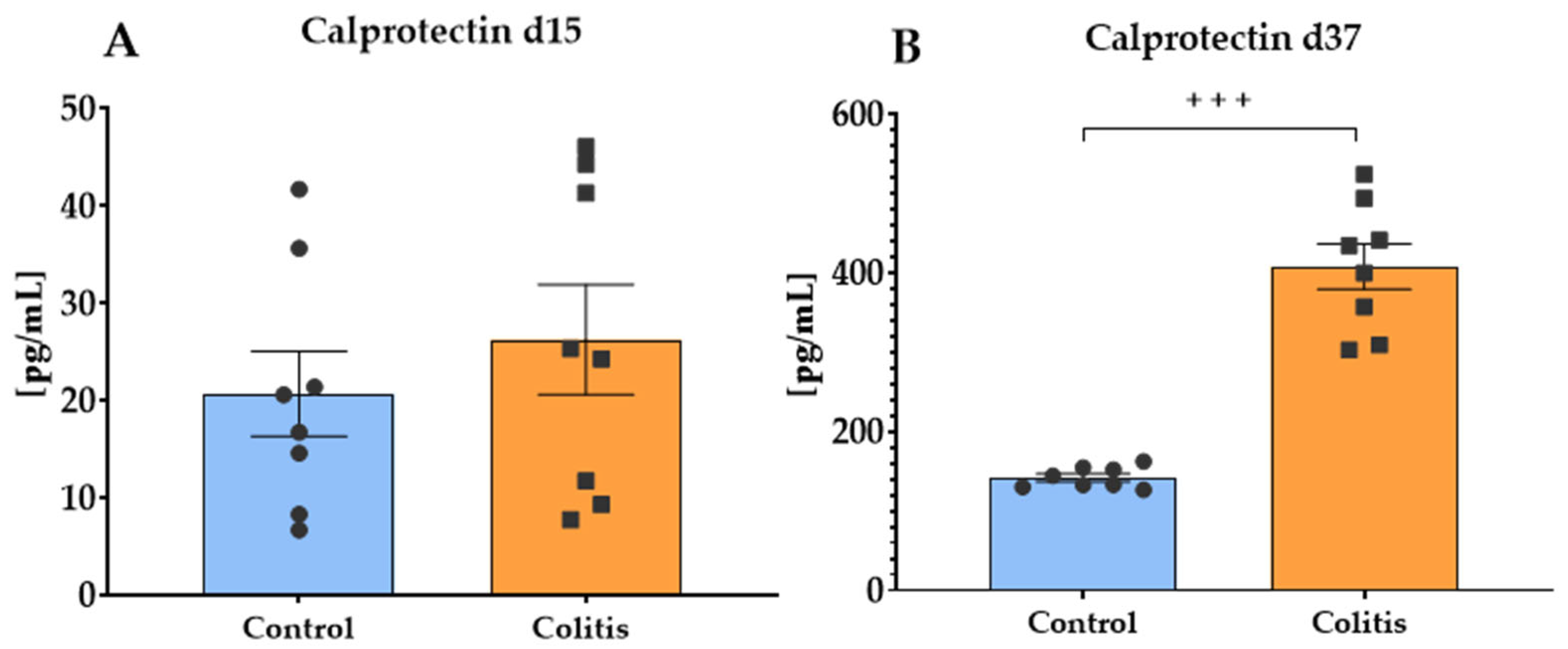
The concentration of calprotectin in feces on day 15 of the study (Figure 3A) did not differ significantly between the control (mean 20.7 pg/mL) and the colitis (mean 26.26 pg/mL) group (p = 0.450). However, on day 37 (Figure 3B), calprotectin concentration was significantly higher in the colitis group (mean 407.89 pg/mL) compared to the control group (mean 140.07 pg/mL) (p = 0.0002).

2.6. Histopathological Evaluation of Porcine Colon

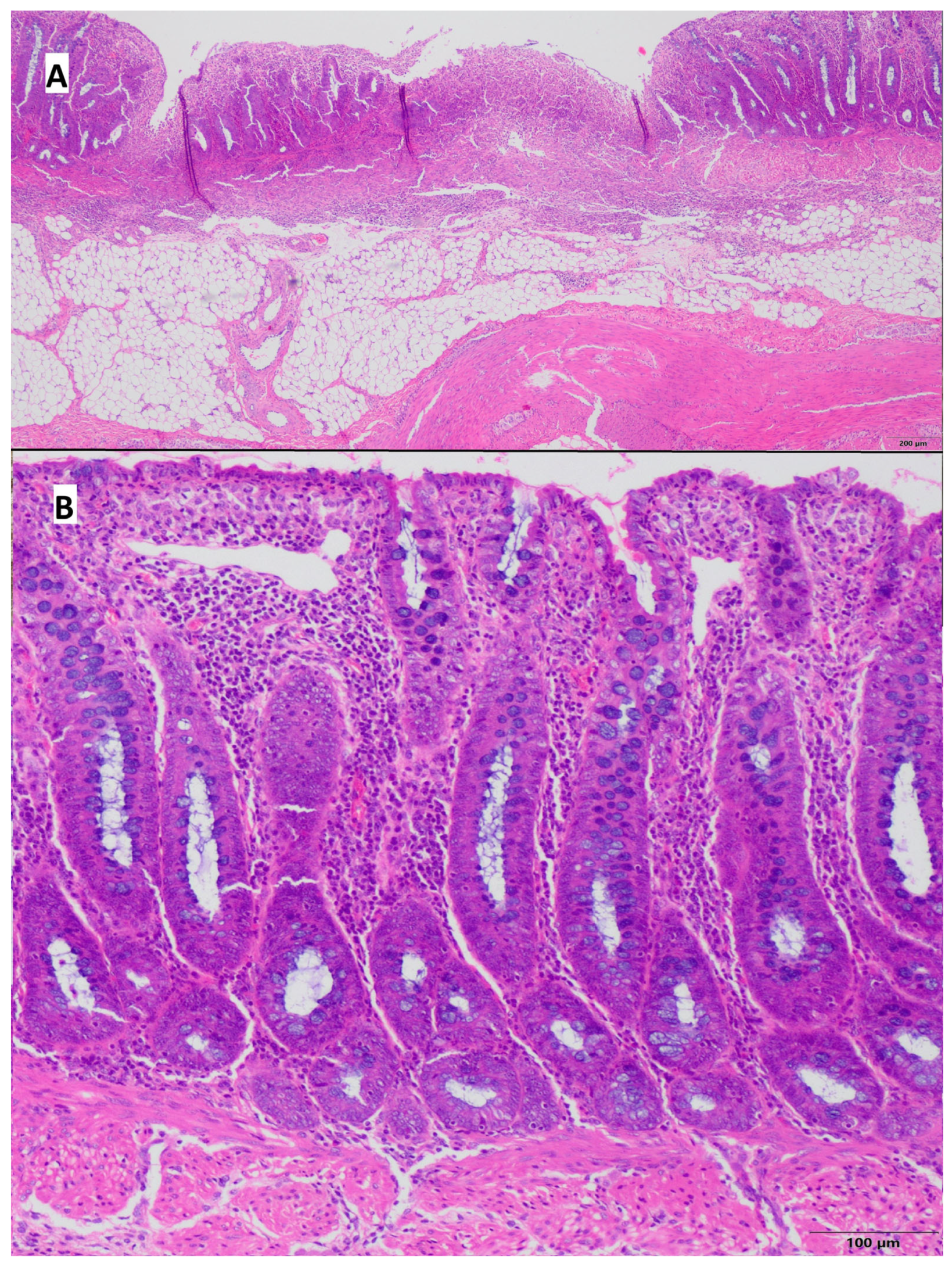
Pathological lesions were restricted to the mucosa, with chronic inflammatory infiltrate consisting of small lymphocytes and plasma cells. In the control group, the average lesions were estimated as 1.0 according to Geboes scoring scale [30], whereas in the colitis group, it was 1.62. Neutrophils in the epithelium of intestinal crypts were present in 3 out of 8 pigs from the control group and in 5 out of 8 pigs from the colitis group; however, these cells were not present in the crypts lumen (crypt abscesses). Alterations in general architecture, including crypts distention, were observed in only 1 of 8 pigs from the control group and in 5 of 8 pigs from the colitis group. Erosions and ulcers were seen only in the colitis group (50% of animals), and they were absent in pigs from the control group. The results are shown in Table 4, Figure 4 and Figure 5. In addition, during tissue collection, macroscopic changes were visible in the colon of pigs at the site of induced inflammation (Supplementary Figure S1).
A histomorphometric analysis of the colon (Figure 6) showed a significant effect of induced inflammation on the length and width of the upper part (top) of the intestinal crypts (p < 0.01), but no effect on the width of the intestinal crypts in the lower part (bottom). In the group with induced inflammation, shorter crypt length and higher width at the top were observed.

2.7. Histomorphometric Analysis of the Small Intestine

There were no significant differences in villus length, mucosa thickness, crypt depth, and muscularis thickness of the duodenum, as well as the proximal and distal parts of the jejunum, between the control and colitis group (p > 0.05). Significant differences were found in muscularis thickness of the middle jejunum (p = 0.03) and the ileum (p = 0.0092), and in crypt depth in the ileum (p = 0.0095). Other histomorphometric parameters in the ileum and middle jejunum were not significantly different between the study groups. The results are shown in Table 5. Additionally, there were also no significant differences in the length of the aforementioned intestinal segments in the two study groups (p > 0.05).

3. Discussion

The present study aimed to confirm the induction of DNBS colitis in a domestic pig model. Given the significant anatomical and physiological differences between humans and rodents [31], the use of rodent models alone has undoubtedly hindered progress and complicated the translation of research findings into effective IBD therapies [32]. In this context, the domestic pig model seems particularly valuable for translational research on IBD. Unlike mouse and rat models, pigs are more similar to humans in anatomy, physiology, body size, immunology, and genome [29,33]. This species has a single-chambered stomach and demonstrates a high degree of similarity to humans regarding inflammatory response, intestinal mucosal and muscular layer thickness, intestinal microbiota composition, nutritional requirements, and expression of immune receptors, among other features [23,25,26]. This characteristic makes this species a much more appropriate model for studying inflammatory diseases of the GIT, especially in the context of gut immunology, microbiota, or morphology. In our study, we used male domestic pigs. According to the available scientific literature, from an epidemiological point of view, Crohn’s disease occurs significantly more often in males than in females [34,35]. The use of male animals further reduces the impact of hormonal fluctuations, allowing for more homogeneous results. However, establishing separate animal models for UC and CD remains challenging due to the similar pathological changes induced by existing protocols [36,37].
In the present study, DNBS was used to induce colonic inflammation. Its mechanism of action is based on activation of the immune system through protein haptenation in the presence of 50% ethanol, which damages the intestinal barrier and allows DNBS to penetrate into the lamina propria [21]. The administration of DNBS results in the recruitment of inflammatory cells, such as neutrophils and macrophages, and increases pro-inflammatory cytokines, characteristic of the inflammatory response seen in CD. In the conducted experiment, DNBS was administered rectally in an ethanol solution at a dose of 80 mg/kg body weight (concentration 8.22% v/v), in accordance with previously described protocols [24,38]. This method is characterized by high reproducibility, the ability to modulate the severity of inflammation by adjusting the dose and frequency of application, and moderate toxicity. Another substance often used to induce colitis is 2,4,6-trinitrobenzenesulfonic acid (TNBS), which acts similarly to DNBS but has higher toxicity and mortality, as well as increased risk of tissue perforation and necrosis, especially at higher doses [39,40,41]. Antoniou et al. [41] suggest that TNBS may better mimic IBD but is more difficult to dose and may have less predictability in large animal models. Furthermore, compared to DNBS, it is considered a dangerous substance due to its strong oxidative properties, which may pose a risk of explosion when in contact with bases such as sodium and potassium hydroxide [21]. Dextran sulfate sodium (DSS) is another commonly used compound for inducing colitis, especially in rodent models. It exerts a direct toxic effect on intestinal epithelial cells, leading to necrosis and an acute inflammatory response [42]. DSS-induced colitis of an acute nature that primarily involves the mucosa and submucosa of the large intestine, without the characteristic transmurality present in CD, is therefore more similar to UC [43]. Moreover, DSS has nephrotoxic and hepatotoxic effects, which limit its use in long-term studies [44]. The above disadvantages of DSS, as well as the variability in concentration and inconsistent exposure to the agent, lead to heterogeneity in results and limit the ability to compare results between study groups [21]. In contrast, DNBS more effectively reproduces chronic and moderate disease courses with relatively low mortality [21,24,38] and may represent a compromise between the properties of DSS and TNBS in large animal models. Moreover, DNBS-induced colitis in pigs more closely replicates the characteristics of intestinal inflammation observed in human CD than DSS-based rodent models, which are generally considered to better mimic UC. It therefore presents a promising model for translational research in CD.
In this study, DNBS administration in pigs resulted in typical clinical signs of colitis, in the form of diarrhea, with occasional presence of blood in the stool and transient abdominal soreness. These symptoms occurred early after inflammation induction and were not observed in the control group. Notably, general clinical condition—including appetite, hydration status, activity, and vital signs—remained stable in all animals throughout the study, resembling the mild to moderate course of CD observed in humans [9,24]. The clinical observation of diarrhea is consistent with the findings of Zizzo et al. [38], who also reported altered stool consistency beginning the day after DNBS administration in rats. Palenca et al. [24] studied a mouse model of colitis induced by DNBS and found that it caused stable inflammation of moderate severity. The main clinical signs observed were diarrhea and changes in stool composition, while the effect on the appetite and activity of the animals was limited. These findings are consistent with our results. Despite GIT-related symptoms, no significant differences in body weight or weight gain were observed between the control and colitis groups. The study by Żyła et al. [45] showed that after TNBS administration, rats exhibited significantly reduced feed intake and body weight gain, which correlated with full clinical CD manifestations, including bloody diarrhea. Likewise, Nielsen et al. [25] observed that DSS-induced colitis in pigs resulted in a 23% reduction in feed intake and a 36% decrease in daily weight gain compared to healthy animals. Symptoms such as diarrhea, bloody stools, and weight loss may mirror the degree of gastrointestinal damage, but our results confirm that DNBS administration in a large animal model leads to mild to moderate inflammation that does not lead to cachexia over a 3-week observation period.
In the typical course of the acute inflammatory phase in IBD, patients exhibit leukocytosis and neutrophilia as a result of increased production of pro-inflammatory cytokines [26]. However, hematological analysis in the present study showed a significant reduction on day 37 in peripheral blood white blood cell (WBC) counts, as well as absolute neutrophil (NEU#) and monocyte (MON#) counts in pigs with DNBS-induced colitis. It should be noted that despite the demonstrated significance, these values were within the hematological norms for pigs. Neutrophils are the most abundant leukocyte population in the blood, comprising 50–60% of circulating leukocytes [46]. The results obtained in the present study suggest different dynamics of the inflammatory process, probably due to the stage of inflammation at week 3 after colitis induction. This interpretation is partly consistent with our previous study by Kopiasz et al. [47] in the rat model of CD, which showed that changes in the number of WBCs and their subtypes significantly depended on the time elapsed after TNBS administration. Studies on the pathogenesis of IBD have shown that, during intestinal inflammation, resident monocytes contribute to the recruitment of neutrophils through producing macrophage-derived chemokines [48,49]. One hypothesis explaining our study’s reduction in blood leukocyte levels may be their massive recruitment from the peripheral blood to the ongoing site of inflammation, as evidenced by the colon’s histopathological findings. Thus, the reduction in circulating leukocytes does not necessarily indicate an improvement in the inflammation, but rather a shift in its localization and nature. This is in line with Witko-Sarsat et al. [50] who suggested that under chronic inflammation conditions, the number of neutrophils in the peripheral blood may paradoxically decrease due to their extensive consumption in the focus of inflammation. Consistent with this interpretation, no significant differences were found in systemic concentrations of TNF-α and IL-1β between groups in the present study. Although these cytokines are considered key pro-inflammatory mediators in IBD, our findings align with other studies using TNBS or DNBS models, in which systemic levels of TNF-α and IL-1β often remain unchanged despite evident local inflammation [51,52]. For instance, in rat models of TNBS-induced colitis, plasma cytokine levels measured up to 7 days post-induction were not significantly elevated, despite marked increases in colonic tissue concentrations. Similar results were reported in DNBS models, where elevated colonic mRNA expression of Tnfa and Il1b was documented, while systemic cytokine expression was not described [53]. These findings support the hypothesis that DNBS-induced colitis in pigs may primarily trigger a local immune response without a strong systemic cytokine reaction. It is also important to consider that ethanol in high doses can have myelotoxic and immunosuppressive effects, leading to a decrease in the number of precursor cells in the bone marrow and impairment of their differentiation [54,55]. Although the ethanol dose was low in our study and administered only once, its local effects may also affect systemic parameters. Furthermore, the possible impact of drugs used for premedication (butorphanol, medetomidine) on hematologic results should also be acknowledged. However, it is worth noting that this scheme and the dosage described are routinely used in studies on pigs conducted in our laboratory. So far, we have not observed deviations in hematological parameters. Moreover, studies by Akbar et al. [56], Samimi et al. [57], Atalan [58], and Sato et al. [59] demonstrated that the use of α2-agonists and opioid drugs (butorphanol) does not significantly affect hematological findings. Nevertheless, the relative contribution of leukocytes, including neutrophils, to the pathogenesis of CD is controversial. While some studies describe their beneficial role, others report their pathological contribution [60,61,62,63]. Their number and role in regulating inflammation strictly depend on the method of colitis induction, the stage of inflammation, and the animal model used.
Calprotectin, which is a calcium-binding protein of the S100 family, is commonly used as a non-invasive marker to assess the degree of intestinal inflammation and disease activity in IBD (active vs. remission) [64]. The results of our study showed that before the administration of DNBS or saline, the fecal calprotectin (FC) concentration did not differ significantly between the two groups. This finding was extremely important for further interpreting the effect of the experimental inflammation model and confirms the homogeneity of the studied group of animals. This result is consistent with the study by Lallès et al. [65], who also found low FC levels in healthy pigs kept in high sanitary conditions. On the other hand, 21 days after DNBS administration, we found a significant, almost 3-fold increase in FC concentration in pigs from the colitis group compared to the control group. Calprotectin, mainly derived from neutrophils and macrophages, is a strong chemotactic factor for neutrophils. During intestinal inflammation, neutrophils are mobilized to migrate to the site of inflammation, causing degranulation and releasing significant amounts of calprotectin into the intestinal lumen [66]. A reduction in the count of #NEU in peripheral blood in our study may also confirm this process. Furthermore, Bjarnason [67] claims that FP concentration provides a non-invasive quantitative measure of neutrophil flow to the intestine. The observed increase in FP levels in the colitis group is consistent with numerous preclinical and clinical studies confirming that calprotectin levels in stool are significantly elevated in patients with IBD, and the degree of elevation is positively correlated with disease severity [68,69,70]. In addition, the study by Amara et al. [71] on DSS-induced colitis in mice also indicates that FC levels correlate with markers of intestinal inflammation, including TNF-α, IL6, and colitis severity. Our results are also consistent with the study by Barbosa et al. [64], who found elevated FC levels in pigs with mucoid or hemorrhagic colitis. The increase in FP revealed in the colitis group confirms that the induction of colitis using DNBS is associated with a marked activation of the inflammatory process in the GIT and that it is a valuable marker in translational IBD research, also due to its high sensitivity and specificity [72].
Plasma liver enzyme activity is commonly used as an indicator of hepatocellular injury and is often elevated in chemically induced colitis models. In our study, ALT activity remained stable across time points and between groups. In contrast, AST activity significantly increased in the colitis group by day 37, compared to the control group and to baseline activities before DNBS administration. These findings are partially consistent with previous studies in TNBS-induced colitis models, where both AST and ALT activities were significantly elevated in plasma, indicating liver function disturbance [73]. The more selective increase in AST observed in our study, without parallel elevation in ALT, may reflect a milder hepatic stress. AST is less liver-specific than ALT and may also originate from muscle [74], although the observed timing and association with colitis induction point toward hepatic origin. The increase in AST may be mediated by gut–liver axis mechanisms, which are well-documented in TNBS and DNBS models. These include intestinal barrier disruption, microbial dysbiosis, and translocation of bacterial products such as lipopolysaccharides (LPS), which can trigger hepatic inflammation [73,75]. Studies have shown that pro-inflammatory cytokines (e.g., TNF-α, IL-1β, IL-6) can be elevated in the liver in such models, contributing to hepatic dysfunction and histological damage. Although our study did not reveal significant systemic elevations of TNF-α or IL-1β, local cytokine production or translocation pathways may still have contributed to hepatic stress. Liver injury is a recognized complication in human IBD. Abnormal liver enzyme activities—particularly elevated AST and ALT—have been reported in up to 30% of IBD patients [76]. Hepatocellular injury, characterized by elevated AST and/or ALT, is one of the more common patterns observed in IBD patients, and it may result from the inflammatory process itself, metabolic dysfunction, or adverse effects of pharmacological treatment [76,77]. Furthermore, the absence of ALT elevation could suggest that the effects of DNBS on the liver are less severe than those reported in TNBS-based models, or that the systemic inflammatory response was not sufficiently robust to cause pronounced liver injury within the study’s timeframe. However, the observed increase in AST highlights the importance of considering the liver function assessment in translational colitis models. It suggests that DNBS-induced colitis in pigs may be suitable for studying such interactions under moderate inflammatory conditions.
Histologic evaluation of the colon, in both CD and UC, is essential to establish a diagnosis and is also valuable in assessing response to therapy [78]. There is also a distinction made for IBD of unspecified type (indeterminate colitis—IC; in cases where there are no clear features to classify the disease into one of the above two entities). UC is characterized by changes that are usually limited to the mucosa in a continuous fashion and to a varying range, extending from the rectum to the more proximal colon [13]. In patients with UC, changes related to intestinal crypts are observed, including the formation of crypt abscesses, crypt architectural distortion (shortening, branching), loss of mucus, metaplasia of Paneth cells, basal lymphoid aggregates, and lamina propria eosinophils. An early symptom indicating UC is also basal plasmocytosis [13]. On the other hand, CD is characterized by involvement of the entire thickness of the intestinal wall (transmural inflammation) and has a multifocal nature (patchy distribution), with an inflammatory cell infiltration in the lamina propria consisting of lymphocytes and plasma cells. A typical feature of CD is also the presence of epithelioid granulomas—such granulomas are visible in 70% of cases of intestinal wall biopsies, but only 15% of cases when the lesions are limited to the mucosa [7]. Our study observed histopathological changes that may be primarily consistent with UC. The most typical feature of the histopathological picture observed in our model was the limitation of changes to the mucosa, which exhibits characteristics typical of UC [13]. Additionally, the inflammatory cell infiltrate was composed of a mixed population of small lymphocytes and plasma cells, which are recognized as changes commonly seen in the early phase of UC (basal plasmocytosis). Mucosal ulcerative changes were observed in 50% of the pigs, with three of these animals having the severity of changes classified as high (3 out of 4 degrees). However, in all cases, the inflammatory infiltrate associated with these lesions did not extend below the submucosal layer, and the inflammatory changes could not be classified as transmural. Such a pattern of change is typical for ulcerative colitis and differs from that observed in CD (in this case, transmural colitis is observed). We also did not see typical inflammatory infiltrates of a granulomatous nature characteristic of CD; therefore, our described model, in histopathological terms, is most similar to what is observed in UC in humans. Furthermore, our morphometric analysis of intestinal crypts objectively revealed a mildly expressed (but statistically significant) shortening of intestinal crypts, which is another morphological feature found in UC [13]. However, in the literature, such changes in crypt histomorphometry are also described as characteristic of CD [79]. Certain morphological differences between our model and UC can be attributed to the fact that our model represents an early stage of the condition. For instance, we observed an absence of colonic crypt abscesses, destruction of goblet cells, and reduced mucus production in our model. These abnormalities, frequently seen in the advanced stages of UC, may not have developed yet in our model.
Erben et al. [80] reported that, among 95 studies on mouse models of IBD, more than half showed inflammation limited to the colon only (65%), while 17% were limited to the small intestine, and 18% involved most or all parts of the colon and small intestine. Clinically, CD affects the small intestine in approximately 30–40% of patients (involving the terminal ileum in 90%), while 40–55% of patients show localization to the ileum and colon, and colonic-only inflammation occurs in only 15–25% of individuals [81]. Therefore, we decided to perform a morphometric analysis of the duodenum, jejunum (proximal, middle, and distal sections), and ileum. Our results showed a significant reduction in the thickness of the muscular layer and the depth of intestinal crypts in the terminal segment of the small intestine—the ileum—in pigs with induced colitis. A reduction in the thickness of the muscle layer may indicate smooth muscle dystrophy, potentially related to chronic inflammatory mediators that can lead to impaired tissue regeneration [82]. In turn, the shortening of crypts may be linked to an imbalance between proliferation and apoptosis of enterocytes. The crypt is a tubular gland formed by the small intestinal epithelium descending into the lamina propria at the base of the villi, and stem cells at their base are responsible for the continuous renewal of the intestinal epithelium [83,84]. Under chronic inflammation conditions, this process is disrupted, resulting in a shortening of the crypts, which often accompanies malabsorption syndrome and malnutrition [85]. Bischoff et al. [28] have reported that increased large intestine permeability may secondarily impair small intestinal barrier function, possibly through weakened tight junctions or endotoxemia. This mechanism may explain the presence of lesions in the ileum, although the main inflammatory stimulus was confined to the large intestine. In addition, it should be mentioned that damage to the small intestine—direct or indirect—can lead to metabolic disorders, which are often observed in patients with IBD. The observed changes in the ileum and segmental inflammation are typical features of CD in humans [8], which are rarely reproduced in rodent models. Due to the similar anatomical length of the gastrointestinal tract and the proportions of the layers of the intestinal wall, the pig model makes it possible to study these features.
The DNBS-induced colitis model in pigs presented in this study shows numerous advantages allowing research on chronic IBD. It successfully reproduces many clinical and histopathological features of this disease. As demonstrated in this study, the model shows high reproducibility and relatively low mortality, making it safe and suitable for long-term experimental IBD protocols. However, as Szkopek et al. [33] rightly point out, there are no perfect experimental animal models, and each model only slightly mimics the clinical condition of the disease entity and requires further improvements and work. As a multifactorial disease, IBD involves interactions between diet, microbiome, environmental factors, genotype, and the intestinal barrier, and no model can holistically capture all these components. Therefore, one of the limiting factors in our study is the lack of a studied microbiome, which is an important aspect to consider in future directions. Another important limiting factor is the short time period (21 days) and single administration of DNBS, which may not capture chronic IBD. It should also be considered that pigs, unlike rodents, have greater space and nutritional requirements, which carry over into higher maintenance costs. Although the DNBS-pig colitis model is not without limitations, its translational value is very high. It allows a complex analysis of the systemic and local inflammatory response under physiologically similar conditions to humans. Given these limitations and the results, we cannot conclusively determine whether the model used is closer to CD or UC, and therefore, it should be treated as an IBD model. However, the model has great potential to contribute to breakthroughs in treating IBD and implementing effective therapies into clinical practice.

4. Materials and Methods

4.1. Animal Experiment

The experiment was conducted on 16 three-month-old Polish White Landrace male pigs, which were randomly divided into two groups: control and colitis. To avoid the risk of bias, the pigs were divided so that the average body weight with standard deviation in each group was similar. Sample size was estimated using G∗Power software [86], version 3.1.9.4 for a one-way ANOVA at α = 0.05 with 95% power, assuming an effect size (f) = 0.35 for two study groups. The effect size was calculated as the minimum expected mean difference between animals (effect size f = delta Mean/SD). SD was obtained from previously published own studies. Since SDs vary greatly for all the parameters listed, we chose the maximum SD (5.71) for the minimum expected difference in means (2.0). The animals were kept individually in boxes equipped with a water supply and feeder, in which they had constant contact with other animals and received environmental enrichments. The pigs were fed a cereal-based, pelleted, standard diet at 4% of their body weight. The diet contained 19 g/100 g crude protein, 65 g/100 g carbohydrates, 3.9% crude fiber, 3% crude fat, and 6% ash, together with 5000 IU/kg vitamin A, 500 IU/kg vitamin D, and 85 mg/kg vitamin E. The conditions in the housing area were monitored daily and were standard for an experimental pig facility (temperature 21–25 °C, light cycle 12/12, air exchanges 15–20/h, humidity 70 +/− 5%). Body weight in both groups was measured weekly throughout the experiment. Pigs’ weight gains were calculated based on the difference in body weights between the evaluated weeks of the experimental period.
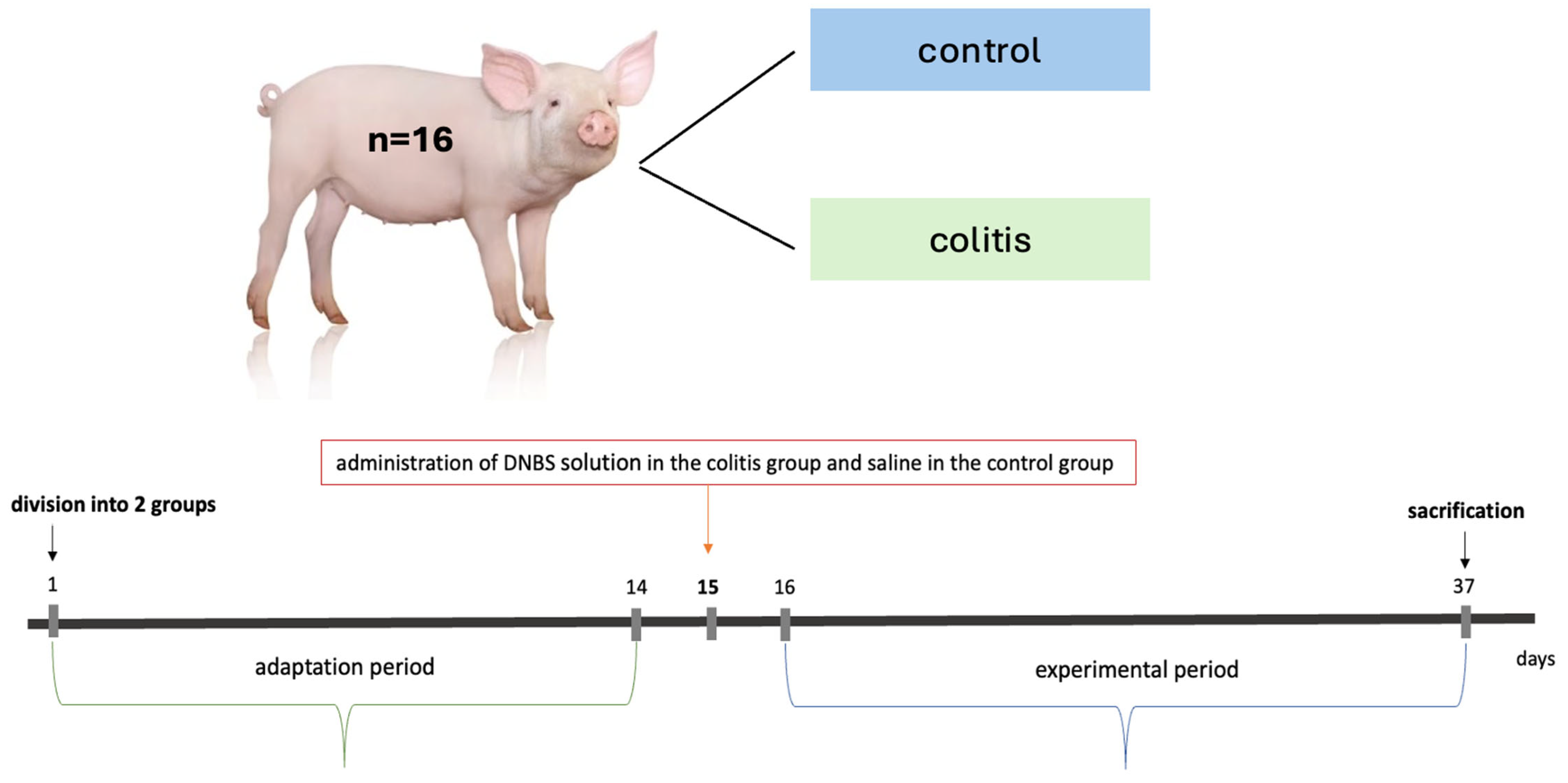
After a two-week adaptation period, the animals were premedicated with a combination of medetomidine (1 mg/m2; “Domitor”, Orion Pharma, Warsaw, Poland) and butorphanol (0.2 mg/kg body weight; “Torphadine”, Dechra, Warsaw, Poland), intramuscularly. Colitis was induced by a single rectal administration of DNBS (2,4-dinitrobenzenesulfonic acid) dissolved in 50% (v/v) ethanol at a dose of 80 mg/kg body weight. The concentration of DNBS was approximately 8.22% v/v. The final density of the ethanolic DNBS solution was approximately 0.977 g/cm3. The DNBS dose was 80 mg/kg body weight, which comes out to 1 mL of ethanolic DNBS solution per kilogram of body weight. The administration was via an intragastric probe made of medical PVC (polyvinyl chloride) with dimensions of 10.6 mm × 76 cm (Sigmed, Cisek, Poland; cat. no. 91951) coated with lignocaine gel under ultrasound control into the descending colon. After administration of DNBS, the animals were kept on the surgical table at an angle of 30° for 30 min. Anesthesia was maintained with inhaled isoflurane (1–1.5%, oxygen flow 2–2.5 L/minute; “Isotek”, Vet-Agro, Lublin, Poland). Animals in the control group were administered a 0.9% NaCl solution in the same volume and way. Then, a 21-day observation period was initiated. A simple diagram of the study design is shown in Figure 7.
The experimental procedures for this study were approved by the II Local Ethics Committee for Animal Experiments in Warsaw, Poland (Resolution No. WAW2/090/2023). All procedures involving animals were performed in accordance with the Polish Law for the Care and Use of Animals, EU regulations (Directive 2010/63/EU), and the Code of Ethics of the World Medical Association (Declaration of Helsinki). The present experiment also complied with the ARRIVE guidelines 2.0 [87].

4.1.1. Evaluation of the Clinical Condition of Animals

For 21 days, the clinical conditions of the animals in both groups were monitored daily after administration of DNBS or 0.9% NaCl. The abdominal pain (animal behavior and palpation examination) and quality and frequency of feces were evaluated on a scoring scale. The scoring scale is shown in Table 6 and Supplementary Table S2. Additional clinical parameters were assessed, including appetite (present, decreased, absent), thirst (present, decreased, absent), locomotor activity (normal, decreased, absent), internal temperature (normal, increased, decreased), number of breaths per minute (normal, increased, decreased), interaction with the environment (normal, decreased or absent), and degree and % dehydration (palpation and total protein with hematocrit test) (Supplementary Table S3).

4.1.2. Sampling Procedure and Autopsy

On days 15 (before DNBS administration) and 37 (end of experiment), blood was collected from the animals’ external jugular vein into EDTA tubes (BD Vacutainer, Becton Dickinson, NJ, USA) for morphological and biochemical analysis. In addition, on the same days, feces samples were collected to determine calprotectin concentration.
At the end of the study, all pigs were sacrificed by a single dose of intravenously injected sodium pentobarbiturate (140 mg/kg body weight; “Morbital”, Biowet, Pulawy, Poland), after prior intramuscular premedication with a mixture of medetomidine (0.1 mg/kg body weight; “Domitor”, Orion Pharma, Warsaw, Poland) and butorphanol (0.2 mg/kg body weight; “Torphadine”, Dechra, Warsaw, Poland). Subsequently, samples from the small intestine were taken for histomorphometry analysis, as well as a section of the colon for histopathological examination and histomorphometry analysis.

4.2. Hematological and Biochemical Examination of the Blood

Blood testing was performed on days 15 and 37 of the experiment using a 5-DIFF Mythic 5VET PRO automatic veterinary hematology analyzer (Cormay, Warsaw, Poland). The following parameters were evaluated: white blood cells count (WBC), percentage and absolute counts of lymphocytes (LYM%, LYM#), monocytes (MON%, MON#), of neutrophils (NEU%, NEU#), eosinophils (EOS%, EOS#), basophils (BASO%, BASO#), red blood cell count (RBC), hemoglobin concentration (HGB), hematocrit (HCT), mean corpuscular volume (MCV), mean corpuscular hemoglobin (MCH), mean corpuscular hemoglobin concentration (MCHC), red blood cell distribution width-coefficient of variation (RDW_CV) and platelet count (PLT). Blood plasma was obtained by centrifugation at 3000× g for 10 min at 4 °C. The resulting plasma samples were subsequently stored at −80 °C until biochemical analyses were performed. Plasma enzyme activities of alanine (ALT) and aspartate (AST) aminotransferases were measured using kinetic assays with commercially available kits (Sigma Aldrich, Saint Louis, MO, USA).

4.3. Assessment of Plasma Cytokine Levels

Tumor necrosis factor-alpha (TNF-α) and interleukin-1 beta (IL-1β) concentrations in plasma were measured using specific quantitative sandwich ELISA kits (Porcine TNF-alpha ELISA Kit (cat. No. RAB0478), Porcine IL-1 beta/IL-1F2 ELISA Kit (cat. No. RAB0276) (Sigma Aldrich, Saint Louis, MO, USA) according to the manufacturer’s instructions.

4.4. Examine Fecal Calprotectin Levels

Fecal calprotectin levels on days 15 and 37 were quantified using a commercially available ELISA test (MyBioSource, San Diego, CA, USA; Cat. No. MBS2801875), according to the manufacturer’s instructions. Briefly, stool samples were homogenized in the provided extraction buffer to achieve a consistent suspension and subsequently centrifuged to remove particulate matter. The resulting supernatants were transferred to 96-well microplates pre-coated with a monoclonal antibody specific to calprotectin. After incubation and washing, a biotinylated detection antibody was added, followed by HRP-conjugated streptavidin. The enzymatic reaction was developed using a TMB substrate and terminated with a stop solution. Optical density was measured at 450 nm using a microplate reader (Multikan Sky, Thermo Scientific, Rockford, IL, USA). Calprotectin concentrations were calculated based on a standard curve generated using known concentrations of recombinant porcine calprotectin provided in the kit.

4.5. Histopathological Evaluation of Porcine Colon and Small Intestine

The colon sections and samples from the small intestine were prepared and stained for routine histopathological and histomorphometric analysis [88]. Briefly, collected macroscopically altered colon specimens, as well as samples from the duodenum, proximal, middle, and distal parts of the jejunum, and the ileum were fixed in 10% neutral buffered formalin for at least 24 h, then processed via standard protocols (dehydration, clearing, and paraffinization). Sections from a paraffin block with colon were cut at 4 µm thickness, while sections from the small intestine were cut at 4.5 µm thickness, and stained with hematoxylin–eosin. Histopathological analysis of the colon was performed according to the Geboes scoring scale (Table 7) [30], which evaluates the composition and intensity of the inflammatory cell infiltrate, crypts destruction, and the presence of erosions and ulcers.
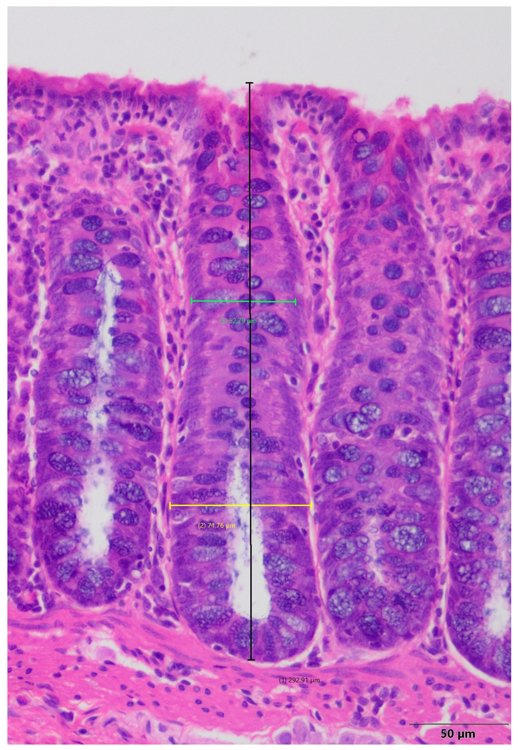
A histomorphometric examination of colonic crypts, including their length and width, was performed to provide a more detailed characterization of inflammation-induced changes in the colon. The samples selected for histomorphometry were those of the highest quality. The histomorphometric analyses were conducted using an Olympus BX41 microscope (Olympus, Tokyo, Japan) coupled to a computer equipped with the CellA® analysis system. For each colon specimen, areas with clearly visible and well-stained crypts with visible histologic elements were selected. In each sample, 30 crypts were analyzed. The software was used to measure the length of the crypt and its width in the upper and lower parts (Figure 8).
In the histomorphometric analysis of the small intestine, parameters such as the thickness of the muscular lamina and mucosa, the length of the villi, and the depth of the crypts were measured [88]. Images were analyzed using a light microscope (Axioskop 40, Zeiss, Jena, Germany) with a digital camera (Coolpix B700, Nikon, Tokyo, Japan). The obtained data were accessed using Axio Vision software version 4.2 (Zeiss, Germany). A minimum of 20 measurements of each parameter were made for each section.

4.6. Statistical Analysis

The data obtained from hematological analyses were analyzed using Statistica software (version 13.3 EN; StatSoft, Krakow, Poland). Before conducting further analyses, the equality of variance and normality of distribution were determined for all datasets. To enable statistical analysis, the following data were transformed to obtain the normal distribution and equal variance by the square root (length and width of the upper part of the intestinal crypts). Based on Cook’s distance analysis, individual values in some parameters were excluded from the statistical analysis. The unpaired t-test was used to indicate the statistical differences between the groups, or the Mann–Whitney test was used to determine if the assumptions of equality of variance and normal distribution were unmet. Statistical significance was established at a p-value of less than 0.05.

5. Summary and Conclusions

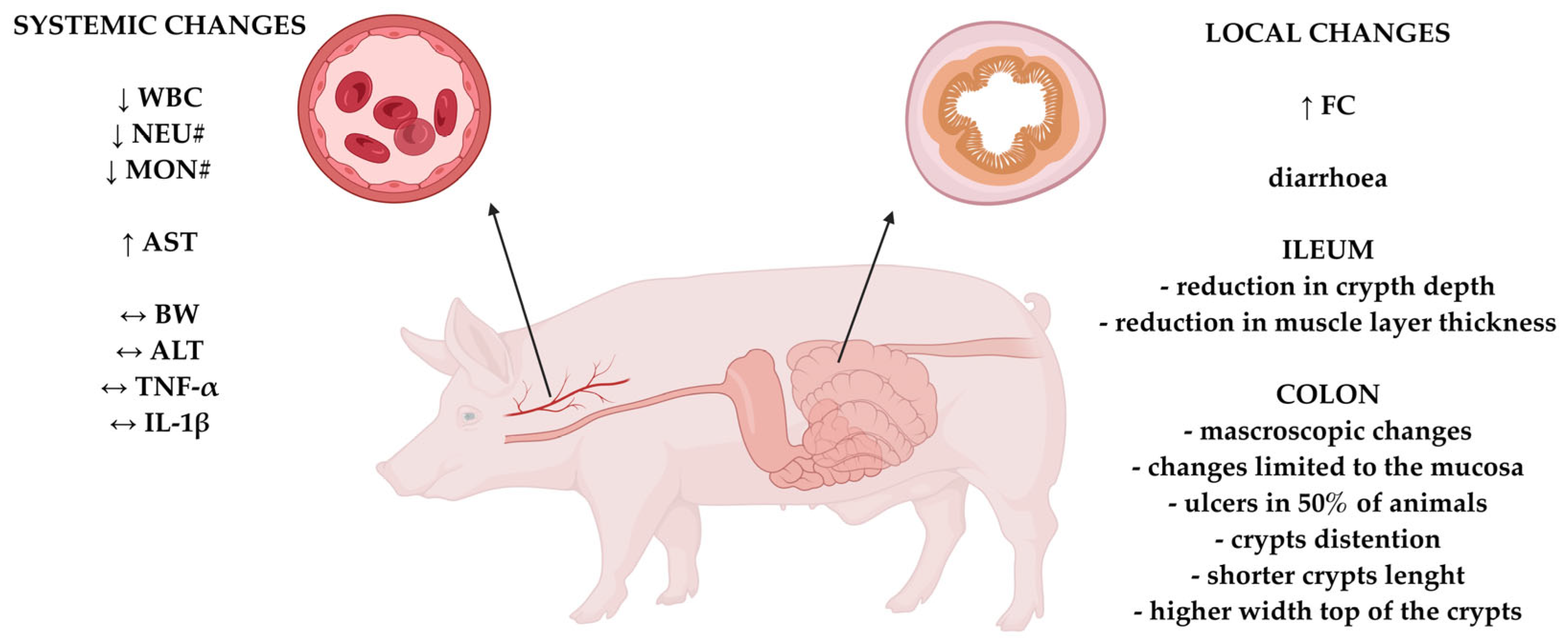
This study demonstrates that rectal administration of DNBS (in 50% (v/v) ethanol at a dose of 80 mg/kg body weight) in domestic pigs induces colonic inflammation with clinical, biochemical, and histopathological features resembling human IBD. The model reproduced key characteristics of IBD—including diarrhea, mucosal ulceration, crypt architecture distortion, and elevated fecal calprotectin—while maintaining good general condition and low mortality, supporting its feasibility for long-term studies (Figure 9). Histologically, the inflammation was primarily limited to the mucosa and submucosa, mimicking ulcerative colitis. However, segmental involvement of the ileum and alterations in crypt depth and muscularis thickness suggest some overlap with Crohn’s disease pathology. Compared to rodent models, the pig model offers greater anatomical and physiological similarity to humans. These findings highlight the translational potential of the DNBS-induced colitis pig model for studying IBD pathophysiology and for preclinical testing of novel therapies, including dietary, probiotic, and pharmacological interventions. These results indicate that we have successfully developed a technically feasible and minimally invasive model of colitis in pigs, which better preserves animal welfare than other available methods. This model can be effectively used to study the pathophysiology of IBD and for preclinical testing of novel therapeutic approaches, including dietary, probiotic, and pharmacological interventions. However, further studies are needed to explore the model’s kinetics over a longer experimental period and/or to assess the effects of multiple doses of DNBS to induce chronic colitis. Such investigations would provide a more comprehensive understanding of the model and allow a better evaluation of its similarity to Crohn’s disease or ulcerative colitis.

Supplementary Materials

The following supporting information can be downloaded at: https://www.mdpi.com/article/10.3390/ijms26189115/s1.

Author Contributions

Conceptualization, D.S., J.W., Ł.K., K.D. and J.G.-O.; methodology, D.S., J.W., Ł.K., K.D. and J.G.-O.; validation, D.S., J.W., Ł.K., K.D. and J.G.-O.; formal analysis, D.S., K.Z., Ł.K., K.D. and R.S.; investigation, D.S., K.Z. and J.W.; resources, D.S., K.Z., Ł.K. and J.W.; data curation, D.S., K.Z., Ł.K. and J.W.; writing—original draft preparation, D.S.; writing—review and editing, Ł.K., K.D., R.S. and J.G.-O.; visualization, D.S., Ł.K., K.D. and J.G.-O.; supervision, J.W. and J.G.-O.; project administration, J.W. and J.G.-O.; funding acquisition, J.G.-O. All authors have read and agreed to the published version of the manuscript.

Funding

This research was funded by the National Science Centre (NCN), Poland. Grant number 2022/47/B/NZ9/03123.

Institutional Review Board Statement

The experimental procedures for this study were approved by the II Local Ethics Committee for Animal Experiments in Warsaw, Poland (Resolution No. WAW2/090/2023, 21 June 2023). All procedures involving animals were performed in accordance with the Polish Law for the Care and Use of Animals, EU regulations (Directive 2010/63/EU), and the Code of Ethics of the World Medical Association (Declaration of Helsinki).

Informed Consent Statement

Not applicable.

Data Availability Statement

The original data presented in the study are openly available in the Repository for Open Data (RepOD) at https://doi.org/10.18150/RGKXLF.

Acknowledgments

Special thanks to veterinarians and veterinary technicians—Aleksandra Sikora, Marta Nałęcz, Izabela Wesołowska—from the Veterinary Center of The KIAPN PAS for their excellent care of the animals participating in this study.

Conflicts of Interest

The authors declare no conflicts of interest.

Abbreviations

IBDChronic inflammatory bowel disease
CDCrohn’s disease
UCUlcerative colitis
GITGastrointestinal tract
DNBS2,4-dinitrobenzenesulfonic acid
TNBS2,4,6-trinitrobenzene sulfonic acid
DSSDextran sulfate sodium
BWBody weight
BWGBody weight gain
APAbdominal pain
FQFFecal quality and frequency
WBCWhite blood cells
NEU#Absolute value of neutrophils
MON#Absolute value of monocytes
IETsIntraepithelial layer
FCFecal calprotectin
TNF-αTumor necrosis factor-alpha
IL-1βInterleukin-1 beta
ALTAlanine aminotransferase
ASTAspartate aminotransferase

References

  1. Ng, S.C.; Shi, H.Y.; Hamidi, N.; Underwood, F.E.; Tang, W.; Benchimol, E.I.; Panaccione, R.; Ghosh, S.; Wu, J.C.Y.; Chan, F.K.L.; et al. Worldwide incidence and prevalence of inflammatory bowel disease in the 21st century: A systematic review of population-based studies. Lancet 2017, 390, 2769–2778. [Google Scholar] [CrossRef]
  2. Feuerstein, J.D.; Cheifetz, A.S. Crohn Disease: Epidemiology, diagnosis, and management. Mayo Clin. Proc. 2017, 92, 1088–1103. [Google Scholar] [CrossRef] [PubMed]
  3. Sanat, Z.M.; Vahedi, H.; Malekzadeh, R.; Fanni, Z. Epidemiologic profile of inflammatory bowel disease in Eastern Mediterranean Region (EMRO) countries: A systematic review and meta-analysis. BMC Public Health 2024, 24, 1395. [Google Scholar] [CrossRef] [PubMed]
  4. Ananthakrishnan, A.N.; Kaplan, G.G.; Ng, S.C. Changing Global Epidemiology of Inflammatory Bowel Diseases: Sustaining Health Care Delivery Into the 21st Century. Clin. Gastroenterol. Hepatol. 2020, 18, 1252–1260. [Google Scholar] [CrossRef] [PubMed]
  5. Windsor, J.W.; Kalpan, G.G. Evolving epidemiology of IBD. Curr. Gastroenterol. Rep. 2019, 21, 40. [Google Scholar] [CrossRef]
  6. Kaplan, G.G.; Windsor, J.W. The four epidemiological stages in the global evolution of inflammatory bowel disease. Nat. Rev. Gastroenterol. Hepatol. 2021, 18, 56–66. [Google Scholar] [CrossRef]
  7. Torres, J.; Mehandru, S.; Colombel, J.; Peyrin-Biroulet, L. Crohn’s disease. Lancet 2017, 389, 1741–1755. [Google Scholar] [CrossRef]
  8. Dollinger, M.; Torres, J.; Vermeire, S. Crohn’s disease. Lancet 2024, 403, 1177–1191. [Google Scholar] [CrossRef]
  9. Plevris, N.; Lees, C.W. Disease monitoring in inflammatory bowel disease: Evolving principles and possibilities. Gastroenterology 2022, 162, 1456–1475. [Google Scholar] [CrossRef]
  10. Baumgart, D.C.; Sandborn, W.J. Inflammatory bowel disease: Clinical aspects and established and evolving therapies. Lancet 2007, 369, 1641–1657. [Google Scholar] [CrossRef]
  11. Seyedian, S.S.; Nokhostin, F.; Malamir, M.D. A review of the diagnosis, prevention, and treatment methods of inflammatory bowel disease. J. Med. Life 2019, 12, 113–122. [Google Scholar] [CrossRef]
  12. Gajendran, M.; Loganathan, P.; Catinella, A.P.; Hashash, J.G. A comprehensive review and update on Crohn’s disease. Disease-a-Month 2018, 64, 20–57. [Google Scholar] [CrossRef]
  13. Gajendran, M.; Loganathan, P.; Jimenez, G.; Catinella, A.P.; Ng, N.; Umapathy, C.; Ziade, N.; Hashash, J.G. A comprehensive review and update on ulcerative colitis. Disease-a-Month 2019, 65, 100851. [Google Scholar] [CrossRef]
  14. Silva, I.; Pinto, R.; Mateus, V. Preclinical study in vivo for new pharmacological approaches in inflammatory bowel disease: A systematic review of chronic model of TNBS-induced colitis. J. Clin. Med. 2019, 8, 1574. [Google Scholar] [CrossRef] [PubMed]
  15. Prajapati, S.K.; Yadav, D.; Katiyar, S.; Jain, S.; Yadav, H. Postbiotics as Mitochondrial Modulators in Inflammatory Bowel Disease: Mechanistic Insights and Therapeutic Potential. Biomolecules 2025, 15, 954. [Google Scholar] [CrossRef] [PubMed]
  16. Sinha, A.; Roy, S. Prospective therapeutic targets and recent advancements in the treatment of inflammatory bowel disease. Immunopharmacol. Immunotoxicol. 2024, 46, 550–563. [Google Scholar] [CrossRef] [PubMed]
  17. Torres, J.; Bonovas, S.; Doherty, G.; Kucharzik, T.; Gisbert, J.P.; Raine, T.; Adamina, M.; Armuzzi, A.; Bachmann, O.; Bager, P.; et al. ECCO guidelines on therapeutics in Crohn’s disease: Medical treatment. J. Crohn’s Colitis 2020, 14, 4–22. [Google Scholar] [CrossRef]
  18. Anka, D.; Senhaji, N.; Aouiss, A.; Khalki, L.; Tijani, Y.; Zaid, N.; Marhoume, F.Z.; Naya, A.; Oudghiri, M.; Kabine, M.; et al. IL-1 and CD40/CD40L platelet complex: Elements of induction of Crohn’s disease and new therapeutic targets. Arch. Pharmacal Res. 2021, 44, 117–132. [Google Scholar] [CrossRef]
  19. Baydi, Z.; Limami, Y.; Khalki, L.; Zaid, N.; Naya, A.; Mtairag, E.M.; Oudghri, M.; Zaid, Y. An update of research animal models of inflammatory bowel disease. Sci. World J. 2021, 2021, 7479540. [Google Scholar] [CrossRef]
  20. Ma, S.; Yeom, J.; Lim, Y.H. Dairy Propionibacterium freudenreichii ameliorates acute colitis by stimulating MUC2 expression in intestinal goblet cell in a DSS-induced colitis rat model. Sci. Rep. 2020, 10, 5523. [Google Scholar] [CrossRef]
  21. Morampudi, V.; Bhinder, G.; Wu, X.; Dai, C.; Sham, H.P.; Vallance, B.A.; Jacobson, K. DNBS/TNBS colitis models: Providing insights into inflammatory bowel disease and effects of dietary fat. J. Vis. Exp. 2014, 84, 51297. [Google Scholar] [CrossRef]
  22. Patterson, J.K.; Lei, X.G.; Miller, D.D. The pig as an experimental model for elucidating the mechanisms governing dietary influence on mineral absorption. Exp. Biol. Med. 2008, 233, 651–664. [Google Scholar] [CrossRef] [PubMed]
  23. Heinritz, S.N.; Mosenthin, R.; Weiss, E. Use of pigs as a potential model for research into dietary modulation of the human gut microbiota. Nutr. Res. Rev. 2013, 26, 191–209. [Google Scholar] [CrossRef] [PubMed]
  24. Palenca, I.; Seguella, L.; Del Re, A.; Franzin, S.B.; Corpetti, C.; Pesce, M.; Rurgo, S.; Steardo, L.; Sarnelli, G.; Esposito, G. N-Palmitoyl-D-Glucosamine Inhibits TLR-4/NLRP3 and Improves DNBS-Induced Colon Inflammation through a PPAR-α-Dependent Mechanism. Biomolecules 2022, 12, 1163. [Google Scholar] [CrossRef] [PubMed]
  25. Nielsen, T.S.; Fredborg, M.; Theil, P.K.; Yue, Y.; Bruhn, L.V.; Andersen, V.; Purup, S. Dietary red meat adversely affects disease severity in a pig model of DSS-induced colitis despite reduction in colonic pro-inflammatory gene expression. Nutrients 2020, 12, 1728. [Google Scholar] [CrossRef]
  26. Wen, C.; Chen, D.; Zhong, R.; Peng, X. Animal models of inflammatory bowel disease: Category and evaluation indexes. Gastroenterol. Rep. 2024, 12, goae021. [Google Scholar] [CrossRef]
  27. Swindle, M.M.; Makin, A.; Herron, A.J.; Clubb, F.J.; Frazier, K.S. Swine as models in biomedical research and toxicology testing. Vet. Pathol. 2012, 49, 344–356. [Google Scholar] [CrossRef]
  28. Bischoff, S.C.; Barbara, G.; Buurman, W.; Ockhuizen, T.; Schulzke, J.; Serino, M.; Tilg, H.; Watson, A.; Wells, J.M. Intestinal permeability—A new target for disease prevention and therapy. BMC Gastroenterol. 2014, 14, 189. [Google Scholar] [CrossRef]
  29. Lunney, J.K.; Van Goor, A.; Walker, K.E.; Hailstock, T.; Franklin, J.; Dai, C. Importance of the pig as a human biomedical model. Sci. Transl. Med. 2021, 13, eabd5758. [Google Scholar] [CrossRef]
  30. Geboes, K.; Riddell, R.; Öst, A.; Jensfelt, B.; Persson, T.; Löfberg, R. A reproducible grading scale for histological assessment of inflammation in ulcerative colitis. Gut 2000, 47, 404–409. [Google Scholar] [CrossRef]
  31. Wang, M.; Donovan, S.M. Human microbiota-associated swine: Current progress and future opportunities. ILAR J. 2015, 56, 63–73. [Google Scholar] [CrossRef]
  32. Yin, L.; Yang, H.; Li, J.; Li, Y.; Ding, X.; Wu, G.; Yin, Y. Pig models on intestinal development and therapeutics. Amino Acids 2017, 49, 2099–2106. [Google Scholar] [CrossRef]
  33. Szkopek, D.; Wychowański, P.; Zaworski, K.; Seklecka, B.; Starzyński, R.; Lipiński, P.; Pierzynowska, K.; Pierzynowski, S.G.; Donaldson, J.; Paczewski, Ł.; et al. Investigating the Influence of a Tooth Absence on Facial Bone Growth Using a Porcine Model. Int. J. Mol. Sci. 2024, 25, 12509. [Google Scholar] [CrossRef] [PubMed]
  34. Greuter, T.; Manser, C.; Pittet, V.; Vavricka, S.R.; Biedermann, L. Gender differences in inflammatory bowel disease. Digestion 2020, 101, 98–104. [Google Scholar] [CrossRef] [PubMed]
  35. Zagórowicz, E.; Walkiewicz, D.; Kucha, P.; Perwieniec, J.; Maluchnik, M.; Wieszczy, P.; Reguła, J. Nationwide data on epidemiology of inflammatory bowel disease in Polan between 2009 and 2020. Pol. Arch. Intern. Med. 2022, 132, 16194. [Google Scholar] [CrossRef] [PubMed]
  36. Breugelmans, T.; Van Spaendonk, H.; De Man, J.G.; De Schepper, H.U.; Jauregui-Amezaga, A.; Macken, E.; Linden, S.K.; Pintelon, I.; Timmermans, J.; De Winter, B.Y.; et al. In-depth study of transmembrane mucins in association with intestinal barrier dysfunction during the course of T cell transfer and DSS-induces colitis. J. Crohn’s Colitis 2020, 14, 974–994. [Google Scholar] [CrossRef]
  37. Xiao, H.; Li, H.; Wen, Y.; Jiang, D.; Zhu, S.; He, X.; Xiong, Q.; Gao, J.; Hou, S.; Huang, S.; et al. Tremella fuciformis polysaccharides ameliorated ulcerative colitis via inhibiting inflammation and enhancing intestinal epithelial barrier function. Int. J. Biol. Macromol. 2021, 180, 633–642. [Google Scholar] [CrossRef]
  38. Zizzo, M.G.; Caldara, G.; Bellanca, A.; Nuzzo, D.; Di Carlo, M.; Serio, R. Preventive effects of guanosine on intestinal inflammation in 2, 4-dinitrobenzene sulfonic acid (DNBS)-induced colitis in rats. Inflammopharmacology 2019, 27, 349–359. [Google Scholar] [CrossRef]
  39. Morris, G.P.; Beck, P.L.; Herridge, M.S.; Depew, W.T.; Szewczuk, M.R.; Wallace, J.L. Hapten-induced model of chronic inflammation and ulceration in the rat colon. Gastroenterology 1989, 96, 795–803. [Google Scholar] [CrossRef]
  40. Valatas, V.; Bamias, G.; Kolios, G. Experimental colitis models: Insights into the pathogenesis of inlammatory bowel disease and translational issues. Eur. J. Pharmacol. 2015, 759, 253–264. [Google Scholar] [CrossRef]
  41. Antoniou, E.; Margonis, G.A.; Angelou, A.; Pikouli, A.; Argiri, P.; Karavokyros, I.; Papalois, A.; Pikoulis, E. The TNBS-induced colitis animal model: An overview. Ann. Med. Surg. 2016, 11, 9–15. [Google Scholar] [CrossRef] [PubMed]
  42. Chassaing, B.; Aitken, J.D.; Malleshappa, M.; Vijay-Kumar, M. Dextran-sulfate sodium (DSS)-induced colitis in mice. Curr. Protoc. Immunol. 2014, 104, 15.25.1–15.25.14. [Google Scholar] [CrossRef] [PubMed]
  43. Gancarcikova, S.; Lauko, S.; Hrckova, G.; Andrejcakova, Z.; Hajduckova, V.; Madar, M.; Fecskeova, L.K.; Mudronova, D.; Mravcova, K.; Strkolcova, G.; et al. Innovative Animal Model of DSS-Induced Ulcerative Colitis in Pseudo Germ-Free Mice. Cells 2020, 9, 2571. [Google Scholar] [CrossRef] [PubMed]
  44. Farombi, E.O.; Adedara, I.A.; Awoyem, O.V.; Njoku, C.R.; Micah, G.O.; Esogwa, C.U.; Owumi, S.E.; Olopade, J.O. Dietary protocatechuic acid ameliorates dextran sulphate sodium-induced ulcerative colitis and hepatotoxicity in rats. Food Funct. 2016, 7, 913–921. [Google Scholar] [CrossRef]
  45. Żyła, E.; Dziendzikowska, K.; Gajewska, M.; Wilczak, J.; Harasym, J.; Gromadzka-Ostrowska, J. Beneficial effects of oat beta-glucan dietary supplementation in colitis depend on its molecular weight. Molecules 2019, 24, 3591. [Google Scholar] [CrossRef]
  46. Sadik, C.D.; Kim, N.D.; Luster, A.D. Neutrophils cascading their way to inflammation. Trends Immunol. 2011, 32, 452–460. [Google Scholar] [CrossRef]
  47. Kopiasz, Ł.; Dziendzikowska, K.; Gajewska, M.; Wilczak, J.; Harasym, J.; Żyła, E.; Kamola, D.; Oczkowski, M.; Królikowski, T.; Gromadzka-Ostrowska, J. Time-Dependent Indirect Antioxidative Effects of Oat Beta-Glucans on Peripheral Blood Parameters in the Animal Model of Colon Inflammation. Antioxidants 2020, 9, 375. [Google Scholar] [CrossRef]
  48. Nathan, C. Neutrophils and immunity: Challenges and opportunities. Nat. Rev. Immunol. 2006, 6, 173–182. [Google Scholar] [CrossRef]
  49. Fournier, B.M.; Parkos, C.A. The role of neutrophils during intestinal inflammation. Mucosal Immunol. 2012, 5, 354–366. [Google Scholar] [CrossRef]
  50. Witko-Sarsat, V.; Rieu, P.; Descamps-Latscha, B.; Lesavre, P.; Halbwachs-Mecarelli, L. Neutrophils: Molecules, functions and pathophysiological aspects. Lab. Investig. 2000, 80, 617–653. [Google Scholar] [CrossRef]
  51. Grijó, N.N.; Borra, R.C.; Sdepanian, V.L. Proinflammatory and Anti-inflammatory Cytokines Present in the Acute Phase of Experimental Colitis Treated with Saccharomyces boulardii. Dig. Dis. Sci. 2010, 55, 2498–2504. [Google Scholar] [CrossRef]
  52. Neilly, P.J.D.; Gardiner, K.R.; Kirk, S.J.; Jennings, G.; Anderson, N.H.; Elia, M.; Rowlands, B.J. Endotoxaemia and cytokine production in experimental colitis. Br. J. Surg. 1995, 82, 1479–1482. [Google Scholar] [CrossRef]
  53. Elissa, N.; Kermarrec, L.; Hussein, H.; Bernstein, C.N.; Ghia, J. Appropriateness of reference genes for normalizing messenger RNA in mouse 2,4-dinitrobenzene sulfonic acid (DNBS)-induced colitis using quantitative real time PCR. Sci. Rep. 2017, 7, 42427. [Google Scholar] [CrossRef] [PubMed]
  54. Siggins, R.W.; Melvan, J.N.; Welsh, D.A.; Bagby, G.J.; Nelson, S.; Zhang, P. Alcohol suppresses the granulopoietic response to pulmonary Streptococcus pneumoniae infection with enhancement of STAT3 signaling. J. Immunol. 2012, 186, 4306–4313. [Google Scholar] [CrossRef] [PubMed]
  55. Shi, X.; Lin, Y.; Gao, B.; Zhang, P. Impairment of hematopoietic precursor cell activation during the granulopoietic response to bacteriemia in mice with chronić-plus-binge alcohol administration. Infect. Immun. 2017, 85, e00369-17. [Google Scholar] [CrossRef] [PubMed]
  56. Akbar, H.; Khan, M.A.; Khan, M.A.; Khan, M.S.; Aslam, S.; Nasir, A.; Anjum, A.A. Effects of different doses of medetomidine on clinical and hematological parameters in dogs. J. Anim. Plant Sci. 2014, 24, 730–737. [Google Scholar]
  57. Samimi, A.S.; Sakhaee, E.; Iranmanesh, F. Evaluation of sedative, analgesic, physiological, and laboratory effects of two doses off medetomidine and xylazine in dromedary calves. J. Vet. Pharmacol. Ther. 2019, 42, 411–419. [Google Scholar] [CrossRef]
  58. Atalan, G. Effect of detomidine-butorphanol-ketamine and atipamezole on clinical, cardiorespiratory, haematological parameters in sheep. Erciyes Üniv. Vet. Fak. Derg. 2019, 16, 1–7. [Google Scholar] [CrossRef]
  59. Sato, S.; Yamano, Y.; Kanno, C.; Maeda, Y.; Takahashi, F. Cardiopulmonary function, anesthetic effects, quality of arousal, hematology, and blood biochemistry during continuous intravenous infusion of a combination solution of xylazine, butorphanol, and propofol in calves. Vet. Res. Commun. 2024, 48, 2295–2308. [Google Scholar] [CrossRef]
  60. Kuhl, A.A.; Kakirman, H.; Janotta, M.; Dreher, S.; Cremer, P.; Pawlowski, N.N.; Loddenkemper, C.; Heimesaat, M.M.; Grollich, K.; Zeitz, M.; et al. Aggravation of different types of experimental colitis by depletion or adhesion blockade of neutrophils. Gastroenterology 2007, 133, 1882–1892. [Google Scholar] [CrossRef]
  61. Zhang, R.; Ito, S.; Nishio, N.; Cheng, Z.; Suzuki, H.; Isobe, K.-I. Up-regulation of Gr1+CD11b+ population in spleen of dextran sulfate sodium administered mice works to repair colitis. Inflamm. Allergy-Drug Targets 2011, 10, 39–46. [Google Scholar] [CrossRef] [PubMed]
  62. Natsui, M.; Kawasaki, K.; Takizawa, H.; Hayashi, S.; Matsuda, Y.; Sugimura, K.; Seki, K.; Narisawa, R.; Sendo, F.; Asakura, H. Selective depletion of neutrophils by a monoclonal antibody, RP-3, suppresses dextran sulphate sodium-induced colitis in rats. J. Gastroenterol. Hepatol. 2008, 12, 801–808. [Google Scholar] [CrossRef]
  63. Buell, M.G.; Berin, M.C. Neutrophil-independence of the initiation of colonic injury: Comparison of results from three models of experimental colitis in the rat. Dig. Dis. Sci. 1994, 39, 2575–2588. [Google Scholar] [CrossRef] [PubMed]
  64. Barbosa, J.A.; Rodrigues, L.A.; Columbus, D.A.; Aguirre, J.C.P.; Harding, J.C.S.; Cantarelli, V.S.; Costa, M.d.O. Experimental infectious challenge in pigs leads to elevated fecal calprotectin levels following colitis, but not enteritis. Porc. Health Manag. 2021, 7, 48. [Google Scholar] [CrossRef] [PubMed]
  65. Lallès, J.-P.; Fagerhol, M.K. Faecal calprotectin: A non invasive marker of inflammation in pigs? ISAH 2005, 1, 405–408. [Google Scholar]
  66. Al-Beltagi, M.; Saeed, N.K.; Bediwy, A.S.; Elbeltagi, R. Fecal calprotectin in pediatric gastrointestinal diseases: Pros and cons. World J. Clin. Pediatr. 2024, 13, 93341. [Google Scholar] [CrossRef]
  67. Bjarnason, I. The use of fecal calprotectin in inflammatory bowel disease. Gastroenterol. Hepatol. 2017, 13, 53–56. [Google Scholar] [PubMed] [PubMed Central]
  68. de Magalhães Costa, M.H.; Sassaki, L.Y.; Chebli, J.M.F. Fecal calprotectin and endoscopic scores: The cornerstones in clinical practice for evaluating mucosal healing in inflammatory bowel diasease. World J. Gastroenterol. 2024, 30, 3022–3035. [Google Scholar] [CrossRef]
  69. Kalla, R.; Kennedy, N.A.; Ventham, N.T.; Boyapati, R.K.; Adams, A.T.; Nimmo, E.R.; Visconti, M.R.; Drummond, H.; Ho, G.; Pattenden, R.; et al. Serum calprotectin: A novel diagnostic and prognostic marker in inflammatory bowel diseases. Am. J. Gastroenterol. 2016, 111, 1796–1805. [Google Scholar] [CrossRef]
  70. Riccutio, A.; Griffiths, A.M. Clinical value of fecal calprotectin. Crit. Rev. Clin. Lab. Sci. 2019, 56, 307–320. [Google Scholar] [CrossRef]
  71. Amara, J.; Saliba, Y.; Hajal, J.; Smayra, V.; Bakhos, J.; Sayegh, R.; Fares, N. Cicardian rhythm disruption aggravates DSS-induced colitis in mice with fecal calprotectin as a marker of colitis severity. Dig. Dis. Sci. 2019, 64, 3122–3133. [Google Scholar] [CrossRef]
  72. Wang, W.; Cao, W.; Zhang, S.; Chen, D.; Liu, L. The role of calprotectin in the diagnosis and treatment of inflammatory bowel disease. Int. J. Mol. Sci. 2025, 26, 1996. [Google Scholar] [CrossRef] [PubMed]
  73. Jang, S.; Jeong, J.; Kim, J.; Han, M.J.; Kim, D. Simultaneous amelioratation of colitis and liver injury in mice by Bifidobacterium longum LC67 and Lactobacillus plantarum LC27. Sci. Rep. 2018, 8, 7500. [Google Scholar] [CrossRef] [PubMed]
  74. Kohli, R.; Harris, D.C.; Whitington, P.F. Relative elevations of serum alanine and aspartate aminotransferase in muscular dystrophy. J. Pediatr. Gastroenterol. Nutr. 2005, 41, 121–124. [Google Scholar] [CrossRef] [PubMed]
  75. Zhu, L.; Zong, X.; Xiao, X.; Cheng, Y.; Fu, J.; Lu, Z.; Jin, M.; Wang, F.; Wang, Y. Multi-omics analysis of the gut-liver axis reveals the mechanism of liver injury in colitis mice. Front. Immunol. 2022, 12, 773070. [Google Scholar] [CrossRef]
  76. Koller, T.; Galambosova, M.; Filakovska, S.; Kubincova, M.; Hlavaty, T.; Toth, J.; Krajcovicova, A.; Payer, J. Drug-induced liver injury in inflammatory bowel disease: 1-year prospective observational study. World J. Gastroenterol. 2017, 23, 4102–4111. [Google Scholar] [CrossRef]
  77. Scott, J.A.; Mysko, C.; Purssell, H.; Athwal, V.S. Investigation of abnormal liver blood tests in patients with inflammatory bowel disease. Frontline Gastroenterol. 2024, 15, 443–444. [Google Scholar] [CrossRef]
  78. Pai, R.K.; Jairath, V. What is the role of histopathology in the evaluation of disease activity in Crohn’s disease? Best Pract. Res. Clin. Gastroenterol. 2019, 38, 101601. [Google Scholar] [CrossRef]
  79. Neri, B.; Mossa, M.; Scucchi, L.; Sena, G.; Palmieri, G.; Biancone, L. Histological scores in inflammatory bowel disease. J. Dig. Dis. 2020, 22, 9–22. [Google Scholar] [CrossRef]
  80. Erben, U.; Loddenkemper, C.; Spieckermann, S.; Heimesaat, M.M.; Siegmund, B.; Kuhl, A.A. Histomorphology of intestinal inflammation in inflammatory bowel diseases (IBD) mouse models and its relevance for IBD in men. Int. J. Clin. Exp. Pathol. 2016, 9, 408–442. [Google Scholar]
  81. Strobel, D.; Goertz, R.S.; Bernatik, T. Diagnostics in inflammatory bowel disease: Ultrasound. World J. Gastroenterol. 2011, 17, 3192–3197. [Google Scholar] [CrossRef]
  82. Kiesler, P.; Fuss, I.J.; Strober, W. Experimental models of inflammatory bowel diseases. Cell. Mol. Gastroenterol. Hepatol. 2015, 1, 154–179. [Google Scholar] [CrossRef]
  83. Clevers, H. The intestinal crypt, a prototype stem cell compartment. Cell 2013, 154, 274–284. [Google Scholar] [CrossRef] [PubMed]
  84. Szkopek, D.; Mendel, M.; Kinsner, M.; Ognik, K.; Szyryńska, N.; Lewczuk, B.; Kozłowski, K.; Kos, I.; Konieczka, P. Cannabidiol and nano-selenium mediate intestinal barrier function by affecting mucosal microstructures, and gut-associated immunological and oxidative stress response in the gut of chickens infected with C. perfringens. Front. Immunol. 2025, 16, 1529449. [Google Scholar] [CrossRef] [PubMed]
  85. Zaworski, K.; Wychowański, P.; Szkopek, D.; Woliński, J.; Donaldson, J.; Pierzynowski, S.; Pierzynowska, K. The Regulatory Role of Pancreatic Enzymes in the Maintenance of Small Intestinal Structure and Enterocyte Turnover with Special Reference to Alpha Amylase. Int. J. Mol. Sci. 2025, 26, 249. [Google Scholar] [CrossRef] [PubMed]
  86. Faul, F.; Erdfelder, E.; Lang, A.G.; Buchner, A. G*power 3: A flexible statistical power analysis program for the social, behavioral, and biomedical sciences. Behav. Res. Methods 2007, 39, 175–191. [Google Scholar] [CrossRef]
  87. du Sert, N.P.; Hurst, V.; Ahluwalia, A.; Alam, S.; Avey, M.T.; Baker, M.; Browne, W.J.; Clark, A.; Cuthill, I.C.; Dirnagl, U.; et al. The ARRIVE guidelines 2.0: Updated guidelines for reporting animal research. PLOS Biol. 2020, 18, e3000410. [Google Scholar] [CrossRef]
  88. Feldman, A.T.; Wolfe, D. Tissue Processing and Hematoxylin and Eosin Staining. In Histopathology; Methods in Molecular Biology; Day, C., Ed.; Humana Press: New York, NY, USA, 2014; Volume 1180, pp. 31–43. [Google Scholar]
Figure 1. (A)—Pig’s body weight during the experiment. (B)—Pig’s body weight gain during the experimental period. Abbreviations: D0—day 0 of the experiment, before administration of saline or DNBS, D7—day 7 of the experiment, before administration of saline or DNBS, D14—day 14 of the experiment, end of adaptation period and DNBS/saline administration, D21—day 21 of the experiment, 1 week after administration of saline or DNBS, D28—day 28 of the experiment, 2 weeks after administration of saline or DNBS, D35—day 35 of the experiment, 3 weeks after administration of saline or DNBS, diff—difference. Results are presented as the mean ± SEM. No significant differences (repeated measures ANOVA, p > 0.05).
Ijms 26 09115 g001
Figure 2. The white blood cell count (WBC, (A)), absolute value of neutrophils (NEU#, (B)), and absolute value of monocytes (MON#, (C)) in the peripheral blood of pigs on day 37 of the experiment. Results are presented as mean ± SEM. Significant differences are indicated by + (p < 0.05).
Ijms 26 09115 g002
Figure 3. Calprotectin concentration on days 15 (A) and 37 (B) of the experiment in pig feces. Results are presented as mean ± SEM. Significant differences are indicated by +++ (p < 0.001).
Ijms 26 09115 g003
Figure 4. Representative microscopic view of colonic mucosa of a pig from the control group—mucosa without erosions and ulceration is visible; hematoxylin–eosin stain, (A)—magnification 40×, (B)—magnification 100×.
Ijms 26 09115 g004
Figure 5. Representative microscopic view of the colonic mucosa of a pig from the colitis group. (A) Erosions and ulcerations of mucosa are visible; however, lesions are restricted to mucosa without involvement of submucosa and muscular layer; hematoxylin–eosin stain, magnification 40×; (B) Area of mucosa not affected by ulceration, diffuse inflammatory infiltrate consisting of small lymphocytes and plasma cells in lamina propria is visible; hematoxylin–eosin stain, magnification 100×.
Ijms 26 09115 g005
Figure 6. A histomorphometric analysis of the crypt length (A), crypt width top (B), and crypt width bottom (C) of the porcine colon. Results are presented as the mean ± SEM. Significant differences are indicated by ++ (p < 0.01).
Ijms 26 09115 g006
Figure 7. A simple diagram of the study.
Ijms 26 09115 g007
Figure 8. Microscopic view of colonic mucosa presenting colonic crypt with measurement points: black line—length of the crypt, yellow line—width of the lower part, green line—width of the upper part; hematoxylin–eosin stain, magnification 200×.
Ijms 26 09115 g008
Figure 9. Diagram summarizing systemic and local changes caused by DNBS-induced local inflammation in the colon. Abbreviation: FC—fecal calprotectin concentration; WBC—white blood cells count; NEU#—neutrophil count; MON#—monocyte count; AST—aspartate aminotransferase activity in blood, BW—body weight; ALT—alanine aminotransferase activity in blood; TNF-α—tumor necrosis factor alpha; IL-1β—interleukin 1 beta. ↓—decrease; ↑—increase; ↔—no change.
Ijms 26 09115 g009
Table 1. Scoring of abdominal pain, fecal quality, and frequency of animals.
Control Group
Pig Number12345678
Days After Administration of NaClAPFQFAPFQFAPFQFAPFQFAPFQFAPFQFAPFQFAPFQF
10000000002010202
20000000002000002
30000000202000000
40000000202000101
50000020201000000
60000010101000000
70000010101000000
80000000001000100
90000010101000000
100000010001000100
110000000000000000
120000000100000000
130000000100000000
140000000100010000
150000000101000000
160000000000000000
170000000000000000
180000000000000000
190000000000000000
200000000001000000
210000000000000000
Colitis Group
Pig Number910111213141516
Days After Administration of DNBSAPFQFAPFQFAPFQFAPFQFAPFQFAPFQFAPFQFAPFQF
10210020202030210
20200020202020002
30200030302020202
40202000202020202
50202020302010202
60302020302000202
70202020202000200
80200000202000200
90201000202000000
100202000202000200
110200000102020200
120200020202010200
130202000102010200
140202010202010200
150200020102020000
160201020102010001
170201010102010000
180001000202010001
190001020202000001
200102020202000001
210102010202000000
Abbreviations: AP—abdominal pain, FQF—fecal quality and frequency. The clinical condition in the control group (saline administration) and the colitis group (DNBS administration) was monitored for 21 days after administration. AP: 0—no abdominal pain, 1—moderate abdominal pain, 2—severe abdominal pain. FQF: 0—feces properly formed, without pathological additions, frequency normal, 1—feces properly formed, but too soft, or feces partially abnormally formed, without pathological additions, frequency normal, 2—diarrhea, without pathological additions, normal or increased frequency, 3—diarrhea with blood or with a lot of mucus, increased frequency.
Table 2. The plasma activities of liver enzymes.
FeatureExperimental GroupStatistical
Significance
Control 15 dColitis 15 dControl 37 dColitis 37 d
AST [mU/mL]35.98 ± 4.5437.19 ± 4.42 ##38.32 ± 3.5654.75 ± 4.79 **** p = 0.0153
ALT [mU/mL]35.55 ± 3.8032.92 ± 3.4056.11 ± 10.2865.44 ± 10.33NS
The data are presented as mean ± SEM, n = 8. Significant differences are indicated by ** (p < 0.01) vs. the control group at the same time point, and ## (p < 0.01) between experimental time points within the colitis group.
Table 3. The plasma levels of cytokines.
FeatureExperimental GroupStatistical
Significance
Control 15 dColitis 15 dControl 37 dColitis 37 d
TNF-α [pg/mL]29.93 ± 9.4425.49 ± 7.5023.75 ± 5.1520.65 ± 4.40NS
IL-1β [pg/mL]85.54 ± 23.6781.08 ± 25.1768.35 ± 16.5054.25 ± 10.11NS
The data are presented as mean ± SEM, n = 8. Abbreviations: TNF-α—tumor necrosis factor-alpha; IL-1β—interleukin-1 beta.
Table 4. Histopathological evaluation of the colon.
Pig NumberGeneral ArchitectureChronic Inflammatory InfiltrateLamina Propria NeutrophilsLamina Propria EosinophilsNeutrophils in
Epithelium
Crypt DestructionErosion and Ulceration
Control Group
10101100
20102100
30101100
41 crypt distension100000
50111000
60101000
70100000
80100000
Colitis Group
91 crypt distension102100
101 crypt distension202101
111 crypt distension211103
121 crypt distension211103
131 crypt distension211103
140101000
150201000
160101000
Histopathological evaluation based on the Geboes scale. General architecture: 0—no abnormality, 1—mild abnormality, 2—mild or moderate diffuse or multifocal abnormalities, 3—severe diffuse or multifocal abnormalities; chronic inflammatory infiltrate: 0—no increase, 1—mild but unequivocal increase, 2—moderate increase, 3—marked increase; lamina propria neutrophils: 0—none, 1—mild but unequivocal increase, 2—moderate increase, 3—marked increase; lamina propria eosinophils: 0—none, 1—mild but unequivocal increase, 2—moderate increase, 3—marked increase; neutrophils in epithelium: 0—none, 1—<5% crypts involved, 2—<50% crypts involved, 3—>50% crypts involved; erosion or ulceration: 1—no erosion, ulceration, or granulation tissue, 1—recovering epithelium + adjacent inflammation, 2—probable erosion (focally stripped), 3—ulcer or granulation tissue.
Table 5. Histomorphometric analysis of the small intestine.
Parameter/GroupControl GroupColitis Group
Duodenum
Villus length [μm]1128.11 ± 10.921104.64 ± 12.60
Mucosa thickness [μm]935.19 ± 4.52910.66 ± 14.24
Crypt depth [μm]320.26 ± 3.46311.73 ± 4.00
Muscularis thickness [μm]653.15 ± 3.71642.64 ± 8.72
Proximal Jejunum
Villus length [μm]984.01 ± 6.32993.55 ± 15.92
Mucosa thickness [μm]914.42 ± 5.70903.10 ± 10.75
Crypt depth [μm]254.78 ± 2.48252.70 ± 2.83
Muscularis thickness [μm]594.75 ± 7.55577.00 ± 12.66
Middle Jejunum
Villus length [μm]949.42 ± 6.59941.34 ± 16.94
Mucosa thickness [μm]842.68 ± 9.78831.30 ± 7.53
Crypt depth [μm]250.91 ± 1.69250.15 ± 3.53
Muscularis thickness [μm]568.00 ± 5.48 a559.31 ± 9.70 b
Distal Jejunum
Villus length [μm]916.30 ± 7.19907.15 ± 9.37
Mucosa thickness [μm]812.72 ± 6.88805.45 ± 12.62
Crypt depth [μm]249.25 ± 2.29246.12 ± 3.24
Muscularis thickness [μm]534.41 ± 3.89530.36 ± 5.19
Ileum
Villus length [μm]864.32 ± 7.64851.66 ± 12.93
Mucosa thickness [μm]760.08 ± 6.56738.85 ± 10.23
Crypt depth [μm]236.38 ± 2.14 a229.16 ± 2.30 b
Muscularis thickness [μm]521.55 ± 3.68 a503.71 ± 6.29 b
Results are presented as the mean ± SEM. Significant differences are indicated by different letters (p < 0.05).
Table 6. Animal condition point scale.
Abdominal Pain
PointsDescription
0No abdominal pain
1Moderate abdominal pain
2Severe abdominal pain
Fecal quality and frequency
0Feces properly formed, without pathological additions, frequency normal
1Feces properly formed, but too soft, or feces partially abnormally formed, without pathological additions, frequency normal
2Diarrhea, without pathological additions, normal or increased frequency
3Diarrhea with blood or with a lot of mucus, increased frequency
Table 7. Geboes scoring scale.
General Architecture
PointsDescription
0No abnormality
1Mild abnormality
2Mild or moderate diffuse or multifocal abnormalities
3Severe diffuse or multifocal abnormalities
Chronic inflammatory infiltrate
0No increase
1Mild but unequivocal increase
2Moderate increase
3Marked increase
Lamina propria neutrophils
0None
1Mild but unequivocal increase
2Moderate increase
3Marked increase
Lamina propria eosinophils
0None
1Mild but unequivocal increase
2Moderate increase
3Marked increase
Epithelium neutrophils
0None
1<5% crypts involved
2<50% crypts involved
3>50% crypts involved
Crypt destruction
0None
1Probable—local excess of neutrophils in part of crypt
2Probable—marked attenuation
3Unequivocal crypt destruction
Erosion or ulceration
0No erosion, ulceration, or granulation tissue
1Recovering epithelium + adjacent inflammation
2Probable erosion—focally stripped
3Unequivocal erosion
4Ulcer or granulation tissue
Disclaimer/Publisher’s Note: The statements, opinions and data contained in all publications are solely those of the individual author(s) and contributor(s) and not of MDPI and/or the editor(s). MDPI and/or the editor(s) disclaim responsibility for any injury to people or property resulting from any ideas, methods, instructions or products referred to in the content.