Review

Genome-Based Mexican Diet Bioactives Target Molecular Pathways in HBV, HCV, and MASLD: A Bioinformatic Approach for Liver Disease Prevention

by
Leonardo Leal-Mercado
1,2,3,
Arturo Panduro
1,2,
Alexis José-Abrego
1,2 and
Sonia Roman
1,2,*
1
Department of Genomic Medicine in Hepatology, Civil Hospital of Guadalajara, “Fray Antonio Alcalde”, Hospital 278, El Retiro, Guadalajara 44280, Jalisco, Mexico
2
Centro Universitario de Ciencias de la Salud, Universidad de Guadalajara, Sierra Mojada 950, Col. Independencia, Guadalajara 44340, Jalisco, Mexico
3
Programa Doctoral de Biología Molecular en Medicina, Centro Universitario de Ciencias de la Salud, Universidad de Guadalajara, Sierra Mojada #950, Col. Independencia, Guadalajara 44340, Jalisco, Mexico
*
Author to whom correspondence should be addressed.
Int. J. Mol. Sci. 2025, 26(18), 8977; https://doi.org/10.3390/ijms26188977
Submission received: 25 June 2025 / Revised: 20 August 2025 / Accepted: 4 September 2025 / Published: 15 September 2025
(This article belongs to the Special Issue Liver Diseases: Causes, Molecular Mechanism and Treatment/Prevention)

Abstract

Viral hepatitis B and C (HBV and HCV) and metabolic dysfunction-associated steatotic liver disease (MASLD) are major public health concerns in Mexico, driving liver cirrhosis and hepatocellular carcinoma. The Genome-based Mexican (GENOMEX) diet, rich in bioactive compounds, may provide a nutritional strategy for preventing and managing liver disease. This study combines a literature review with integrative bioinformatic analyses to map the antiviral and hepatoprotective mechanisms activated by GENOMEX-derived bioactives and assess their therapeutic potential for preventing and managing liver disease. A literature-based review integrated with bioinformatics to identify the pathways activated by nutrients and bioactive compounds of the GENOMEX diet against HBV, HCV, and MASLD, incorporating data from in silico, in vitro, in vivo, and clinical studies, was conducted. An integrative bioinformatic approach, incorporating the Comparative Toxicogenomic Database and Functional Enrichment Analysis (STRING, DAVID, and Enrichr), was used to identify links between genes, nutrients, and bioactive compounds, with a subset of Mexican food staples included in the GENOMEX diet. The GENOMEX diet includes bioactive nutrients that may modulate molecular pathways related to immune response, oxidative stress, nutrient metabolism, and inflammation. Through integrative analysis, we identified key molecular targets—including TNF, PPARA, TP53, and IL6—that are implicated in viral replication, MASLD progression, and hepatocarcinogenesis. Functional enrichment revealed that these traditional Mexican foods and their nutrients are associated with genes and pathways involved in viral infection, metabolic dysfunction, fibrosis, and liver cancer. These findings highlight that the gene–nutrient interactions of the Mexican staple food in the GENOMEX diet can be integrated into nutritional strategies to prevent and manage HBV, HCV, and MASLD, while reducing fibrosis and HCC progression. These strategies are especially relevant in regions where antiviral treatments are limited due to high costs, antiviral resistance, and an escalating mismatch between the population’s evolutionary genetics and modern environment.

1. Introduction

Chronic viral hepatitis and metabolic diseases are significant global health concerns, each affecting hundreds of millions of individuals worldwide. Firstly, as of 2022, approximately 304 million people globally live with chronic hepatitis B (HBV) or hepatitis C (HCV) infection. These viral infections mutually account for over one million deaths annually, mainly due to cirrhosis and hepatocellular carcinoma (HCC) [1,2,3]. Secondly, the global prevalence of metabolic syndrome and its associated comorbidities, including hypertension, cardiovascular and renal diseases, type 2 diabetes, metabolic dysfunction-associated steatohepatitis (MASH), and the inflammatory progression of metabolic dysfunction-associated steatotic liver disease (MASLD), has steadily risen over the last decades [4]. It has been documented that metabolic syndrome is present in approximately 25% of the population in high-income countries, with prevalence increasing with age [5].
Furthermore, as part of the ongoing epidemiological transition in Mexico—from a predominance of communicable diseases to an increasing burden of non-communicable chronic diseases—it has become increasingly common to encounter patients with overlapping viral hepatitis and MASLD [6]. In both cases, pathways linking infectious and metabolic chronic inflammation activate cytokines, induce oxidative stress, lead to mitochondrial dysfunction, and promote dysbiosis. Furthermore, both conditions weaken the immune system due to persistent immune responses that trigger liver fibrosis/cirrhosis, which may ultimately lead to HCC [7]. These conditions pose additional challenges in managing liver health that require attention, as metabolic conditions can exacerbate liver disease progression in hepatitis patients [8,9,10].
In Mexico, the impact of these diseases—whether occurring alone or in combination—is magnified by restricted access to antiviral therapies, high healthcare costs, structural economic inequities, drug-resistant strains, and increasing rates of intravenous drug use, a growing risk factor for hepatitis B and C transmission in Mexico [11,12,13,14]. Simultaneously, Mexico is experiencing a nutritional transition, resulting in high rates of overweight and obesity of over 75% among adults. This shift has contributed to a heightened burden of cardiometabolic diseases, including MASLD, even in normal-weight individuals [15,16,17]. Mexicans are considered a population with elevated genetic and metabolic susceptibility to MASLD [18]. Shen et al. (2024) reported a disproportionately high prevalence of MASLD among Mexican Americans in the United States [19]. In Mexico, the estimated national prevalence of MASLD reached 41.3% in 2023 [20]. Additionally, recent findings from our research group revealed that among metabolically at-risk individuals, 57% were at risk for MASH. Notably, 67.8% of these patients, including 46% of those with normal weight, exhibited liver fibrosis or histopathological evidence of liver damage [16].
As a result, cardiometabolic conditions, including type 2 diabetes and liver disease, stand among the leading causes of morbidity and mortality nationwide [21]. Notably, chronic viral and metabolic-derived diseases require medical and nutritional counseling to reduce the risk of long-term liver damage. However, there are no official nutritional guidelines for managing viral hepatitis B and C, nor are there updated MASLD/MASH recommendations tailored to the Mexican population [22,23,24,25]. In this sense, earlier studies have highlighted the antiviral and hepatoprotective properties of specific nutrients and bioactive compounds found in the Traditional Mexican diet, which have been explored through the regionalized and genome-based dietary model known as the GENOMEX diet. This intervention combines traditional Mexican food staples with genomic, cultural, and clinical profiling to offer genome-based nutritional strategies. Previous studies have shown that the GENOMEX diet improves metabolic risk factors in individuals with obesity and dyslipidemia, and that its bioactive components exhibit antiviral activity against hepatitis B and C viruses [26,27,28,29,30]. Thus, searching for alternative strategies based on the population’s genomic and food culture background could potentially contribute to offering a culturally relevant, evidence-based framework for improving liver health in Mexico.
Genomic research has provided innovative nutrigenetic and nutrigenomic approaches focusing on nutrients and bioactive compounds that activate or inhibit metabolic pathways [31]. Furthermore, bioinformatics enables the comprehensive integration of molecular interactions across the genomic landscape, revealing network characteristics intricately linked to the pathogenesis of diseases [32]. This study aims to conduct a literature-based review integrated with bioinformatics to identify the antiviral and hepatoprotective pathways activated by ingredients in the traditional Mexican diet, exploring their potential clinical implications for the prevention and management of liver diseases.

2. Materials and Methods

2.1. Literature Review

2.1.1. Data Sources for the Literature Review

A literature review was conducted using PubMed and Google Scholar databases to identify relevant studies on bioactive compounds in Mexican foods and their effects on s HBV, HCV and MASLD. Studies were included if they met the following criteria: (i) they presented experimental, mechanistic, or clinical evidence on the effects of nutrients or bioactive compounds; (ii) they focused specifically on traditional Mexican foods or their derived ingredients; and (iii) they addressed outcomes related to HBV, HCV, or MASLD. Both human studies and experimental models (in vitro, in vivo, or in silico) were eligible. Studies were excluded if they did not investigate Mexican foods or their nutrients or bioactives, lacked relevant experimental data, addressed unrelated health conditions, were duplicates, or were not original research articles (e.g., reviews, commentaries, or editorials). Only peer-reviewed original articles published in English or Spanish were considered. The review and screening process was independently performed by two researchers with expertise in nutrigenomics and liver disease. Disagreements were resolved through discussion and consensus.
The search strategy employed Medical Subject Headings (MeSH) terms to establish a systematic framework, refined through Boolean operator combinations in PubMed and Google Scholar. This approach prioritized the identification of high-quality, peer-reviewed studies. The literature review was categorized into two sections to ensure analytical clarity: (1) antiviral nutrients and bioactive compounds and (2) anti-MASLD nutrients and bioactive compounds. This approach facilitated a systemic integration of therapeutic mechanisms specific to viral hepatitis and metabolic liver disease.

2.1.2. Search Strategy for Antiviral Nutrients

For the identification of antiviral nutrients, two independent literature searches were performed using Google Scholar and PubMed, each with a tailored Boolean operator. In Google Scholar, the Boolean operator used was as follows: (“Hepatitis B” OR “Hepatitis C” OR “Viral Hepatitis” OR “Hepatitis B Virus” OR “Hepatitis C Virus” OR “Hepatitis, Viral, Chronic”) AND (“Diet” OR “Nutrients” OR “Bioactive compounds”) AND (“Mexican diet” OR “Traditional Mexican foods”) AND (“clinical trial” OR “randomized controlled trial” OR “In silico” OR “In vivo” OR “In vitro”). For PubMed, the same search terms were applied, except that the category (“Mexican diet” OR “Traditional Mexican foods”) was replaced by (“Mexico”). The PubMed search returned four results, while the Google Scholar search yielded 69 results. After applying the predefined inclusion and exclusion criteria, a total of 27 articles were selected for qualitative analysis.

2.1.3. Search Strategy for Anti-MASLD Nutrients

For the identification of Mexican nutrients and bioactive compounds with therapeutic potential against MASLD, two independent literature searches were performed using Google Scholar and PubMed, each with a tailored Boolean operator. In Google Scholar, the Boolean operator used was as follows: (“MASLD” OR “Non-alcoholic Fatty Liver Disease” OR “Fatty Liver”) AND (“Insulin Resistance” OR “Diabetes mellitus” OR “Type 2 Diabetes”) AND (“Diet” OR “Nutrients” OR “Bioactive compounds”) AND (“Mexican diet” OR “Traditional Mexican foods”) AND (“clinical trial” OR “randomized controlled trial” OR “In silico” OR “In vivo” OR “In vitro”). For PubMed, the same search terms were applied, except that the category (“Mexican diet” OR “Traditional Mexican foods”) was replaced by (“Mexico”). The PubMed search returned six results, while the Google Scholar search yielded 134 results. After applying the predefined inclusion and exclusion criteria, a total of 41 articles were selected for qualitative analysis.

2.1.4. Classification of the Biological Effect of Antiviral and Anti-MASLD Nutrients

The biological effects of antiviral and anti-MASLD nutrients retrieved from the literature review were categorized into distinct functional groups: antiviral activity, which included the inhibition of viral entry and replication, as well as the enhancement of immune responses; antioxidants and anti-inflammatory properties, such as the upregulation of endogenous antioxidant systems and the reduction in inflammatory markers; metabolic effects, which included hypoglycemic activity (lowering blood glucose levels), insulin-sensitizing properties (enhancing insulin sensitivity and reducing insulin resistance), and hypolipidemic effects (decreasing total cholesterol, triglycerides, and VLDL while increasing HDL levels); anthropometric improvements through reducing body fat percentage and body weight; anorexigenic effects by enhancing satiety via prebiotic activity (promoting microbiota eubiosis); hepatoprotective effects (modulating liver enzymes, steatosis, and fibrosis); and anti-carcinogenic properties (upregulating tumor suppressor genes).

2.2. Integrative Bioinformatic Analysis

2.2.1. Selection of Ingredients and Nutrients

For the Integrative Bioinformatic Analysis, we prioritized nutrients and bioactive compounds from five traditional Mexican dietary staples, selected based on their documented biological effects in prior literature, to enhance the clarity of genomic data integration and visualization. Four of these ingredients (maize, beans, chili, and tomato) are core components of the milpa diet, a traditional agricultural and culinary system central to Mexican cuisine and ubiquitously consumed across the country [33]. Avocado was additionally included for its unique nutrient profile, particularly its anti-carcinogenic compounds such as manganese, potassium, vitamin E, and monounsaturated fatty acids, which are linked to hepatoprotective and metabolic benefits [34].

2.2.2. Identification of Gene Interactions with Nutrients and Bioactive Compounds

Using the Comparative Toxicogenomic Database (CTD) (https://ctdbase.org/, accessed on 4 March 2025), we retrieved the interacting genes linked to the identified nutrients and bioactive compounds. The analysis was targeted to deploy the top 10 genes involved in viral hepatitis, immune response, inflammation, nutrient metabolism, antioxidant mechanisms, insulin signaling, fibrosis, apoptosis, and cancer-related pathways [35]. These top 10 genes were selected for each nutrient or bioactive compound based on the highest number of curated chemical–gene interactions reported in CTD, which reflects both their biological relevance and frequency of interaction. In CTD, “top interacting genes” refers to those genes with the most curated interactions with a given chemical, derived from manually curated evidence across vertebrate and invertebrate species in the published literature.

2.2.3. Functional Enrichment Analysis

For the Functional Enrichment Analysis, we utilized the bioinformatics tools STRING (https://string-db.org/, accessed on 4 March 2025), DAVID (https://davidbioinformatics.nih.gov/ortholog.jsp, accessed on 4 March 2025), and Enrichr (https://maayanlab.cloud/Enrichr/, accessed on 4 March 2025) to identify KEGG pathways influenced by the top 10 genes (to optimize visualization and enhance the clarity and interpretability of genomic data integration) interacting with nutrients and bioactive compounds derived from the selected ingredients [36,37,38,39]. All enrichment tools used the Fisher exact test, based on the hypergeometric distribution, to evaluate gene overrepresentation. DAVID applied the EASE score, a conservative variant of the Fisher exact test. The results were filtered using False Discovery Rate (FDR) correction with the Benjamini-Hochberg method (−Log FDR), using the Homo sapiens whole genome as the reference background. Pathways with FDR-adjusted p values < 0.05 were considered statistically significant.

2.2.4. Data Visualization of the Integrative Bioinformatic Analysis

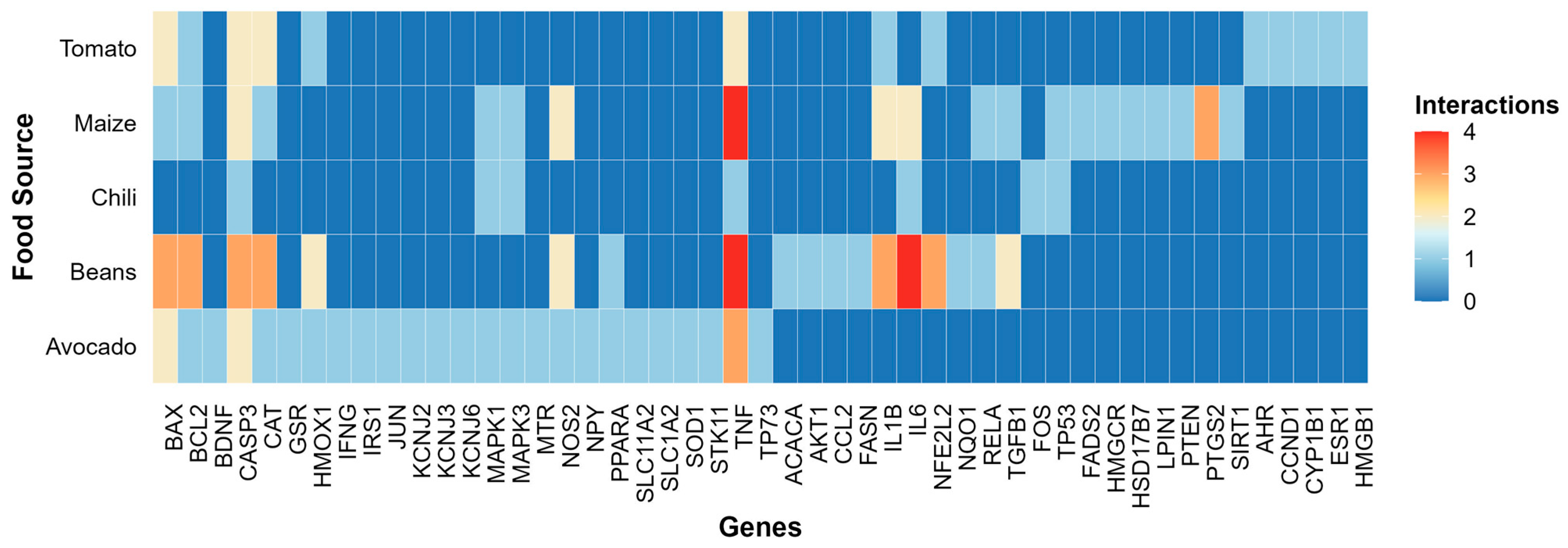
For comprehensive data visualization, we employed three complementary approaches, all conducted in RStudio (version 4.3.1) [40]. First, a heatmap was generated to display the frequency of co-occurrence between food sources and their associated genes, based on interactions retrieved from the CTD database. The interaction matrix was constructed by counting gene–food pairings and reshaping the data using tidyr [41] and reshape2 packages [42]. The heatmap was plotted using the ggplot2 package [43], with a visually optimized color scale from RColorBrewer [44] to enhance contrast and legibility.
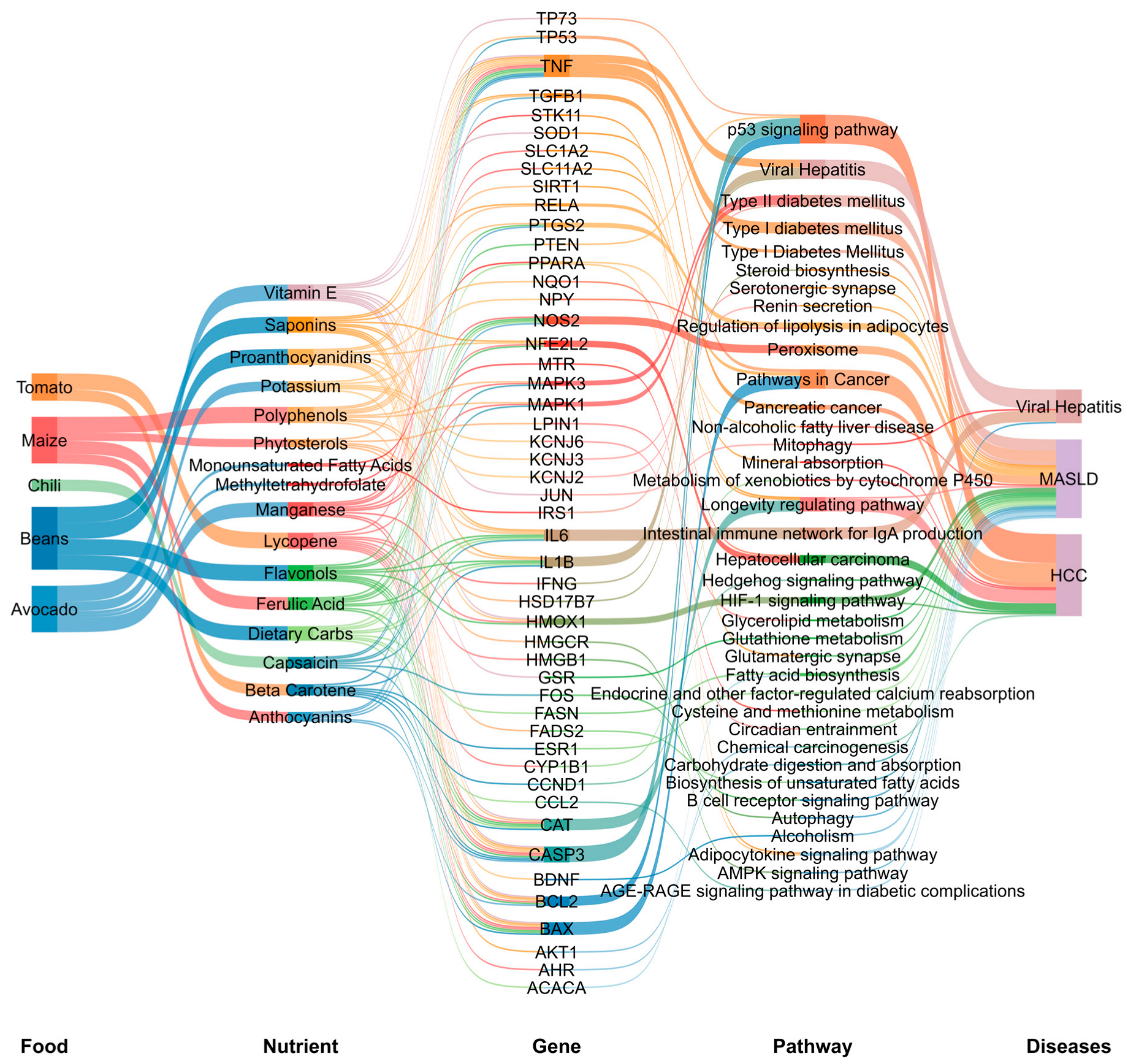
Second, we generated a five-level Sankey diagram using the ggsankey package [45] to visualize the relationships between food sources, nutrients, genes, KEGG pathways (https://www.genome.jp/kegg/, accessed on 4 March 2025) [39], and potential liver diseases. A custom color palette from RColorBrewer was used to distinguish node categories.
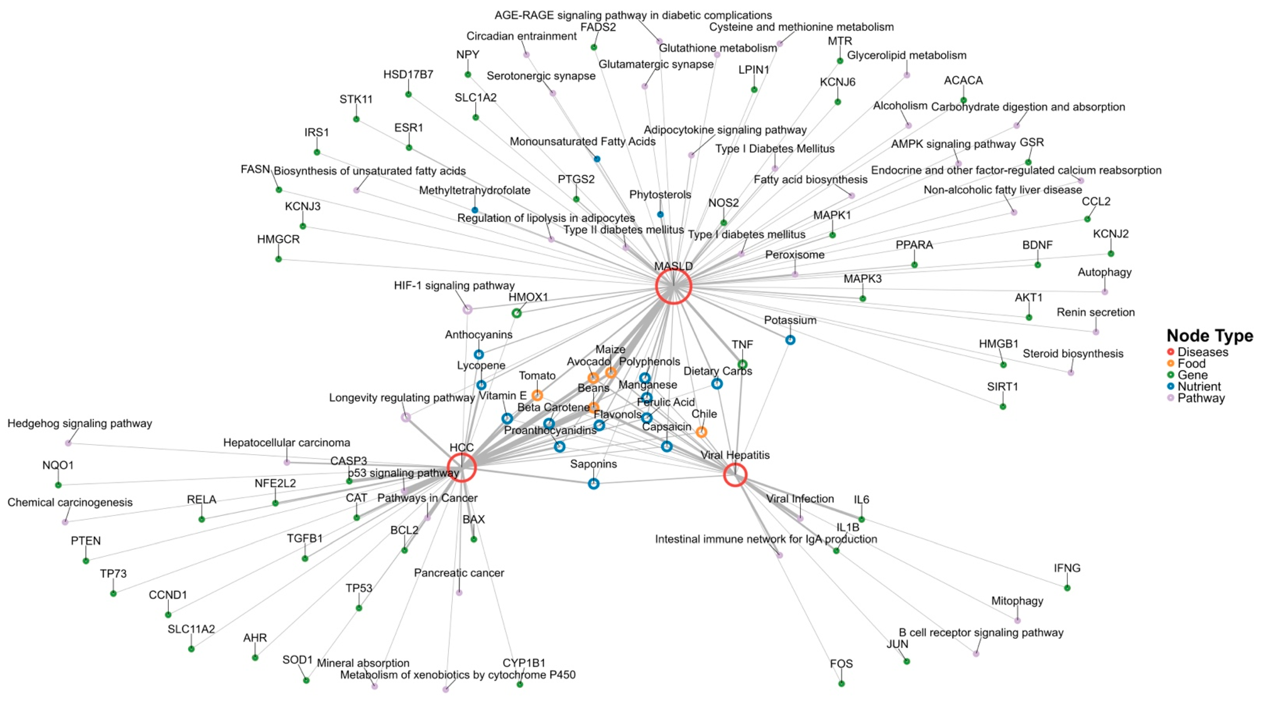
Finally, we constructed an undirected, weighted co-occurrence network plot to illustrate integrated associations between foods, nutrients, genes, KEGG pathways (https://www.genome.jp/kegg/, accessed on 4 March 2025), and liver diseases. The network was created using the igraph and ggraph packages [35,36,46]. Input data were reshaped with tidyr, and edge weights were computed based on co-occurrence frequencies between nodes and disease terms. Node-representing distinct biological or nutritional entities were color-coded by category and scaled in size according to degree centrality. Edge thickness reflects interaction frequency. The layout was computed using the Fruchterman–Reingold force-directed algorithm to cluster highly connected nodes visually. All nodes were labeled for clarity and interpretability.
Figure 1 summarizes the workflow for identifying antiviral and anti-MASLD nutrients/bioactive compounds through a literature review, followed by the computational pipeline used in the Integrative Bioinformatic Analysis.

3. Results

3.1. Literature Review of Nutrients in the Mexican Diet Against HBV, HCV, and MASLD

We categorized the literature review into two thematic sections: antiviral nutrients (Table S1; References [47,48,49,50,51,52,53,54,55,56,57,58,59,60,61,62,63,64,65,66,67,68,69,70,71,72,73,74]) and anti-MASLD nutrients/bioactive compounds (Table S2; References [75,76,77,78,79,80,81,82,83,84,85,86,87,88,89,90,91,92,93,94,95,96,97,98,99,100,101,102,103,104,105,106,107,108,109,110,111,112,113,114,115,116,117,118,119]) to improve clarity and organization. The initial search yielded 73 scientific papers for antiviral nutrients (4 from PubMed and 69 from Google Scholar), of which 27 articles were selected based on the predefined methodology. Similarly, for anti-MASLD nutrients, the initial search retrieved 140 scientific articles (6 from PubMed and 134 from Google Scholar), with 41 studies ultimately chosen according to the established criteria.
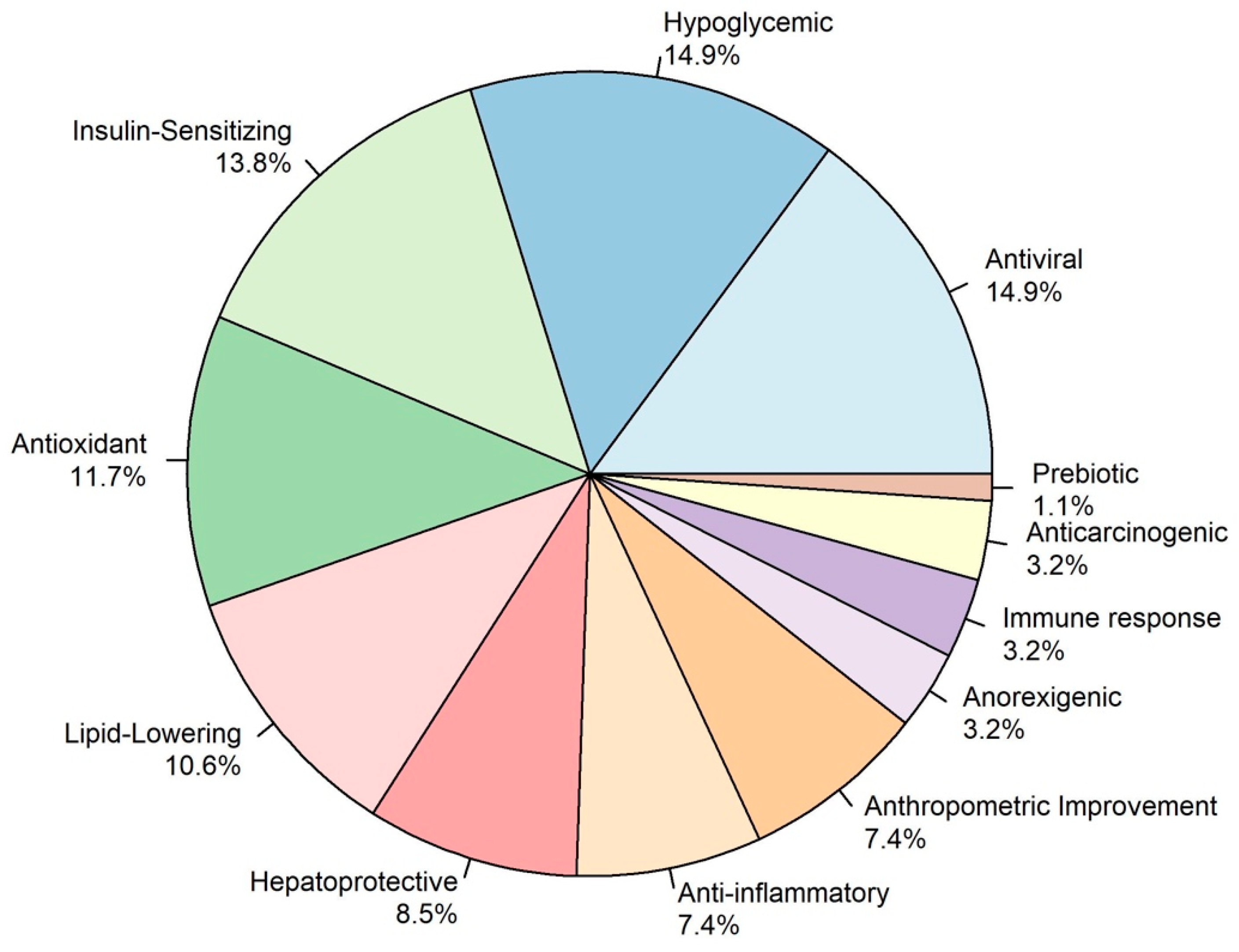
Figure 2 illustrates the biological effects of traditional Mexican ingredients, proportionally categorized by their therapeutic contributions. These effects underscore the potential of regional nutrients and bioactive compounds targeting viral hepatitis, MASLD, and preventing associated chronic liver damage and progression to cirrhosis and HCC [28,29].

3.1.1. Antiviral Nutrients

As illustrated in Figure 2 and depicted in Table S1 several nutrients have potential anti-HBV and anti-HCV activity (14.9%) which been described to disrupt HBV replication and mitigate its oncogenic progression. For instance, resveratrol in Mexican peanuts has shown inhibitory effects on HBV replication and tumor volume reduction in HBV-induced HCC in both in vitro and in vivo models [28,47]. Vitamin E, found in avocado and sunflower seeds, has demonstrated a reduction in HBV DNA levels and inflammatory cytokines in randomized controlled trials [48,49]. Lactoferrin, a bioactive protein in milk, blocks HBV entry into hepatocytes through HBsAg binding, as shown in vitro [50,51]. Similarly, selenium, present in tuna and sardines, has been shown in clinical trials to activate p53 and reduce HBV transcription, lowering HCC risk [52].
Furthermore, curcumin—derived from turmeric—has been shown, in vitro and in vivo, to suppress HBV transcription and replication, lower cccDNA levels, and inhibit NF-κB–mediated inflammatory signaling [53,54]. Luteolin-7-O-glucoside, a compound found in Mexican oregano, decreases HBV RNA and DNA levels, inhibits the secretion of HBsAg, and exhibits antioxidant and immunomodulatory activity in vitro [55]. Likewise, Moringa oleifera extracts have been shown, in vitro, to reduce cccDNA levels, inhibit NFκB signaling pathways, and exert antifibrotic effects. In addition, chlorogenic acid—present in sunflower seeds and coffee—has been shown in vitro to lower the secretion of HBsAg and HBeAg and slow the progression of liver fibrosis [56,57,58,59,60].
Lastly, epigallocatechin-3-gallate (EGCG) from cacao degrades sodium taurocholate cotransporting polypeptide (NTCP) receptors, which are key entry points for HBV, and stimulates the formation of autophagosomes that help eliminate viral components, as demonstrated in vitro [61,62,63,64]. Together, these Mexican ingredients provide a complementary and potentially cost-effective strategy to counteract HBV infection, mitigate liver injury, and reduce the risk of progression to cirrhosis or HCC.
On the other hand, various foods supply nutrients that may directly or indirectly inhibit HCV replication and mitigate liver damage, as summarized in Table S1. Docosahexaenoic acid (DHA), found in fish and seafood such as sardines, has been shown in vitro, to counteract lipid disruptions induced by HCV core proteins, thereby reducing viral replication. Likewise, eicosapentaenoic acid (EPA), also derived from fish, exhibits anti-inflammatory properties and impedes viral propagation in similar experimental settings [65,66]. Gallic acid in cloves and oregano diminishes HCV expression through antioxidant mechanisms, as demonstrated in vitro [67]. Meanwhile, vitamin E—found in sunflower seeds and avocado—has been shown to reduce ALT levels and inflammatory markers in a randomized controlled trial [68].
Other micronutrients, such as vitamin A (from liver and carrots) and vitamin D3 (from fish and eggs), support interferon-mediated antiviral pathways and inhibit viral replication, as demonstrated in vitro and prospective cohort studies, respectively [69,70,71]. Vitamin B12 (from green leafy vegetables), dietary iron (from marjoram, leafy greens, and beans), and zinc (from agave and sesame seeds) have all demonstrated antiviral activity against HCV in vitro. Specifically, B12 targets internal ribosome entry sites [72], iron inhibits HCV polymerase activity [73], and zinc interferes with RNA synthesis to suppress replication [74]. These nutrients may be further explored as part of nutritional therapeutic strategies to manage HCV infections in the Mexican population.

3.1.2. Anti-MASLD Effect

As illustrated in Figure 2 and Table S2, this literature review revealed the potential therapeutic biological effects of the traditional Mexican ingredients, including hypoglycemic (14.9%), insulin-sensitizing (13.8%), antioxidant (11.7%), lipid-lowering (10.6%), anti-inflammatory, and anthropometric improvements (7.4%). In a minor proportion, anorexigenic (3.2%), anti-carcinogenic (3.2%), and prebiotic (1.1%) effects were observed. All these effects potentially mitigate liver damage progression to MASH, cirrhosis, and HCC.
The following section outlines the principal therapeutic potentials of nutrients and bioactive compounds derived from Zea mays (maize), Phaseolus vulgaris (common bean), Solanum lycopersicum (tomato), Capsicum annuum (chili pepper), and Persea americana (avocado). These species hold profound culinary and cultural significance in Mexican cuisine, with accumulating scientific evidence underscoring their roles in modulating metabolic, inflammatory, and oxidative pathways. Table S2 expands this analysis by including additional Mexican foods, including Theobroma cacao (cacao), Pachyrhizus erosus (jicama), Agave spp. (agave), Psidium guajava (guayaba), Opuntia Ficus Indica (nopal), Opuntia Robusta fruit (prickly pear fruit), Opuntia cochenillifera (Nopal Cladodes), Carica papaya (papaya), Arachis hypogaea (peanut), Salvia hispanica (chia), Cucurbita maxima (pumpkin seeds), Helianthus annuus (sunflower seeds), Linum usitatissimum (flaxseed), Carya Illinoensis (pecan), Amaranthus spp. (quelites), and Portulaca oleracea (purslane).
Hypoglycemic and Insulin-Sensitizing Effect
The regulation of blood glucose levels and insulin sensitivity is fundamental to metabolic health, particularly for type 2 diabetes mellitus, MASLD, and MASH. Several traditional Mexican foods exhibit hypoglycemic and insulin-sensitizing properties, primarily through enhanced glucose uptake, modulation of insulin signaling pathways, inhibition of carbohydrate digestion, and regulation of metabolic enzymes [120,121].
Beans (Phaseolus vulgaris), a staple in Mexican cuisine, contain insoluble fiber, anthocyanins, α-amylase inhibitors, and lectins, all of which contribute to lowered fasting glucose levels, enhanced GLP-1 secretion, and improved glucose homeostasis. Additionally, beans slow carbohydrate digestion and absorption, reducing glycemic variability. These effects have been demonstrated through in vitro, in silico, and clinical randomized double blind controlled trials [31,90,91,95,96,97]. Cacao (Theobroma cacao) enhances GLP-1 expression and insulin secretion through its procyanidins and epicatechins, contributing to improved postprandial glucose control and enhanced insulin sensitivity in randomized control trials and in vitro essays [85,88].
Avocado (Persea americana), a rich source of monounsaturated fatty acids (MUFAs), phytosterols, perseitol, and avocatin B, has been associated with improved glucose utilization, lower fasting blood glucose levels, and modulation of insulin signaling pathways—such as AKT phosphorylation [115,116]. These effects have been demonstrated both in vivo and in randomized, double-blind, controlled clinical trials [98,99,100]. Chia seeds (Salvia hispanica L.), rich in α-linolenic acid (ALA), fiber, quercetin, and myricetin, have been shown to promote GLUT-4 translocation in muscle tissue and improve insulin sensitivity. These mechanisms contribute to better fasting glucose control and enhanced metabolic efficiency, as shown in vivo and in randomized, double-blind clinical trials [101,102,103].
Tomato (Solanum lycopersicum) also exhibits hypoglycemic effects, primarily attributed to its fiber, lycopene, and β-carotene content. These bioactive compounds help slow carbohydrate absorption, resulting in reduced fasting glucose levels and improved postprandial glycemic control, as demonstrated in vivo, in randomized controlled trials, and in case–control studies [117,118,119]. Chili (Capsicum spp.) exerts insulin-sensitizing effects through its active compound, capsaicin, which promotes glucose uptake in muscle cells by activating AMPK and p38 MAPK pathways. Furthermore, chili consumption has been associated with increased insulin secretion, supporting glycemic regulation, as demonstrated in vitro assays and crossover clinical trials [104,105,106,107].
Lipid-Lowering Effect
Dyslipidemia, characterized by elevated triglycerides, total cholesterol, and low-density lipoprotein cholesterol (LDL-C), along with reduced high-density lipoprotein cholesterol (HDL-C), is a key contributor to MASLD, MASH, and cardiovascular disease [116,122]. Several traditional Mexican foods possess bioactive compounds that regulate lipid metabolism by promoting fatty acid oxidation, inhibiting cholesterol synthesis, enhancing lipid transport, and reducing hepatic lipid accumulation.
Tomato (Solanum Lycopersicum), a rich source of lycopene, β-carotene, and dietary fiber, has been associated with reductions in triglycerides, total cholesterol, and LDL-C. Lycopene functions as a peroxisome proliferator-activated receptor gamma (PPARγ) inhibitor, modulating lipid metabolism by suppressing adipogenesis and enhancing lipoprotein lipase (LPL) activity, thereby promoting triglyceride clearance, as demonstrated in vivo and in clinical trials [117,118,119]. Avocado (Persea americana), another widely consumed food, is an excellent source of monounsaturated fatty acids, phytosterols, and soluble fiber. These compounds reduce total cholesterol, LDL-C, and small dense LDL particles while increasing HDL-C. Avocado also inhibits the activity of cholesterol ester transfer protein (CETP), which plays a role in lipoprotein remodeling and lipid transport [98,115,116].
Blue corn (Zea mays), rich in anthocyanins and polyphenols, has been shown to lower total cholesterol, triglycerides, and LDL-C, while enhancing HDL-C synthesis. These bioactive compounds modulate key cholesterol transporters, including ATP-binding cassette transporter A1 (ABCA1) and liver X receptor alpha (LXRα), facilitating cholesterol efflux and improving lipid homeostasis. These effects have been demonstrated through in silico, in vitro, and in vivo studies [31,89,90,91,92,93,94,123]. Beans (Phaseolus vulgaris) have been associated with reductions in plasma triglycerides, total cholesterol, and LDL-C levels. These lipid-lowering effects are primarily attributed to their high content of insoluble fiber, which facilitates bile acid excretion, and their capacity to modulate lipid absorption in the intestine. These findings have been supported by evidence from in vitro, in silico, and randomized double-blind clinical trials [90,91,95,96,97,124].
Antioxidant and Anti-Inflammatory Effects
Chronic oxidative stress and inflammation are central to the progression of MASLD, MASH, insulin resistance, and cardiovascular diseases. Oxidative stress occurs when there is an imbalance between the production of reactive oxygen species (ROS) and the body’s antioxidant defense mechanisms, resulting in lipid peroxidation, mitochondrial dysfunction, and DNA damage. Simultaneously, persistent low-grade inflammation, driven by cytokine dysregulation, contributes to insulin resistance, fibrosis, and hepatic injury [116,125]. Several traditional Mexican foods contain bioactive compounds that exert antioxidant and anti-inflammatory effects by neutralizing free radicals, enhancing the activity of endogenous antioxidant enzymes, and suppressing pro-inflammatory pathways.
The anthocyanins and phenolic compounds found in blue corn (Zea mays) have similarly been linked to reductions in oxidative damage, as evidenced by decreased MDA levels and increased hepatic superoxide dismutase (SOD1) expression [31,89,90,91,92,93,94,123]. These findings suggest a protective role against oxidative stress-related metabolic dysfunction. Beyond their antioxidant activity, several traditional Mexican foods also exert anti-inflammatory effects by modulating key cytokine pathways. Tomato (Solanum lycopersicum), rich in lycopene and β-carotene, has been shown to reduce levels of pro-inflammatory cytokines—particularly tumor necrosis factor-alpha (TNF-α)—in clinical trials [117,118,119]. Chili (Capsicum spp.), through capsaicin, a bioactive compound with anti-inflammatory properties, inhibits nuclear factor-kappa B (NF-κB) signaling. By downregulating the expression of pro-inflammatory mediators, capsaicin helps mitigate chronic inflammation associated with metabolic disease [104,105,106,107]. These effects reduce systemic inflammation, which is particularly relevant in conditions such as MASLD and insulin resistance.
Hepatoprotective and Potential Anti-Carcinogenic Effect
Liver health is crucial for maintaining metabolic homeostasis, and disruptions in hepatic function contribute to MASLD/MASH, fibrosis, and cirrhosis [116,125]. Several traditional Mexican foods contain bioactive compounds that exert hepatoprotective effects by reducing liver enzyme levels, preventing fibrosis, regulating lipid metabolism, and mitigating oxidative damage to hepatocytes.
Experimental studies have shown that blue corn (Zea mays, white and blue varieties) exerts hepatoprotective effects by reducing liver weight, hepatic steatosis, and inflammatory foci. Its anthocyanins and polyphenols mitigate lipid accumulation and oxidative damage in hepatocytes, thereby preventing liver injury associated with metabolic dysfunction [31,89,90,91,92,93,94,123]. In parallel, Opuntia robusta fruit (prickly pear), a traditional Mexican cactus fruit rich in betacyanins and betalains, has demonstrated potent hepatoprotective and anti-carcinogenic properties. In vivo models revealed significant reductions in serum AST and ALT levels, as well as decreased caspase-3 activity, indicating attenuation of liver damage and apoptosis. Notably, Opuntia consumption led to increased hepatic P53 expression, highlighting its potential role in cancer prevention [86]. Together, these findings underscore the relevance of native Mexican foods as promising dietary strategies for preserving liver health and preventing liver-related carcinogenesis.
Gut Microbiota Modulation by Mexican Foods on Liver Health
Recent evidence highlights the importance of gut microbiota modulation in the prevention and management of MASLD through traditional dietary components. Prebiotic fibers from culturally relevant Mexican foods—such as beans, chia seeds, maize, quelites, and avocado—promote the growth of beneficial bacteria, stimulate SCFA production, and enhance gut barrier integrity, thereby reducing endotoxin translocation and liver inflammation. Clinical and preclinical studies consistently report that prebiotics help decrease hepatic steatosis, inflammation, fibrosis, and liver enzyme abnormalities [126,127,128,129,130].
Additionally, omega-3 polyunsaturated fatty acids (PUFAs) found in fish, chia, and avocado further support a hepatoprotective microbiota profile by enriching SCFA-producing taxa such as Lachnospiraceae, Prevotella, Roseburia, and Ruminococcus [131]. In contrast, high intake of saturated fatty acids (SFAs)—characteristic of ultra-processed foods in the obesogenic Mexican environment—has been linked to reduced microbial diversity and a lower abundance of fiber-degrading bacteria like Acetivibrio cellulolyticus and Clostridium spp., contributing to hepatic steatosis and metabolic dysfunction [132]. These findings underscore the value of traditional Mexican diets, rich in fiber and PUFAs, as a culturally relevant strategy to counteract the liver-related consequences of modern dietary patterns.
Findings from this review highlight the therapeutic potential of traditional Mexican foods in delaying MASLD progression through multiple mechanisms. Their bioactive compounds exert hypoglycemic, lipid-lowering, antioxidant, anti-inflammatory, hepatoprotective, and prebiotic effects, while also modulating the gut–liver axis. Collectively, these effects underscore the value of culturally relevant dietary strategies rooted in traditional Mexican cuisine to prevent chronic liver injury and its progression to MASH, cirrhosis, and hepatocellular carcinoma.

3.2. Integrative Bioinformatic Analysis

3.2.1. Gene–Nutrient and Bioactive Compound Interactions

The criteria for ingredient selection are detailed in Section 2. Figure 3 illustrates gene interaction networks for maize, beans, chili, tomato, and avocado, emphasizing critical molecular pathways. Prominent genes showing frequent interactions—including TNF, IL6, IL1B, and PTGS2—are central to immune system regulation. Beans emerged as the ingredient with the most robust gene interactions, particularly involving genes associated with immune response (TNF, IL-6, and IL-1β), apoptosis regulation (BAX, BCL-2), oxidative stress response (NFE2L2), and fibrotic processes (TGFB1).
Avocado displays a moderate but broad impact, influencing genes related to immune response and apoptosis, including TNF, IFNG, BAX, and CASP3. Maize primarily interacts with genes involved in immune modulation and apoptotic pathways, such as TNF, PTGS2, CASP3, IL1B, and IL6. Tomatoes primarily interact with the regulation of apoptosis and oxidative stress, as well as with BAX, CASP3, and CAT. Chili exhibits a distinct interaction pattern, influencing tumor suppression, cell proliferation, survival, and apoptosis-related genes, including TP53, CASP3, and MAPK1/2.

3.2.2. Functional Enrichment Analysis Visualization

Functional Enrichment Analysis, illustrated in the Sankey diagram (Figure 4), reveals a complex network of interactions between bioactive compounds from these traditional Mexican foods, their target genes, and key KEGG pathways linked to liver diseases, viral hepatitis, MASLD, and HCC.
The Functional Enrichment Analysis highlights that bioactive compounds from maize, beans, chili, tomato, and avocado exert pleiotropic effects on key metabolic, antioxidant, immune, fibrogenic, and carcinogenic pathways (Figure 4). These compounds modulate the expression of genes involved in lipid metabolism (PPARA, IRS1, and FASN), inflammatory signaling (TNF, IL-6, RELA, IFN-γ, and IL-1β), and apoptotic regulation (BAX, BCL-2, and CASP3). The main interacting KEGG pathways were related to carcinogenesis (P53, cancer pathways, and hepatocellular carcinoma), viral hepatitis, and metabolic dysfunction (diabetes mellitus type 1 and 2, HIF1A signaling, peroxisomes, and Lipolysis). These findings align with the evidence from the literature review, where maize, beans, chili, tomato, and avocado were shown to improve insulin sensitivity and have hypoglycemic, lipid-lowering, antioxidant, and anti-inflammatory effects.
The network plot (Figure 5) illustrates the complex relationship between Mexican foods and key biological processes associated with viral hepatitis, MASLD, and HCC. In the context of viral hepatitis, the predominant genes and pathways identified were those involved in modulating the immune response. For MASLD, the most relevant genetic interactions and pathways were linked to metabolic dysfunction, oxidative stress regulation, and inflammation. In the case of HCC, the analysis highlighted tumor suppression pathways and genes associated with apoptosis, cell proliferation, and antioxidant defense mechanisms.
Additionally, the network analysis revealed that avocado, chili, tomato, maize, and beans were all interconnected with viral hepatitis, MASLD, and HCC, though the bioactive nutrients driving these interactions varied. Specifically, vitamin E was a common link between viral hepatitis and HCC, while potassium was associated with both viral hepatitis and MASLD. Similarly, lycopene and anthocyanins were shared between MASLD and HCC, suggesting their dual role in metabolic and oncogenic pathways. Notably, folate, monounsaturated fatty acids (MUFAs) and phytosterols were exclusively associated with MASLD, indicating their potential role in metabolic regulation and liver health.
Overall, the convergence of data from genomic enrichment and nutritional studies highlights the potential of the traditional Mexican diet as a nutritional strategy for preventing liver disease. The combination of diverse bioactive compounds within this dietary pattern provides synergistic effects that modulate key molecular pathways involved in metabolic regulation, antioxidation, inflammation, fibrosis, and carcinogenesis.

3.2.3. Integration of Nutrigenomic Interactions of Traditional Mexican Foods

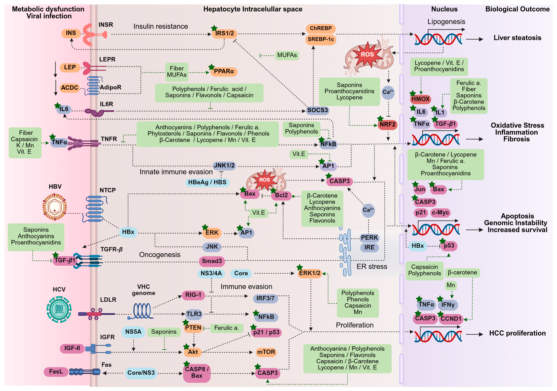
Figure 6 illustrates the complex molecular interplay between viral infections (HBV and HCV), metabolic dysfunction, and HCC, highlighting the modulatory role of bioactive compounds derived from traditional Mexican foods as identified through enrichment analysis.
These interactions span key pathways related to immune evasion, insulin resistance, oxidative stress, inflammation, uncontrolled apoptosis, and tumor proliferation, all of which are central to the pathogenesis of chronic liver disease. Viral hepatitis generates liver damage by hijacking host immune signaling. HBV and HCV evade innate immunity by suppressing RIG-I, TLR3, and NFκB pathways, leading to persistent inflammation and hepatocyte injury [28,29]. The viral proteins HBsAg, HBx, and NS5A modulate key cellular pathways, including ERK, JNK, and mTOR, thereby contributing to viral persistence and oncogenesis [3,133,134,135]. However, bioactive compounds such as anthocyanins, polyphenols, flavonoids, saponins, and proanthocyanidins can interfere with these mechanisms, potentially enhancing antiviral immune responses while mitigating inflammation [136,137,138,139,140].
Insulin signaling (INS/INSR) is impaired by pro-inflammatory cytokines, such as IL-6 and TNF-α, which promote hepatic lipid accumulation and fibrosis [141,142]. Nutrients such as fiber, capsaicin, potassium, manganese, and vitamin E may counteract these effects by improving insulin sensitivity and modulating inflammatory pathways [143,144,145,146,147]. Additionally, monounsaturated fatty acids (MUFAs) may activate PPARα, a key regulator of lipid metabolism and generator of peroxisomes, thereby reducing hepatic steatosis and preventing metabolic stress [148,149].
Oxidative stress plays a central role in the progression of liver disease, with excessive ROS production depleting NRF2-mediated antioxidant responses [150]. Excessive oxidative stress stimulates TGF-β signaling, promoting fibrosis and chronic inflammation through the actions of IL-6 and TNF-α [151,152,153,154]. Bioactive molecules, such as ferulic acid, lycopene, vitamin E, and proanthocyanidins, may help neutralize oxidative damage, enhance hepatic antioxidant defenses (HMOX and NQO1), and prevent fibrotic remodeling. These compounds contribute to hepatoprotection by maintaining redox homeostasis and inhibiting fibrogenic pathways [155,156,157,158].
Apoptosis regulation is also crucial in liver pathology, as it balances cell survival and programmed cell death. Dysregulation in apoptotic pathways, characterized by increased BAX, CASP3, and Jun expression, leads to excessive hepatocyte loss. In contrast, oncogenic signals, such as HBx, suppress p53, thereby fostering genomic instability and promoting tumor progression [159,160,161,162,163]. Lycopene, β-carotene, polyphenols, saponins, and flavonoids may restore apoptotic balance by preventing mitochondrial dysfunction, reducing oncogenic mutations, and promoting controlled cell death [34,156,164,165,166].
HCC progression is partly fueled by the activation of mTOR and ERK1/2, which stimulate cell proliferation and survival [167]. The suppression of tumor suppressors (p21/p53) and the overexpression of CCND1 (Cyclin D1) accelerate oncogenic transformation [34,159,168]. However, dietary bioactive compounds such as capsaicin, polyphenols, manganese, and β-carotene exert antiproliferative effects, potentially inhibiting inflammatory mediators (TNF-α, CASP3), reducing pro-tumorigenic signals, and regulating autophagy, thereby limiting HCC progression [31,104,105,106,107].

4. Discussion

The gene–nutrient interactions of nutrients and bioactive compounds derived from the traditional Mexican foods analyzed in this study have clinical implications for preventing and managing HBV, HCV, MASLD, and ultimately HCC. The Integrative Bioinformatic Analysis revealed that food-derived molecules regulating key pathways linked to liver disease pathogenesis could serve as a basis for implementing therapeutic strategies to enhance synergistic outcomes when combined with antiviral therapies. Such integrative approaches may diminish viral persistence, alleviate insulin resistance, suppress chronic inflammatory cascades, and attenuate fibrotic progression and HCC risk. Therefore, by building on prior evidence from the literature review (summarized in Figure 2 and Tables S1 and S2) and the Integrative Bioinformatics Analysis, we propose nutrigenomic culinary recommendations rooted in traditional Mexican cuisine to address viral hepatitis, MASLD, and HCC. This framework bridges ancestral dietary patterns with precision nutrition to mitigate liver diseases.
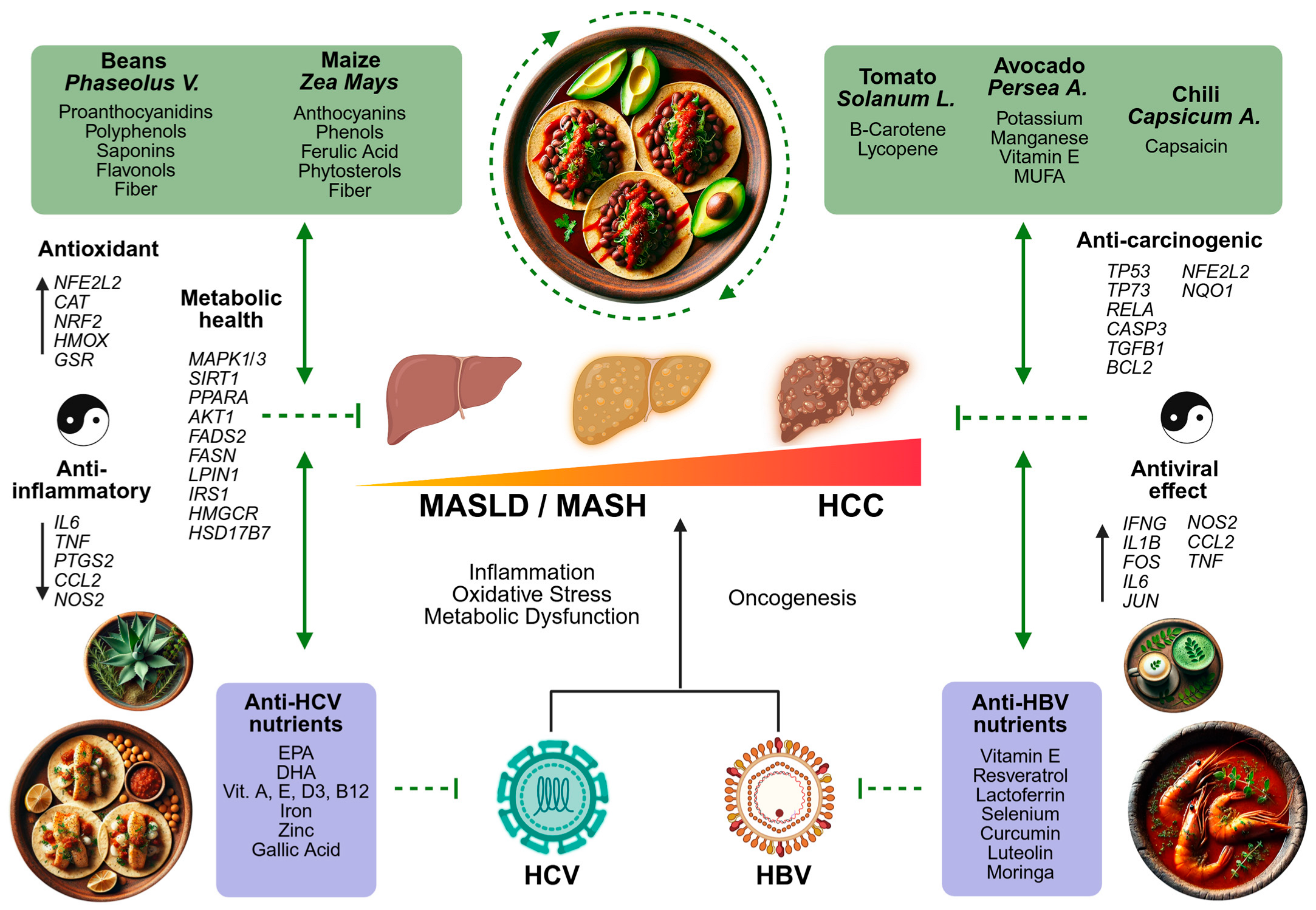
Figure 7 presents a comprehensive nutrigenomic framework that integrates the antiviral, anti-MASLD, and anti-HCC capacities of staple Mexican foods, illustrating how their bioactive compounds can be combined into culturally relevant dishes to enhance liver health benefits. On the upper left side, beans and maize are highlighted for their antioxidant, anti-inflammatory, and metabolic-regulating effects, including reductions in oxidative stress, improved lipid metabolism, and modulation of inflammatory cytokines. In the upper right section, tomatoes are notable for their β-carotene and lycopene, which confer anti-carcinogenic activity by inhibiting cell proliferation and promoting apoptosis. Avocados, on the other hand, deliver potassium, manganese, vitamin E, and MUFAs, conferring both hepatoprotective and anti-carcinogenic benefits. Additionally, chili (Capsicum spp.) provides capsaicin, which has been shown to exert antiviral and tumor-suppressing actions.
In the lower section, Figure 7 links virus-specific nutrients (for both HBV and HCV) to their immunomodulatory and antiviral mechanisms, including EPA, DHA, vitamin A, E, and D3, as well as B12, iron, zinc, resveratrol, lactoferrin, selenium, curcumin, luteolin, and moringa extracts. Thus, this integrative model illustrates not only how these traditional Mexican foods target overlapping molecular pathways that underlie viral hepatitis, metabolic dysfunction, and HCC but also how they can be combined into whole-food dishes that align with both ancestral dietary practices and modern nutrigenomic insights for optimal liver health.
HBV and HCV remain significant global contributors to liver disease, ranging from chronic hepatitis to cirrhosis and HCC [1,169]. These viruses, though adaptive signatures in their genome and replication strategies, share common mechanisms of liver damage, including immune evasion, metabolic disruption, and the promotion of chronic inflammation, fibrosis, and carcinogenesis. Given the central role of immune evasion, metabolic disruption, and chronic inflammation in the pathogenesis of HBV and HCV, the bioactive compounds in the traditional Mexican diet emerge as potential modulators of these mechanisms.
For viral hepatitis, traditional Mexican ingredients like avocado, sunflower seeds, oregano, sardines, and leafy greens, such as purslane or amaranth leaves, provide bioactive compounds that help counteract the infection. Vitamin E (avocado, sunflower seeds) blocks HBV entry (NTCP receptor) and reduces cccDNA. Luteolin-7-O-glucoside (oregano) and curcumin suppress NFκB inflammation, while EGCG (cocoa) enhances viral clearance. Selenium (sardines, tuna) activates p53 for DNA repair, and DHA inhibits HCV polymerase. Vitamin B12 (leafy greens) disrupts HCV replication. A traditional antiviral dish could include grilled sardines or seasonal Mexican fish with an oregano rub, accompanied by guacamole salsa (rich in vitamin E) and a stew of leafy greens, served with agua fresca (fruit-flavored waters) infused with moringa, lime, and chia seeds. This meal supports the immune system, reduces viral replication, and helps control hepatic inflammation, providing a dietary approach to support antiviral therapies.
MASLD has become a significant cause of chronic liver disease and liver transplantation globally [170], driven mainly by an epidemiological shift linked to Westernized lifestyles [171,172,173]. This nutritional transition, characterized by a decline in the traditional diet rich in maize, beans, tomatoes, chili, avocado, and other staples, has been replaced by ultra-processed foods high in obesogens, sugars, and unhealthy fats, contributing to obesity, type 2 diabetes, and metabolic dysfunction-associated liver diseases [174,175].
The global nutrition transition intensifies ecosystem and human health dysfunctions by disrupting the delicate balance of gene–environment interactions [18,176,177,178,179]. Traditional diets have historically played a crucial role in supporting immune and metabolic health through millennial gene–nutrient adaptations [180]. However, this shift exemplifies an evolutionary mismatch: human metabolism co-evolved alongside nutrient-dense, ancestral diets, yet modern pathogenic dietary patterns dysregulate immune, antioxidant, and metabolic pathways [181,182,183,184]. This dysregulation fosters insulin resistance, oxidative stress, chronic inflammation, and progressive liver damage, further exacerbating the burden of metabolic and liver diseases [159,185]. In Mexico, approximately half of the adult population is affected by MASLD and faces a heightened risk of developing MASH, highlighting a critical public health issue [16,186].
For MASLD, incorporating Mexican ingredients such as jicama, beans, maize, agave, tomatoes, chili peppers, purslane, and chia seeds into traditional Mexican recipes can help slow the progression of metabolic dysfunction. Fiber and fructans (inulin and fructooligosaccharides) enhance insulin sensitivity and reduce hepatic steatosis, while anthocyanins and polyphenols support lipid metabolism and mitigate oxidative stress. MUFAs and phytosterols may lower LDL, raise HDL, and reduce liver fat. Additionally, micronutrients such as folate, vitamin E, potassium, and manganese help modulate inflammation and improve insulin responsiveness. Consequently, a traditional MASLD-focused dish could feature a white or blue variety of maize sopes with smashed black beans and chili–tomato sauce, accompanied by a purslane stew and fresh jicama slices and lime water sweetened with agave inulin and chia seeds for dessert.
For HCC, traditional Mexican ingredients such as peanuts, cocoa, sunflower seeds, tomatoes, blue corn, beans, chili, avocado, and international turmeric offer bioactive compounds to combat hepatocellular carcinogenesis. Resveratrol (peanuts) and curcumin (turmeric) induce apoptosis (via BAX/CASP3) and suppress oncogenic NFκB/mTOR pathways. Chlorogenic acid (found in sunflower seeds) and epicatechins (found in cocoa) inhibit tumor growth by blocking proliferation signals. Lycopene (tomato) and anthocyanins (blue corn and black beans) reduce oxidative DNA damage and modulate p53/BCL2 balance to promote controlled cell death, while β-carotene (tomato) inhibits cyclin D1 to curb uncontrolled division. Capsaicin (chili) exerts antiproliferative effects through MAPK pathway inhibition, and phytosterols (avocado) enhance TP53 tumor suppressor activity.
A traditional HCC-focused meal might be mole poblano (a sauce blending cocoa, different species of chili, and peanuts) served with maize tortillas and black beans, accompanied by a tomato–avocado salad, and a chia–cacao atole (corn-based drink) sweetened with agave inulin. This combination may potentiate synergistic anti-carcinogenic mechanisms, aligning with cultural and culinary practices, and offers a dietary strategy to complement HCC prevention.
These findings not only highlight the functional role of Mexican native foods in liver health but also support their inclusion in dietary guidelines aimed at reducing the burden of liver diseases in Mexico. However, despite these properties, clinical practice guidelines in Mexico continue to promote dietary patterns such as the Mediterranean and DASH diets, which are not culturally rooted in the Mexican context [22,23,24,25,187]. Notably, the 2023 Dietary Guidelines from the National Institute of Public Health mark a shift in advocating for a healthy Mexican diet that aligns with local ecosystems and cultural heritage [188].
Nevertheless, traditional Mexican staples like maize face economic and legal threats due to trade agreements, such as the United States–Mexico–Canada Agreement (USMCA or T-MEC), which is leading to the monopolization of genetically modified maize that resists pesticides and herbicides, thereby displacing native Mexican varieties, including blue maize [189,190,191]. This displacement undermines the nutritional diversity provided by autochthonous crops, which are rich in anthocyanins, polyphenols, and other bioactive compounds that may have shaped and sustained the metabolic health of the Mexican population [26,30,180].
Paradoxically, while avocado (Persea americana) was included due to its rich nutrient composition, which has demonstrated hepatoprotective properties and potential to prevent HCC [18,34,164], the increasing international demand for avocado has led to environmental degradation, deforestation, and unsustainable land use in Mexico. Moreover, economic profits remain heavily concentrated in American agribusiness, while the resulting regional and local environmental burdens increasingly affect the most vulnerable populations [192]. Therefore, this selection highlights the importance of striking a balance between nutritional benefits and sustainable agricultural practices, thereby preserving both ecosystem integrity and traditional food systems.
Although this study provides an integrative nutrigenomic perspective, several limitations should be acknowledged. As expected, the strength of evidence inherently varies across in silico, in vitro, and in vivo studies, and such evidence is not uniformly available for all traditional Mexican foods included in this analysis. Moreover, the literature may be biased toward compounds with previously documented biological activity, potentially overlooking lesser-known foods or nutrients. This work is not a systematic literature review; instead, it offers an exploratory, Integrative Bioinformatic Analysis based on curated literature sources and bioinformatic tools to generate nutrigenomic hypotheses. As such, the selection of studies may be subject to selection bias and limitations inherent to the databases used. While bioinformatic tools enable high-throughput hypothesis generation, they rely on existing curated datasets and may not fully capture the complexity of nutrient–gene interactions in physiological contexts. Therefore, in silico predictions should be interpreted with caution and validated through mechanistic studies, including experimental models and human clinical trials.
Additionally, many of the bioactive compounds identified have been studied primarily in isolated or supplement form, which may not accurately reflect their behavior when consumed as part of whole-food matrices. Their bioavailability, metabolic fate, and synergistic interactions could differ within traditional dietary contexts. Moreover, there is a notable lack of dietary intervention trials conducted specifically in Mexican populations. The broader implementation of GENOMEX diet strategies may also face important challenges, including persistent socioeconomic inequalities, shifts in agricultural practices, and the ongoing nutritional transition that is displacing traditional diets and contributing to the erosion of native crops such as maize. Nutrient–gene interactions are further shaped by a range of population-specific factors, including ancestry-related polymorphisms, epigenetic regulation, and microbiome diversity, which may modulate the biological effects of traditional foods. Since this study was developed in the context of the Mexican population, the extrapolation of findings to other populations with different genetic, environmental, and cultural backgrounds is limited. Collectively, these factors constrain the immediate translational applicability of the current findings and emphasize the need for rigorous, context-sensitive validation.
Despite these limitations, this study underscores several strengths. It offers a novel integrative approach, combining a narrative literature review with bioinformatic enrichment to elucidate nutrigenomic mechanisms linking traditional Mexican foods to pathways implicated in HBV, HCV, and MASLD. Rather than ranking foods, the analysis demonstrates how multi-component foods engage liver disease-related pathways, situating these mechanisms within a culturally grounded dietary pattern consumed for millennia by Native Mexican populations. By focusing on the genomic and cultural context of the Mexican population, this work highlights the potential of culturally and genomically tailored nutritional interventions. Notably, research groups in Mexico have already demonstrated the efficacy of this dietary pattern, with clinical studies reporting improvements in adherence, self-efficacy, and cardiometabolic health markers, further supporting its role in mitigating the burden of chronic liver and metabolic diseases [26,27,193].
While the GENOMEX diet model shares functional similarities with other heritage-based dietary patterns—such as the antioxidant and anti-inflammatory benefits of the Mediterranean and traditional Asian diets—its aim is not to replace or compete with these frameworks. Instead, the GENOMEX diet offers a population-specific, nutrigenomic approach grounded in the evolutionary, genomic, cultural, and ecological context of Mexico. Just as Mediterranean diets are best suited to Mediterranean populations, the GENOMEX diet highlights the health potential of native Mexican foods for the Mexican population. This model also serves as an invitation for other regions to reexamine their traditional food systems through a nutrigenomic lens, contributing to more inclusive, culturally adapted public health strategies. Hence, we invite other Latin American populations to reclaim their ancestral and traditional ingredients, resist nutritional and epistemological colonialism, and establish sovereign public health strategies designed specifically for their populations. These strategies should honor and integrate their cultural and genomic heritage, fostering health policies that are effective and deeply rooted in each nation’s identity, biodiversity, and history.

5. Conclusions

This study demonstrates that integrating traditional Mexican foods into dietary strategies provides a potential nutrigenomic solution to mitigate viral hepatitis B and C, MASLD, and reduce the risk of developing liver fibrosis and HCC in Mexican populations. This approach addresses urgent public health priorities such as curbing the cardiometabolic disease epidemic and alleviating the burden of viral hepatitis. This implication is particularly relevant in countries where antiviral therapies remain inaccessible due to cost barriers, genetic polymorphisms linked to treatment resistance and risk of cardiometabolic disease, and an escalating mismatch between the population’s evolutionary genetics and modern dietary patterns.

Supplementary Materials

The following supporting information can be downloaded at https://www.mdpi.com/article/10.3390/ijms26188977/s1.

Author Contributions

Conceptualization, S.R. and A.P.; methodology, L.L.-M., A.J.-A., A.P., and S.R.; software, L.L.-M.; formal analysis, L.L.-M.; investigation, L.L.-M., A.P., A.J.-A., and S.R.; writing—original draft preparation, L.L.-M.; writing—review and editing, L.L.-M., A.P., A.J.-A., and S.R.; validation, A.J.-A.; visualization, S.R. All authors have read and agreed to the published version of the manuscript.

Funding

This research did not receive external funding.

Data Availability Statement

The original contributions presented in this study are included in the article/Supplementary Materials. Further inquiries can be directed to the corresponding author.

Acknowledgments

Leonardo Leal-Mercado is a PhD candidate in Molecular Biology in Medicine at the Centro Universitario de Ciencias de la Salud, Universidad de Guadalajara (Health Sciences Center, University of Guadalajara), supported by a CONAHCYT-Mexico scholarship.

Conflicts of Interest

The authors declare no conflicts of interest.

Abbreviations

The following abbreviations are used in this manuscript:
HBVHepatitis B virus
HCVHepatitis C virus
HCCHepatocellular carcinoma
MASHMetabolic dysfunction-associated steatohepatitis
MASLDMetabolic dysfunction-associated steatotic liver disease
GENOMEXGenome-based Mexican (diet)
VLDLVery-low-density lipoprotein
HDLHigh-density lipoprotein
CTDComparative Toxicogenomic Database
HBsAgHepatitis B surface antigen
EGCGEpigallocatechin-3-gallate
NTCPSodium taurocholate cotransporting polypeptide
DHADocosahexaenoic acid
EPAEicosapentaenoic acid
ALAα-linolenic acid
LDL-CLow-density lipoprotein cholesterol
HDL-CHigh-density lipoprotein cholesterol
LPLLipoprotein lipase
PPARγPeroxisome proliferator-activated receptor gamma
ABCA1ATP-binding cassette transporter A1
LXRαLiver X receptor alpha
CETPCholesterol ester transfer protein
ROSReactive oxygen species
SOD1Superoxide dismutase
TNF-αTumor necrosis factor-alpha
NF-κBNuclear factor-kappa B
MUFAsMonounsaturated fatty acids

References

  1. Saraceni, C.; Birk, J. A Review of Hepatitis B Virus and Hepatitis C Virus Immunopathogenesis. J. Clin. Transl. Hepatol. 2021, 9, 409–418. [Google Scholar] [CrossRef]
  2. Global Hepatitis Report 2024: Action for Access in Low- and Middle-Income Countries. Available online: https://www.who.int/publications/i/item/9789240091672 (accessed on 17 February 2025).
  3. Jose-Abrego, A.; Roman, S.; Laguna-Meraz, S.; Panduro, A. Host and HBV Interactions and Their Potential Impact on Clinical Outcomes. Pathogens 2023, 12, 1146. [Google Scholar] [CrossRef]
  4. Zhang, H.; Zhou, X.-D.; Shapiro, M.D.; Lip, G.Y.H.; Tilg, H.; Valenti, L.; Somers, V.K.; Byrne, C.D.; Targher, G.; Yang, W.; et al. Global Burden of Metabolic Diseases, 1990–2021. Metabolism 2024, 160, 155999. [Google Scholar] [CrossRef]
  5. Jaroszewicz, J.; Flisiak, R. Metabolic Syndrome and Hepatitis C Infection—Brothers in Arms. Liver Int. 2013, 33, 1135–1137. [Google Scholar] [CrossRef]
  6. Jarcuska, P.; Abdel-Razik, A.; Flisiak, R.; Singh, R.B. Chronic Viral Hepatitis and Metabolic Syndrome/Cardiovascular Risk. Can. J. Gastroenterol. Hepatol. 2018, 2018, 7369314. [Google Scholar] [CrossRef]
  7. Wang, C.-C.; Cheng, P.-N.; Kao, J.-H. Systematic Review: Chronic Viral Hepatitis and Metabolic Derangement. Aliment. Pharmacol. Ther. 2020, 51, 216–230. [Google Scholar] [CrossRef]
  8. Huang, S.-C.; Liu, C.-J.; Kao, J.-H. Impact of Metabolic Disorders on Chronic Hepatitis B. Clin. Liver Dis. 2024, 23, e0130. [Google Scholar] [CrossRef]
  9. Medhioub, M.; Khsiba, A.; Bachali, A.; Bibi, A.; Hamzaoui, L.; Azouz, M.M. Risk of Metabolic Syndrome in Patients with Chronic Hepatitis C. Tunis. Med. 2023, 101, 362–366. [Google Scholar]
  10. Seo, J.A. Metabolic Syndrome: A Warning Sign of Liver Fibrosis. J. Obes. Metab. Syndr. 2022, 31, 1–3. [Google Scholar] [CrossRef]
  11. Langness, J.A.; Tabano, D.; Wieland, A.; Tise, S.; Pratt, L.; Harrington, L.A.; Lin, S.; Ghuschcyan, V.; Nair, K.V.; Everson, G.T.; et al. Curing Chronic Hepatitis C: A Cost Comparison of the Combination Simeprevir Plus Sofosbuvir vs. Protease-Inhibitor-Based Triple Therapy. Ann. Hepatol. 2017, 16, 366–374. [Google Scholar] [CrossRef]
  12. Jose-Abrego, A.; Laguna-Meraz, S.; Roman, S.; Mariscal-Martinez, I.M.; Panduro, A. Hepatitis C Virus Resistance-Associated Substitutions in Mexico. Viruses 2025, 17, 169. [Google Scholar] [CrossRef]
  13. Esquivel, G.; Cruces, G. The Dynamics of Income Inequality in Mexico since NAFTA [with Comment]. Economía 2011, 12, 155–188. [Google Scholar] [CrossRef]
  14. Laguna-Meraz, S.; Jose-Abrego, A.; Roman, S.; Leal-Mercado, L.; Panduro, A. Risk Factors Associated with Hepatitis C Subtypes and the Evolutionary History of Subtype 1a in Mexico. Viruses 2024, 16, 1259. [Google Scholar] [CrossRef]
  15. Stevens, G.; Dias, R.H.; Thomas, K.J.; Rivera, J.A.; Carvalho, N.; Barquera, S.; Hill, K.; Ezzati, M. Characterizing the Epidemiological Transition in Mexico: National and Subnational Burden of Diseases, Injuries, and Risk Factors. PLoS Med. 2008, 5, e125. [Google Scholar] [CrossRef]
  16. Sepulveda-Villegas, M.; Roman, S.; Rivera-Iñiguez, I.; Ojeda-Granados, C.; Gonzalez-Aldaco, K.; Torres-Reyes, L.A.; Jose-Abrego, A.; Panduro, A. High Prevalence of Nonalcoholic Steatohepatitis and Abnormal Liver Stiffness in a Young and Obese Mexican Population. PLoS ONE 2019, 14, e0208926. [Google Scholar] [CrossRef] [PubMed]
  17. Campos-Nonato, I.; Galván-Valencia, Ó.; Hernández-Barrera, L.; Oviedo-Solís, C.I.; Barquera, S. Prevalencia de Obesidad y Factores de Riesgo Asociados En Adultos Mexicanos: Resultados de La Ensanut 2022. Inst. Nac. Salud Pública 2023, 65, s238–s247. [Google Scholar] [CrossRef]
  18. Panduro, A.; Roman, S.; Mariscal-Martinez, I.M.; Jose-Abrego, A.; Gonzalez-Aldaco, K.; Ojeda-Granados, C.; Ramos-Lopez, O.; Torres-Reyes, L.A. Personalized Medicine and Nutrition in Hepatology for Preventing Chronic Liver Disease in Mexico. Front. Nutr. 2024, 11, 1379364. [Google Scholar] [CrossRef]
  19. Shen, T.-H.; Wu, C.-H.; Lee, Y.-W.; Chang, C.-C. Prevalence, Trends, and Characteristics of Metabolic Dysfunction-Associated Steatotic Liver Disease among the US Population Aged 12–79 Years. Eur. J. Gastroenterol. Hepatol. 2024, 36, 636–645. [Google Scholar] [CrossRef]
  20. Palma-Lara, I.; Ortiz-López, M.G.; Bonilla-Delgado, J.; Pérez-Escobar, J.; Godínez-Aguilar, R.; Luévano-Contreras, C.; Espinosa-García, A.M.; Pérez-Durán, J.; García Alonso-Themann, P.; Nolasco-Quiroga, M.; et al. A Landscape of Liver Cirrhosis and Transplantation in Mexico: Changing Leading Causes and Transplant as Response. Ann. Hepatol. 2025, 30, 101562. [Google Scholar] [CrossRef] [PubMed]
  21. INEGI. Estadísticas de Defunciones Registradas (EDR); INEGI 2023 Preliminar; INEGI: Aguascalientes, Mexico, 2024. [Google Scholar]
  22. Chávez-Manzanera, E.A.; Vera-Zertuche, J.M.; Kaufer-Horwitz, M.; Vázquez-Velázquez, V.; Flores-Lázaro, J.R.; Mireles-Zavala, L.; Calzada-León, R.; Garnica-Cuellar, J.C.; Sánchez-Muñoz, V.; Ramírez-Butanda, E.; et al. Mexican Clinical Practice Guidelines for Adult Overweight and Obesity Management. Curr. Obes. Rep. 2024, 13, 643–666. [Google Scholar] [CrossRef] [PubMed]
  23. Centro Nacional para la Prevención y Control del VIH y el sida. Guía para la Prevención y Atención de las Hepatitis Virales en México 2023. Available online: http://www.gob.mx/censida/documentos/guia-para-la-prevencion-y-atencion-de-las-hepatitis-virales-en-mexico-2023 (accessed on 17 February 2025).
  24. Uscanga Domínguez, L.; Bielsa Fernández, M.V.; Huerta Iga, F.; Lizardi Cervera, J.; Muñoz Espinosa, L.; López Tarabay, C.; Rodríguez Hernández, H.; Torre Delgadillo, A.; Lilia Tostado Ramos, C. Guías clínicas de diagnóstico y tratamiento de hepatopatía grasa no alcohólica. Generalidades. Rev. Gastroenterol. Mex. 2008, 73, 126–128. [Google Scholar] [PubMed]
  25. IMSS. Diagnóstico y Tratamiento Farmacológico de la Diabetes Mellitus Tipo 2 en el Primer Nivel de Atención. In Guía de Evidencias y Recomendaciones: Guía de Práctica Clínica; IMSS: Mexico City, Mexico, 2018. [Google Scholar]
  26. Ojeda-Granados, C.; Panduro, A.; Gonzalez-Aldaco, K.; Sepulveda-Villegas, M.; Rivera-Iñiguez, I.; Roman, S. Tailoring Nutritional Advice for Mexicans Based on Prevalence Profiles of Diet-Related Adaptive Gene Polymorphisms. J. Pers. Med. 2017, 7, 16. [Google Scholar] [CrossRef] [PubMed]
  27. Ojeda-Granados, C.; Panduro, A.; Rivera-Iñiguez, I.; Sepúlveda-Villegas, M.; Roman, S. A Regionalized Genome-Based Mexican Diet Improves Anthropometric and Metabolic Parameters in Subjects at Risk for Obesity-Related Chronic Diseases. Nutrients 2020, 12, 645. [Google Scholar] [CrossRef] [PubMed]
  28. Jose-Abrego, A.; Rivera-Iñiguez, I.; Torres-Reyes, L.A.; Roman, S. Anti-Hepatitis B Virus Activity of Food Nutrients and Potential Mechanisms of Action. Ann. Hepatol. 2023, 28, 100766. [Google Scholar] [CrossRef]
  29. Gonzalez-Aldaco, K.; Torres-Reyes, L.A.; Ojeda-Granados, C.; Leal-Mercado, L.; Roman, S.; Panduro, A. Metabolic Dysfunction-Associated Steatotic Liver Disease in Chronic Hepatitis C Virus Infection: From Basics to Clinical and Nutritional Management. Clin. Pract. 2024, 14, 2542–2558. [Google Scholar] [CrossRef]
  30. Roman, S.; Campos-Medina, L.; Leal-Mercado, L. Personalized Nutrition: The End of the One-Diet-Fits-All Era. Front. Nutr. 2024, 11, 1370595. [Google Scholar] [CrossRef]
  31. Escalante-Araiza, F.; Gutiérrez-Salmeán, G. Traditional Mexican Foods as Functional Agents in the Treatment of Cardiometabolic Risk Factors. Crit. Rev. Food Sci. Nutr. 2021, 61, 1353–1364. [Google Scholar] [CrossRef]
  32. Elizondo-Solis, C.; Rojas-Gutiérrez, S.; Martínez-Canales, R.; Montoya-Rosales, A.; Hernández-García, M.; Salazar-Cepeda, C.; Ramírez, K.; Gelinas-Martín del Campo, M.; Salinas-Carmona, M.; Rosas-Taraco, A.; et al. Integrative Bioinformatics Analysis of Immune Activation and Gene Networks in Pediatric Septic Arthritis. Comput. Biol. Chem. 2025, 115, 108287. [Google Scholar] [CrossRef]
  33. Zizumbo-Villarreal, D.; Flores-Silva, A.; Colunga-García Marín, P. The Archaic Diet in Mesoamerica: Incentive for Milpa Development and Species Domestication. Econ. Bot. 2012, 66, 328–343. [Google Scholar] [CrossRef]
  34. George, E.S.; Sood, S.; Broughton, A.; Cogan, G.; Hickey, M.; Chan, W.S.; Sudan, S.; Nicoll, A.J. The Association between Diet and Hepatocellular Carcinoma: A Systematic Review. Nutrients 2021, 13, 172. [Google Scholar] [CrossRef]
  35. Davis, A.P.; Wiegers, T.C.; Sciaky, D.; Barkalow, F.; Strong, M.; Wyatt, B.; Wiegers, J.; McMorran, R.; Abrar, S.; Mattingly, C.J. Comparative Toxicogenomics Database’s 20th Anniversary: Update 2025. Nucleic Acids Res. 2025, 53, D1328–D1334. [Google Scholar] [CrossRef]
  36. von Mering, C.; Jensen, L.J.; Snel, B.; Hooper, S.D.; Krupp, M.; Foglierini, M.; Jouffre, N.; Huynen, M.A.; Bork, P. STRING: Known and Predicted Protein-Protein Associations, Integrated and Transferred across Organisms. Nucleic Acids Res. 2005, 33, D433–D437. [Google Scholar] [CrossRef]
  37. Huang, D.W.; Sherman, B.T.; Lempicki, R.A. Systematic and Integrative Analysis of Large Gene Lists Using DAVID Bioinformatics Resources. Nat. Protoc. 2009, 4, 44–57. [Google Scholar] [CrossRef]
  38. Kuleshov, M.V.; Jones, M.R.; Rouillard, A.D.; Fernandez, N.F.; Duan, Q.; Wang, Z.; Koplev, S.; Jenkins, S.L.; Jagodnik, K.M.; Lachmann, A.; et al. Enrichr: A Comprehensive Gene Set Enrichment Analysis Web Server 2016 Update. Nucleic Acids Res. 2016, 44, W90–W97. [Google Scholar] [CrossRef]
  39. Kanehisa, M.; Furumichi, M.; Sato, Y.; Matsuura, Y.; Ishiguro-Watanabe, M. KEGG: Biological Systems Database as a Model of the Real World. Nucleic Acids Res. 2025, 53, D672–D677. [Google Scholar] [CrossRef]
  40. R Core Team. R: The R Project for Statistical Computing; R Foundation for Statistical Computing: Vienna, Austria, 2025; Available online: https://www.r-project.org/ (accessed on 20 February 2025).
  41. Wickham, H.; Averick, M.; Bryan, J.; Chang, W.; McGowan, L.D.; François, R.; Grolemund, G.; Hayes, A.; Henry, L.; Hester, J.; et al. Welcome to the Tidyverse. J. Open Source Softw. 2019, 4, 1686. [Google Scholar] [CrossRef]
  42. Wickham, H. Reshaping Data with the Reshape Package. J. Stat. Softw. 2007, 21, 1–20. [Google Scholar] [CrossRef]
  43. Wickham, H. ggplot2; Use R! Springer International Publishing: Cham, Switzerland, 2016; ISBN 978-3-319-24275-0. [Google Scholar]
  44. Neuwirth, E. R Package RColorBrewer, version 1.1-3. ColorBrewer Palettes. R Foundation for Statistical Computing: Vienna, Austria, 2022. Available online: https://CRAN.R-project.org/package=RColorBrewer (accessed on 20 February 2025).
  45. Sjoberg, D. Davidsjoberg/Ggsankey 2025. An Implementation of Grammar of Graphics for Graphs and Networks. Available online: https://ggraph.data-imaginist.com/ (accessed on 20 February 2025).
  46. Csárdi, G.; Nepusz, T.; Traag, V.; Horvát, S.; Zanini, F.; Noom, D.; Müller, K.; Salmon, M.; Antonov, M. R Package, version 2.3. igraph: Network Analysis and Visualization. R Foundation for Statistical Computing: Vienna, Austria, 2025.
  47. Park, S.; Lim, J.; Kim, J.R.; Cho, S. Inhibitory Effects of Resveratrol on Hepatitis B Virus X Protein-Induced Hepatocellular Carcinoma. J. Vet. Sci. 2017, 18, 419–429. [Google Scholar] [CrossRef]
  48. Andreone, P.; Fiorino, S.; Cursaro, C.; Gramenzi, A.; Margotti, M.; Di Giammarino, L.; Biselli, M.; Miniero, R.; Gasbarrini, G.; Bernardi, M. Vitamin E as Treatment for Chronic Hepatitis B: Results of a Randomized Controlled Pilot Trial. Antivir. Res. 2001, 49, 75–81. [Google Scholar] [CrossRef]
  49. Fiorino, S.; Bacchi-Reggiani, M.L.; Leandri, P.; Loggi, E.; Andreone, P. Vitamin E for the Treatment of Children with Hepatitis B e Antigen-Positive Chronic Hepatitis: A Systematic Review and Meta-Analysis. World J. Hepatol. 2017, 9, 333–342. [Google Scholar] [CrossRef]
  50. Hara, K.; Ikeda, M.; Saito, S.; Matsumoto, S.; Numata, K.; Kato, N.; Tanaka, K.; Sekihara, H. Lactoferrin Inhibits Hepatitis B Virus Infection in Cultured Human Hepatocytes. Hepatol. Res. 2002, 24, 228. [Google Scholar] [CrossRef]
  51. Luo, Y.; Xiang, K.; Liu, J.; Song, J.; Feng, J.; Chen, J.; Dai, Y.; Hu, Y.; Zhuang, H.; Zhou, Y. Inhibition of In Vitro Infection of Hepatitis B Virus by Human Breastmilk. Nutrients 2022, 14, 1561. [Google Scholar] [CrossRef]
  52. Yu, S.Y.; Zhu, Y.J.; Li, W.G. Protective Role of Selenium against Hepatitis B Virus and Primary Liver Cancer in Qidong. Biol. Trace Elem. Res. 1997, 56, 117–124. [Google Scholar] [CrossRef]
  53. Kim, H.J.; Yoo, H.S.; Kim, J.C.; Park, C.S.; Choi, M.S.; Kim, M.; Choi, H.; Min, J.S.; Kim, Y.S.; Yoon, S.W.; et al. Antiviral Effect of Curcuma longa Linn Extract against Hepatitis B Virus Replication. J. Ethnopharmacol. 2009, 124, 189–196. [Google Scholar] [CrossRef]
  54. Waiyaput, W.; Payungporn, S.; Issara-Amphorn, J.; Panjaworayan, N.T.-T. Inhibitory Effects of Crude Extracts from Some Edible Thai Plants against Replication of Hepatitis B Virus and Human Liver Cancer Cells. BMC Complement. Altern. Med. 2012, 12, 246. [Google Scholar] [CrossRef]
  55. Cui, X.-X.; Yang, X.; Wang, H.-J.; Rong, X.-Y.; Jing, S.; Xie, Y.-H.; Huang, D.-F.; Zhao, C. Luteolin-7-O-Glucoside Present in Lettuce Extracts Inhibits Hepatitis B Surface Antigen Production and Viral Replication by Human Hepatoma Cells In Vitro. Front. Microbiol. 2017, 8, 2425. [Google Scholar] [CrossRef]
  56. Feustel, S.; Ayón-Pérez, F.; Sandoval-Rodriguez, A.; Rodríguez-Echevarría, R.; Contreras-Salinas, H.; Armendáriz-Borunda, J.; Sánchez-Orozco, L.V. Protective Effects of Moringa Oleifera on HBV Genotypes C and H Transiently Transfected Huh7 Cells. J. Immunol. Res. 2017, 2017, 6063850. [Google Scholar] [CrossRef]
  57. Hamza, A.A. Ameliorative Effects of Moringa Oleifera Lam Seed Extract on Liver Fibrosis in Rats. Food Chem. Toxicol. 2010, 48, 345–355. [Google Scholar] [CrossRef]
  58. Wang, G.-F.; Shi, L.-P.; Ren, Y.-D.; Liu, Q.-F.; Liu, H.-F.; Zhang, R.-J.; Li, Z.; Zhu, F.-H.; He, P.-L.; Tang, W.; et al. Anti-Hepatitis B Virus Activity of Chlorogenic Acid, Quinic Acid and Caffeic Acid In Vivo and In Vitro. Antivir. Res. 2009, 83, 186–190. [Google Scholar] [CrossRef]
  59. Chen, C.-L.; Chang, W.-C.; Yi, C.-H.; Hung, J.-S.; Liu, T.-T.; Lei, W.-Y.; Hsu, C.-S. Association of Coffee Consumption and Liver Fibrosis Progression in Patients with HBeAg-Negative Chronic Hepatitis B: A 5-Year Population-Based Cohort Study. J. Formos. Med. Assoc. 2019, 118, 628–635. [Google Scholar] [CrossRef]
  60. Jang, E.S.; Jeong, S.-H.; Lee, S.H.; Hwang, S.H.; Ahn, S.Y.; Lee, J.; Park, Y.S.; Hwang, J.H.; Kim, J.-W.; Kim, N.; et al. The Effect of Coffee Consumption on the Development of Hepatocellular Carcinoma in Hepatitis B Virus Endemic Area. Liver Int. 2013, 33, 1092–1099. [Google Scholar] [CrossRef]
  61. Huang, H.-C.; Tao, M.-H.; Hung, T.-M.; Chen, J.-C.; Lin, Z.-J.; Huang, C. (−)-Epigallocatechin-3-Gallate Inhibits Entry of Hepatitis B Virus into Hepatocytes. Antivir. Res. 2014, 111, 100–111. [Google Scholar] [CrossRef]
  62. Karamese, M.; Aydogdu, S.; Karamese, S.A.; Altoparlak, U.; Gundogdu, C. Preventive Effects of a Major Component of Green Tea, Epigallocathechin-3-Gallate, on Hepatitis-B Virus DNA Replication. Asian Pac. J. Cancer Prev. 2015, 16, 4199–4202. [Google Scholar] [CrossRef]
  63. Pang, J.; Zhao, K.; Wang, J.; Ma, Z.; Xiao, X. Green Tea Polyphenol, Epigallocatechin-3-Gallate, Possesses the Antiviral Activity Necessary to Fight against the Hepatitis B Virus Replication In Vitro. J. Zhejiang Univ. Sci. B 2014, 15, 533–539. [Google Scholar] [CrossRef]
  64. Zhong, L.; Hu, J.; Shu, W.; Gao, B.; Xiong, S. Epigallocatechin-3-Gallate Opposes HBV-Induced Incomplete Autophagy by Enhancing Lysosomal Acidification, Which Is Unfavorable for HBV Replication. Cell Death Dis. 2015, 6, e1770. [Google Scholar] [CrossRef]
  65. Leu, G.-Z.; Lin, T.-Y.; Hsu, J.T.A. Anti-HCV Activities of Selective Polyunsaturated Fatty Acids. Biochem. Biophys. Res. Commun. 2004, 318, 275–280. [Google Scholar] [CrossRef]
  66. Miyoshi, H.; Moriya, K.; Tsutsumi, T.; Shinzawa, S.; Fujie, H.; Shintani, Y.; Fujinaga, H.; Goto, K.; Todoroki, T.; Suzuki, T.; et al. Pathogenesis of Lipid Metabolism Disorder in Hepatitis C: Polyunsaturated Fatty Acids Counteract Lipid Alterations Induced by the Core Protein. J. Hepatol. 2011, 54, 432–438. [Google Scholar] [CrossRef] [PubMed]
  67. Govea-Salas, M.; Rivas-Estilla, A.M.; Rodríguez-Herrera, R.; Lozano-Sepúlveda, S.A.; Aguilar-Gonzalez, C.N.; Zugasti-Cruz, A.; Salas-Villalobos, T.B.; Morlett-Chávez, J.A. Gallic Acid Decreases Hepatitis C Virus Expression through Its Antioxidant Capacity. Exp. Ther. Med. 2016, 11, 619–624. [Google Scholar] [CrossRef]
  68. Bunchorntavakul, C.; Wootthananont, T.; Atsawarungruangkit, A. Effects of Vitamin E on Chronic Hepatitis C Genotype 3: A Randomized, Double-Blind, Placebo-Controlled Study. J. Med. Assoc. Thail. 2014, 97 (Suppl. 11), S31–S40. [Google Scholar]
  69. Hamamoto, S.; Fukuda, R.; Ishimura, N.; Rumi, M.A.K.; Kazumori, H.; Uchida, Y.; Kadowaki, Y.; Ishihara, S.; Kinoshita, Y. 9-Cis Retinoic Acid Enhances the Antiviral Effect of Interferon on Hepatitis C Virus Replication through Increased Expression of Type I Interferon Receptor. J. Lab. Clin. Med. 2003, 141, 58–66. [Google Scholar] [CrossRef]
  70. Murayama, A.; Kato, T. Inhibition of Hepatitis C Virus by Vitamin D. In Vitamins and Hormones; Litwack, G., Ed.; Hormones, Regulators and Viruses; Academic Press: Cambridge, MA, USA, 2021; Volume 117, Chapter 9; pp. 227–238. [Google Scholar]
  71. Eltayeb, A.A.; Abdou, M.A.A.; Abdel-aal, A.M.; Othman, M.H. Vitamin D Status and Viral Response to Therapy in Hepatitis C Infected Children. World J. Gastroenterol. 2015, 21, 1284–1291. [Google Scholar] [CrossRef]
  72. Li, D.; Lott, W.B.; Martyn, J.; Haqshenas, G.; Gowans, E.J. Differential Effects on the Hepatitis C Virus (HCV) Internal Ribosome Entry Site by Vitamin B12 and the HCV Core Protein. J. Virol. 2004, 78, 12075–12081. [Google Scholar] [CrossRef]
  73. Fillebeen, C.; Rivas-Estilla, A.M.; Bisaillon, M.; Ponka, P.; Muckenthaler, M.; Hentze, M.W.; Koromilas, A.E.; Pantopoulos, K. Iron Inactivates the RNA Polymerase NS5B and Suppresses Subgenomic Replication of Hepatitis C Virus. J. Biol. Chem. 2005, 280, 9049–9057. [Google Scholar] [CrossRef]
  74. Yuasa, K.; Naganuma, A.; Sato, K.; Ikeda, M.; Kato, N.; Takagi, H.; Mori, M. Zinc Is a Negative Regulator of Hepatitis C Virus RNA Replication. Liver Int. 2006, 26, 1111–1118. [Google Scholar] [CrossRef]
  75. Santoso, P.; Amelia, A.; Rahayu, R. Jicama (Pachyrhizus erosus) Fiber Prevents Excessive Blood Glucose and Body Weight Increase without Affecting Food Intake in Mice Fed with High-Sugar Diet. J. Adv. Vet. Anim. Res. 2019, 6, 222–230. [Google Scholar] [CrossRef] [PubMed]
  76. Park, C.J.; Lee, H.-A.; Han, J.-S. Jicama (Pachyrhizus erosus) Extract Increases Insulin Sensitivity and Regulates Hepatic Glucose in C57BL/Ksj-Db/Db Mice. J. Clin. Biochem. Nutr. 2016, 58, 56–63. [Google Scholar] [CrossRef][Green Version]
  77. Ristic-Medic, D.; Perunicic-Pekovic, G.; Rasic-Milutinovic, Z.; Takic, M.; Popovic, T.; Arsic, A.; Glibetic, M. Effects of Dietary Milled Seed Mixture on Fatty Acid Status and Inflammatory Markers in Patients on Hemodialysis. Sci. World J. 2014, 2014, 563576. [Google Scholar] [CrossRef]
  78. McKay, D.L.; Eliasziw, M.; Chen, C.Y.O.; Blumberg, J.B. A Pecan-Rich Diet Improves Cardiometabolic Risk Factors in Overweight and Obese Adults: A Randomized Controlled Trial. Nutrients 2018, 10, 339. [Google Scholar] [CrossRef] [PubMed]
  79. König, A.; Schwarzinger, B.; Stadlbauer, V.; Lanzerstorfer, P.; Iken, M.; Schwarzinger, C.; Kolb, P.; Schwarzinger, S.; Mörwald, K.; Brunner, S.; et al. Guava (Psidium guajava) Fruit Extract Prepared by Supercritical CO2 Extraction Inhibits Intestinal Glucose Resorption in a Double-Blind, Randomized Clinical Study. Nutrients 2019, 11, 1512. [Google Scholar] [CrossRef] [PubMed]
  80. Tiwari, A.K.; Jyothi, A.L.; Tejeswini, V.B.; Madhusudana, K.; Kumar, D.A.; Zehra, A.; Agawane, S.B. Mitigation of Starch and Glucose-Induced Postprandial Glycemic Excursion in Rats by Antioxidant-Rich Green-Leafy Vegetables’ Juice. Pharmacogn. Mag. 2013, 9, S66–S73. [Google Scholar] [CrossRef]
  81. Sarker, U.; Oba, S. Nutritional and Bioactive Constituents and Scavenging Capacity of Radicals in Amaranthus hypochondriacus. Sci. Rep. 2020, 10, 19962. [Google Scholar] [CrossRef]
  82. Montoya-Rodríguez, A.; de Mejía, E.G.; Dia, V.P.; Reyes-Moreno, C.; Milán-Carrillo, J. Extrusion Improved the Anti-Inflammatory Effect of Amaranth (Amaranthus hypochondriacus) Hydrolysates in LPS-Induced Human THP-1 Macrophage-like and Mouse RAW 264.7 Macrophages by Preventing Activation of NF-κB Signaling. Mol. Nutr. Food Res. 2014, 58, 1028–1041. [Google Scholar] [CrossRef] [PubMed]
  83. Rosas-Campos, R.; Meza-Rios, A.; Rodriguez-Sanabria, J.S.; la Rosa-Bibiano, R.D.; Corona-Cervantes, K.; García-Mena, J.; Santos, A.; Sandoval-Rodriguez, A.; Armendariz-Borunda, J. Dietary Supplementation with Mexican Foods, Opuntia ficus indica, Theobroma cacao, and Acheta domesticus: Improving Obesogenic and Microbiota Features in Obese Mice. Front. Nutr. 2022, 9, 987222. [Google Scholar] [CrossRef] [PubMed]
  84. Indrianingsih, A.W.; Wulanjati, M.P.; Windarsih, A.; Bhattacharjya, D.K.; Suzuki, T.; Katayama, T. In Vitro Studies of Antioxidant, Antidiabetic, and Antibacterial Activities of Theobroma cacao, Anonna muricata and Clitoria ternatea. Biocatal. Agric. Biotechnol. 2021, 33, 101995. [Google Scholar] [CrossRef]
  85. Kawakami, Y.; Watanabe, Y.; Mazuka, M.; Yagi, N.; Sawazaki, A.; Koganei, M.; Natsume, M.; Kuriki, K.; Morimoto, T.; Asai, T.; et al. Effect of Cacao Polyphenol-Rich Chocolate on Postprandial Glycemia, Insulin, and Incretin Secretion in Healthy Participants. Nutrition 2021, 85, 111128. [Google Scholar] [CrossRef]
  86. Villa-Jaimes, G.S.; Aguilar-Mora, F.A.; González-Ponce, H.A.; Avelar-González, F.J.; Saldaña, M.C.M.; Buist-Homan, M.; Moshage, H. Biocomponents from Opuntia Robusta and Opuntia Streptacantha Fruits Protect against Diclofenac-Induced Acute Liver Damage In Vivo and In Vitro. J. Funct. Foods 2022, 89, 104960. [Google Scholar] [CrossRef]
  87. Magaña-Cerino, J.M.; Guzmán, T.J.; Soto-Luna, I.C.; Betanzos-Cabrera, G.; Gurrola-Díaz, C.M. Cladodes from Nopalea cochenillifera (L.) Salm-Dyck (Cactaceae) attenuate postprandial glycaemia without markedly influencing α-glucosidase activity. Nat. Prod. Res. 2022, 36, 1105–1108. [Google Scholar] [CrossRef]
  88. Santos, H.O.; Macedo, R.C.O. Cocoa-Induced (Theobroma cacao) Effects on Cardiovascular System: HDL Modulation Pathways. Clin. Nutr. ESPEN 2018, 27, 10–15. [Google Scholar] [CrossRef]
  89. Magaña-Cerino, J.M.; Tiessen, A.; Soto-Luna, I.C.; Peniche-Pavía, H.A.; Vargas-Guerrero, B.; Domínguez-Rosales, J.A.; García-López, P.M.; Gurrola-Díaz, C.M. Consumption of Nixtamal from a New Variety of Hybrid Blue Maize Ameliorates Liver Oxidative Stress and Inflammation in a High-Fat Diet Rat Model. J. Funct. Foods 2020, 72, 104075. [Google Scholar] [CrossRef]
  90. Damián-Medina, K.; Salinas-Moreno, Y.; Milenkovic, D.; Figueroa-Yáñez, L.; Marino-Marmolejo, E.; Higuera-Ciapara, I.; Vallejo-Cardona, A.; Lugo-Cervantes, E. In Silico Analysis of Antidiabetic Potential of Phenolic Compounds from Blue Corn (Zea mays L.) and Black Bean (Phaseolus vulgaris L.). Heliyon 2020, 6, e03632. [Google Scholar] [CrossRef]
  91. Orona-Tamayo, D.; Valverde, M.E.; Paredes-López, O. Bioactive Peptides from Selected Latin American Food Crops—A Nutraceutical and Molecular Approach. Crit. Rev. Food Sci. Nutr. 2019, 59, 1949–1975. [Google Scholar] [CrossRef]
  92. Kong, B.; Xiong, Y.L. Antioxidant Activity of Zein Hydrolysates in a Liposome System and the Possible Mode of Action. J. Agric. Food Chem. 2006, 54, 6059–6068. [Google Scholar] [CrossRef]
  93. Higuchi, N.; Hira, T.; Yamada, N.; Hara, H. Oral Administration of Corn Zein Hydrolysate Stimulates GLP-1 and GIP Secretion and Improves Glucose Tolerance in Male Normal Rats and Goto-Kakizaki Rats. Endocrinology 2013, 154, 3089–3098. [Google Scholar] [CrossRef] [PubMed]
  94. Luna-Vital, D.A.; Gonzalez de Mejia, E. Anthocyanins from Purple Corn Activate Free Fatty Acid-Receptor 1 and Glucokinase Enhancing in Vitro Insulin Secretion and Hepatic Glucose Uptake. PLoS ONE 2018, 13, e0200449. [Google Scholar] [CrossRef] [PubMed]
  95. Nchanji, E.B.; Ageyo, O.C. Do Common Beans (Phaseolus vulgaris L.) Promote Good Health in Humans? A Systematic Review and Meta-Analysis of Clinical and Randomized Controlled Trials. Nutrients 2021, 13, 3701. [Google Scholar] [CrossRef]
  96. Escobedo, A.; Rivera-León, E.A.; Luévano-Contreras, C.; Urías-Silvas, J.E.; Luna-Vital, D.A.; Morales-Hernández, N.; Mojica, L. Common Bean Baked Snack Consumption Reduces Apolipoprotein B-100 Levels: A Randomized Crossover Trial. Nutrients 2021, 13, 3898. [Google Scholar] [CrossRef] [PubMed]
  97. Spadafranca, A.; Rinelli, S.; Riva, A.; Morazzoni, P.; Magni, P.; Bertoli, S.; Battezzati, A. Phaseolus vulgaris Extract Affects Glycometabolic and Appetite Control in Healthy Human Subjects. Br. J. Nutr. 2013, 109, 1789–1795. [Google Scholar] [CrossRef] [PubMed]
  98. Wang, L.; Tao, L.; Hao, L.; Stanley, T.H.; Huang, K.-H.; Lambert, J.D.; Kris-Etherton, P.M. A Moderate-Fat Diet with One Avocado per Day Increases Plasma Antioxidants and Decreases the Oxidation of Small, Dense LDL in Adults with Overweight and Obesity: A Randomized Controlled Trial. J. Nutr. 2020, 150, 276–284. [Google Scholar] [CrossRef]
  99. Ahmed, N.; Tcheng, M.; Roma, A.; Buraczynski, M.; Jayanth, P.; Rea, K.; Akhtar, T.A.; Spagnuolo, P.A. Avocatin B Protects Against Lipotoxicity and Improves Insulin Sensitivity in Diet-Induced Obesity. Mol. Nutr. Food Res. 2019, 63, e1900688. [Google Scholar] [CrossRef]
  100. Wang, L.; Bordi, P.L.; Fleming, J.A.; Hill, A.M.; Kris-Etherton, P.M. Effect of a Moderate Fat Diet with and without Avocados on Lipoprotein Particle Number, Size and Subclasses in Overweight and Obese Adults: A Randomized, Controlled Trial. J. Am. Heart Assoc. 2015, 4, e001355. [Google Scholar] [CrossRef]
  101. Orona-Tamayo, D.; Valverde, M.E.; Nieto-Rendón, B.; Paredes-López, O. Inhibitory Activity of Chia (Salvia hispanica L.) Protein Fractions against Angiotensin I-Converting Enzyme and Antioxidant Capacity. LWT-Food Sci. Technol. 2015, 64, 236–242. [Google Scholar] [CrossRef]
  102. Fernández-Martínez, E.; Lira-Islas, I.G.; Cariño-Cortés, R.; Soria-Jasso, L.E.; Pérez-Hernández, E.; Pérez-Hernández, N. Dietary Chia Seeds (Salvia hispanica) Improve Acute Dyslipidemia and Steatohepatitis in Rats. J. Food Biochem. 2019, 43, e12986. [Google Scholar] [CrossRef]
  103. Vuksan, V.; Jenkins, A.L.; Brissette, C.; Choleva, L.; Jovanovski, E.; Gibbs, A.L.; Bazinet, R.P.; Au-Yeung, F.; Zurbau, A.; Ho, H.V.T.; et al. Salba-Chia (Salvia hispanica L.) in the Treatment of Overweight and Obese Patients with Type 2 Diabetes: A Double-Blind Randomized Controlled Trial. Nutr. Metab. Cardiovasc. Dis. 2017, 27, 138–146. [Google Scholar] [CrossRef]
  104. Batiha, G.E.-S.; Alqahtani, A.; Ojo, O.A.; Shaheen, H.M.; Wasef, L.; Elzeiny, M.; Ismail, M.; Shalaby, M.; Murata, T.; Zaragoza-Bastida, A.; et al. Biological Properties, Bioactive Constituents, and Pharmacokinetics of Some Capsicum spp. and Capsaicinoids. Int. J. Mol. Sci. 2020, 21, 5179. [Google Scholar] [CrossRef]
  105. Panchal, S.K.; Bliss, E.; Brown, L. Capsaicin in Metabolic Syndrome. Nutrients 2018, 10, 630. [Google Scholar] [CrossRef]
  106. Kim, S.-H.; Hwang, J.-T.; Park, H.S.; Kwon, D.Y.; Kim, M.-S. Capsaicin Stimulates Glucose Uptake in C2C12 Muscle Cells via the Reactive Oxygen Species (ROS)/AMPK/P38 MAPK Pathway. Biochem. Biophys. Res. Commun. 2013, 439, 66–70. [Google Scholar] [CrossRef]
  107. Scheau, C.; Badarau, I.A.; Caruntu, C.; Mihai, G.L.; Didilescu, A.C.; Constantin, C.; Neagu, M. Capsaicin: Effects on the Pathogenesis of Hepatocellular Carcinoma. Molecules 2019, 24, 2350. [Google Scholar] [CrossRef]
  108. López-Velázquez, G.; Parra-Ortiz, M.; Mora, I.D.l.M.-D.l.; García-Torres, I.; Enríquez-Flores, S.; Alcántara-Ortigoza, M.A.; Angel, A.G.-D.; Velázquez-Aragón, J.; Ortiz-Hernández, R.; Cruz-Rubio, J.M.; et al. Effects of Fructans from Mexican Agave in Newborns Fed with Infant Formula: A Randomized Controlled Trial. Nutrients 2015, 7, 8939–8951. [Google Scholar] [CrossRef]
  109. Padilla-Camberos, E.; Barragán-Álvarez, C.P.; Diaz-Martinez, N.E.; Rathod, V.; Flores-Fernández, J.M. Effects of Agave Fructans (Agave tequilana Weber var. azul) on Body Fat and Serum Lipids in Obesity. Plant Foods Hum. Nutr. 2018, 73, 34–39. [Google Scholar] [CrossRef] [PubMed]
  110. Regalado-Rentería, E.; Aguirre-Rivera, J.R.; Godínez-Hernández, C.I.; García-López, J.C.; Oros-Ovalle, A.C.; Martínez-Gutiérrez, F.; Martinez-Martinez, M.; Ratering, S.; Schnell, S.; Ruíz-Cabrera, M.Á.; et al. Effects of Agave Fructans, Inulin, and Starch on Metabolic Syndrome Aspects in Healthy Wistar Rats. ACS Omega 2020, 5, 10740–10749. [Google Scholar] [CrossRef] [PubMed]
  111. Dehghan, P.; Pourghassem Gargari, B.; Asgharijafarabadi, M. Effects of High Performance Inulin Supplementation on Glycemic Status and Lipid Profile in Women with Type 2 Diabetes: A Randomized, Placebo-Controlled Clinical Trial. Health Promot. Perspect. 2013, 3, 55–63. [Google Scholar] [CrossRef][Green Version]
  112. Jones, J.B.; Provost, M.; Keaver, L.; Breen, C.; Ludy, M.J.; Mattes, R.D. A Randomized Trial on the Effects of Flavorings on the Health Benefits of Daily Peanut Consumption. Am. J. Clin. Nutr. 2014, 99, 490–496. [Google Scholar] [CrossRef]
  113. Miglio, C.; Peluso, I.; Raguzzini, A.; Villaño, D.V.; Cesqui, E.; Catasta, G.; Toti, E.; Serafini, M. Fruit Juice Drinks Prevent Endogenous Antioxidant Response to High-Fat Meal Ingestion. Br. J. Nutr. 2014, 111, 294–300. [Google Scholar] [CrossRef]
  114. Nieto Calvache, J.; Cueto, M.; Farroni, A.; de Escalada Pla, M.; Gerschenson, L.N. Antioxidant Characterization of New Dietary Fiber Concentrates from Papaya Pulp and Peel (Carica papaya L.). J. Funct. Foods 2016, 27, 319–328. [Google Scholar] [CrossRef]
  115. Ardina, K.P.; Pramaningtyas, M.D.; Hendrawati, A.; Adnan, L.; Adrian, H.; Agus, D.; Nariski, G.; Lucky, R. Effect of Administration of Avocado Juice (Persea americana Mill) on Rat-Induced Malondialdehid (Rattus norvegicus) Levels. Metab.-Clin. Exp. 2022, 128, 155098. [Google Scholar] [CrossRef]
  116. Adnan, M.L.; Buana, R.L.; Cholili, D.A.; Sudarto, H.A.; Safitri, A.A.D.; Ramadhan, T.; Pramaningtyas, M.D. Body Weight Change in Hypercolestrolemic Rats Model After Intervention with Avocado (Persea americana Mill) Juice. Metab.-Clin. Exp. 2022, 128, 154996. [Google Scholar] [CrossRef]
  117. Saito, Y.; Nitta, A.; Imai, S.; Kajiyama, S.; Miyawaki, T.; Ozasa, N.; Kajiyama, S.; Hashimoto, Y.; Fukui, M. Tomato Juice Preload Has a Significant Impact on Postprandial Glucose Concentration in Healthy Women: A Randomized Cross-over Trial. Asia Pac. J. Clin. Nutr. 2020, 29, 491–497. [Google Scholar] [CrossRef] [PubMed]
  118. Tsitsimpikou, C.; Tsarouhas, K.; Kioukia-Fougia, N.; Skondra, C.; Fragkiadaki, P.; Papalexis, P.; Stamatopoulos, P.; Kaplanis, I.; Hayes, A.W.; Tsatsakis, A.; et al. Dietary Supplementation with Tomato-Juice in Patients with Metabolic Syndrome: A Suggestion to Alleviate Detrimental Clinical Factors. Food Chem. Toxicol. 2014, 74, 9–13. [Google Scholar] [CrossRef] [PubMed]
  119. Chaudhary, P.; Sharma, A.; Singh, B.; Nagpal, A.K. Bioactivities of Phytochemicals Present in Tomato. J. Food Sci. Technol. 2018, 55, 2833–2849. [Google Scholar] [CrossRef]
  120. Ferdous, S.-E.; Ferrell, J.M. Pathophysiological Relationship between Type 2 Diabetes Mellitus and Metabolic Dysfunction-Associated Steatotic Liver Disease: Novel Therapeutic Approaches. Int. J. Mol. Sci. 2024, 25, 8731. [Google Scholar] [CrossRef]
  121. Li, Y.; Yang, P.; Ye, J.; Xu, Q.; Wu, J.; Wang, Y. Updated Mechanisms of MASLD Pathogenesis. Lipids Health Dis. 2024, 23, 117. [Google Scholar] [CrossRef] [PubMed]
  122. Bril, F.; Berg, G.; Barchuk, M.; Nogueira, J.P. Practical Approaches to Managing Dyslipidemia in Patients with Metabolic Dysfunction-Associated Steatotic Liver Disease. J. Lipid Atheroscler. 2025, 14, 5–29. [Google Scholar] [CrossRef] [PubMed]
  123. Costa, I.S.; Medeiros, A.F.; Piuvezam, G.; Medeiros, G.C.B.S.; Maciel, B.L.L.; Morais, A.H.A. Insulin-Like Proteins in Plant Sources: A Systematic Review. Diabetes Metab. Syndr. Obes. 2020, 13, 3421–3431. [Google Scholar] [CrossRef] [PubMed]
  124. He, S.; Simpson, B.K.; Sun, H.; Ngadi, M.O.; Ma, Y.; Huang, T. Phaseolus vulgaris Lectins: A Systematic Review of Characteristics and Health Implications. Crit. Rev. Food Sci. Nutr. 2018, 58, 70–83. [Google Scholar] [CrossRef]
  125. Schwärzler, J.; Grabherr, F.; Grander, C.; Adolph, T.E.; Tilg, H. The Pathophysiology of MASLD: An Immunometabolic Perspective. Expert. Rev. Clin. Immunol. 2024, 20, 375–386. [Google Scholar] [CrossRef]
  126. Scarpellini, E.; Scarcella, M.; Tack, J.F.; Scarlata, G.G.M.; Zanetti, M.; Abenavoli, L. Gut Microbiota and Metabolic Dysfunction-Associated Steatotic Liver Disease. Antioxidants 2024, 13, 1386. [Google Scholar] [CrossRef]
  127. Cui, C.; Gao, S.; Shi, J.; Wang, K. Gut-Liver Axis: The Role of Intestinal Microbiota and Their Metabolites in the Progression of Metabolic Dysfunction-Associated Steatotic Liver Disease. Gut Liver 2025, 19, 479–507. [Google Scholar] [CrossRef]
  128. Saeed, H.; Díaz, L.A.; Gil-Gómez, A.; Burton, J.; Bajaj, J.S.; Romero-Gomez, M.; Arrese, M.; Arab, J.P.; Khan, M.Q. Microbiome-Centered Therapies for the Management of Metabolic Dysfunction-Associated Steatotic Liver Disease. Clin. Mol. Hepatol. 2025, 31, S94–S111. [Google Scholar] [CrossRef]
  129. Castelnuovo, G.; Perez-Diaz-Del-Campo, N.; Guariglia, M.; Poggiolini, I.; Armandi, A.; Rosso, C.; Caviglia, G.P.; Bugianesi, E. Prebiotics Targeting Gut-Liver Axis to Treat Non-Alcoholic Fatty Liver Disease. Minerva Gastroenterol. 2024, 70, 446–453. [Google Scholar] [CrossRef]
  130. Nagashimada, M.; Honda, M. Effect of Microbiome on Non-Alcoholic Fatty Liver Disease and the Role of Probiotics, Prebiotics, and Biogenics. Int. J. Mol. Sci. 2021, 22, 8008. [Google Scholar] [CrossRef]
  131. Costantini, L.; Molinari, R.; Farinon, B.; Merendino, N. Impact of Omega-3 Fatty Acids on the Gut Microbiota. Int. J. Mol. Sci. 2017, 18, 2645. [Google Scholar] [CrossRef]
  132. Schoeler, M.; Ellero-Simatos, S.; Birkner, T.; Mayneris-Perxachs, J.; Olsson, L.; Brolin, H.; Loeber, U.; Kraft, J.D.; Polizzi, A.; Martí-Navas, M.; et al. The Interplay between Dietary Fatty Acids and Gut Microbiota Influences Host Metabolism and Hepatic Steatosis. Nat. Commun. 2023, 14, 5329. [Google Scholar] [CrossRef]
  133. Jm, P. Pathophysiology of Hepatitis C Virus Infection and Related Liver Disease. Trends Microbiol. 2004, 12, 96–102. [Google Scholar] [CrossRef]
  134. Moradpour, D.; Penin, F.; Rice, C.M. Replication of Hepatitis C Virus. Nat. Rev. Microbiol. 2007, 5, 453–463. [Google Scholar] [CrossRef] [PubMed]
  135. Pazienza, V.; Clément, S.; Pugnale, P.; Conzelman, S.; Foti, M.; Mangia, A.; Negro, F. The Hepatitis C Virus Core Protein of Genotypes 3a and 1b Downregulates Insulin Receptor Substrate 1 through Genotype-specific Mechanisms. Hepatology 2007, 45, 1164. [Google Scholar] [CrossRef] [PubMed]
  136. Mohammadi Pour, P.; Fakhri, S.; Asgary, S.; Farzaei, M.H.; Echeverría, J. The Signaling Pathways, and Therapeutic Targets of Antiviral Agents: Focusing on the Antiviral Approaches and Clinical Perspectives of Anthocyanins in the Management of Viral Diseases. Front. Pharmacol. 2019, 10, 1207. [Google Scholar] [CrossRef] [PubMed]
  137. Montenegro-Landívar, M.F.; Tapia-Quirós, P.; Vecino, X.; Reig, M.; Valderrama, C.; Granados, M.; Cortina, J.L.; Saurina, J. Polyphenols and Their Potential Role to Fight Viral Diseases: An Overview. Sci. Total Environ. 2021, 801, 149719. [Google Scholar] [CrossRef]
  138. Naderi, M.; Salavatiha, Z.; Gogoi, U.; Mohebbi, A. An Overview of Anti-Hepatitis B Virus Flavonoids and Their Mechanisms of Action. Front. Cell Infect. Microbiol. 2024, 14, 1356003. [Google Scholar] [CrossRef]
  139. Mieres-Castro, D.; Mora-Poblete, F. Saponins: Research Progress and Their Potential Role in the Post-COVID-19 Pandemic Era. Pharmaceutics 2023, 15, 348. [Google Scholar] [CrossRef]
  140. Takeshita, M.; Ishida, Y.-I.; Akamatsu, E.; Ohmori, Y.; Sudoh, M.; Uto, H.; Tsubouchi, H.; Kataoka, H. Proanthocyanidin from Blueberry Leaves Suppresses Expression of Subgenomic Hepatitis C Virus RNA. J. Biol. Chem. 2009, 284, 21165–21176. [Google Scholar] [CrossRef]
  141. Krogh-Madsen, R.; Plomgaard, P.; Møller, K.; Mittendorfer, B.; Pedersen, B.K. Influence of TNF-Alpha and IL-6 Infusions on Insulin Sensitivity and Expression of IL-18 in Humans. Am. J. Physiol. Endocrinol. Metab. 2006, 291, E108–E114. [Google Scholar] [CrossRef]
  142. Bansal, S.K.; Bansal, M.B. Pathogenesis of MASLD and MASH—Role of Insulin Resistance and Lipotoxicity. Aliment. Pharmacol. Ther. 2024, 59 (Suppl. S1), S10–S22. [Google Scholar] [CrossRef] [PubMed]
  143. Kaźmierczak-Siedlecka, K.; Maciejewska-Markiewicz, D.; Sykulski, M.; Gruszczyńska, A.; Herman-Iżycka, J.; Wyleżoł, M.; Katarzyna Petriczko, K.; Palma, J.; Jakubczyk, K.; Janda-Milczarek, K.; et al. Gut Microbiome—How Does Two-Month Consumption of Fiber-Enriched Rolls Change Microbiome in Patients Suffering from MASLD? Nutrients 2024, 16, 1173. [Google Scholar] [CrossRef]
  144. Barbhuiya, P.A.; Ahmed, A.; Dutta, P.P.; Sen, S.; Pathak, M.P. Mitigating Metabolic Dysfunction-Associated Steatotic Liver Disease (MASLD): The Role of Bioactive Phytoconstituents in Indian Culinary Spices. Curr. Nutr. Rep. 2025, 14, 20. [Google Scholar] [CrossRef] [PubMed]
  145. Rowe, J.W.; Tobin, J.D.; Rosa, R.M.; Andres, R. Effect of Experimental Potassium Deficiency on Glucose and Insulin Metabolism. Metabolism 1980, 29, 498–502. [Google Scholar] [CrossRef]
  146. Zhang, Q.; Song, S.; Jiang, R.; Zhang, J.; Na, L. Protective Effect of Manganese Treatment on Insulin Resistance in HepG2 Hepatocytes. Nutr. Hosp. 2023, 40, 746–754. [Google Scholar] [CrossRef] [PubMed]
  147. Asbaghi, O.; Nazarian, B.; Yousefi, M.; Anjom-Shoae, J.; Rasekhi, H.; Sadeghi, O. Effect of Vitamin E Intake on Glycemic Control and Insulin Resistance in Diabetic Patients: An Updated Systematic Review and Meta-Analysis of Randomized Controlled Trials. Nutr. J. 2023, 22, 10. [Google Scholar] [CrossRef]
  148. Kliewer, S.A.; Sundseth, S.S.; Jones, S.A.; Brown, P.J.; Wisely, G.B.; Koble, C.S.; Devchand, P.; Wahli, W.; Willson, T.M.; Lenhard, J.M.; et al. Fatty Acids and Eicosanoids Regulate Gene Expression through Direct Interactions with Peroxisome Proliferator-Activated Receptors α and γ. Proc. Natl. Acad. Sci. USA 1997, 94, 4318–4323. [Google Scholar] [CrossRef]
  149. Pawar, A.; Jump, D.B. Unsaturated Fatty Acid Regulation of Peroxisome Proliferator-Activated Receptor α Activity in Rat Primary Hepatoctes. J. Biol. Chem. 2003, 278, 35931–35939. [Google Scholar] [CrossRef]
  150. Akl, M.G.; Li, L.; Widenmaier, S.B. Protective Effects of Hepatocyte Stress Defenders, Nrf1 and Nrf2, against MASLD Progression. Int. J. Mol. Sci. 2024, 25, 8046. [Google Scholar] [CrossRef]
  151. Liu, R.-M.; Desai, L.P. Reciprocal Regulation of TGF-β and Reactive Oxygen Species: A Perverse Cycle for Fibrosis. Redox Biol. 2015, 6, 565–577. [Google Scholar] [CrossRef] [PubMed]
  152. Dooley, S.; ten Dijke, P. TGF-β in Progression of Liver Disease. Cell Tissue Res. 2012, 347, 245–256. [Google Scholar] [CrossRef]
  153. Zhang, X.L.; Topley, N.; Ito, T.; Phillips, A. Interleukin-6 Regulation of Transforming Growth Factor (TGF)-Beta Receptor Compartmentalization and Turnover Enhances TGF-Beta1 Signaling. J. Biol. Chem. 2005, 280, 12239–12245. [Google Scholar] [CrossRef] [PubMed]
  154. Liu, Z.; Zhang, Y.; Zhang, L.; Zhou, T.; Li, Y.; Zhou, G.; Miao, Z.; Shang, M.; He, J.; Ding, N.; et al. Duality of Interactions Between TGF-β and TNF-α During Tumor Formation. Front. Immunol. 2022, 12, 810286. [Google Scholar] [CrossRef] [PubMed]
  155. Gawish, R.A.; Samy, E.M.; Aziz, M.M. Ferulic Acid Protects against Gamma-Radiation Induced Liver Injury via Regulating JAK/STAT/Nrf2 Pathways. Arch. Biochem. Biophys. 2024, 753, 109895. [Google Scholar] [CrossRef]
  156. Saeed, N.M.; Mansour, A.M.; Allam, S. Lycopene Induces Insulin Signaling and Alleviates Fibrosis in Experimental Model of Non-Alcoholic Fatty Liver Disease in Rats. PharmaNutrition 2020, 14, 100225. [Google Scholar] [CrossRef]
  157. Di Sario, A.; Candelaresi, C.; Omenetti, A.; Benedetti, A. Vitamin E in Chronic Liver Diseases and Liver Fibrosis. Vitam. Horm. 2007, 76, 551–573. [Google Scholar] [CrossRef]
  158. Amer, M.A.; Othman, A.I.; EL-Missiry, M.A.; Farag, A.A.; Amer, M.E. Proanthocyanidins Attenuated Liver Damage and Suppressed Fibrosis in CCl4-Treated Rats. Environ. Sci. Pollut. Res. Int. 2022, 29, 91127–91138. [Google Scholar] [CrossRef]
  159. Phoolchund, A.G.S.; Khakoo, S.I. MASLD and the Development of HCC: Pathogenesis and Therapeutic Challenges. Cancers 2024, 16, 259. [Google Scholar] [CrossRef]
  160. LeFort, K.R.; Rungratanawanich, W.; Song, B.-J. Contributing Roles of Mitochondrial Dysfunction and Hepatocyte Apoptosis in Liver Diseases through Oxidative Stress, Post-Translational Modifications, Inflammation, and Intestinal Barrier Dysfunction. Cell Mol. Life Sci. 2024, 81, 34. [Google Scholar] [CrossRef]
  161. Thapaliya, S.; Wree, A.; Povero, D.; Inzaugarat, M.E.; Berk, M.; Dixon, L.; Papouchado, B.G.; Feldstein, A.E. Caspase 3 Inactivation Protects against Hepatic Cell Death and Ameliorates Fibrogenesis in a Diet Induced NASH Model. Dig. Dis. Sci. 2014, 59, 1197–1206. [Google Scholar] [CrossRef]
  162. Lebeaupin, C.; Blanc, M.; Vallée, D.; Keller, H.; Bailly-Maitre, B. BAX Inhibitor-1: Between Stress and Survival. FEBS J. 2020, 287, 1722–1736. [Google Scholar] [CrossRef] [PubMed]
  163. Elmore, L.W.; Hancock, A.R.; Chang, S.-F.; Wang, X.W.; Chang, S.; Callahan, C.P.; Geller, D.A.; Will, H.; Harris, C.C. Hepatitis B Virus X Protein and P53 Tumor Suppressor Interactions in the Modulation of Apoptosis. Proc. Natl. Acad. Sci. USA 1997, 94, 14707–14712. [Google Scholar] [CrossRef] [PubMed]
  164. Blanco Carcache, P.J.; Clinton, S.K.; Kinghorn, A.D. Discovery of Natural Products for Cancer Prevention. Cancer J. 2024, 30, 313–319. [Google Scholar] [CrossRef]
  165. Dong, S.; Guo, X.; Han, F.; He, Z.; Wang, Y. Emerging Role of Natural Products in Cancer Immunotherapy. Acta Pharm. Sin. B 2022, 12, 1163–1185. [Google Scholar] [CrossRef] [PubMed]
  166. García, E.R.; Gutierrez, E.A.; de Melo, F.C.S.A.; Novaes, R.D.; Gonçalves, R.V. Flavonoids Effects on Hepatocellular Carcinoma in Murine Models: A Systematic Review. Evid. Based Complement. Altern. Med. 2018, 2018, 6328970. [Google Scholar] [CrossRef] [PubMed]
  167. Sun, E.J.; Wankell, M.; Palamuthusingam, P.; McFarlane, C.; Hebbard, L. Targeting the PI3K/Akt/mTOR Pathway in Hepatocellular Carcinoma. Biomedicines 2021, 9, 1639. [Google Scholar] [CrossRef]
  168. Zhang, H. CCND1 Silencing Suppresses Liver Cancer Stem Cell Differentiation through Inhibiting Autophagy. Hum. Cell 2020, 33, 140–147. [Google Scholar] [CrossRef]
  169. Santos-López, G.; Panduro, A.; Sosa-Jurado, F.; Fierro, N.A.; Lira, R.; Márquez-Domínguez, L.; Cerbón, M.; Méndez-Sánchez, N.; Roman, S. Advances in the Elimination of Viral Hepatitis in Mexico: A Local Perspective on the Global Initiative. Pathogens 2024, 13, 859. [Google Scholar] [CrossRef]
  170. Battistella, S.; D’Arcangelo, F.; Grasso, M.; Zanetto, A.; Gambato, M.; Germani, G.; Senzolo, M.; Russo, F.P.; Burra, P. Liver Transplantation for Non-Alcoholic Fatty Liver Disease: Indications and Post-Transplant Management. Clin. Mol. Hepatol. 2023, 29, S286–S301. [Google Scholar] [CrossRef]
  171. Barquera, S.; Rivera, J.A. Obesity in Mexico: Rapid Epidemiological Transition and Food Industry Interference in Health Policies. Lancet Diabetes Endocrinol. 2020, 8, 746–747. [Google Scholar] [CrossRef]
  172. Sánchez-Ortiz, N.A.; Unar-Munguía, M.; Bautista-Arredondo, S.; Shamah-Levy, T.; Colchero, M.A. Changes in Apparent Consumption of Staple Food in Mexico Associated with the Gradual Implementation of the NAFTA. PLoS Glob. Public Health 2022, 2, e0001144. [Google Scholar] [CrossRef]
  173. Ojeda-Granados, C.; Barchitta, M.; La Rosa, M.C.; La Mastra, C.; Roman, S.; Panduro, A.; Agodi, A.; Maugeri, A. Evaluating Dietary Patterns in Women from Southern Italy and Western Mexico. Nutrients 2022, 14, 1603. [Google Scholar] [CrossRef]
  174. Roman, S.; Ojeda-Granados, C.; Ramos-Lopez, O.; Panduro, A. Genome-Based Nutrition: An Intervention Strategy for the Prevention and Treatment of Obesity and Nonalcoholic Steatohepatitis. World J. Gastroenterol. 2015, 21, 3449–3461. [Google Scholar] [CrossRef] [PubMed]
  175. Heindel, J.J.; Lustig, R.H.; Howard, S.; Corkey, B.E. Obesogens: A Unifying Theory for the Global Rise in Obesity. Int. J. Obes. 2024, 48, 449–460. [Google Scholar] [CrossRef] [PubMed]
  176. Astrup, A.; Dyerberg, J.; Selleck, M.; Stender, S. Nutrition Transition and Its Relationship to the Development of Obesity and Related Chronic Diseases. Obes. Rev. Off. J. Int. Assoc. Study Obes. 2008, 9 (Suppl. S1), 48–52. [Google Scholar] [CrossRef]
  177. Wells, J.C.; Sawaya, A.L.; Wibaek, R.; Mwangome, M.; Poullas, M.S.; Yajnik, C.S.; Demaio, A. The Double Burden of Malnutrition: Aetiological Pathways and Consequences for Health. Lancet 2020, 395, 75–88. [Google Scholar] [CrossRef]
  178. Popkin, B.M.; Adair, L.S.; Ng, S.W. Global Nutrition Transition and the Pandemic of Obesity in Developing Countries. Nutr. Rev. 2012, 70, 3–21. [Google Scholar] [CrossRef]
  179. Bodirsky, B.L.; Dietrich, J.P.; Martinelli, E.; Stenstad, A.; Pradhan, P.; Gabrysch, S.; Mishra, A.; Weindl, I.; Le Mouël, C.; Rolinski, S.; et al. The Ongoing Nutrition Transition Thwarts Long-Term Targets for Food Security, Public Health and Environmental Protection. Sci. Rep. 2020, 10, 19778. [Google Scholar] [CrossRef] [PubMed]
  180. Ojeda-Granados, C.; Abondio, P.; Setti, A.; Sarno, S.; Gnecchi-Ruscone, G.A.; González-Orozco, E.; De Fanti, S.; Jiménez-Kaufmann, A.; Rangel-Villalobos, H.; Moreno-Estrada, A.; et al. Dietary, Cultural, and Pathogens-Related Selective Pressures Shaped Differential Adaptive Evolution among Native Mexican Populations. Mol. Biol. Evol. 2022, 39, msab290. [Google Scholar] [CrossRef]
  181. Brassington, L.; Arner, A.M.; Watowich, M.M.; Damstedt, J.; Ng, K.S.; Lim, Y.A.L.; Venkataraman, V.V.; Wallace, I.J.; Kraft, T.S.; Lea, A.J. Integrating the Thrifty Genotype and Evolutionary Mismatch Hypotheses to Understand Variation in Cardiometabolic Disease Risk. Evol. Med. Public Health 2024, 12, 214–226. [Google Scholar] [CrossRef] [PubMed]
  182. Lea, A.J.; Clark, A.G.; Dahl, A.W.; Devinsky, O.; Garcia, A.R.; Golden, C.D.; Kamau, J.; Kraft, T.S.; Lim, Y.A.L.; Martins, D.J.; et al. Applying an Evolutionary Mismatch Framework to Understand Disease Susceptibility. PLoS Biol. 2023, 21, e3002311. [Google Scholar] [CrossRef]
  183. Román, S.; Ojeda-Granados, C.; Panduro, A. Genética y evolución de la alimentación de la población en México. Rev. Endocrinol. Nutr. 2013, 21, 42–51. [Google Scholar]
  184. Corbett, S.; Courtiol, A.; Lummaa, V.; Moorad, J.; Stearns, S. The transition to modernity and chronic disease: Mismatch and natural selection. Nat. Rev. Genet. 2018, 19, 419–430. [Google Scholar] [CrossRef]
  185. Barrera, F.; Uribe, J.; Olvares, N.; Huerta, P.; Cabrera, D.; Romero-Gómez, M. The Janus of a Disease: Diabetes and Metabolic Dysfunction-Associated Fatty Liver Disease. Ann. Hepatol. 2024, 29, 101501. [Google Scholar] [CrossRef]
  186. Ruiz-Manriquez, J.; Olivas-Martinez, A.; Chávez-García, L.C.; Fernández-Ramírez, A.; Moctezuma-Velazquez, C.; Kauffman-Ortega, E.; Castro-Narro, G.; Astudillo-García, F.; Escalona-Nandez, I.; Aguilar-Salinas, C.A.; et al. Prevalence of Metabolic-Associated Fatty Liver Disease in Mexico and Development of a Screening Tool: The MAFLD-S Score. Gastro Hep Adv. 2022, 1, 352–358. [Google Scholar] [CrossRef]
  187. Ojeda-Granados, C.; Roman, S. Mediterranean Diet or Genome-Based Nutrition Diets in Latin America’s Clinical Practice Guidelines for Managing Chronic Liver Diseases? Ann. Hepatol. 2021, 20, 100291. [Google Scholar] [CrossRef] [PubMed]
  188. Unar-Munguía, M.; Cervantes-Armenta, M.A.; Rodríguez-Ramírez, S.; Bonvecchio Arenas, A.; Fernández Gaxiola, A.C.; Rivera, J.A. Mexican National Dietary Guidelines Promote Less Costly and Environmentally Sustainable Diets. Nat. Food 2024, 5, 703–713. [Google Scholar] [CrossRef]
  189. Fernández, A.T.; Wise, T.A.; Garvey, E. Achieving Mexico’s Maize Potential; Research in Agricultural & Applied Economics; Tufts University: Medford, MA, USA, 2012. [Google Scholar] [CrossRef]
  190. Álvarez-Buylla Roces, M.E.; Calderón, A.E.; Delgado Valerio, P.; Piñeyro Nelson, A.; Castro del Campo, N.; Consejo Nacional de Humanidades, Ciencias y Tecnologías (Conahcyt); Secretaría Ejecutiva de la Comisión Intersecretarial de Bioseguridad y Organismos Genéticamente Modificados (Cibiogem). Contaminación Transgénica en Maíz Para la Alimentación Humana del Pueblo de México; Secretaria de Ciencia, Humanidades, Tecnología e Innovación (SECIHTI): Ciudad de México, México; Available online: https://www.youtube.com/live/xnVd_1v2jak?si=lPt6FzCedLGLtAzv (accessed on 26 September 2024).
  191. Santillán-Fernández, A.; Salinas-Moreno, Y.; Valdez-Lazalde, J.R.; Carmona-Arellano, M.A.; Vera-López, J.E.; Pereira-Lorenzo, S. Relationship between Maize Seed Productivity in Mexico between 1983 and 2018 with the Adoption of Genetically Modified Maize and the Resilience of Local Races. Agriculture 2021, 11, 737. [Google Scholar] [CrossRef]
  192. De la Vega-Rivera, A.; Merino-Pérez, L. Socio-Environmental Impacts of the Avocado Boom in the Meseta Purépecha, Michoacán, Mexico. Sustainability 2021, 13, 7247. [Google Scholar] [CrossRef]
  193. Rivera-Iñiguez, I.; Panduro, A.; Villaseñor-Bayardo, S.J.; Sepulveda-Villegas, M.; Ojeda-Granados, C.; Roman, S. Influence of a Nutrigenetic Intervention on Self-Efficacy, Emotions, and Rewarding Behaviors in Unhealthy Eating among Mexicans: An Exploratory Pilot Study. Nutrients 2022, 14, 213. [Google Scholar] [CrossRef] [PubMed]
Figure 1. Schematic representation of the methodological workflow applied to conduct the literature review and Integrative Bioinformatic Analysis. Abbreviations: Anti-MASLD, anti-Metabolic dysfunction-Associated Steatotic Liver Disease; CTD, Comparative Toxicogenomic Database; STRING, Search Tool for the Retrieval of Interacting Genes/Proteins; DAVID, Database for Annotation, Visualization and Integrated Discovery; KEGG, Kyoto Encyclopedia of Genes and Genomes.
Ijms 26 08977 g001
Figure 2. The effect of traditional Mexican ingredients on markers related to HBV, HCV, and MASLD, as found in the literature review. The pie chart illustrates the frequency distribution of identified biological effects from nutrients and bioactive compounds found in Mexican foods. Percentages represent the cumulative proportion of reported effects per food, as detailed in Tables S1 and S2. Abbreviations: HBV, Hepatitis B Virus; HCV, Hepatitis C Virus; MASLD, Metabolic Dysfunction-Associated Steatotic Liver Disease.
Ijms 26 08977 g002
Figure 3. A heatmap illustrates the interactions between food sources and genes. The rows represent different food sources (tomato, maize, chili, beans, and avocado), while the columns represent the interacting genes. The color gradient reflects the number of interactions, with darker shades of red indicating a higher frequency of interactions and blue representing no interaction. Abbreviations: BAX, BCL2 Associated X Protein; BCL2, B-cell Lymphoma 2; BDNF, Brain-Derived Neurotrophic Factor; CASP3, Caspase 3; CAT, Catalase; GSR, Glutathione-Disulfide Reductase; HMOX1, Heme Oxygenase 1; IFNG, Interferon Gamma; IRS1, Insulin Receptor Substrate 1; JUN, Jun Proto-Oncogene; KCNJ2, Potassium Inwardly Rectifying Channel Subfamily J Member 2; KCNJ3, Potassium Inwardly Rectifying Channel Subfamily J Member 3; KCNJ6, Potassium Inwardly Rectifying Channel Subfamily J Member 6; MAPK1, Mitogen-Activated Protein Kinase 1; MAPK3, Mitogen-Activated Protein Kinase 3; MTR, 5-Methyltetrahydrofolate-Homocysteine Methyltransferase; NOS2, Nitric Oxide Synthase 2; NPY, Neuropeptide Y; PPARA, Peroxisome Proliferator-Activated Receptor Alpha; SLC11A2, Solute Carrier Family 11 Member 2; SLC1A2, Solute Carrier Family 1 Member 2; SOD1, Superoxide Dismutase 1; STK11, Serine/Threonine Kinase 11; TNF, Tumor Necrosis Factor; TP73, Tumor Protein p73; ACACA, Acetyl-CoA Carboxylase Alpha; AKT1, AKT Serine/Threonine Kinase 1; CCL2, C-C Motif Chemokine Ligand 2; FASN, Fatty Acid Synthase; IL1B, Interleukin 1 Beta; IL6, Interleukin 6; NFE2L2, Nuclear Factor, Erythroid 2 Like 2 (NRF2); NQO1, NAD(P)H Quinone Dehydrogenase 1; RELA, RELA Proto-Oncogene, NF-κB Subunit; TGFB1, Transforming Growth Factor Beta 1; FOS, Fos Proto-Oncogene; TP53, Tumor Protein p53; FADS2, Fatty Acid Desaturase 2; HMGCR, 3-Hydroxy-3-Methylglutaryl-CoA Reductase; HSD17B7, Hydroxysteroid 17-Beta Dehydrogenase 7; LPIN1, Lipin 1; PTEN, Phosphatase and Tensin Homolog; PTGS2, Prostaglandin-Endoperoxide Synthase 2 (COX-2); SIRT1, Sirtuin 1; AHR, Aryl Hydrocarbon Receptor; CCND1, Cyclin D1; CYP1B1, Cytochrome P450 Family 1 Subfamily B Member 1; ESR1, Estrogen Receptor 1; HMGB1, High Mobility Group Box 1.
Ijms 26 08977 g003
Figure 4. Sankey diagram linking Mexican food bioactive compounds, target genes, KEGG pathways, and liver diseases. The diagram displays the relationships between five culturally relevant foods (tomato, maize, chili, beans, and avocado) and their most relevant bioactive compounds (nutrient panel), the top 10 predicted target genes per nutrient (CTD), and enriched KEGG pathways (STRING, DAVID, and Enrichr) with liver diseases (MASLD, HCC, and viral hepatitis). Node categories are arranged from left to right: Food → Nutrient → Gene → Pathway → Disease. Flow thickness represents the number of connections between elements (greater thickness = more frequent associations). The color palette is applied solely to enhance interpretability and visual contrast between nodes, and does not convey biological meaning. Abbreviatures: CTD, Comparative Toxicogenomics Database; KEGG, Kyoto Encyclopedia of Genes and Genomes; STRING, Search Tool for the Retrieval of Interacting Genes/Proteins; DAVID, Database for Annotation, Visualization and Integrated Discovery; MASLD, Metabolic dysfunction-Associated Steatotic Liver Disease; HCC, Hepatocellular Carcinoma; TP73, Tumor Protein p73; TP53, Tumor Protein p53; TNF, Tumor Necrosis Factor; TGFB1, Transforming Growth Factor Beta 1; STK11, Serine/Threonine Kinase 11; SOD1, Superoxide Dismutase 1; SLC1A2, Solute Carrier Family 1 Member 2; SLC11A2, Solute Carrier Family 11 Member 2; SIRT1, Sirtuin 1; RELA, RELA Proto-Oncogene, NF-κB Subunit; PTGS2, Prostaglandin-Endoperoxide Synthase 2 (COX-2); PTEN, Phosphatase and Tensin Homolog; PPARA, Peroxisome Proliferator-Activated Receptor Alpha; NQO1, NAD(P)H Quinone Dehydrogenase 1; NPY, Neuropeptide Y; NOS2, Nitric Oxide Synthase 2; NFE2L2, Nuclear Factor, Erythroid 2 Like 2 (NRF2); MTR, 5-Methyltetrahydrofolate-Homocysteine Methyltransferase; MAPK3, Mitogen-Activated Protein Kinase 3; MAPK1, Mitogen-Activated Protein Kinase 1; LPIN1, Lipin 1; KCNJ6, Potassium Inwardly Rectifying Channel Subfamily J Member 6; KCNJ3, Potassium Inwardly Rectifying Channel Subfamily J Member 3; KCNJ2, Potassium Inwardly Rectifying Channel Subfamily J Member 2; JUN, Jun Proto-Oncogene; IRS1, Insulin Receptor Substrate 1; IL6, Interleukin 6; IL1B, Interleukin 1 Beta; IFNG, Interferon Gamma; HSD17B7, Hydroxysteroid 17-Beta Dehydrogenase 7; HMOX1, Heme Oxygenase 1; HMGCR, 3-Hydroxy-3-Methylglutaryl-CoA Reductase; HMGB1, High Mobility Group Box 1; GSR, Glutathione-Disulfide Reductase; FOS, Fos Proto-Oncogene; FASN, Fatty Acid Synthase; FADS2, Fatty Acid Desaturase 2; ESR1, Estrogen Receptor 1; CYP1B1, Cytochrome P450 Family 1 Subfamily B Member 1; CCND1, Cyclin D1; CCL2, C-C Motif Chemokine Ligand 2; CAT, Catalase; CASP3, Caspase 3; BDNF, Brain-Derived Neurotrophic Factor; BCL2, B-cell Lymphoma 2; BAX, BCL2 Associated X Protein; AKT1, AKT Serine/Threonine Kinase 1; AHR, Aryl Hydrocarbon Receptor; ACACA, Acetyl-CoA Carboxylase Alpha.
Ijms 26 08977 g004
Figure 5. Network plot linking traditional Mexican foods, bioactive nutrients, target genes, enriched KEGG pathways, and liver diseases. The undirected network was generated using the Fruchterman–Reingold force-directed algorithm to cluster highly connected nodes visually. Nodes represent distinct biological or nutritional entities and are color-coded by category: red = diseases, orange = foods, blue = nutrients, green = genes, and purple = pathways. Node size is proportional to degree centrality (number of direct connections), highlighting hub elements within the network. Gray edges denote documented associations, with edge thickness proportional to co-occurrence frequency (weight) between each node and disease term. This representation emphasizes integrative links and multi-target relationships through which diet-derived bioactives may influence liver disease-related molecular pathways. Abbreviations: MASLD, Metabolic dysfunction-Associated Steatotic Liver Disease; TP73, Tumor Protein p73; TP53, Tumor Protein p53; TNF, Tumor Necrosis Factor; TGFB1, Transforming Growth Factor Beta 1; STK11, Serine/Threonine Kinase 11; SOD1, Superoxide Dismutase 1; SLC1A2, Solute Carrier Family 1 Member 2; SLC11A2, Solute Carrier Family 11 Member 2; SIRT1, Sirtuin 1; RELA, RELA Proto-Oncogene, NF-κB Subunit; PTGS2, Prostaglandin-Endoperoxide Synthase 2; PTEN, Phosphatase and Tensin Homolog; PPARA, Peroxisome Proliferator-Activated Receptor Alpha; NQO1, NAD(P)H Quinone Dehydrogenase 1; NPY, Neuropeptide Y; NOS2, Nitric Oxide Synthase 2; NFE2L2, Nuclear Factor Erythroid 2 Like 2; MTR, 5-Methyltetrahydrofolate-Homocysteine Methyltransferase; MAPK3, Mitogen-Activated Protein Kinase 3; MAPK1, Mitogen-Activated Protein Kinase 1; LPIN1, Lipin 1; KCNJ6, Potassium Inwardly Rectifying Channel Subfamily J Member 6; KCNJ3, Potassium Inwardly Rectifying Channel Subfamily J Member 3; KCNJ2, Potassium Inwardly Rectifying Channel Subfamily J Member 2; JUN, Jun Proto-Oncogene; IRS1, Insulin Receptor Substrate 1; IL6, Interleukin 6; IL1B, Interleukin 1 Beta; IFNG, Interferon Gamma; HSD17B7, Hydroxysteroid 17-Beta Dehydrogenase 7; HMOX1, Heme Oxygenase 1; HMGCR, 3-Hydroxy-3-Methylglutaryl-CoA Reductase; HMGB1, High Mobility Group Box 1; GSR, Glutathione-Disulfide Reductase; FOS, Fos Proto-Oncogene; FASN, Fatty Acid Synthase; FADS2, Fatty Acid Desaturase 2; ESR1, Estrogen Receptor 1; CYP1B1, Cytochrome P450 Family 1 Subfamily B Member 1; CCND1, Cyclin D1; CCL2, C-C Motif Chemokine Ligand 2; CAT, Catalase; CASP3, Caspase 3; BDNF, Brain-Derived Neurotrophic Factor; BCL2, B-Cell Lymphoma 2; BAX, BCL2 Associated X Protein; AKT1, AKT Serine/Threonine Kinase 1; AHR, Aryl Hydrocarbon Receptor; ACACA, Acetyl-CoA Carboxylase Alpha; IgA, Immunoglobulin A.
Ijms 26 08977 g005
Figure 6. Nutrigenomic interactions of traditional Mexican foods in key hepatocyte pathways: potential implications for MASLD, viral hepatitis, and HCC. The diagram integrates viral infection mechanisms and metabolic dysfunction pathways within hepatocytes, highlighting molecular nodes targeted by bioactive compounds from Mexican food staples. Bioactive components such as polyphenols, dietary fiber, saponins, capsaicin, anthocyanins, lycopene, β-carotene, ferulic acid, phytosterols, flavonols, proanthocyanidins, manganese, monounsaturated fatty acids (MUFAs), and vitamin E are mapped to specific signaling cascades. Color coding indicates pathway categories: yellow = metabolic regulation, blue = immune modulation, red = oxidative stress response, and purple = proliferation/apoptosis. Green boxes denote bioactive compounds acting on each molecular target. Arrows indicate activation or inhibitory effects based on the literature and bioinformatic evidence, providing a mechanistic link between dietary patterns and modulation of liver disease pathways. Abreviations: INS, Insulin; INSR, Insulin Receptor; IRS1/2, Insulin Receptor Substrate 1/2; ChREBP, Carbohydrate Response Element-Binding Protein; SREBP-1c, Sterol Regulatory Element-Binding Protein 1c; ROS, Reactive Oxygen Species; LEP, Leptin; ACDC, Adiponectin; AdipoR, Adiponectin Receptor; LEPR, Leptin Receptor; PPARA, Peroxisome Proliferator-Activated Receptor Alpha; SOCS3, Suppressor of Cytokine Signaling 3; NRF2, Nuclear Factor Erythroid 2 Like 2; IL6, Interleukin 6; TNFα, Tumor Necrosis Factor Alpha; TNFR, Tumor Necrosis Factor Receptor; HMOX, Heme Oxygenase; IL1, Interleukin 1; TGF-β1, Transforming Growth Factor Beta 1; JUN, Jun Proto-Oncogene; BAX, BCL2 Associated X Protein; CASP3, Caspase 3; p21, Cyclin-Dependent Kinase Inhibitor 1A; c-Myc, Myc Proto-Oncogene; CCND1, Cyclin D1; IFNγ, Interferon Gamma; NFκB, Nuclear Factor Kappa B; AP1, Activator Protein 1; Vit. E, Vitamin E; PERK, Protein Kinase R-Like Endoplasmic Reticulum Kinase; IRE, Inositol-Requiring Enzyme 1; ER, Endoplasmic Reticulum; ERK, Extracellular Signal-Regulated Kinase; HBx, Hepatitis B Virus X Protein; NTCP, Sodium Taurocholate Cotransporting Polypeptide; HBV, Hepatitis B Virus; HCV, Hepatitis C Virus; VHC, Viral Hepatitis C; LDLr, Low-Density Lipoprotein Receptor; IGFR, Insulin-Like Growth Factor Receptor; Fas, Fas Receptor; FasL, Fas Ligand; IGF-II, Insulin-Like Growth Factor II; NS5A, Non-Structural Protein 5A; Core/NS3, Hepatitis C Virus Core and Non-Structural Protein 3; CASP8, Caspase 8; mTOR, Mechanistic Target of Rapamycin; p53, Tumor Protein p53; IRF3/7, Interferon Regulatory Factors 3 and 7; ERK1/2, Extracellular Signal-Regulated Kinases 1 and 2; TLR3, Toll-Like Receptor 3; PTEN, Phosphatase and Tensin Homolog; Ferulic a., Ferulic Acid; HCC, Hepatocellular Carcinoma; HBeAf, Hepatitis B e Antigen; HBs, Hepatitis B Surface Antigen; JNK1/2, c-Jun N-terminal Kinases 1 and 2; BCL2, B-cell Lymphoma 2.
Ijms 26 08977 g006
Figure 7. Nutrigenomic framework of traditional Mexican foods for modulating pathways involved in viral hepatitis and MASLD/MASH. The upper green panels present the traditional Mexican foods analyzed in the Functional Enrichment Analysis—beans (Phaseolus V.), maize (Zea mays), tomato (Solanum L.), avocado (Persea americana), and chili (Capsicum annuum)—along with their principal gene interactions identified through bioinformatic integration. These genes are implicated in antioxidant defense, metabolic regulation, anti-inflammatory activity, and anticancer pathways. The central section shows the key molecular targets linking these foods to protective effects against oxidative stress, inflammation, metabolic dysfunction, and carcinogenesis. The lower blue panels depict additional nutrients and bioactive compounds with activity against HCV and HBV. These antiviral compounds can be incorporated as complementary ingredients alongside the foods from the enrichment analysis to design culturally relevant dishes aimed at preventing liver injury and slowing its progression from both viral and metabolic etiologies. Abbreviatures: MUFA, Monounsaturated Fatty Acids; NFE2L2, Nuclear Factor Erythroid 2 Like 2 (NRF2); CAT, Catalase; HMOX1, Heme Oxygenase 1; GSR, Glutathione-Disulfide Reductase; MAPK1/3, Mitogen-Activated Protein Kinase 1 and 3 (ERK2/ERK1); SIRT1, Sirtuin 1; PPARA, Peroxisome Proliferator-Activated Receptor Alpha; AKT1, AKT Serine/Threonine Kinase 1; FADS2, Fatty Acid Desaturase 2; FASN, Fatty Acid Synthase; LPIN1, Lipin 1; IRS2, Insulin Receptor Substrate 2; HMGCR, 3-Hydroxy-3-Methylglutaryl-CoA Reductase; HSD17B7, Hydroxysteroid 17-Beta Dehydrogenase 7; IL6, Interleukin 6; TNF, Tumor Necrosis Factor; PTGS2, Prostaglandin-Endoperoxide Synthase 2 (COX-2); CCL2, C-C Motif Chemokine Ligand 2; NOS2, Nitric Oxide Synthase 2; HCV, Hepatitis C Virus; HBV, Hepatitis B Virus; HCC, Hepatocellular Carcinoma; MASLD, Metabolic dysfunction-Associated Steatotic Liver Disease; MASH, Metabolic dysfunction-Associated Steatohepatitis; EPA, Eicosapentaenoic Acid; DHA, Docosahexaenoic Acid; Vitamin A, Retinol; Vitamin E, α-Tocopherol; Vitamin D3, Cholecalciferol; Vitamin B12, Cobalamin; TP53, Tumor Protein p53; TP73, Tumor Protein p73; RELA, RELA Proto-Oncogene, NF-κB Subunit; CASP3, Caspase 3; TGFB1, Transforming Growth Factor Beta 1; BCL2, B-cell Lymphoma 2; NQO1, NAD(P)H Quinone Dehydrogenase 1; IFNG, Interferon Gamma; IL1B, Interleukin 1 Beta; FOS, Fos Proto-Oncogene; JUN, Jun Proto-Oncogene.
Ijms 26 08977 g007
Disclaimer/Publisher’s Note: The statements, opinions and data contained in all publications are solely those of the individual author(s) and contributor(s) and not of MDPI and/or the editor(s). MDPI and/or the editor(s) disclaim responsibility for any injury to people or property resulting from any ideas, methods, instructions or products referred to in the content.