Communication

Cellular Lyso-Gb3 Is a Biomarker for Mucolipidosis II

1
Department of Molecular and Genetic Medicine, Kawasaki Medical School, Kurashiki 701-0192, Japan
2
Lipidome Lab Co., Ltd., Akita 010-0825, Japan
3
Department of Pediatrics, University of Osaka, Suita 565-0871, Japan
*
Author to whom correspondence should be addressed.
Int. J. Mol. Sci. 2025, 26(13), 6275; https://doi.org/10.3390/ijms26136275 (registering DOI)
Submission received: 8 June 2025 / Revised: 25 June 2025 / Accepted: 27 June 2025 / Published: 29 June 2025
(This article belongs to the Special Issue Mucopolysaccharidoses: Diagnosis, Treatment and Management 4.0)

Abstract

Lysosomal storage diseases are caused by defective lysosomal function, such as impaired lysosomal enzyme activities, which include more than 70 different diseases. Although biomarkers and therapies have been developed to date for some of them, many others remain challenging to diagnose and treat. In this study, an elevated level of Globotriaosylsphingosine (Lyso-Gb3), an already known biomarker for Fabry disease, was confirmed in the knock-out cells of the GLA, GNPTAB, and PSAP genes and models for Fabry, mucolipidosis II/III (ML II/III), and combined saposin deficiency, respectively. Lyso-Gb3 was high in ML II/III patient skin fibroblasts compared with normal cells and was decreased after total lysosomal enzyme supplementation. There have been no useful biomarkers reported in ML II/III until now. This study shows that Lyso-Gb3 is elevated in ML II/III patient cells and is decreased by treatment, indicating that Lyso-Gb3 is a potential biomarker for ML II/III.

1. Introduction

Lysosomal storage diseases (LSDs) are inborn errors of metabolism caused by defective lysosomal functions. LSDs include more than 70 diseases, many of which are caused by lysosomal enzyme deficiency, and the others contain impairments of lysosomal targeting of lysosomal hydrolases, lysosomal transporters, lysosomal environments for degradation, and so on [1].
Lysosomal dysfunction leads to the accumulation of undegraded substrates, resulting in a variety of clinical phenotypes. Deficiency of a lysosomal enzyme leads to the primary accumulation of the enzyme’s substrates. Furthermore, in lysosomes filled with a massive accumulation of undegraded substrates, various hydrolytic enzymatic reactions are thought to be impaired, resulting in secondary accumulation of various substrates due to a general decline in lysosomal function. Indeed, we have reported the disruption of the lysosomal acidic environment in mucolipidosis II/III (ML II/III) and lysosomal acid lipase deficiency (LAL-D) [2,3].
The disruption of intracellular vesicular trafficking is also known to cause LSDs. We reported that a genetic mutation in VPS33A, which is a component of protein complexes for tethering intracellular vesicles, leads to mucopolysaccharidosis-plus syndrome [4]. This novel disease manifests a similar clinical phenotype to LSDs and is speculated to be due to impaired transport of substrates to be degraded by lysosomes [5]. ML II/III is caused by multiple deficiencies of lysosomal enzymes due to the disruption of intracellular trafficking of lysosomal enzymes. In ML II/III, abnormalities in the intracellular transport of lipids and in the distribution of mannose 6-phosphate (M6P) receptors that function in lysosomal enzyme transport are reported [2].
These complex mechanisms of lysosomal dysfunction make it difficult to objectively evaluate the effectiveness of specific treatments for LSDs. Appropriate biomarkers that precisely reflect the severity and progression of diseases, or the effectiveness of therapies, are indispensable for the diagnosis and future development of treatments for LSDs such as ML II/III, in which no valuable biomarkers are available.
In this study, we focused on one of the sphingolipids, Globotriaosylsphingosine (Lyso-Gb3). Lyso-Gb3 is a known biomarker for the diagnosis and disease assessment of Fabry disease, a type of LSD [6,7,8,9]. Fabry disease is caused by a deficiency in the GLA gene that codes for α-galactosidase A, a lysosomal hydrolase. Sphingolipids such as globotriaosylceramide (Gb3) and galactosylceramide are the substrates of α-galactosidase A and accumulate in tissues throughout the body. In particular, Lyso-Gb3, the lyso form of Gb3, has been suggested to be related to the organ damage caused by Fabry disease, such as by promoting the proliferation of smooth muscle, thickening vascular walls, and damaging renal podocytes [10]. Therefore, considering the pathological overlap in sphingolipidoses and other LSDs, we decided to measure Lyso-Gb3 cross-sectionally in LSD model cells with uniform genetic backgrounds and in patient specimens to clarify the profile of Lyso-Gb3 accumulation.

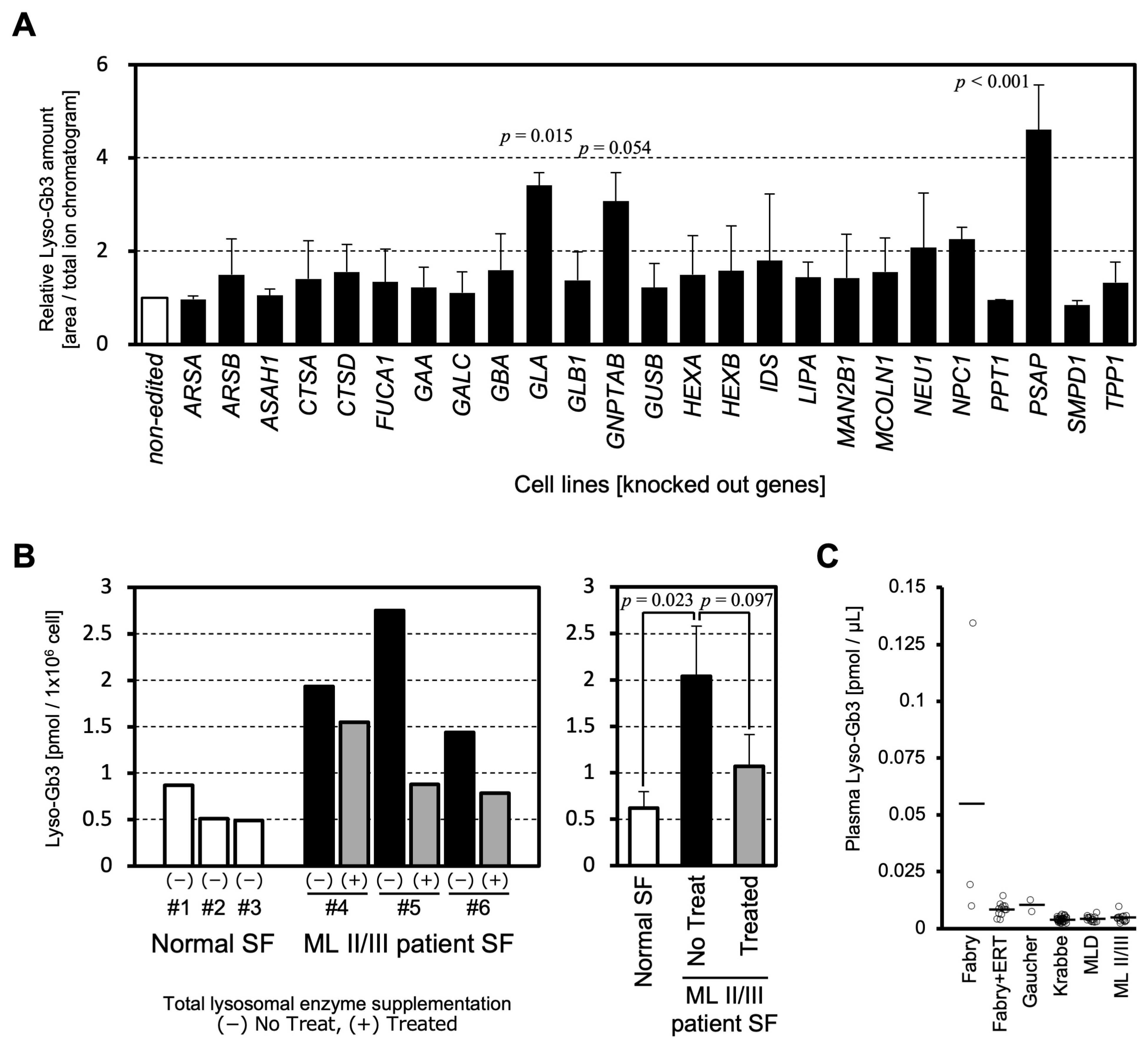
2. Results and Discussion

To systematically compare whether lysosomal dysfunction in various LSDs affects lipid metabolism and leads to Lyso-Gb3 accumulation, we established disease model cells by knockout (KO) of 25 lysosomal enzymes or lysosome-related genes using CRISPR/Cas9 in the HeLa cell line: ARSA KO (metachromatic leukodystrophy), ARSB KO (mucopolysaccharidosis VI), ASAH1 KO (Farber disease), CTSA KO (galactosialidosis), CTSD KO (neuronal ceroid lipofuscinosis 10), FUCA1 KO (fucosidosis), GAA KO (Pompe disease), GALC KO (Krabbe disease), GBA KO (Gaucher disease), GLA KO (Fabry disease), GLB1 KO (GM1 gangliosidosis), GNPTAB KO (ML II/III), GUSB KO (mucopolysaccharidosis VII), HEXA KO (Tay-Sachs disease), HEXB KO (Sandhoff disease), IDS KO (mucopolysaccharidosis II), LIPA KO (LAL-D), MAN2B1 KO (α-mannosidosis), MCOLN1 KO (mucolipidosis IV), NEU1 KO (sialidosis), NPC1 KO (Niemann-Pick disease type C), PPT1 KO (neuronal ceroid lipofuscinosis 1), PSAP KO (combined saposin deficiency), SMPD1 KO (Niemann-Pick disease type A/B), and TPP1 KO (neuronal ceroid lipofuscinosis 2). We collected cell pellets and quantified Lyso-Gb3 in the cells by LC-MS/MS. The results showed that Lyso-Gb3 levels were more than three times higher than those in non-edited cells in three cell lines, including GLA KO, GNPTAB KO, and PSAP KO cells. These cells showed a statistically significant or a tendency to increase in Lyso-Gb3 compared to non-edited cells. NEU1 KO and NPC1 KO cells showed approximately two-fold increases, and the other cell lines showed no obvious increase in Lyso-Gb3 compared to non-edited cells (Figure 1A).
Lyso-Gb3 has already been established as a useful clinical biomarker for Fabry disease [7,8,9]. Therefore, the detection of increased Lyso-Gb3 in the GLA KO cells demonstrates the validity of the model cell and the measuring system.
Prosaposin, encoded by the PSAP gene, is the precursor of saposins A, B, C, and D, which are produced by post-translational cleavage [11,12]. Deficiency of PSAP causes combined saposin deficiency, which is a fatal infantile storage disorder with hepatosplenomegaly and severe neurologic symptoms [12,13]. Each saposin is a co-factor for the hydrolytic reaction of lysosomal enzymes. Because SapB supports the degradative functions of α-galactosidase A (encoded by GLA), it is conceivable that the loss of prosaposin caused the intracellular accumulation of Lyso-Gb3 [14].
ML II/III is caused by a biallelic mutation in GNPTAB [15,16,17], leading to a defect of GlcNAc 1-phosphotransferase that works for the addition of the M6P residues on lysosomal enzymes. M6P residues are necessary for the targeting of lysosomal enzymes to lysosomes [18]. Since α-galactosidase A (encoded by GLA) and lysosomal acid lipase (encoded by LIPA) are also transported to lysosomes via the M6P-dependent pathway, M6P-modified enzymes are used in enzyme replacement therapy for Fabry and LAL-D. It is speculated that Lyso-Gb3 accumulation in ML II cells is caused by a combined effect of a lack of lysosomal enzymes and the disruption of acidic environments inside lysosomes [2].
The amounts of Lyso-Gb3 were investigated in ML II/III patient specimens. Lyso-Gb3 was significantly elevated in ML II/III patient skin fibroblasts compared with normal skin fibroblasts (Figure 1B). The cellular accumulation of Lyso-Gb3 showed a tendency to decrease (p = 0.097) after total lysosomal enzyme supplementation (Figure 1B). These results indicate that the levels of Lyso-Gb3 have biomarker properties that change in response to treatment.
Lyso-Gb3 in patient plasma was measured from residual specimens used for the diagnosis of LSDs. Among the diseases targeted in Figure 1A, LSDs, for which multiple measurement data were obtained, are shown in Figure 1C. Although an increase in Lyso-Gb3 was observed in some Fabry disease patients’ plasma, no clear increase in Lyso-Gb3 was observed in other LSDs measured in this study, such as Gaucher, Krabbe, metachromatic leukodystrophy (MLD), and ML II/III. These data are consistent with a previous report [19]. Due to the limited number of samples analyzed, further analysis is awaited to reach a conclusion. Interestingly, Lyso-Gb3 does not appear to leak into the plasma of ML II/III patients (Figure 1C), although cellular accumulation of Lyso-Gb3 is comparable between the Fabry and ML II/III models. The difference in plasma Lyso-Gb3 in patients with Fabry disease and ML II/III has important implications for the pathomechanism of these LSDs.
Recently, measurements of lysosomal enzyme activity in lymphocytes in dried blood spots have been used in newborn screening of LSDs. B lymphocytes in ML II have been found to have an accumulation of storage material and impaired function [20]. Our results suggest that measurements of Lyso-Gb3 in blood cells may be a biomarker for ML II/III, even though Lyso-Gb3 does not change in plasma.

3. Materials and Methods

3.1. Knockout (KO) Cell Lines

Lysosome-related genes were knocked out using CRISPR/Cas9 in HeLa Kyoto, a cell line for which whole genome analysis has been performed [21]. HeLa Kyoto cells were originally established at Kyoto University based on canonical HeLa cells, were provided to EMBL, and are now distributed worldwide without any limitation (RRID: CVCL_1922). Plasmids expressing each CRISPR guide RNA and Cas9 protein were prepared based on px458 (pSpCas9(BB)-2A-GFP (PX458)), which was a gift from Feng Zhang (Addgene plasmid #48138; http://n2t.net/addgene:48138 (accessed on 3 October 2017); RRID: Addgene_48138) [22]. Guide sequences for gene targeting were designed using online tools, either the Benchling CRISPR Guide RNA Design Tool (https://www.benchling.com/crispr/ (accessed on 6 July 2020)) or CRISPRdirect (https://crispr.dbcls.jp/ (accessed on 24 June 2022)) (Table 1). The double-stranded DNA coding guide RNA sequences were prepared by annealing synthesized complementary DNA oligo pairs on a thermal cycler after phosphorylation by the T4 polynucleotide kinase (TaKaRa Bio, Shiga, Japan) and then cloned at the BbsI site of the px458 plasmid. The targeting plasmid was transfected into HeLa Kyoto cells with the Effectene Transfection reagent (Qiagen, Hilden, Germany). After 48 h of transfection, GFP-positive cells were sorted into 96-well plates as a single cell culture by FACSAria III™ Cell Sorter (BD Biosciences, CA, USA) and expanded. Genomic DNA from each clone was prepared using the QuickExtract™ DNA Extraction Solution (Lucigen, Oxford, UK). The genomic sequence of each targeted site was PCR-amplified with gene-specific primer sets, and genome editing was analyzed by Sanger sequencing. KO clones were characterized by phenotyping either lysosomal enzyme activities, substrate accumulation, or autophagic flux and confirmed by genotyping. The established LSD model cell lines were maintained in Dulbecco’s Modified Eagle Medium (DMEM) (Merck, MA, USA) supplemented with 10% fetal bovine serum FBS, 100 units/mL penicillin, and 100 µg/mL streptomycin (Nacalai-Tesque, Kyoto, Japan). The cell pellets for LC-MS/MS analysis were prepared by harvesting cells from multiple dishes in subconfluent conditions with trypsin–EDTA digestion, followed by several PBS(-) washes.

3.2. Patient Specimens

ML II/III patient skin fibroblasts (SFs) and LSD patient plasma were obtained from residual specimens of diagnostic research after obtaining informed consent. The use of the patients’ specimens was approved by the institutional ethics review board (Kawasaki Medical School: approval Number: 5889). Patient SF lines were established from a 5 mm square of buttock skin piece from each patient. The tissue was excised by punching and then shredded with a clean scalpel and scissors. The disrupted skin tissue was directly transferred and cultured in the AmnioMAX-II™ medium (Thermo Fisher, Waltham, MA, USA). After weeks of expansion, grown colonies were dispersed by trypsin–EDTA digestion and thereafter cultured in the normal DMEM as described above in the HeLa Kyoto part. Genotypes of ML II/III patient SFs were #4 c.310C>T (p.Q104X)/c.2522delA (p.K848fs), #5, and #6 c.1120T>C (p.F374L)/c.3565C>T (p.R1189X). For healthy controls, three commercially available normal SFs were purchased from Kurabo (Kurashiki, Japan), Thermo Fisher Scientific Inc. (Waltham, MA, USA), and Lonza (Basel, Switzerland).
In vitro therapeutic intervention for ML II/III SFs via total lysosomal enzyme supplementation was performed as described before [2]. In short, the normal SF was incubated with ammonium chloride, and M6P-tagged lysosomal enzymes exhaled in the culture supernatant were collected. Ammonium chloride was removed using a molecular weight cut-off filter, and the lysosomal enzyme mixture was purified. ML II/III patient SFs were treated with a conditioned medium containing the M6P-tagged lysosomal enzyme mixture for 72 h to replenish lysosomal enzymes.

3.3. LC-MS/MS Analysis for Lyso-Gb3

The internal standard reagent was purchased from Avanti Polar Lipid (Alabaster, CA, USA). Methanol, isopropanol, and chloroform of ultra-performance liquid chromatography (UPLC)/MS quality were obtained from FUJIFILM Wako Pure Chemical Corporation (Osaka, Japan). Ultrapure water was obtained from a Milli-Q water system (Millipore, Billerica, MA, USA). Briefly, cell lines were mixed with 1.0 mL of methanol containing the internal standards. The samples were briefly sonicated and incubated on ice for 0.5 h. The samples were then centrifuged for 3 min at 10,000× g. Liquid phases were collected in measuring vials. LC-MS/MS analysis was performed using the Xevo TQ-XS mass spectrometer with an ACQUITY UPLC H-Class system (Waters). The lipids were separated on a Waters X-Bridge C18 column (3.5 mm, 150 mm × 1.0 mm internal diameter) at 40 °C using a gradient solvent system as follows: mobile phase A was isopropanol/methanol/water (5/1/4 v/v/v) supplemented with 5 mM ammonium formate and 0.05% ammonium hydroxide (28% in water); mobile phase B was isopropanol supplemented with 5 mM ammonium formate and 0.05% ammonium hydroxide (28% in water) with a flow rate of 80 mL/min. Lyso-Gb3 metabolites were measured using multiple reaction monitoring (MRM) in the positive ion mode. The peak areas of the individual species were normalized to those of internal/surrogate standards, which were added to the samples before lipid extraction. Raw LC-MS/MS data were processed using analytical software (MassLynx 4.2; Waters). The quantification and annotation methods used in this study correspond to “absolute quantification Level 2” and the “Fatty Acyl/Alkyl Level“ defined by the Lipidomics Standard initiative, respectively [23].

Author Contributions

S.T.: Investigation, writing—original draft, and writing—review and editing. H.N.: Investigation, validation, visualization, and writing—review and editing. T.S.: Investigation and resources. N.S.: Investigation, resources, and supervision. T.O.: Conceptualization, funding acquisition, methodology, project administration, supervision, validation, visualization, writing—original draft, and writing—review and editing. All authors have read and agreed to the published version of the manuscript.

Funding

This research was supported by the JSPS KAKENHI grant (JP17H05088), the AMED grant (JP23ym0126810, JP24ym0126810 via Okayama University), and the Sanofi LSD research grant (2022) via the Japanese Society for Inherited Metabolic Diseases to T.O.

Institutional Review Board Statement

The use of the patients’ specimens was approved by the institutional ethics review board (Kawasaki Medical School: approval Number: 5889, approved on 20 December 2022).

Informed Consent Statement

This study uses existing human materials (i.e., residual materials used for diagnostic purposes). These patient samples do not contain any personal information. No additional human materials were obtained for the purposes of this study. Written informed consent was obtained for the diagnosis of the disease, but the residual samples used in this study are completely anonymized, and it is impossible to obtain consent to participate in this study. All procedures are conducted in accordance with Japanese ethical guidelines and based on the principles of the Declaration of Helsinki.

Data Availability Statement

The datasets generated and/or analyzed during the current study are not publicly available but are available from the corresponding author upon reasonable request.

Acknowledgments

We thank Yukiko Kawakami (Kawasaki Medical School) and Keiko Tsujimoto (University of Osaka) for their technical assistance.

Conflicts of Interest

H.N. was employed by the company Lipidome Lab Co., Ltd. The remaining authors declare that the research was conducted in the absence of any commercial or financial relationships that could be construed as potential conflicts of interest.

Abbreviations

The following abbreviations are used in this manuscript:
LSDlysosomal storage disease
Lyso-Gb3globotriaosylsphingosine
ML II/IIImucolipidosis II/III
LAL-Dlysosomal acid lipase deficiency
M6Pmannose 6-phosphate
Gb3globotriaosylceramide
KOknockout
SFskin fibroblast
ERTenzyme replacement therapy

References

  1. Platt, F.M.; d’Azzo, A.; Davidson, B.L.; Neufeld, E.F.; Tifft, C.J. Lysosomal storage diseases. Nat. Rev. Dis. Primers 2018, 4, 27. [Google Scholar] [CrossRef] [PubMed]
  2. Otomo, T.; Higaki, K.; Nanba, E.; Ozono, K.; Sakai, N. Lysosomal storage causes cellular dysfunction in mucolipidosis II skin fibroblasts. J. Biol. Chem. 2011, 286, 35283–35290. [Google Scholar] [CrossRef] [PubMed]
  3. Moriwaki, T.; Terawaki, S.; Otomo, T. Impaired lysosomal acidity maintenance in acid lipase-deficient cells leads to defective autophagy. J. Biol. Chem. 2024, 300, 105743. [Google Scholar] [CrossRef] [PubMed]
  4. Kondo, H.; Maksimova, N.; Otomo, T.; Kato, H.; Imai, A.; Asano, Y.; Kobayashi, K.; Nojima, S.; Nakaya, A.; Hamada, Y.; et al. Mutation in VPS33A affects metabolism of glycosaminoglycans: A new type of mucopolysaccharidosis with severe systemic symptoms. Hum. Mol. Genet. 2017, 27, 173–183. [Google Scholar] [CrossRef]
  5. Cyske, Z.; Gaffke, L.; Pierzynowska, K.; Węgrzyn, G. Mucopolysaccharidosis-Plus Syndrome: Is This a Type of Mucopolysaccharidosis or a Separate Kind of Metabolic Disease? Int. J. Mol. Sci. 2024, 25, 9570. [Google Scholar] [CrossRef] [PubMed]
  6. Desnick, R.J.; Ioannou, Y.A.; Eng, C.M. The Online Metabolic and Molecular Bases of Inherited Disease, Part 16: Lysosomal Disorders, α-Galactosidase A Deficiency: Fabry Disease. Available online: https://ommbid.mhmedical.com/content.aspx?bookid=2709&sectionid=225546984 (accessed on 20 June 2025).
  7. Aerts, J.M.; Groener, J.E.; Kuiper, S.; Donker-Koopman, W.E.; Strijland, A.; Ottenhoff, R.; van Roomen, C.; Mirzaian, M.; Wijburg, F.A.; Linthorst, G.E.; et al. Elevated globotriaosylsphingosine is a hallmark of Fabry disease. Proc. Natl. Acad. Sci. USA 2008, 105, 2812–2817. [Google Scholar] [CrossRef] [PubMed]
  8. Maruyama, H.; Miyata, K.; Mikame, M.; Taguchi, A.; Guili, C.; Shimura, M.; Murayama, K.; Inoue, T.; Yamamoto, S.; Sugimura, K.; et al. Effectiveness of plasma lyso-Gb3 as a biomarker for selecting high-risk patients with Fabry disease from multispecialty clinics for genetic analysis. Genet. Med. 2018, 21, 44–52. [Google Scholar] [CrossRef] [PubMed]
  9. Ramaswami, U.; West, M.L.; Tylee, K.; Castillon, G.; Braun, A.; Ren, M.; Doobaree, I.U.; Howitt, H.; Nowak, A. The use and performance of lyso-Gb3 for the diagnosis and monitoring of Fabry disease: A systematic literature review. Mol. Genet. Metab. 2025, 145, 109110. [Google Scholar] [CrossRef] [PubMed]
  10. Sanchez-Niño, M.D.; Sanz, A.B.; Carrasco, S.; Saleem, M.A.; Mathieson, P.W.; Valdivielso, J.M.; Ruiz-Ortega, M.; Egido, J.; Ortiz, A. Globotriaosylsphingosine actions on human glomerular podocytes: Implications for Fabry nephropathy. Nephrol. Dial. Transplant. 2011, 26, 1797–1802. [Google Scholar] [CrossRef] [PubMed]
  11. Kishimoto, Y.; Hiraiwa, M.; O’Brien, J.S. Saposins: Structure, function, distribution, and molecular genetics. J. Lipid Res. 1992, 33, 1255–1267. [Google Scholar] [CrossRef] [PubMed]
  12. Sandhoff, K.; Kolter, T.; Harzer, K.; Schepers, U.; Remmel, N. The Online Metabolic and Molecular Bases of Inherited Disease, Part 16: Lysosomal Disorders, Sphingolipid Activator Proteins. Available online: https://ommbid.mhmedical.com/content.aspx?bookid=2709&sectionid=225543732 (accessed on 20 June 2025).
  13. Hulková, H.; Cervenková, M.; Ledvinová, J.; Tochácková, M.; Hrebícek, M.; Poupetová, H.; Befekadu, A.; Berná, L.; Paton, B.C.; Harzer, K.; et al. A novel mutation in the coding region of the prosaposin gene leads to a complete deficiency of prosaposin and saposins, and is associated with a complex sphingolipidosis dominated by lactosylceramide accumulation. Hum. Mol. Genet. 2001, 10, 927–940. [Google Scholar] [CrossRef] [PubMed]
  14. Eckhardt, M. Pathology and current treatment of neurodegenerative sphingolipidoses. Neuromolecular Med. 2011, 12, 362–382. [Google Scholar] [CrossRef] [PubMed]
  15. Tiede, S.; Storch, S.; Lübke, T.; Henrissat, B.; Bargal, R.; Raas-Rothschild, A.; Braulke, T. Mucolipidosis II is caused by mutations in GNPTA encoding the alpha/beta GlcNAc-1-phosphotransferase. Nat. Med. 2005, 11, 1109–1112. [Google Scholar] [CrossRef] [PubMed]
  16. Khan, S.A.; Tomatsu, S.C. Mucolipidoses Overview: Past, Present, and Future. Int. J. Mol. Sci. 2020, 21, 6812. [Google Scholar] [CrossRef] [PubMed]
  17. Braulke, T.; Raas-Rothschild, A.; Kornfeld, S. The Online Metabolic and Molecular Bases of Inherited Disease, Part 16: Lysosomal Disorders, I-Cell Disease and Pseudo-Hurler Polydystrophy: Disorders of Lysosomal Enzyme Phosphorylation and Localization. Available online: https://ommbid.mhmedical.com/content.aspx?bookid=2709&sectionid=225544648 (accessed on 20 June 2025).
  18. Kornfeld, S. Trafficking of lysosomal enzymes. FASEB J. 1987, 1, 462–468. [Google Scholar] [CrossRef] [PubMed]
  19. Spiewak, J.; Doykov, I.; Papandreou, A.; Hällqvist, J.; Mills, P.; Clayton, P.T.; Gissen, P.; Mills, K.; Heywood, W.E. New Perspectives in Dried Blood Spot Biomarkers for Lysosomal Storage Diseases. Int. J. Mol. Sci. 2023, 24, 10177. [Google Scholar] [CrossRef] [PubMed]
  20. Otomo, T.; Schweizer, M.; Kollmann, K.; Schumacher, V.; Muschol, N.; Tolosa, E.; Mittrücker, H.W.; Braulke, T. Mannose 6 phosphorylation of lysosomal enzymes controls B cell functions. J. Cell Biol. 2015, 208, 171–180. [Google Scholar] [CrossRef] [PubMed]
  21. Landry, J.J.; Pyl, P.T.; Rausch, T.; Zichner, T.; Tekkedil, M.M.; Stütz, A.M.; Jauch, A.; Aiyar, R.S.; Pau, G.; Delhomme, N.; et al. The genomic and transcriptomic landscape of a HeLa cell line. G3 2014, 3, 1213–1224. [Google Scholar] [CrossRef] [PubMed]
  22. Ran, F.A.; Hsu, P.D.; Wright, J.; Agarwala, V.; Scott, D.A.; Zhang, F. Genome engineering using the CRISPR-Cas9 system. Nat. Protoc. 2013, 8, 2281–2308. [Google Scholar] [CrossRef] [PubMed]
  23. Lipidomics Standards Initiative Consortium. Lipidomics needs more standardization. Nat. Metab. 2019, 1, 745–747. [Google Scholar] [CrossRef] [PubMed]
Figure 1. Lyso-Gb3 in LSDs. (A) Relative amount of Lyso-Gb3 in LSD model cells. Values “area/total ion chromatogram” were obtained by LC-MS/MS in each cell line. Labels at the bottom indicate genes knocked out in HeLa cells, except for the non-edited cell line. Data from three different genotype clones (Table 1). Values are normalized to non-edited HeLa cells, and the mean and SDs are shown. The absolute quantification values of Lyso-Gb3 from the non-edited and GNPTAB KO cells were measured in a separate experiment to be 0.30 and 0.70 pmol/106 cells, respectively. Statistical significances between all KO cell lines were tested using one-way ANOVA, followed by Dunnett’s post hoc test for comparison with the control group. Only p-values less than 0.1 are shown on top of the bar graph. (B) Lyso-Gb3 amount in skin fibroblasts (SFs). Three normal SFs (#1–3) and three ML II/III patient SFs (#4–6) were analyzed for Lyso-Gb3. Parts of the ML II/III skin fibroblasts were treated with a total lysosomal enzyme mixture. The right panel represents the mean values and SDs for the normal and patient SFs (the no treat group and the treated group) from the left panel. Statistical analysis was performed using one-way ANOVA followed by Tukey’s post hoc test among the three groups of samples. p-values less than 0.1 are shown. (C) Lyso-Gb3 amount in patient plasma. Each data point is indicated by a small circle. The horizontal bars represent the means for each data group. Fabry patients without enzyme replacement therapy (ERT) (n = 3); Fabry patients undergoing ERT (n = 12); Gaucher patients (n = 2); Krabbe patients (n = 33); metachromatic leukodystrophy (MLD) patients (n = 15); and ML II/III patients (n = 14).
Ijms 26 06275 g001
Table 1. List of CRISPR guide RNA sequences and variants for knockout HeLa cell lines used in this study.
GeneCRISPR Guide (5′ to 3′)Clone #1Clone #2Clone #3
ARSAGGGAGTCCCCAAATGGCCCGdelC/del7bpdel13bp/insGdel16bp homo
ARSBGCTGCGTGTAGTAGTTGTCCdel2/del11bpdelC/delCACCinsC/del8bp
ASAH1TCAAGATTTATGGTGTACCAinsT homodel7bp homoinsT/delG
CTSACTTTGAGGTAGCCGGAGTACdel11 homodel11bp/del23bpdelTC/delCTCC
CTSDGTCCATCCGCCGGACCATGTdel10bp homodelCA/del40bpdelT homo
FUCA1GAACTTGGCTTCGTCGAACCdelT/insGdelTC/del8bpinsT homo
GAAAGGGATGTAGCAACAGCCGCdelGC homoCTGT > GCTGC homoinsG/delCTG
GALCGTAATTTACTAGAAGTCGGGinsT/delCinsG homoinsGA homo
GBAAGACCAATGGAGCGGTGAAT TGTGGTGAGTACTGTTGGCG *del4bp/del5bpinsA/insCCinsC/delA
GLAGCTAGCTGGCGAATCCCATGdelG homodelG homodelG/insGG
GLB1CCCGTGTGCCCCGCTTCTACdel8/del19bpinsAT/del19bpdelACT/del26bp
GNPTABACACGTAGAGCCCATACCTGdelGT/del8bpdelG homodelAG/del12bp
GUSBGTGGTACCGGCGGCCGCTGTdelGC homoinsT/insGinsC homo
HEXAGTTGTCTCTGTAGTCACACCdelCC/del13bpinsC/delTCACdelAC/del5bp
HEXBGAGGGGCCCGCCGTGGAATTdelT/del8bpdelT/insTdelT homo
IDSGAACGTTCTTCTCATCATCGinsA homodelT/delCdelA/delTC
LIPATCCCATGAGGAATTCGGTTAdelC homodel13bp homoinsC/del5bp
MAN2B1ACGTAAATGAAGCGACGGGTdelCC homoinsCG/del5delC homo
MCOLN1AGTATTTGAGACGACGGCGAdelC homodelC homodelC/delGC
NEU1CCAAGTTCATCGCCCTGCGGdel5 homodelG/delGdelC homo
NPC1GCGCTGGACACAGTAGCAGCdel12bp homoinsT/del8bpdel7bp homo
PPT1GCAGCAAGGCTACAATGCTAdelTG/del5bpdelTG homodel5bp/del8bp
PSAPTGAAGACGGCGTCCGACTGCinsT/delACdel7bp homodelT/ del5bp
SMPD1GTTCTTTGGCCACACTCATGinsC/del14bpinsC homoinsT/del10bp
TPP1TGTGGAAAGACTCTCGGAGCdelG/delGGdelGG homodelGG/delTCGG
* Two different guides were used.
Disclaimer/Publisher’s Note: The statements, opinions and data contained in all publications are solely those of the individual author(s) and contributor(s) and not of MDPI and/or the editor(s). MDPI and/or the editor(s) disclaim responsibility for any injury to people or property resulting from any ideas, methods, instructions or products referred to in the content.

