Review

Primary Plasma Cell Leukemia: Recent Advances in Molecular Understanding and Treatment Approaches

1
Division of Hematology, Department of Internal Medicine, Aichi Medical University, Nagakute 480-1195, Japan
2
Department of Biochemistry, Aichi Medical University, Nagakute 480-1195, Japan
3
Department of Food and Nutritional Environment, College of Human Life and Environment, Kinjo Gakuin University, Nagoya 463-8521, Japan
*
Author to whom correspondence should be addressed.
Int. J. Mol. Sci. 2025, 26(13), 6166; https://doi.org/10.3390/ijms26136166
Submission received: 12 May 2025 / Revised: 20 June 2025 / Accepted: 25 June 2025 / Published: 26 June 2025
(This article belongs to the Special Issue New Advances in Molecular Research in Leukemia)

Abstract

Primary plasma cell leukemia (pPCL) is a rare and aggressive plasma cell dyscrasia. According to revised diagnostic criteria, pPCL is defined by the presence of ≥5% circulating plasma cells (CPCs) in the peripheral blood of patients with newly diagnosed multiple myeloma (NDMM). pPCL is characterized by a distinct cytogenetic profile, including frequent t(11;14), MAF/MAB translocations, 1q gain, and del(17p). While t(11;14) is generally associated with a favorable prognosis, the coexistence of multiple high-risk cytogenetic abnormalities is linked to poorer outcomes. Tandem autologous hematopoietic stem cell transplantation and novel anti-myeloma agents have improved survival in some patients; however, overall prognosis remains poor, particularly in those ineligible for transplantation. Venetoclax and emerging immunotherapies, such as CAR-T cells and bispecific antibodies, show promise and merit clinical trials focused on pPCL-enriched cohorts. Additionally, recent findings associating even minimal CPCs with adverse outcomes in NDMM support broader inclusion criteria in future trials. A deeper understanding of pPCL’s molecular pathology is critical for the development of effective targeted therapies. This article reviews recent advances in the molecular understanding of and treatment strategies for pPCL.

1. Introduction

Plasma cell leukemia (PCL), which is defined as the presence of plasma cells in peripheral blood [1,2], is classified as primary PCL (pPCL), which occurs in patients with newly diagnosed multiple myeloma (NDMM), and secondary PCL, which develops in those with relapsed refractory MM (RRMM) [3,4]. PCL suggests the presence of MM cells with a growth advantage, which can survive without the support of the bone marrow microenvironment. Studies have shown that even low levels of circulating plasma cells (CPCs), defined as <5%, are associated with poor prognosis in patients with NDMM [5,6,7,8,9].
pPCL is very rare, accounting for approximately 1–2% of NDMM cases [10,11]. pPCL progresses rapidly and can lead to early death if left untreated; therefore, immediate diagnosis and treatment are critical in patients with pPCL. The clinical and cytogenetic features of pPCL are distinct from those of NDMM [1,2,12,13,14]. The clinical outcomes of pPCL remain unfavorable despite the introduction of novel anti-MM agents [11,15,16]. However, given its rarity, pPCL is usually excluded from prospective randomized clinical trials for patients with NDMM and the establishment of standard optimal therapeutic regimens has been challenging. Emerging immunotherapeutics—such as chimeric antigen receptor T cells (CAR-Ts), bispecific antibodies (BsAbs), and antibody–drug conjugates (ADCs) targeting B-cell maturation antigen (BCMA) and other markers—have shown high efficacy in patients with heavily pretreated MM [17,18], and are expected to offer therapeutic benefit in pPCL as well. In this review, we summarize the recent advances in the diagnosis, molecular understanding and treatment approaches of pPCL.

2. Diagnostic Criteria and Clinical Features of pPCL

Before the revised diagnostic criteria were updated in 2021 [4], the initial diagnostic criteria for pPCL, which were proposed by Kyle et al. in the 1970s, required the presence of ≥20% CPCs with an absolute CPC count of ≥2 × 10⁹/L in the peripheral blood [1]. In the 2010s, following the development of novel agents for MM, studies reported that patients with a CPC level of 5–19% exhibited poor overall survival (OS) comparable to those observed in patients with ≥20% CPCs [19,20]. The newly proposed criteria for the diagnosis of pPCL, published by the International Myeloma Working Group in 2021, include a lower percentage of CPCs (≥5%) as a threshold [4]. Subsequently, several studies have confirmed that the revised diagnostic criteria are appropriate for the diagnosis of pPCL, in terms of prognostic value [21,22]. However, with more improvements in anti-myeloma treatment, several recent studies have reported that even a CPC level of <5% is an independent poor prognostic factor for NDMM (Figure 1) [5,6,9]. The current diagnostic criterion for pPCL, a cutoff value of 5%, is arbitrary, and recent studies have demonstrated the prognostic value of CPCs as a continuous variable [8,23]. More suitable or nuanced diagnostic criteria that better reflect the biological and clinical spectrum of NDMM with CPCs are required.
CPCs harboring t(11;14) often exhibit a lymphocyte-like appearance in smears and may be difficult to morphologically distinguish; therefore, flow cytometry is recommended for the detection of CPCs [24].
The incidence of pPCL is very low, with an estimated rate of 1% in patients with NDMM [25,26]. The median age at pPCL diagnosis is approximately 60 years, which is younger than that of the patients with NDMM. Severe anemia, thrombocytopenia with platelet counts of <100,000/L, renal insufficiency, hypercalcemia, high lactate dehydrogenase levels, advanced International Staging System stage, and extramedullary disease are more common in patients with pPCL [25,27,28]. Compared with typical NDMM, pPCL progresses rapidly and can lead to early death, requiring immediate diagnosis and intervention with antitumor therapeutics. In patients with pPCL, positron emission tomography/computed tomography (PET/CT) is recommended for the detection of extramedullary lesions, an important cause of early death [29,30].

3. Biologic Features of pPCL

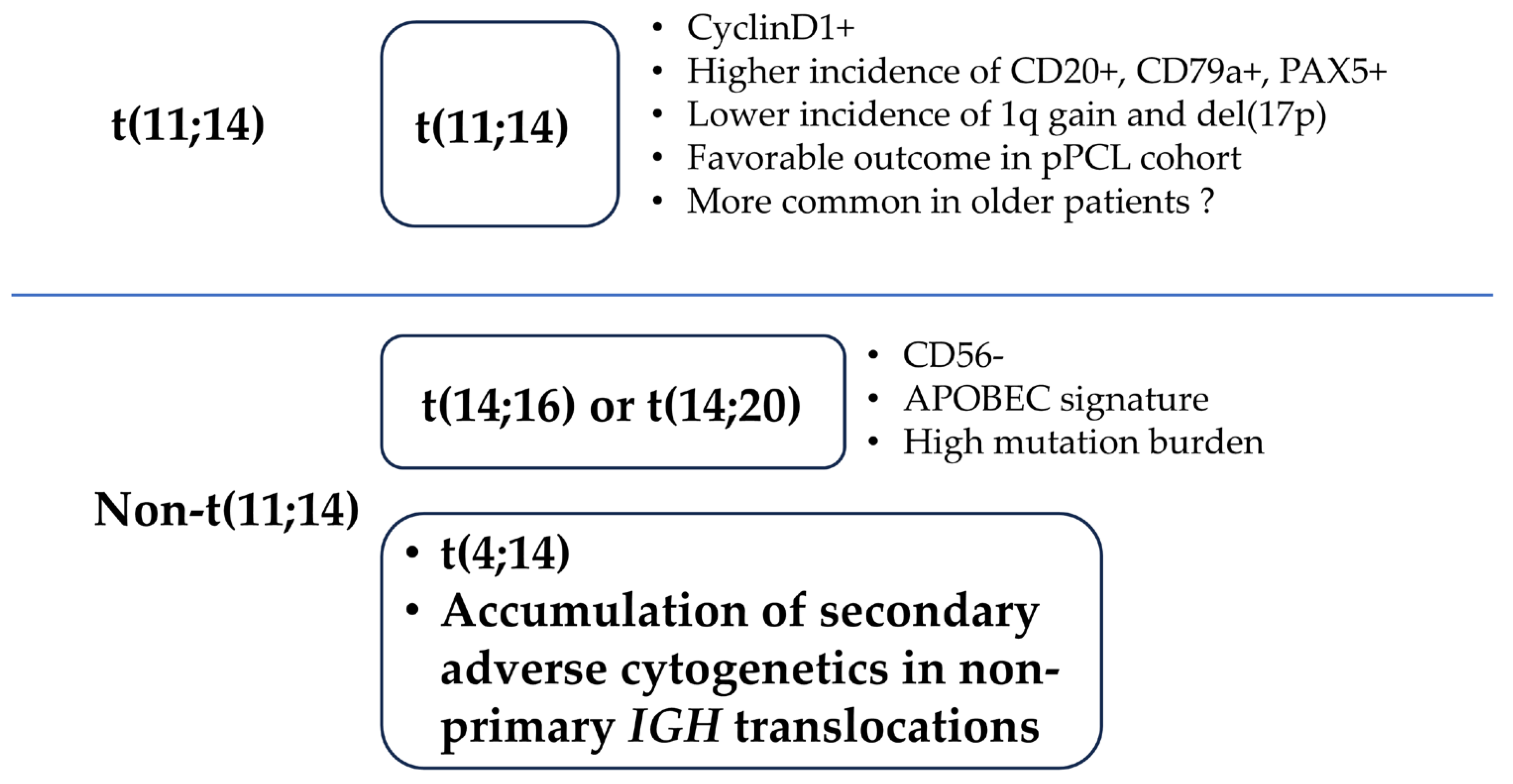
Although pPCL shares genomic abnormalities with MM, the genetic profile, including the cytogenetics, of pPCL is distinct from that of NDMM [3,14]. The incidence of t(11;14), t(14;16), t(14;20), del(1p), del(13q), del(17p), 1q gain, and hypodiploidy are higher in patients with pPCL than in those with NDMM [12,13,27,31,32]. Except for the higher frequency of t(11;14), pPCL has similar cytogenetic features to those of high-risk NDMM (i.e., a high frequency of high-risk cytogenetics and multiple high-risk cytogenetics) [33]. Recently, van de Donk et al. reported that the cytogenetic profile differed between the patients aged ≤65 years and those aged >66 years among the patients with pPCL enrolled in the EMN12/HOVON-129 study [34]. The authors reported that the frequency of t(11;14) was lower whereas the frequencies of t(14;16) and del(17p) were higher in patients with pPCL aged ≤65 years (Table 1) [34]. These findings might suggest that patients are categorized into those who develop pPCL in association with t(11;14), which is more commonly observed in older individuals, and those who develop pPCL in association with high MAF/MAFB expression and/or poor prognostic secondary abnormalities, which are more commonly observed in younger individuals (Figure 2).
Whole-exome sequencing has revealed that patients with pPCL exhibit a highly heterogeneous mutational profile [35]. The number of nonsynonymous mutations is higher in pPCL than in NDMM, which is potentially due to a greater prevalence of the APOBEC mutational signature induced by MAF/MAFB translocations [35]. TP53 mutations, including the bi-allelic inactivation of TP53, are more common in patients with pPCL than in those with NDMM [32,35].
In pPCL, malignant plasma cells typically express plasma cell markers, including CD38 and CD138. Compared to NDMM, pPCL cells more frequently express CD19, CD20, CD27, CD44, and CD45, and less frequently express CD56, CD117, and human leukocyte antigen-DR (HLA-DR) [12,36]. The infrequent expression of CD56 may correlate with the higher incidence of MAF/MAFB translocations in pPCL. The downregulation of adhesion molecules, such as CD44 and CD56, can disrupt the adhesion of plasma cells to the bone marrow stroma, facilitating their spread to extramedullary sites, including the peripheral blood. The lack of CD27 expression may be linked to nuclear factor kappa B-mediated antiapoptotic pathways [37]. The loss of HLA-DR expression in pPCL may reflect immune evasion by the tumor. HLA-DR is essential for presenting antigens to CD4+ T cells, and its absence impairs immune recognition, which might contribute to the aggressive behavior and rapid progression of pPCL.
Paiva et al. reported differences in the immune phenotypes of CPCs and myeloma cells in the bone marrow of the same patients [38]. CPCs constituted a unique subpopulation of clonal myeloma cells in the bone marrow, characterized by the downregulation of integrins (CD11a/CD11c/CD29/CD49d/CD49e), adhesion (CD33/CD56/CD117/CD138), and activation molecules (CD28/CD38/CD81). In addition, this paper reported that CPCs are quiescent but clonogenic and, interestingly, exhibit a fluctuating circadian rhythm.
The gene expression profile of pPCL is also distinct from that of NDMM, including the higher expression of TAGLN2, RUNX4, PHF19, DEPTOR, FAS, FAM72D, and TOX2 and the lower expression of VCAM1, CXCL12, PPBP, CD163, and DKK1 [32,35]. Hofste Op Bruinink et al. reported that pPCL was identified on the basis of a specific tumor transcriptome signature, which was also present in patients with high-risk NDMM, who constitute around 10% of patients, despite not being clinically leukemic [39]. Such patients might be in the pre-stage of pPCL.
pPCL patients with t(11;14) exhibit a lower incidence of 1q gain and del(17p), and higher expression of immature B-cell markers such as PAX5 and CD79A, compared to those without t(11;14), suggesting distinct oncogenic mechanisms [32,40]. In a study of patients with NDMM and CPC levels of 0.105–5%, Xia et al. reported that those with mutations in TP53, BRAF, DNMT3A, and TENT5C, and the genes related to the interleukin 6/Janus kinase/signal transducer and the activator of the transcription 3 (IL-6/JAK/STAT) pathway tended to have higher levels of CPCs [41]. This suggests that pPCL cells can proliferate in an IL-6-independent manner, aided by mutations that promote cell survival and growth.
Patients with pPCL often present with disseminated extramedullary lesions (EMDs) [30,42], which appear to be caused by the hematogenous metastasis of MM cells. On the other hand, some NDMM patients have tumorous EMDs without clear leukemic status. Compared with typical NDMM, both pPCL and NDMM with tumorous EMD show a poor prognosis [42,43], but pPCL generally shows a poorer prognosis and is more aggressive, with a higher tumor burden and more rapid progression than NDMM with tumorous EMDs. This may be related to the higher chromosomal instability and higher mutation load in pPCL, which result in more aggressive tumors. Garcés et al. reported that CPCs were detected in the blood of all MM patients using advanced detection techniques (next-generation flow, NGF) (n = 53), showing high concordance with bone marrow myeloma cells in copy number changes at chromosomal arm levels (>95%) but low in translocations (39%). High-risk cytogenetic abnormalities and >82% of mutations were also captured. About 22% of CPCs originated from different marrow or extramedullary sites, indicating spatial heterogeneity [43].
Del(17p) is known to be frequently observed both in pPCL and tumorous EMD, but molecular abnormalities specific to pPCL that are not observed in tumorous EMD are not well characterized. Rojas et al. performed transcriptome analysis of pPCL and NDMM, both of which have del(17p), and reported that the abnormal expression of spliceosome components was significantly observed in pPCL with del(17p) compared to NDMM with del(17p). This suggests that the intracellular mRNA processing pathway including RNA splicing machinery is different between pPCL with del(17p) and NDMM with del(17p) [44].
The clinical features and molecular pathology of pPCL overlap those of ultrahigh-risk NDMM, but the frequency of patients with t(11;14), which is a favorable marker in NDMM, is clearly higher in pPCL [33,45]. This suggests once again that pPCL could be classified into two types: one that develops in association with t(11;14) and another that is a leukemic type of ultrahigh-risk NDMM (Figure 2).
Research on the molecular pathology of pPCL supports the development of targeted therapies. As pPCL is an aggressive tumor with highly complex and multifaced molecular abnormalities, targeted drugs may be more effective when used in combination with other anti-myeloma agents rather than as monotherapy. In the future, progress in basic research directly related to pPCL treatment is expected.

4. Treatment and Perspectives

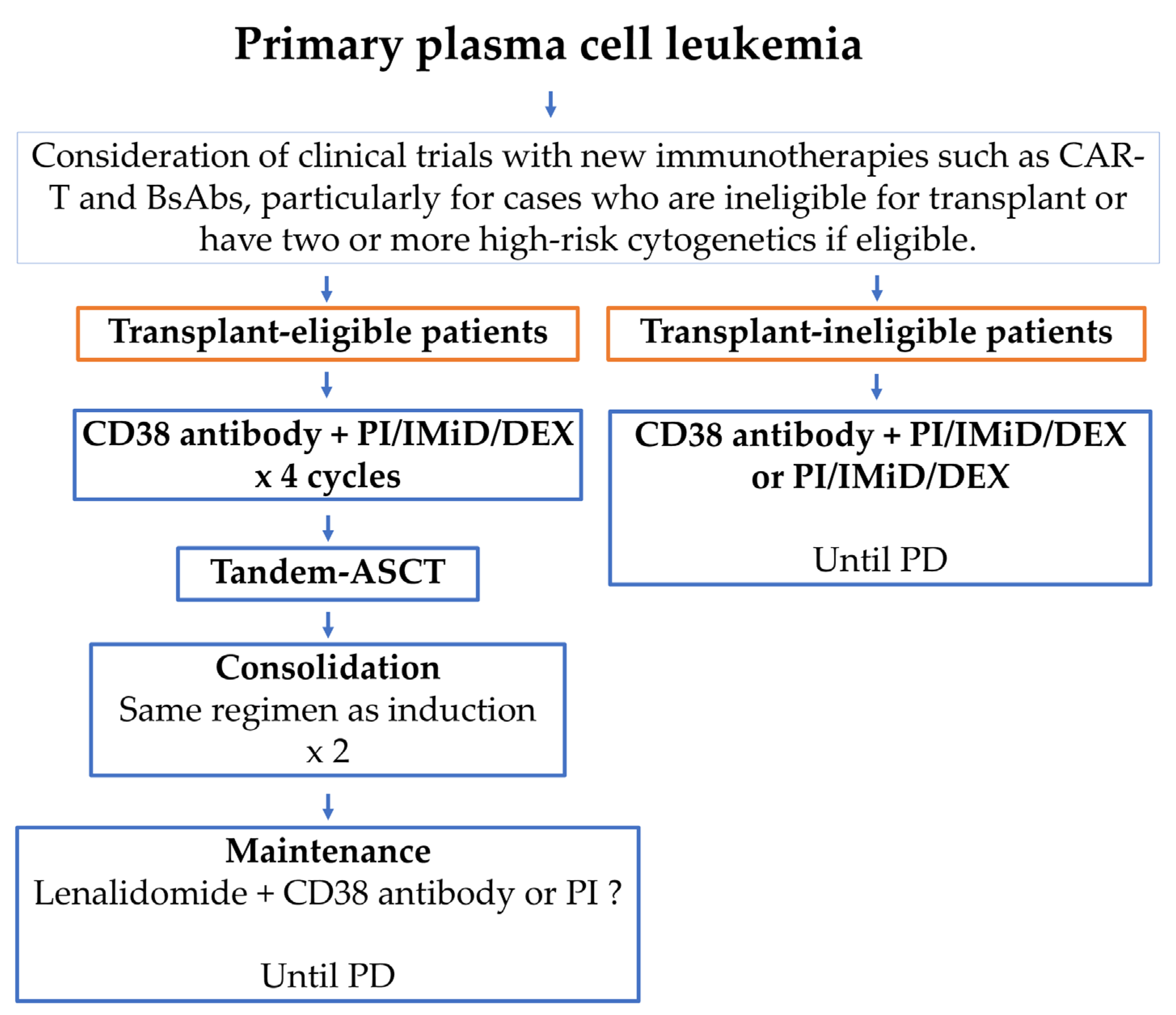
Historically, pPCL has been associated with high early mortality and extremely poor OS. Conventional MM chemotherapy in pPCL has shown a response rate of less than 50% and a 5-year OS of less than 10% [46]. Novel anti-MM agents—such as CD38 antibodies, proteasome inhibitors (PIs), and immunomodulatory drugs (IMiDs)—have improved response rates, the quality of response, progression-free survival (PFS), and OS in pPCL compared to conventional chemotherapy, although not to the extent seen in NDMM [7,22,47,48,49]. Currently, CD38 antibody-based quadruplet regimens—including a CD38 antibody, PI, IMiD, and dexamethasone (DEX)—are recommended as the optimal frontline therapy for patients with pPCL (Figure 3) [22,30]. Autologous hematopoietic stem cell transplantation (ASCT), especially tandem ASCT followed by consolidation and maintenance therapy, has been shown to reduce early relapse and improve survival, and is therefore recommended for eligible patients [3]. There is no established post-transplant treatment regimen for CD38 antibody-based quadruple therapy; however, since treatment failure in pPCL can lead directly to death, consolidation therapy is recommended to be the same as the induction therapy that has been demonstrated to be effective [30]. This will help deepen the response and reduce the risk of early recurrence. Maintenance therapy will be continued for a long period of time until progression or intolerance, and will consist of a two-drug combination therapy based on lenalidomide excluding steroids [30].
Tandem ASCT may be particularly beneficial for pPCL patients who achieve complete remission (CR) before transplantation [50,51]. Regarding allogeneic SCT (allo-SCT), myeloablative conditioning allo-SCT has resulted in long-term survival with a plateau phase in approximately 20% of patients; however, this approach carries a high risk of treatment-related mortality (TRM) [52]. In contrast, reduced-intensity conditioning (RIC) allo-SCT is associated with lower TRM but a higher relapse rate in pPCL [52]. Tandem ASCT/RIC-allo-SCT may be an option when a graft-versus-myeloma effect is desired [52]. The disease status prior to first transplantation may guide the choice of transplant modality: patients achieving CR may benefit from tandem ASCT, while those not achieving CR may benefit more from tandem ASCT/RIC-allo-SCT [51]. The role of SCT in the era of CD38 antibody-based quadruplets and novel immunotherapies (including CAR-T, BsAbs and ADCs) has not been fully defined in pPCL. Although transplantation can improve survival, many pPCL patients are ineligible due to age, comorbidities, or delayed diagnosis and ineffective initial treatment—even among younger patients. Therefore, early detection and effective initial therapy are critical to enabling transplantation and improving outcomes.
As in NDMM, achieving minimal residual disease (MRD) negativity—rather than merely CR—may be a more meaningful prognostic indicator in pPCL [6,9,34]. In the EMN12/HOVON-129 study, 28% of younger patients (10 out of 36) with pPCL (CPCs ≥ 20%) achieved MRD negativity [34]. These MRD-negative patients were overrepresented among long-term survivors and had superior outcomes. This highlights MRD negativity as a strong prognostic indicator even in high-risk diseases like pPCL. In addition, it has been reported that achieving MRD negativity improved PFS in transplant-eligible NDMM patients at any level of CPCs detectable by flow cytometry [6,7]. Sustained MRD negativity may serve as a strong surrogate marker, as pPCL cells tend to proliferate more rapidly than those of typical NDMM. The optimal timing for MRD assessment in pPCL has not been established, but considering the aggressiveness of pPCL, it is recommended to perform MRD assessment multiple times after induction therapy, post-ASCT, after consolidation therapy, and during maintenance therapy. In pPCL, MRD positivity may be associated with early clinical relapse, and therefore it is recommended to start treatment as early as possible. In particular, in cases with MRD positivity after ASCT, the induction or consolidation therapy may have been insufficient. Therefore, it may be appropriate to promptly initiate salvage treatment with CAR-T cells or bispecific antibodies, which have distinct mechanisms of action from the prior therapy.
Due to its rarity and frequent exclusion from large NDMM trials, most available data on pPCL treatment outcomes are derived from small retrospective studies. To date, only three prospective phase II trials have been conducted for newly diagnosed pPCL (all patients had ≥20% CPCs) (Table 2) [46]. The Gruppo Italiano Malattle Ematologiche d’Adulto (GIMEMA) trial investigated lenalidomide and dexamethasone in 23 patients with pPCL, including 15 transplant-eligible (TE) and 8 transplant-ineligible (TIE) patients [46]. With a median follow-up of 34 months, the median PFS and OS were 27 months and not-reached, respectively, in TE patients and 2 and 12 months, respectively, in TIE patients. Among the 15 TE patients, 9 underwent SCT (single ASCT [n = 4], double ASCT [n = 4], ASCT/RIC-allo [n = 1]). Survival was better among those who received transplantation.
The Intergroupe Francophone du Myélome (IFM) trial investigated the efficacy of bortezomib-based induction therapy followed by SCT in patients with newly diagnosed pPCL. The treatment regimen included four alternating cycles of PAD (bortezomib + doxorubicin + dexamethasone) and VCD (bortezomib + cyclophosphamide + dexamethasone), followed by ASCT. Eligible patients then received RIC-allo-SCT (=tandem auto/RIC-allo), while the remaining patients received a second ASCT (=tandem auto/auto) along with consolidation therapy using VRD (bortezomib + lenalidomide + dexamethasone) and one year of lenalidomide maintenance [53]. Forty transplant-eligible (TE) patients (aged 70 years or younger) were enrolled. One patient died from septic shock before the start of induction therapy, and 35 patients completed the planned four cycles of PAD/VCD induction. Following induction, 27 patients (69%) achieved stable disease or better, including 14 patients (36%) who achieved VGPR or better, and 25 patients proceeded to the first ASCT. Of the 23 patients who achieved VGPR or better after the first ASCT, 16 patients (70%)—all younger than 66 years and with an available donor—underwent RIC-allo-SCT (=tandem auto/RIC-allo), while the remaining 7 underwent a second ASCT (=tandem auto/auto) followed by post-transplant therapy. With a median follow-up of 28 months, the median OS and PFS were 36 and 15 months, respectively, in the overall cohort. From the date of the second SCT, the median OS and PFS were 36 and 17 months, respectively, in the RIC-allo-SCT group (=tandem auto/RIC-allo), while the median OS and PFS were not reached in the ASCT plus post-transplant therapy group (=tandem auto/auto). These results indicate that bortezomib-based induction followed by SCT provides a high response rate and improved PFS compared to historical cohorts, and that tandem auto/auto with VRD/lenalidomide may offer superior outcomes over tandem auto/RIC-allo.
The EMN12/HOVON-129 trial included 61 patients with pPCL who were treated with a regimen including carfilzomib and lenalidomide in combination with SCT, if eligible [34]. Patients aged 18–65 years (younger patients) and those aged 66 years or older (older patients) were treated in age-specific cohorts. Patients aged 65 years or younger received four cycles of induction therapy with carfilzomib, lenalidomide, and dexamethasone (KRd), followed by either tandem ASCT (=tandem auto/auto) or ASCT/RIC-allo-SCT (=tandem auto/RIC-allo). Patients in the tandem ASCT group received four cycles of consolidation therapy with KRd and maintenance therapy with KR until disease progression or unacceptable toxicity. Patients eligible for allo-SCT with an available donor underwent a single RIC-allo-SCT (=tandem auto/RIC-allo), followed by carfilzomib maintenance and KR until progression or undue toxicity. Patients aged 66 years or older received eight cycles of KRd induction therapy, followed by KR maintenance. In the younger cohort, the ORR, ≥VGPR, and CR rates were 86%, 83%, and 50%, respectively. Among the 20 evaluable patients, 16 (80%) achieved MRD negativity. At a median follow-up of 43 months, the median PFS and OS were 15 and 28 months, respectively; outcomes were comparable between the tandem ASCT (=tandem auto/auto) and ASCT/RIC-allo-SCT (=tandem auto/RIC-allo) groups. In the older cohort, the ORR, ≥VGPR, and CR rates were 80%, 68%, and 36%, respectively. Among the eight evaluable patients, five (63%) achieved MRD negativity. With a median follow-up of 32 months, the median PFS and OS were 13 and 24 months, respectively—results superior to those previously reported in transplant-ineligible patients. These three studies suggest that novel anti-myeloma drugs (particularly PI/IMiD/DEX triplets), followed by tandem ASCT with consolidation and maintenance therapy, are more likely to improve outcomes in pPCL. However, the overall prognosis of pPCL remains unsatisfactory compared to that of NDMM.
The rapidly evolving treatment paradigm for MM is expected to drive advances in the management of pPCL. CAR-T therapy and BsAbs therapies have shown high efficacy in heavily treated refractory or relapsed MM, and are promising therapeutic options for pPCL [54]. In daily clinical practice, these therapies have already been shown to be highly effective in refractory or relapsed pPCL. Several case studies have reported the efficacy of CAR-T and BsAbs in refractory or relapsed pPCL [55,56]. Ongoing phase II clinical trials are currently evaluating the efficacy and safety of BCMA-targeted CAR-T therapy as a front-line treatment for newly diagnosed pPCL (NCT05979363 and NCT05870917) (Table 3). Xu et al. reported at ASH 2024 that all four patients who completed the full “sandwich” regimen—comprising CAR-T1/ASCT/CAR-T2/R-maintenance—for transplant-eligible, newly diagnosed pPCL achieved MRD-negative complete responses (NCT05870917) [57]. Currently, there are no clinical trials evaluating BsAbs in the front-line setting for pPCL. In CAR-T cell therapy, the interval from apheresis to infusion—referred to as the manufacturing turnaround time—is typically around one month. Given that some pPCL patients are refractory to induction therapy, the earlier use of BsAbs is also being considered. CT071, an anti-GPRC5D CAR-T product with a turnaround time of as little as two days from apheresis to infusion, represents an attractive treatment option for patients with rapidly progressing pPCL (NCT05838131) (Table 3). As pPCL has a high tumor burden and is an extramedullary disease itself, therapies with CAR-T and BsAbs may not improve outcomes as much as they do for MM. However, when used as first-line therapy, these therapies may avoid T-cell exhaustion, immune dysfunction, and a highly immunosuppressive tumor microenvironment, and are therefore expected to be more effective than anticipated. pPCL with t(11;14) has a lower frequency of high-risk cytogenetic abnormalities of NDMM, and is expected to have lower intrinsic tumor aggressiveness, resulting in novel immunotherapies potentially being more effective in these cases.
Venetoclax, a BCL-2 inhibitor, also shows promise in pPCL with t(11;14) [32]. BCL-2 is an anti-apoptotic protein that promotes cancer cell survival, and myeloma cells with t(11;14) are particularly dependent on BCL-2. Venetoclax blocks BCL-2, activates the apoptotic pathway, and selectively eliminates BCL-2-dependent tumor cells. As a result, venetoclax is especially effective in t(11;14)-positive multiple myeloma, often inducing deep and durable responses [58,59]. Venetoclax, either as monotherapy or in combination with daratumumab and other agents, may be a viable treatment option for this subset of pPCL patients [60,61].
Although long-term survival has been achieved in a subset of patients treated with novel anti-MM agents in combination with tandem ASCT followed by continuous post-transplant therapy, the overall prognosis of pPCL remains poor. There is an urgent need to improve outcomes, particularly for patients without t(11;14), with ≥2 high-risk cytogenetic abnormalities, and those who are ineligible for transplantation. Clinical trials evaluating new therapeutics, especially emerging immunotherapies, are essential for improving the prognosis of pPCL.
In summary, future clinical trials should explore the earlier-line integration of CAR-T cells and BsAbs into the CD38-based quadruplet and tandem ASCT strategy for pPCL. Venetoclax trials are also warranted for patients with t(11;14). These studies will help identify novel prognostic markers, support risk-adapted treatment approaches, and guide the development of targeted therapies. MRD-guided strategies for treatment escalation or de-escalation may further advance pPCL management.

5. Conclusions

A rare plasma cell dyscrasia, pPCL is characterized by a distinct genetic profile, rapid progression, and resistance to conventional MM chemotherapies, with a limited response to novel anti-MM drugs compared to NDMM. In pPCL, t(11;14), MAF/MAFB translocations, and the disruption of TP53 are more frequently observed. Patients with t(11;14) and those without it exhibit different molecular features, outcomes, and age distributions. Therefore, it may be possible to develop a new classification system based on the presence or absence of t(11;14) and to establish new treatment strategies, including the use of venetoclax. In pPCL, due to its refractory and aggressive nature, continuous intensified treatment is essential for long-term survival beginning immediately after diagnosis. Currently, CD38-based quadruplet therapy followed by tandem ASCT with consolidation and maintenance therapy is recommended for eligible patients and has provided long-term survival in a subset of the pPCL population. However, many patients with pPCL are not eligible for transplantation due to delayed diagnosis, the inadequate efficacy of initial therapy, age, and frailty. Therefore, early diagnostic systems (e.g., the establishment of regional referral systems and the detection of CPCs using flow cytometry in patients with NDMM) and the development of well-tolerated and effective treatments are important to improve outcomes in pPCL. There is an urgent need for clinical trials to evaluate the efficacy and safety of CAR-T, BsAbs, ADCs, and venetoclax in patients with pPCL. In addition, the further elucidation of the molecular pathology of pPCL and the development of targeted drugs that can reduce or overcome drug resistance will support the advancement of effective treatment strategies in the future.

Author Contributions

I.H. designed the study and wrote the manuscript. S.K., A.O., and A.T. provided intellectual expertise. All authors have read and agreed to the published version of the manuscript.

Funding

This study was supported in part by grants from the Ministry of Education, Culture, Sports and Technology of Japan (25K11678 to I.H.).

Institutional Review Board Statement

Not applicable.

Informed Consent Statement

Not applicable.

Data Availability Statement

Not applicable.

Acknowledgments

The authors thank Yuka Oohigashi and Taeko Nakamura for their valuable secretarial assistance.

Conflicts of Interest

I.H. has received honoraria from and/or membership of an entity’s board of directors, speakers’ bureau, or advisory committees from Janssen, Takeda, Ono, Bristol-Myers Squibb, Pfizer, and Sanofi.

Abbreviations

The following abbreviations are used in this manuscript:
ADCAntibody–drug conjugate
BCMAB-cell maturation antigen
BsAbBispecific antibody
CAR-TChimeric antigen receptor T cell
CPCCirculating plasma cell
CRComplete remission
GIMEMAGruppo Italiano Malattle Ematologiche d’Adulto
FCMFlow cytometry
IFMIntergroupe Francophone du Mye′ lome
SCTHematopoietic stem cell transplantation
MMMultiple myeloma
MRDMinimal residual disease
OSOverall survival
pPCLPrimary plasma cell leukemia
PFSProgression-free survival
RICReduced-intensity conditioning
TETransplantation-eligible
TIETransplantation-ineligible

References

  1. Kyle, R.A.; Maldonado, J.E.; Bayrd, E.D. Plasma cell leukemia. Report on 17 cases. Arch. Intern. Med. 1974, 133, 813–818. [Google Scholar] [CrossRef] [PubMed]
  2. Dimopoulos, M.A.; Palumbo, A.; Delasalle, K.B.; Alexanian, R. Primary plasma cell leukaemia. Br. J. Haematol. 1994, 88, 754–759. [Google Scholar] [CrossRef] [PubMed]
  3. Van de Donk, N.W.; Lokhorst, H.M.; Anderson, K.C.; Richardson, P.G. How I treat plasma cell leukemia. Blood 2012, 120, 2376–2389. [Google Scholar] [CrossRef] [PubMed]
  4. Fernández de Larrea, C.; Kyle, R.; Rosiñol, L.; Paiva, B.; Engelhardt, M.; Usmani, S.; Caers, J.; Gonsalves, W.; Schjesvold, F.; Merlini, G.; et al. Primary plasma cell leukemia: Consensus definition by the International Myeloma Working Group according to peripheral blood plasma cell percentage. Blood Cancer J. 2021, 11, 192. [Google Scholar] [CrossRef]
  5. Shah, R.A.; Mohite, S.; Baladandayuthapani, V.; Thomas, S.K.; Weber, D.M.; Wang, M.; Alexanian, R.; Qazilbash, M.H.; Popat, U.R.; Champlin, R.E.; et al. Circulating Plasma Cells by Routine Complete Blood Count Identify Patients with Similar Outcome as Plasma Cell Leukemia. Blood 2013, 122, 5356. [Google Scholar] [CrossRef]
  6. Bertamini, L.; Oliva, S.; Rota-Scalabrini, D.; Paris, L.; Morè, S.; Corradini, P.; Ledda, A.; Gentile, M.; De Sabbata, G.; Pietrantuono, G.; et al. High Levels of Circulating Tumor Plasma Cells as a Key Hallmark of Aggressive Disease in Transplant-Eligible Patients with Newly Diagnosed Multiple Myeloma. J. Clin. Oncol. 2022, 40, 3120–3131. [Google Scholar] [CrossRef]
  7. Garcés, J.-J.; Cedena, M.-T.; Puig, N.; Burgos, L.; Perez, J.J.; Cordon, L.; Flores-Montero, J.; Sanoja-Flores, L.; Calasanz, M.-J.; Ortiol, A.; et al. Circulating Tumor Cells for the Staging of Patients with Newly Diagnosed Transplant-Eligible Multiple Myeloma. J. Clin. Oncol. 2022, 40, 3151–3161. [Google Scholar] [CrossRef]
  8. Jelinek, T.; Bezdekova, R.; Zihala, D.; Sevcikova, T.; Anilkumar Sithara, A.; Pospisilova, L.; Sevcikova, S.; Polackova, P.; Stork, M.; Knechtova, Z.; et al. More Than 2% of Circulating Tumor Plasma Cells Defines Plasma Cell Leukemia-like Multiple Myeloma. J. Clin. Oncol. 2023, 41, 1383–1392. [Google Scholar] [CrossRef]
  9. Van de Donk, N. How We Manage Newly Diagnosed Multiple Myeloma with Circulating Tumor Cells. J. Clin. Oncol. 2023, 41, 1342–1349. [Google Scholar] [CrossRef]
  10. Sant, M.; Allemani, C.; Tereanu, C.; De Angelis, R.; Capocaccia, R.; Visser, O.; Marcos-Gragera, R.; Maynadié, M.; Simonetti, A.; Lutz, J.M.; et al. Incidence of hematologic malignancies in Europe by morphologic subtype: Results of the HAEMACARE project. Blood 2010, 116, 3724–3734. [Google Scholar] [CrossRef]
  11. Gonsalves, W.I.; Rajkumar, S.V.; Go, R.S.; Dispenzieri, A.; Gupta, V.; Singh, P.P.; Buadi, F.K.; Lacy, M.Q.; Kapoor, P.; Dingli, D.; et al. Trends in survival of patients with primary plasma cell leukemia: A population-based analysis. Blood 2014, 124, 907–912. [Google Scholar] [CrossRef] [PubMed]
  12. García-Sanz, R.; Orfão, A.; González, M.; Tabernero, M.D.; Bladé, J.; Moro, M.J.; Fernández-Calvo, J.; Sanz, M.A.; Pérez-Simón, J.A.; Rasillo, A.; et al. Primary Plasma Cell Leukemia: Clinical, Immunophenotypic, DNA Ploidy, and Cytogenetic Characteristics. Blood 1999, 93, 1032–1037. [Google Scholar] [CrossRef] [PubMed]
  13. Avet-Loiseau, H.; Daviet, A.; Brigaudeau, C.; Callet-Bauchu, E.; Terré, C.; Lafage-Pochitaloff, M.; Désangles, F.; Ramond, S.; Talmant, P.; Bataille, R. Cytogenetic, interphase, and multicolor fluorescence in situ hybridization analyses in primary plasma cell leukemia: A study of 40 patients at diagnosis, on behalf of the Intergroupe Francophone du Myélome and the Groupe Français de Cytogénétique Hématologique. Blood 2001, 97, 822–825. [Google Scholar] [CrossRef] [PubMed]
  14. Rojas, E.A.; Gutiérrez, N.C. Genomics of Plasma Cell Leukemia. Cancers 2022, 14, 1594. [Google Scholar] [CrossRef]
  15. Mina, R.; Joseph, N.S.; Kaufman, J.L.; Gupta, V.A.; Heffner, L.T.; Hofmeister, C.C.; Boise, L.H.; Dhodapkar, M.V.; Gleason, C.; Nooka, A.K.; et al. Survival outcomes of patients with primary plasma cell leukemia (pPCL) treated with novel agents. Cancer 2019, 125, 416–423. [Google Scholar] [CrossRef]
  16. Nandakumar, B.; Kumar, S.K.; Dispenzieri, A.; Buadi, F.K.; Dingli, D.; Lacy, M.Q.; Hayman, S.R.; Kapoor, P.; Leung, N.; Fonder, A.; et al. Clinical Characteristics and Outcomes of Patients with Primary Plasma Cell Leukemia in the Era of Novel Agent Therapy. Mayo Clin. Proc. 2021, 96, 677–687. [Google Scholar] [CrossRef]
  17. Van de Donk, N.; Usmani, S.Z.; Yong, K. CAR T-cell therapy for multiple myeloma: State of the art and prospects. Lancet Haematol. 2021, 8, e446–e461. [Google Scholar] [CrossRef]
  18. Sidana, S.; Patel, K.K.; Peres, L.C.; Bansal, R.; Kocoglu, M.H.; Shune, L.; Atrash, S.; Smith, K.; Midha, S.; Ferreri, C.; et al. Safety and efficacy of standard-of-care ciltacabtagene autoleucel for relapsed/refractory multiple myeloma. Blood 2025, 145, 85–97. [Google Scholar] [CrossRef]
  19. Granell, M.; Calvo, X.; Garcia-Guiñón, A.; Escoda, L.; Abella, E.; Martínez, C.M.; Teixidó, M.; Gimenez, M.T.; Senín, A.; Sanz, P.; et al. Prognostic impact of circulating plasma cells in patients with multiple myeloma: Implications for plasma cell leukemia definition. Haematologica 2017, 102, 1099–1104. [Google Scholar] [CrossRef]
  20. Ravi, P.; Kumar, S.K.; Roeker, L.; Gonsalves, W.; Buadi, F.; Lacy, M.Q.; Go, R.S.; Dispenzieri, A.; Kapoor, P.; Lust, J.A.; et al. Revised diagnostic criteria for plasma cell leukemia: Results of a Mayo Clinic study with comparison of outcomes to multiple myeloma. Blood Cancer J. 2018, 8, 116. [Google Scholar] [CrossRef]
  21. Jung, S.-H.; Kim, K.; Yoon, S.E.; Moon, J.H.; Kim, D.; Kim, H.J.; Kim, M.K.; Kim, K.H.; Lee, H.J.; Lee, J.H.; et al. Validation of the revised diagnostic criteria for primary plasma cell leukemia by the Korean Multiple Myeloma Working Party. Blood Cancer J. 2022, 12, 157. [Google Scholar] [CrossRef] [PubMed]
  22. Katodritou, E.; Kastritis, E.; Dalampira, D.; Delimpasi, S.; Spanoudakis, E.; Labropoulou, V.; Ntanasis-Stathopoulos, I.; Gkioka, A.I.; Giannakoulas, N.; Kanellias, N.; et al. Improved survival of patients with primary plasma cell leukemia with VRd or daratumumab-based quadruplets: A multicenter study by the Greek myeloma study group. Am. J. Hematol. 2023, 98, 730–738. [Google Scholar] [CrossRef] [PubMed]
  23. Li, A.Y.; Kamangar, F.; Holtzman, N.G.; Rapoport, A.P.; Kocoglu, M.H.; Atanackovic, D.; Badros, A.Z. A Clinical Perspective on Plasma Cell Leukemia: A Single-Center Experience. Cancers 2024, 16, 2149. [Google Scholar] [CrossRef] [PubMed]
  24. Evans, L.A.; Jevremovic, D.; Nandakumar, B.; Dispenzieri, A.; Buadi, F.K.; Dingli, D.; Lacy, M.Q.; Hayman, S.R.; Kapoor, P.; Leung, N.; et al. Utilizing multiparametric flow cytometry in the diagnosis of patients with primary plasma cell leukemia. Am. J. Hematol. 2020, 95, 637–642. [Google Scholar] [CrossRef]
  25. Ramsingh, G.; Mehan, P.; Luo, J.; Vij, R.; Morgensztern, D. Primary plasma cell leukemia: A Surveillance, Epidemiology, and End Results database analysis between 1973 and 2004. Cancer 2009, 115, 5734–5739. [Google Scholar] [CrossRef]
  26. Fernández de Larrea, C.; Kyle, R.A.; Durie, B.G.; Ludwig, H.; Usmani, S.; Vesole, D.H.; Hajek, R.; San Miguel, J.F.; Sezer, O.; Sonneveld, P.; et al. Plasma cell leukemia: Consensus statement on diagnostic requirements, response criteria and treatment recommendations by the International Myeloma Working Group. Leukemia 2013, 27, 780–791. [Google Scholar] [CrossRef]
  27. Usmani, S.Z.; Nair, B.; Qu, P.; Hansen, E.; Zhang, Q.; Petty, N.; Waheed, S.; Shaughnessy, J.D.; Alsayed, Y.; Heuck, C.J.; et al. Primary plasma cell leukemia: Clinical and laboratory presentation, gene-expression profiling and clinical outcome with Total Therapy protocols. Leukemia 2012, 26, 2398–2405. [Google Scholar] [CrossRef]
  28. Musto, P.; Statuto, T.; Valvano, L.; Grieco, V.; Nozza, F.; Vona, G.; Bochicchio, G.B.; La Rocca, F.; D’Auria, F. An update on biology, diagnosis and treatment of primary plasma cell leukemia. Expert. Rev. Hematol. 2019, 12, 245–253. [Google Scholar] [CrossRef]
  29. Visram, A.; Suska, A.; Jurczyszyn, A.; Gonsalves, W.I. Practical management and assessment of primary plasma cell leukemia in the novel agent era. Cancer Treat. Res. Commun. 2021, 28, 100414. [Google Scholar] [CrossRef]
  30. Musto, P.; Engelhardt, M.; van de Donk, N.W.C.J.; Gay, F.; Terpos, E.; Einsele, H.; Fernández de Larrea, C.; Sgherza, N.; Bolli, N.; Katodritou, E.; et al. European Myeloma Network Group review and consensus statement on primary plasma cell leukemia. Ann. Oncol. 2025, 36, 361–374. [Google Scholar] [CrossRef]
  31. Chang, H.; Qi, X.; Yeung, J.; Reece, D.; Xu, W.; Patterson, B. Genetic aberrations including chromosome 1 abnormalities and clinical features of plasma cell leukemia. Leuk. Res. 2009, 33, 259–262. [Google Scholar] [CrossRef] [PubMed]
  32. Cazaubiel, T.; Leleu, X.; Perrot, A.; Manier, S.; Buisson, L.; Maheo, S.; Do Souto Ferreira, L.; Lannes, R.; Pavageau, L.; Hulin, C.; et al. Primary Plasma Cell Leukemia displaying t(11;14) have specific genomic, transcriptional and clinical feature. Blood 2022, 139, 2666–2672. [Google Scholar] [CrossRef]
  33. Hanamura, I. Multiple myeloma with high-risk cytogenetics and its treatment approach. Int. J. Hematol. 2022, 115, 762–777. [Google Scholar] [CrossRef]
  34. Van de Donk, N.; Minnema, M.C.; van der Holt, B.; Schjesvold, F.; Wu, K.L.; Broijl, A.; Roeloffzen, W.W.H.; Gadisseur, A.; Pietrantuono, G.; Pour, L.; et al. Treatment of primary plasma cell leukaemia with carfilzomib and lenalidomide-based therapy (EMN12/HOVON-129): Final analysis of a non-randomised, multicentre, phase 2 study. Lancet Oncol. 2023, 24, 1119–1133. [Google Scholar] [CrossRef]
  35. Schinke, C.; Boyle, E.M.; Ashby, C.; Wang, Y.; Lyzogubov, V.; Wardell, C.; Qu, P.; Hoering, A.; Deshpande, S.; Ryan, K.; et al. Genomic analysis of primary plasma cell leukemia reveals complex structural alterations and high-risk mutational patterns. Blood Cancer J. 2020, 10, 70. [Google Scholar] [CrossRef]
  36. Kraj, M.; Kopeć-Szlęzak, J.; Pogłód, R.; Kruk, B. Flow cytometric immunophenotypic characteristics of 36 cases of plasma cell leukemia. Leuk. Res. 2011, 35, 169–176. [Google Scholar] [CrossRef]
  37. Guikema, J.E.; Vellenga, E.; Abdulahad, W.H.; Hovenga, S.; Bos, N.A. CD27-triggering on primary plasma cell leukaemia cells has anti-apoptotic effects involving mitogen activated protein kinases. Br. J. Haematol. 2004, 124, 299–308. [Google Scholar] [CrossRef]
  38. Paiva, B.; Paino, T.; Sayagues, J.-M.; Garayoa, M.; San-Segundo, L.; Martín, M.; Mota, I.; Sanchez, M.-L.; Bárcena, P.; Aires-Mejia, I.; et al. Detailed characterization of multiple myeloma circulating tumor cells shows unique phenotypic, cytogenetic, functional, and circadian distribution profile. Blood 2013, 122, 3591–3598. [Google Scholar] [CrossRef]
  39. Hofste Op Bruinink, D.; Kuiper, R.; van Duin, M.; Cupedo, T.; van der Velden, V.H.J.; Hoogenboezem, R.; van der Holt, B.; Beverloo, H.B.; Valent, E.T.; Vermeulen, M.; et al. Identification of High-Risk Multiple Myeloma with a Plasma Cell Leukemia-like Transcriptomic Profile. J. Clin. Oncol. 2022, 40, 3132–3150. [Google Scholar] [CrossRef]
  40. Almodovar Diaz, A.A.; Alouch, S.; Dispenzieri, A.; Yadav, U.; Buadi, F.; Dingli, D.; Muchtar, E.; Leung, N.; Kourelis, T.; Warsame, R.M.; et al. Impact of Multiple High-Risk Cytogenetic Abnormalities on the Survival Outcomes of Patients with Primary Plasma Cell Leukemia. Blood 2024, 144, 3315. [Google Scholar] [CrossRef]
  41. Xia, Y.; Shen, N.; Zhang, R.; Wu, Y.; Shi, Q.; Li, J.; Chen, L.; Xu, M.; Jin, Y. High-risk multiple myeloma predicted by circulating plasma cells and its genetic characteristics. Front. Oncol. 2023, 13, 1083053. [Google Scholar] [CrossRef] [PubMed]
  42. Rodriguez-Otero, P.; Paiva, B.; San-Miguel, J.F. Roadmap to cure multiple myeloma. Cancer Treat. Rev. 2021, 100, 102284. [Google Scholar] [CrossRef] [PubMed]
  43. Garcés, J.-J.; Bretones, G.; Burgos, L.; Valdes-Mas, R.; Puig, N.; Cedena, M.-T.; Alignani, D.; Rodriguez, I.; Puente, D.Á.; Álvarez, M.-G.; et al. Circulating tumor cells for comprehensive and multiregional non-invasive genetic characterization of multiple myeloma. Leukemia 2020, 34, 3007–3018. [Google Scholar] [CrossRef]
  44. Rojas, E.A.; Corchete, L.A.; Mateos, M.V.; García-Sanz, R.; Misiewicz-Krzeminska, I.; Gutiérrez, N.C. Transcriptome analysis reveals significant differences between primary plasma cell leukemia and multiple myeloma even when sharing a similar genetic background. Blood Cancer J. 2019, 9, 90. [Google Scholar] [CrossRef] [PubMed]
  45. Kaiser, M.F.; Hall, A.; Walker, K.; Sherborne, A.; De Tute, R.M.; Newnham, N.; Roberts, S.; Ingleson, E.; Bowles, K.; Garg, M.; et al. Daratumumab, Cyclophosphamide, Bortezomib, Lenalidomide, and Dexamethasone as Induction and Extended Consolidation Improves Outcome in Ultra-High-Risk Multiple Myeloma. J. Clin. Oncol. 2023, 41, 3945–3955. [Google Scholar] [CrossRef]
  46. Musto, P.; Simeon, V.; Martorelli, M.C.; Petrucci, M.T.; Cascavilla, N.; Di Raimondo, F.; Caravita, T.; Morabito, F.; Offidani, M.; Olivieri, A.; et al. Lenalidomide and low-dose dexamethasone for newly diagnosed primary plasma cell leukemia. Leukemia 2014, 28, 222–225. [Google Scholar] [CrossRef]
  47. Musto, P. Progress in the Treatment of Primary Plasma Cell Leukemia. J. Clin. Oncol. 2016, 34, 2082–2084. [Google Scholar] [CrossRef]
  48. Gundesen, M.T.; Lund, T.; Moeller, H.E.H.; Abildgaard, N. Plasma Cell Leukemia: Definition, Presentation, and Treatment. Curr. Oncol. Rep. 2019, 21, 8. [Google Scholar] [CrossRef]
  49. Saba, L.; Landau, K.S.; Liang, H.; Fu, C.-L.; Chaulagain, C.P. Real world analysis on the determinants of survival in primary plasma cell leukemia in the United States. Leukemia 2024, 38, 435–437. [Google Scholar] [CrossRef]
  50. Dhakal, B.; Patel, S.; Girnius, S.; Bachegowda, L.; Fraser, R.; Davila, O.; Kanate, A.S.; Assal, A.; Hanbali, A.; Bashey, A.; et al. Hematopoietic cell transplantation utilization and outcomes for primary plasma cell leukemia in the current era. Leukemia 2020, 34, 3338–3347. [Google Scholar] [CrossRef]
  51. Musto, P.; Wäsch, R. Plasma cell leukemia: Another piece of the puzzle. Haematologica 2023, 108, 941–944. [Google Scholar] [CrossRef] [PubMed]
  52. Tuazon, S.A.; Holmberg, L.A.; Nadeem, O.; Richardson, P.G. A clinical perspective on plasma cell leukemia; current status and future directions. Blood Cancer J. 2021, 11, 23. [Google Scholar] [CrossRef]
  53. Royer, B.; Minvielle, S.; Diouf, M.; Roussel, M.; Karlin, L.; Hulin, C.; Arnulf, B.; Macro, M.; Cailleres, S.; Brion, A.; et al. Bortezomib, Doxorubicin, Cyclophosphamide, Dexamethasone Induction Followed by Stem Cell Transplantation for Primary Plasma Cell Leukemia: A Prospective Phase II Study of the Intergroupe Francophone du Myélome. J. Clin. Oncol. 2016, 34, 2125–2132. [Google Scholar] [CrossRef] [PubMed]
  54. Elmaleh, H.; Li, H.; Raza, S.; Mazzoni, S.; Samaras, C.J.; Faiman, B.M.; Khouri, J.; Valent, J.; Anwer, F.; Williams, L.S. Impact of T-Cell Redirecting Therapies on Survival Outcomes in Ultra-High-Risk Multiple Myeloma and Plasma Cell Leukemia. Blood 2024, 144, 7092. [Google Scholar] [CrossRef]
  55. Li, C.; Cao, W.; Que, Y.; Wang, Q.; Xiao, Y.; Gu, C.; Wang, D.; Wang, J.; Jiang, L.; Xu, H.; et al. A phase I study of anti-BCMA CAR T cell therapy in relapsed/refractory multiple myeloma and plasma cell leukemia. Clin. Transl. Med. 2021, 11, e346. [Google Scholar] [CrossRef]
  56. Bernardi, C.; Beauverd, Y.; Tran, T.A.; Maulini, M.; Mappoura, M.; Morin, S.; Simonetta, F.; Cairoli, A.; Auner, H.W.; Samii, K.; et al. Anti-BCMA and GPRC5D bispecific antibodies in relapsed/refractory primary plasma cell leukemia: A case report. Front. Immunol. 2024, 15, 1495233. [Google Scholar] [CrossRef]
  57. Xu, J.; Zhang, S.; Yan, W.; Liu, Y.; Lyu, R.; Du, C.; Sui, W.; Qiu, L.-G.; An, G. A Phase 2 Study of CART-ASCT-CART2 Sandwich Regimen Treatment in Patients with Primary Plasma Cell Leukemia. Blood 2024, 144, 3389. [Google Scholar] [CrossRef]
  58. Moreau, P.; Chanan-Khan, A.A.; Roberts, A.W.; Agarwal, A.B.; Facon, T.; Kumar, S.; Touzeau, C.; Cordero, J.; Ross, J.; Munasinghe, W.; et al. Venetoclax Combined with Bortezomib and Dexamethasone for Patients with Relapsed/Refractory Multiple Myeloma. Blood 2016, 128, 975. [Google Scholar] [CrossRef]
  59. Kumar, S.; Vij, R.; Kaufman, J.L.; Mikhael, J.; Facon, T.; Pegourie, B.; Benboubker, L.; Gasparetto, C.; Amiot, M.; Moreau, P.; et al. Venetoclax Monotherapy for Relapsed/Refractory Multiple Myeloma: Safety and Efficacy Results from a Phase I Study. Blood 2016, 128, 488. [Google Scholar] [CrossRef]
  60. Jelinek, T.; Mihalyova, J.; Kascak, M.; Duras, J.; Popkova, T.; Benkova, K.; Richterova, P.; Plonkova, H.; Zuchnicka, J.; Broskevicova, L.; et al. Single-agent venetoclax induces MRD-negative response in relapsed primary plasma cell leukemia with t(11;14). Am. J. Hematol. 2019, 94, E35–E37. [Google Scholar] [CrossRef]
  61. Gonsalves, W.I.; Buadi, F.K.; Kumar, S.K. Combination therapy incorporating Bcl-2 inhibition with Venetoclax for the treatment of refractory primary plasma cell leukemia with t (11;14). Eur. J. Haematol. 2018, 100, 215–217. [Google Scholar] [CrossRef] [PubMed]
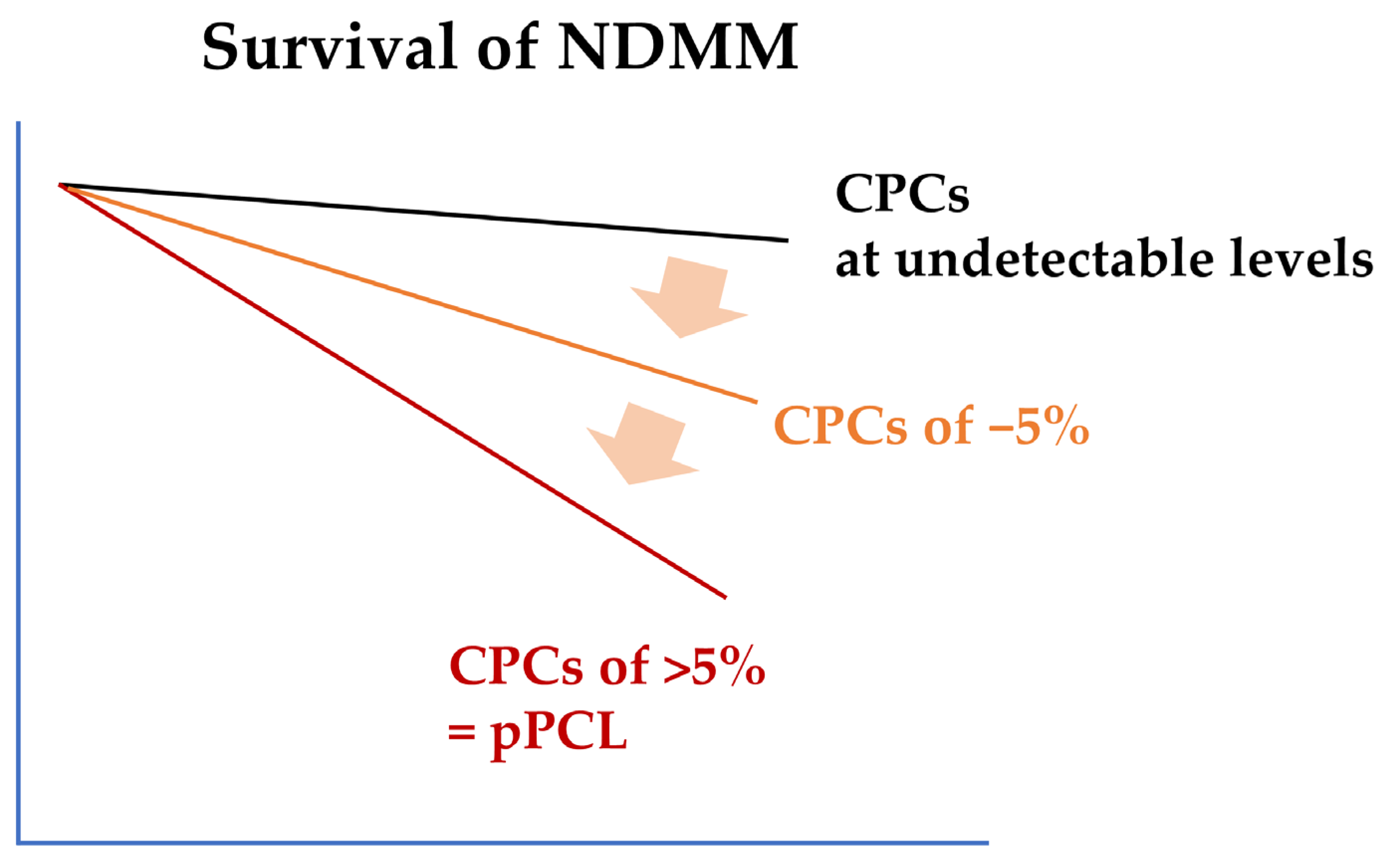
Figure 1. A schematic diagram showing the relationship between CPC levels and overall survival in NDMM. In NDMM, even low CPC levels are a poor prognostic factor. CPC is a continuous variable, and survival rates decrease as the proportion of CPCs increases. Currently, patients with CPCs of 5% or more are defined as pPCL. CPC, circulating plasma cell; NDMM, newly diagnosed multiple myeloma; pPCL, primary plasma cell leukemia.
Ijms 26 06166 g001
Figure 2. A schematic diagram of the proposed molecular pathology of pPCL based on chromosomal abnormalities. pPCL is classified into two types: one type that develops based on t(11;14) and another type that develops based on other factors. pPCL: primary plasma cell leukemia; PAX5: paired box protein 5; APOBEC: apolipoprotein B mRNA editing enzyme, catalytic polypeptide-like.
Ijms 26 06166 g002
Figure 3. Treatment approach for patients with newly diagnosed pPCL. CAR-T, chimeric antigen receptor T cell; BsAb, bispecific antibody; PI, proteasome inhibitor; IMiD, immunomodulatory drug; DEX, dexamethasone; ASCT, autologous hematopoietic stem cell transplantation; PD, progressive disease.
Ijms 26 06166 g003
Table 1. Cytogenetic and clinical characteristics in primary plasma cell leukemia at diagnosis.
Cytogenetic AbnormalityEMN12/HOVON129
n = 61, %
IFM
n = 32, %
GIMEMA
n = 23, %
Age, yearsOverall
(52–74)
52–63
(n = 36)
69–74
(n = 25)
70 years old and younger44–80
t(4;14) 694613
t(14;16)111941630
t(14;20)N/AN/AN/AN/A4
t(11;14)3226555030
Hyperdiploidy813179N/A
1q ≥ 3 copies5161525343
del(1p)2636016
(homozygous deletion of CDKN2C)
N/A
del(13q)5471525970
del(17p)3147182830
del(17p), t(4;14), or t(14;16)405927N/AN/A
≥2 high-risk cytogenetics
(del(17p), t(4;14), or t(14;16))
243414N/A38
(del(17p), t(4;14), t(14;16), t(14;20), 1q gain)
Clinical characteristics
IgG40315637N/A
IgA1119012N/A
IgM1042N/A
IgD6685N/A
Light chain only39443232N/A
EMD other than PB141712128
ISS stage 22125174317
ISS stage 36564744361
IFM, Intergroupe Francophone du Mye′ lome; GIMEMA, Gruppo Italiano Malattle Ematologiche d’Adulto; EMD, extramedullary disease; PB, peripheral blood; ISS, International Staging System.
Table 2. Summary of phase II prospective trials for primary plasma cell leukemia.
Study Name or Group (Years)PatientsTreatmentsActual Transplant (s)
(Number)
Results
Response, %Median PFS/OS (months)
GIMEMA [46]
(2009–2011)
N = 23
TE 15, TIE 8
Induction
Lenalidomide + dexamethasone
Single ASCT 4
Double ASCT 4
ASCT/RIC-allo 1
Overall
ORR 73 (CR 13, VGPR 26)
Transplant-eligible (n = 15)
PFS/OS 21/not reached
Transplant-ineligible (n = 8)
PFS/OS 10/12
IFM [53]
(2010–2013)
N = 40
All patients were TE
Median age 57 years (range, 27 to 71)
Induction
Bortezomib + dexamethasone + doxorubicin/cyclophosphamide (PAD/VCd)
ASCT/RIC-allo 16
ASCT/ASCT 9
(One patient received a syngeneic transplant conditioned with high-dose melphalan.)
Overall
ORR 69 (CR 10, VGPR 26)
After induction (n = 39)
ORR 69 (sCR 0, CR 10, VGPR 26, PR 23) Refractory disease 25, early death 5 in induction
After 1st ASCT (n = 26)
ORR 92 (sCR 4, CR 34, VGPR 38, PR 16, PD 8)
Overall
PFS/OS 15/36
ASCT/ASCT (n = 7)
(From the second transplant)
PFS/OS not reached/not reached
ASCT/RIC-allo (n = 17)
(From the second transplant)
PFS/OS 11/28
EMN12/HOVON129 [34] (2015–2021)N = 61
TE 36
TIE 25
TE (n = 36)
Induction
4 cycles of carfilzomib + lenalidomide + dexamethasone (KRd)
Consolidation
4 cycles of KRd in ASCT/ASCT
Maintenance
KR in ASCT/ASCT and ASCT/RIC-allo (until PD)
1st ASCT 24
ASCT/ASCT 12
ASCT/RIC-allo 5
After induction (n = 36)
ORR 83 (sCR 3, CR 11, VGPR 61, PR 8, PD 8)
After 1st ASCT (n = 24)
ORR 96 (sCR 13, CR 21, VGPR 63, PD 1)
After 2nd ASCT (n = 12)
ORR 100 (sCR 8, CR 17, VGPR 75)
After RIC-allo (n = 5)
ORR 100 (sCR 40, CR 40, VGPR 20)
Overall cohort of TE
PFS/OS 15/28
(Early mortality rate 8% at 6 months)
After 1st ASCT (n = 24)
PFS/OS 26/not reached
(From date of first ASCT)
After 2nd ASCT (n = 12)
PFS/OS at 2 years
58%/82%
(From the second transplant)
After RIC-allo (n = 5)
PFS/OS at 2 years
60%/53%
(From the second transplant)
TIE (n = 25)
Induction
8 cycles of carfilzomib + lenalidomide + dexamethasone (KRd)
Maintenance
KR until PD
NoneAfter induction cycle 1–4 (n = 25)
ORR 80 (sCR 12, CR 12, VGPR 44, PR 12); 2 early death, 2 excessive toxicity
After induction cycle 5–8 (n = 19)
ORR 95 (sCR 21, CR 21, VGPR 47, PR 5, PD 1)
PFS/OS 13/24
(Early mortality rate 16% at 6 months)
GIMEMA, Gruppo Italiano Malattle Ematologiche d’Adulto; IFM, Intergroupe Francophone du Mye′ lome; pPCL, primary plasma cell leukemia; TE, transplant-eligible; TIE, transplant-ineligible; ASCT, autologous hematopoietic stem cell transplantation; RIC, reduced intensity conditioning; allo, allogeneic hematopoietic stem cell transplantation; PD, progressive disease; ORR, overall response rate; sCR, stringent complete remission; CR, complete remission; VGPR, very good partial remission; PR, partial remission; OS, overall survival; PFS, progression-free survival.
Table 3. Selected clinical trials of CAR-T for pPCL.
NCT NumberStudy TitleCandidates and
Targets of CAR-T
Study Phase and
Treatment Flow
Brief SummaryComments
NCT05979363A Study of Bortezomib, Lenalidomide and Dexamethasone (VRd) Followed by BCMA CAR-T Therapy in Transplant-Ineligible Patients With Primary Plasma Cell LeukemiaNewly diagnosed pPCL
Anti-BCMA CAR-T Cells
Phase 2
VRd x3→CAR-T
→VR x3
→VR maintenance
A single-arm, open-label study to evaluate the efficacy and safety of VRD-based regimen combined with BCMA CAR-T in transplant-ineligible patients with primary plasma cell leukemia.CART without ASCT in patients (age ≤ 75 years old).
NCT05870917A Study of VRD-based Regimen Combined With CART-ASCT-CART2 Treatment in Patients With Primary Plasma Cell LeukemiaNewly diagnosed pPCL
Anti-BCMA CAR-T Cells
Phase 2
VRd x3→1st CAR-T
→VR x3→2nd CAR-T
→R maintenance
A single-arm, open-label study to evaluate the efficacy and safety of a VRD-based regimen (Bortezomib, Lenalidomide, and Dexamethasone) combined with CART-ASCT-CART2 in Chinese patients with newly diagnosed primary plasma cell leukemia.CART-ASCT-CART2 sandwich regimen in transplant-eligible patients (age ≤ 65 years old). [57]
NCT05838131A Clinical Trial to Explore the Safety and Efficacy of CT071 Injection in Patients With Relapsed/Refractory Multiple Myeloma or Primary Plasma Cell LeukemiaRelapsed/refractory MM and relapsed/refractory pPCL
Anti-GPRC5D CAR-T Cells (CT071)
Phase 1/2 studyA single-arm, open-label, dose-finding, first-in-human clinical trial. The main aim of this study is to preliminarily evaluate the safety and tolerability of CT071 after infusion, and explore the dose range of CT071 in patients with relapsed/refractory multiple myeloma or primary plasma cell leukemia, so as to determine the possible recommended therapeutic dose (RD).Shortened manufacturing process to around 30 h in vein-to-vein time.
pPCL, primary plasma cell leukemia; CAR-T, chimeric antigen receptor—T cell; MM, multiple myeloma; BCMA, B-cell maturation antigen; GPRC5D, G protein-coupled receptor, class C, group 5 member D.
Disclaimer/Publisher’s Note: The statements, opinions and data contained in all publications are solely those of the individual author(s) and contributor(s) and not of MDPI and/or the editor(s). MDPI and/or the editor(s) disclaim responsibility for any injury to people or property resulting from any ideas, methods, instructions or products referred to in the content.