Article

Post-Transcriptional Regulation of the MiaA Prenyl Transferase by CsrA and the Small RNA CsrB in Escherichia coli

1
Department of Microbiology, College of Medicine, Howard University, Washington, DC 20059, USA
2
Department of Biology, Claflin University, Orangeburg, SC 29115, USA
3
Department of Biology, Howard University, Washington, DC 20059, USA
*
Author to whom correspondence should be addressed.
Int. J. Mol. Sci. 2025, 26(13), 6068; https://doi.org/10.3390/ijms26136068
Submission received: 5 March 2025 / Revised: 16 May 2025 / Accepted: 19 May 2025 / Published: 24 June 2025
(This article belongs to the Special Issue Role of RNA Decay in Bacterial Gene Regulation)

Abstract

MiaA is responsible for the addition of the isopentyl modification to adenine 37 in the anticodon stem loop of specific tRNAs in Escherichia coli. Mutants in miaA have pleotropic effects on the cell in E. coli and play a role in virulence gene regulation. In addition, MiaA is necessary for stress response gene expression by promoting efficient decoding of UUX-leucine codons, and genes with elevated UUX-leucine codons may be a regulatory target for i6A-modified tRNAs. Understanding the temporal nature of the i6A modification status of tRNAs would help us determine the regulatory potential of MiaA and its potential interplay with leucine codon frequency. In this work, we set out to uncover additional information about the synthesis of the MiaA. MiaA synthesis is primarily driven at the transcriptional level from multiple promoters in a complex operon. However, very little is known about the post-transcriptional regulation of MiaA, including the role of sRNAs in its synthesis. To determine the role of small RNAs (sRNAs) in the regulation of miaA, we constructed a chromosomal miaA-lacZ translational fusion driven by the arabinose-responsive PBAD promoter and used it to screen against an Escherichia coli sRNA library (containing sRNAs driven by the IPTG-inducible PLac promoter). Our genetic screen and quantitative β-galactosidase assays identified CsrB and its cognate protein CsrA as potential regulators of miaA expression in E. coli. Consistent with our hypothesis that CsrA regulates miaA post-transcriptional gene expression through binding to the miaA mRNA 5′ UTR, and CsrB binds and regulates miaA post-transcriptional gene expression through sequestration of CsrA levels, a deletion of csrA significantly reduced expression of the reporter fusion as well as reducing miaA mRNA levels. These results suggest that under conditions where CsrA is inhibited, miaA mRNA translation and thus MiaA-dependent tRNA modification may be limited.

1. Introduction

MiaA is a tRNA Isopentenyl Transferase (IPT or IPTase) that catalyzes the prenylation of adenine 37 in the anticodon stem loop of tRNAs that read codons beginning with uridine [1,2,3]. The resulting N6-(isopentenyl) adenosine 37 (i6A37) is the precursor for subsequent methylthiolation by the MiaB enzyme, resulting in the 2-methylthio-N6-(isopentenyl) adenosine (ms2i6A37) in E. coli [1,2,3]. The function of MiaA has been the subject of study for several decades [1,2,3,4,5,6,7]. Previous studies demonstrated a role for i6A37, and other RNA modifications, in translational fidelity by preventing translational aberrations such as ribosome pausing and ribosomal frameshifting [8,9,10,11,12,13,14,15]. In addition, E. coli miaA mutants decrease cellular growth rates and promote spontaneous mutants, specifically GC—TA transversions [2,3,16]. The MiaA amino acid sequence is highly conserved with homologues in both prokaryotes and eukaryotes [17,18,19]. MiaA levels influence translational frameshifting to alter the global proteome, fitness, and virulence potential of Extraintestinal Pathogenic E. coli (ExPEC) [20]. MiaA promotes virulence in Shigella flexneri [21]. Acinetobacter baumannii miaA mutations exhibit Colistin resistance [22]. Streptomyces albus requires miaA for proper morphological development and metabolic regulation [23,24].
We previously identified MiaA as a regulatory factor necessary for the full expression of the stationary phase and general stress response sigma factor RpoS in E. coli K12 (σS) [25]. MiaA affects the expression of two additional stress response genes in E. coli K12: Hfq and IraP, which are also involved in the regulation of RpoS [26,27]. The MiaA (i6A)-sensitive genes identified in E. coli K12 thus far have higher UUX-leucine codon usage than the average genome wide UUX-leucine codon usage [25,27]. MiaA promotes the expression of its targets by promoting efficient UUX-leucine decoding [27]. While these studies have expanded our understanding of i6A modification function, we still do not know the physiological or metabolic conditions that control i6A levels in E. coli. We reasoned that further characterization of MiaA synthesis would assist us in understanding the regulation of i6A levels in the cell and we sought to achieve that in this work.
The miaA gene is contained in a complex operon, immediately upstream of hfq, the gene encoding the global RNA chaperone that also serves as a host factor for bacteriophage Qβ replication [28,29,30]. The transcription of miaA is driven by two promoters, one of which is a heat shock promoter (miaAP2(hs)) that is recognized by the heat shock-responsive alternative sigma factor, σ32 [30,31]. However, we know much less about the post-transcriptional regulatory factors that influence miaA expression. There are several clues in the literature that point to potential post-transcriptional regulation of MiaA. First, the transcript driven by the miaAP2(h) promoter has a 270 nucleotide 5′ untranslated region (UTR) [30,31]. Long 5′ UTRs are often associated with post-transcriptional regulatory processes. Second, a null mutation in hfq resulted in elevated levels of multiple transcripts from the miaA superoperon [32]. Third, MiaA transcript levels are increased in the absence of RNase E and/or RNaseIII [31]. RNase E is an endoribonuclease that is essential for growth and works with the 3′ to 5′ exoribonuclease polynucleotide phosphorylase (PNPase) to process rRNA and tRNA and the coordinated turnover of sRNAs and their mRNA targets [33,34,35,36]. These previous results support the idea that MiaA expression is regulated at the post-transcriptional level. Post-transcriptional regulation of miaA by Hfq and RNase would likely be mediated by sRNAs. Yet, prior to this work, no sRNA regulators of miaA have been identified. To close this gap, we executed a targeted screen of a plasmid sRNA library on a PBAD-miaA27P2-lacZ translational fusion strain, as previously described [37,38,39]. We identified several candidate sRNA repressors of the miaAP2(hs) transcript including SdsR, ArcZ, GcvB, Spot42, and CsrB. While SdsR and CsrB had the most dramatic inhibitory effect of all candidate sRNAs regulators of the miaAP2(hs) transcript in our genetic screen, the effect of CsrB on miaA expression was the focus of subsequent experiments for this study.
CsrB is an sRNA that acts to sequester the activity of the RNA-binding protein CsrA [40]. CsrA is a pleiotropic regulator of carbon metabolism and a global RNA-binding protein involved in direct post-transcriptional regulation of gene expression in E. coli, following binding to the 5′ untranslated regions [41,42,43]. CsrA regulates the expression of pgaABCD and flhDC operons to influence biofilm formation and motility/flagellar synthesis, respectively, in E. coli [44,45]. CsrA binds to several mRNA transcripts and subsequently regulates the expression of these genes in E. coli. The regulatory processes, metabolic impact, and virulence-promoting activities of CsrA have been identified in many other bacteria including Escherichia coli, Salmonella Typhimurium, Pseudomonas sp, Serratia sp., Campylobacter jejuni, Vibrio cholera, Yersinia pseudotuberculosis, Erwinia amylovora, Legionella pneumophila, Bacillus subtilis, Staphylococcus aureus, Clostridiodes difficile, and Acinetobacter baumannii [46,47,48,49,50,51,52,53,54,55,56,57,58,59,60,61]. CsrA activity was previously demonstrated to be regulated through sequestration by the sRNAs CsrB and CsrC. However, recent studies have demonstrated an expanded number of 10 direct CsrA sRNA-binding partners in vitro [62]. Four of them bind to CsrA in vivo [62]. In addition, recent studies suggest that CsrA binds sRNAs and promotes sRNA–mRNA complex formation, placing it in the category of RNA chaperones such as Hfq and ProQ [57,63,64]. Since CsrA works with CsrB to regulate gene expression, we tested the effect of CsrA on MiaA expression. We show that CsrA is necessary for miaAP2(hs) translation. We further extended the prior work on RNases and MiaA expression, demonstrating that RNase E and PNPase from the Degradosome contribute to stabilization of the miaA mRNA transcript.

2. Results

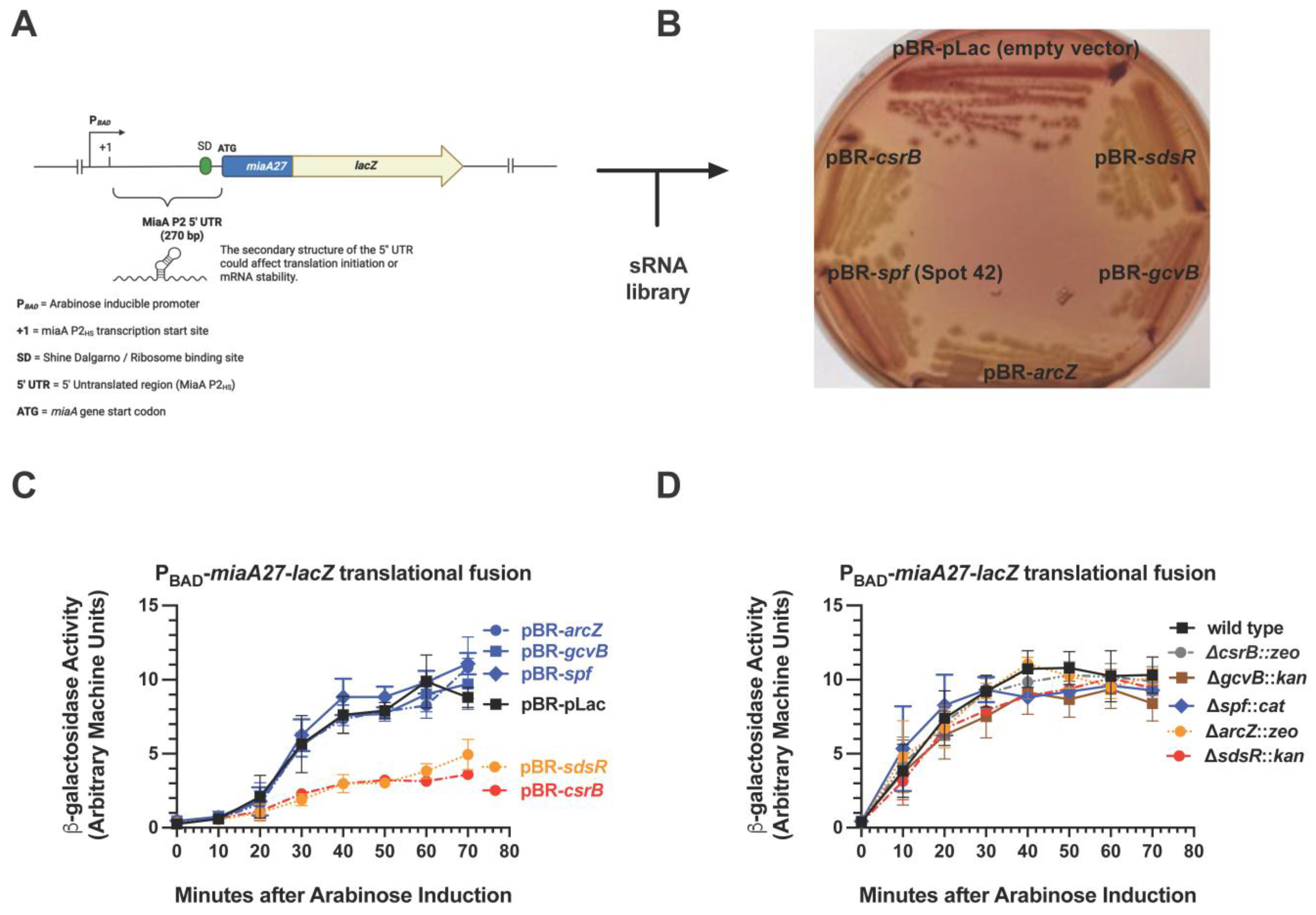
2.1. CsrB Was Selected as a Multi-Copy Repressor of MiaA Translation in a Targeted Screen of sRNA Regulators

We constructed a chromosomal miaA-lacZ translational fusion, whose expression is driven by the arabinose-responsive PBAD promoter (Figure 1A). It was used for screening a plasmid-based sRNA library containing 30 Escherichia coli sRNAs that were cloned downstream of an IPTG-inducible promoter (Mandin and Gottesman, 2009 [38]). The screening was carried out on Mac-Lac-Amp plates supplemented with arabinose to a final concentration of 0.002% to induce basal transcription from the PBAD promoter without producing a strong Lac+ phenotype, as previously described [65]. This facilitated the identification of sRNA repressors more easily through our genetic screen. We identified five candidate sRNA regulators of miaA: SdsR, ArcZ, GcvB, Spot42, and CsrB, with CsrB showing the strongest inhibitory effect (Figure 1B). To further test our hypothesis that post-transcriptional regulation of miaA is modulated by one or more sRNAs, we executed quantitative β-galactosidase assays of the PBAD-miaA-lacZ translational fusion strains carrying plasmids expressing SdsR, ArcZ, GcvB, Spot42, or CsrB (Figure 1C). Over-expression of ArcZ, GcvB, and Spot42 did not affect PBAD-miaA-lacZ activity as compared to the vector control (Figure 1C). There was a 2-fold decrease in the activity of PBAD-miaA-lacZ translational fusion activity upon over-expression of CsrB or SdsR (Figure 1C), suggesting that CsrB and SdsR sRNAs are involved in the post-transcriptional repression of miaA expression. We also tested the impact of deleting the sRNA candidates on the expression of the PBAD-miaA-lacZ translational fusion (Figure 1D). Individual deletions of gcvB, spf, arcZ, or sdsR had no effect on the expression of the activity of the PBAD-miaA-lacZ translational fusion following arabinose induction (Figure 1D). The csrB mutant demonstrated a modest increase in activity at 40 and 50 min after arabinose induction (Figure 1D and Figure 2A).

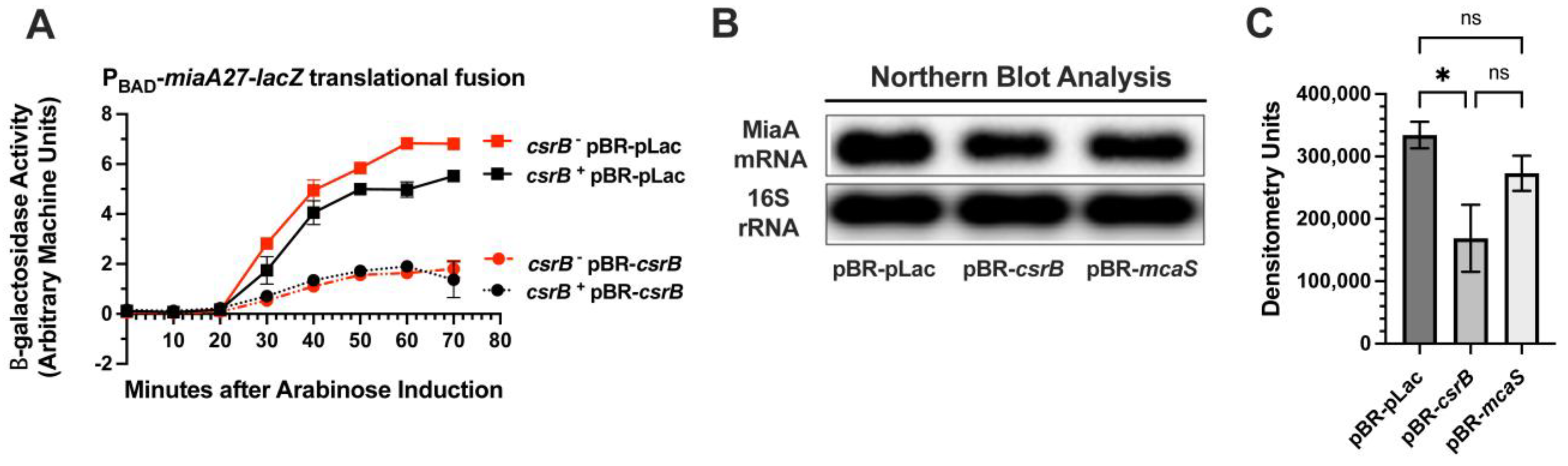
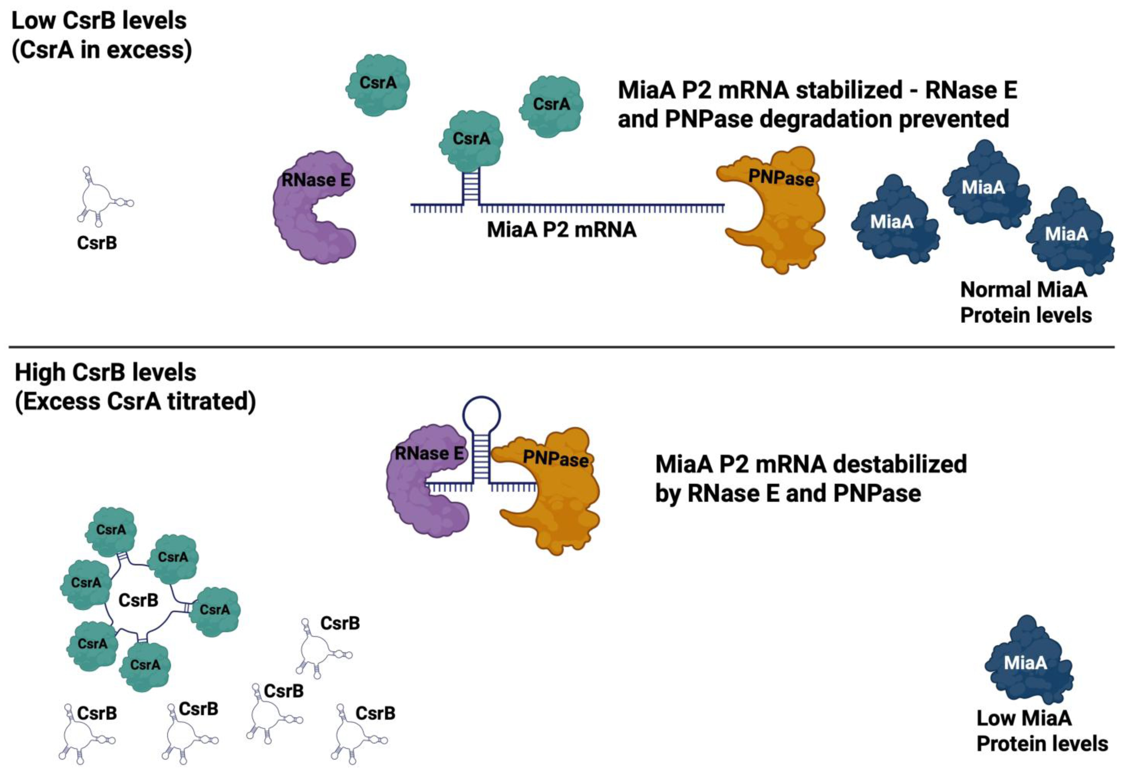
2.2. CsrB Affects MiaA mRNA Levels and Translation

We transformed the ΔPBAD-miaA-lacZ translational fusion strain with pBR-pLac or pBR-csrB to execute a complementation assay (Figure 2A). We did observe complementation of the csrB mutant with the csrB plasmid. CsrB repression of miaA-lacZ was slightly more pronounced in the csrB background, causing a 7-fold vs. 5-fold inhibition (Figure 2A). We then measured the steady-state levels of MiaA mRNA upon over-expression of CsrB and McaS. We decided to measure the effect of McaS on MiaA mRNA levels since it also acts to sequester CsrA [66] (Figure 2B). Consistent with decreased activity of the PBAD-miaA-lacZ translational fusion, we observed a decrease in the miaA transcript level upon over-expression of CsrB (Figure 2B,C), with CsrB proving to be a much more effective regulator of the miaA RNA level. However, over-expression of McaS did not affect miaA mRNA levels (Figure 2B,C).
Figure 2. The effect of CsrB on miaA expression. (A) Quantitative β-galactosidase assay analysis of PBAD-miaA27(P2HS)-lacZ translational fusion activity showing repression by CsrB sRNA. β-galactosidase assays were repeated at least three times and data points represent the mean plus and minus the standard error of the mean (mean ± sem). β-galactosidase activity is quantified by the use of arbitrary machine units as described in Section 4. (B) Northern blot analysis of MiaA steady-state levels following over-expression of CsrB (KMT792) or McaS (KMT799) in comparison to the empty vector. Northern blots were repeated at least three times. (C) Quantitative densitometry of Northern blot analysis. Densitometry signals were acquired using a Fluorochem R Fluorescent/Chemiluminescent imager. Each data point represents an average of at least three experiments and error bars represent the standard error of the mean (mean ± sem) based on the densitometry of Northern blot signals in Section (B). Statistical analysis was executed using One-Way ANOVA with Tukey’s Multiple Comparisons test on GraphPad Prism 9 (* p-value = 0.05, ns = not significant).
Ijms 26 06068 g002

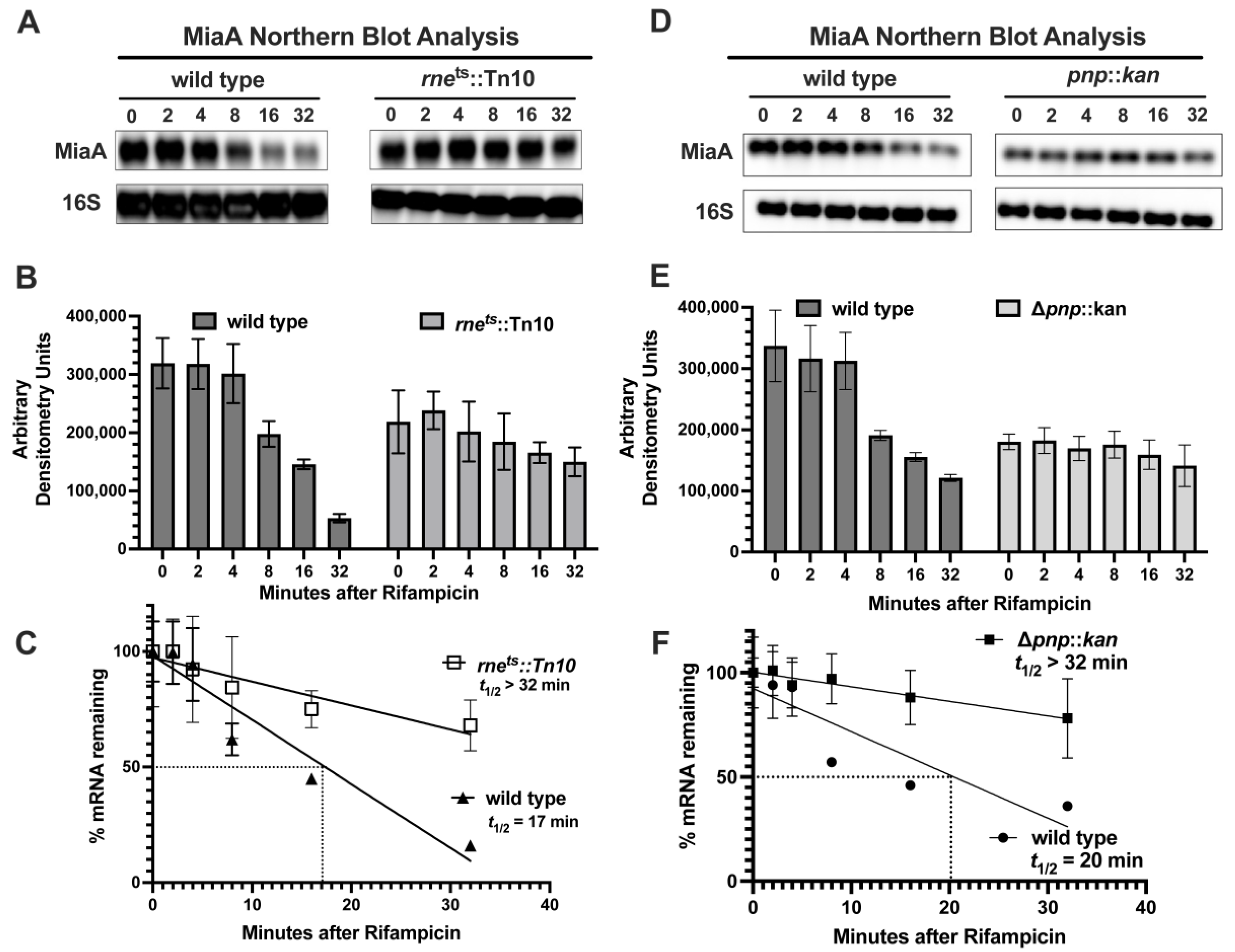
2.3. MiaA Is Regulated at the Level of mRNA Stability by PNPase and RNase E

Since very little is known regarding the regulation of MiaA mRNA stability, we decided to test the role of RNases in miaA mRNA turnover. Previous studies demonstrated an increase in mRNA levels of the miaA operon under non-permissive conditions in a temperature-sensitive RNase E mutant [31]. We tested the roles of RNase E and PNPase, both of which are components of the RNA Degradosome, in miaA mRNA turnover. Specifically, we compared miaA mRNA recycling in the cell, in WT isogenic wild-type, PNPase mutant (pnpA), and temperature-sensitive RNase E mutants (rnets) (Figure 3). The wild-type and rnets strains were grown at 32 °C to mid-log phase and then shifted to 43.5 °C, the non-permissive condition for the RNase E ts mutant. Then, we immediately added rifampicin to the cultures to halt transcription. We then isolated total RNA at times of 0, 2, 4, 8, 16, and 32 min after rifampicin treatment and measured miaA mRNA levels by Northern blot (Figure 3A,B). Upon semi-quantitative densitometric analysis of the Northern blot analysis and statistical analysis of the miaA mRNA in this experiment (Figure 3B,C), we determined that the t1/2 of the miaA mRNA increased in the rnets (>32 min) vs. the wild-type control (17 min) (Figure 3C). We grew wild-type and ΔpnpA::kan mutants in rich media at 37 °C to mid-log, added rifampicin, and isolated total RNA at 0, 2, 4, 8, 16, and 32 min post-rifampicin treatment (Figure 3D,E). The t1/2 of the miaA mRNA was >32 min and 20 min in the ΔpnpA::kan and wild-type genetic backgrounds, respectively (Figure 3E,F). The longer half-life of the miaA transcript in the rnets allele and ΔpnpA::kan mutant, in comparison to the wild-type control, suggests that RNase E and PNPase are involved in the post-transcriptional regulation of miaA at the level of mRNA stability.

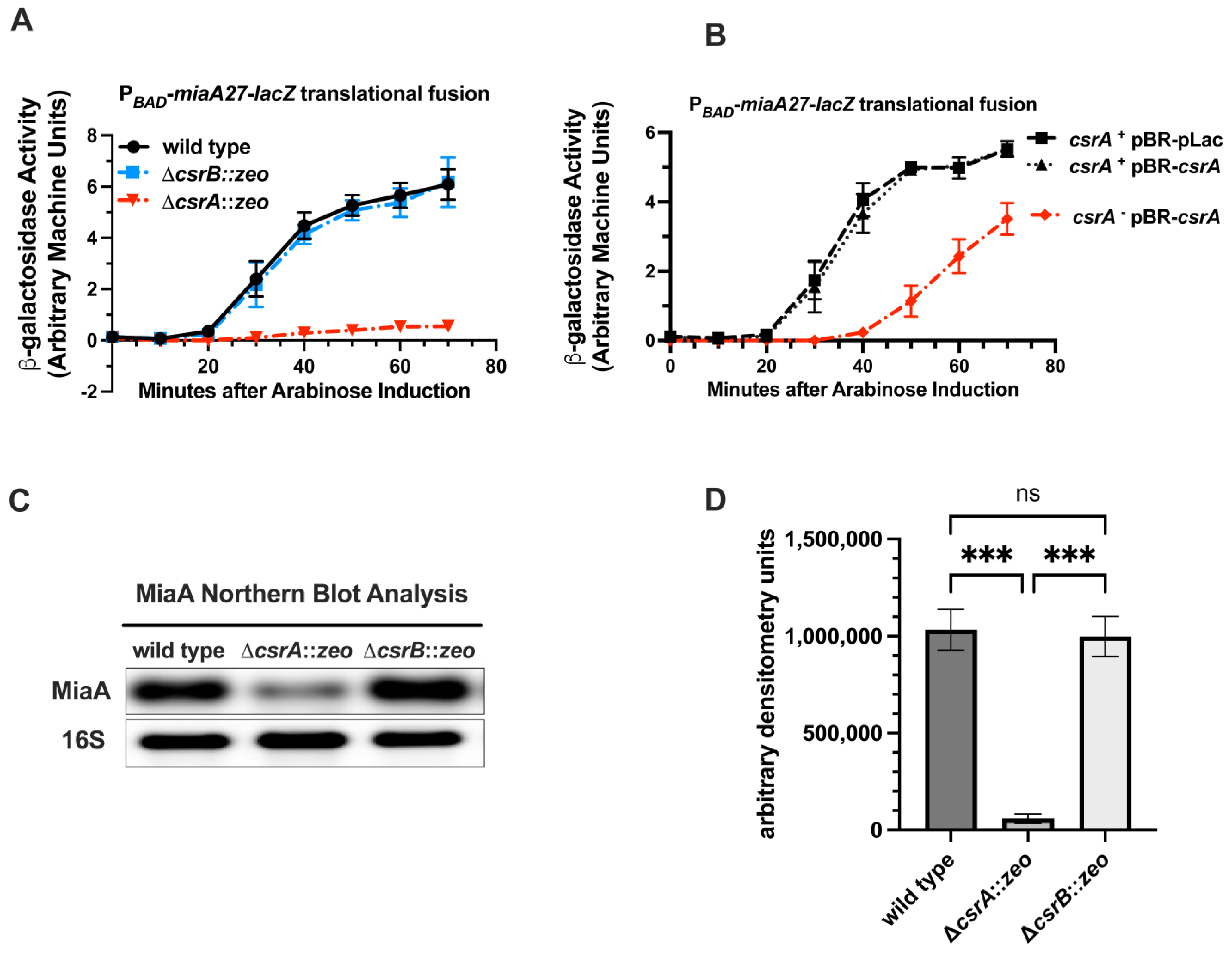
2.4. CsrA Is Necessary for the Full Expression of MiaA

The RNA-binding protein CsrA is part of a global regulatory system that controls bacterial gene expression at the post-transcriptional level [40,41,62,67,68,69,70,71]. CsrA regulates translation of target proteins by binding to target sequences in the 5′ UTR of the target genes. The availability of CsrA is regulated by sequestration of CsrB and CsrC sRNAs. CsrB and CsrC have multiple CsrA binding sites, each binding to approximately 18 CsrA subunits and inhibiting the activity of CsrA. Since CsrB represses the expression of miaA, and CsrB acts to sequester and inhibit CsrA activity, we hypothesized that the regulatory effect of CsrB on miaA may be through CsrA. To test our hypothesis, we measured the activity of the PBAD-miaA27-lacZ translational fusion in a csrA genetic background with over-expression of CsrA (Figure 4). In the csrA background, the β-galactosidase activity of the PBAD-miaA27-lacZ strain was decreased by approximately 6–10-fold at 70 min after arabinose induction, and was essentially non-detectable, in comparison to the csrA+ and csrB genetic backgrounds (Figure 4A). The β-galactosidase activity of the csrA PBAD-miaA27-lacZ strain was partially rescued by over-expression of plasmid-based csrA (Figure 4B). Given the decrease in the activity of the PBAD-miaA27-lacZ fusion, in the csrA mutant, we decided to measure miaA mRNA levels in the absence of csrA. We measured miaA mRNA levels in the wild-type, csrA, and csrB genetic backgrounds. The miaA mRNA levels were decreased, in a statistically significant manner, by approximately 20-fold in the absence of csrA while remaining virtually unchanged in the absence of csrB (Figure 4C,D). This result is consistent with miaA mRNA down-regulation upon over-expression of CsrB (Figure 1C) and confirms that the CsrA-CsrB system regulates post-transcriptional expression of miaA.
Figure 3. Effects of RNase E and PNPase on miaA mRNA stability. (A) Northern blot analysis of miaA mRNA stability. A temperature-sensitive mutant RNase E (rne-3071 zce-726::Tn10) allele was transduced from KMT621 into MG1655 (KMT665) by bacteriophage P1 transduction and selected for tetracycline (TetR) resistance KMT801. Wild-type and rnets strains were grown in rich media (LB) at 30 °C to an OD600 of 0.3. Sample aliquots of 600 μL were collected for total RNA isolation and Northern blot analysis at zero minutes, before transferring cultures to 43 °C. Rifampicin was added, and the samples were collected at 2, 4, 8, 16, and 32 min for total RNA isolation and analysis by agarose Northern blot. Experiments were repeated at least three times and the blots shown are representative blots of the triplicate experiments. (B) Quantitative densitometry of Northern blot in Section (A). Statistical analysis includes mean and standard error of the mean (mean ± s.e.m.). (C) Half-life calculations of Northern blot executed in Section (A). Quantitative Northern blot data from Section (B) were subjected to linear regression analysis using GraphPad Prism 9. (D) Δpnp::kan mutation was transduced from KMT624 into MG1655 (KMT665) by bacteriophage P1 transduction and selected for kanamycin (kanR) resistance (KMT800). Wild-type and pnpA strains (KMT665 and KMT800) were grown in rich media (LB) to an OD600 of 0.3. Then, 600 μL aliquots of the sample were collected for total RNA isolation and Northern blot analysis at zero minutes. Rifampicin was added, and samples were collected at 2, 4, 8, and 16 min for total RNA isolation and analysis by agarose Northern blot. (E) Quantitative densitometry of Northern blot in Section (A). Statistical analysis includes mean and standard error of the mean (mean ± s.e.m.) (F) Half-life calculations of Northern blot executed in Section (D). Quantitative Northern blot data from Section (B) were subjected to linear regression analysis using GraphPad Prism 9.
Ijms 26 06068 g003

3. Discussion

3.1. RNA Modifications and the Bacterial Epitranscriptome

RNA modifications have long been recognized as essential for maintaining translational fidelity and the structural stability of tRNAs and rRNAs. In bacterial systems, parttRNA modifications in particular have been extensively studied for their role in ensuring accurate and efficient protein synthesis [72,73,74,75]. While early work focused on a few bacterial model organisms, recent studies have broadened this view to include diverse species, providing insights into bacterial pathogenesis and the regulation of virulence [76,77,78,79,80]. Additionally, the potential for targeting tRNA modification pathways as antimicrobial strategies is now under active investigation [81,82]. More recent findings have further expanded the scope of the bacterial epitranscriptome to include modifications in mRNAs, suggesting regulatory functions beyond the canonical roles in tRNA and rRNA [83]. Studies in eukaryotic systems have also highlighted an expanded list of RNA species that contain RNA modifications. These include extensive mRNA modifications within the coding and noncoding regions [84]. Beyond mRNAs, RNA modifications have been found within eukaryotic regulatory RNA species such as miRNA, CircRNAs, and lncRNAs [85,86,87]. They are thought to play roles in post-transcriptional regulatory circuits by modulating miRNA interactions with the 3′ UTRs of mRNAs, modulating mRNA stability. Several different RNA modifications play regulatory roles in cellular physiology [88,89,90,91,92,93]. Work by our group and others has demonstrated that RNA modifications may play regulatory roles in gene expression by promoting the expression of stress response genes, in a manner dependent upon codon bias in bacteria and bacteriophages [25,26,27,94,95,96]. Subsequent reports from several groups have also demonstrated codon-biased gene regulation in bacterial pathogens [25,26,27,95,96]. In all these studies, the role of the modification was established by mutating the gene(s) encoding the modification enzymes. To add to these discoveries, understanding the conditions whereby RNA modifications are synthesized will assist us in understanding their impact on the physiology of the cell.

3.2. New Regulators of MiaA Expression

We previously demonstrated that miaA is necessary for the expression of RpoS, IraP, and Hfq [25,26,27]. The miaA requirement for optimal expression of RpoS and IraP is related to UUX-leucine decoding, suggesting that MiaA may promote the expression of stress response genes during leucine starvation [25,26,27]. Over-expression of leucine tRNAs was able to suppress the decreased expression of rpoS in miaA mutants. Interestingly, a previous study demonstrated that miaA mutants are synthetically lethal with leuX (leucine tRNA) mutants during heat shock [97]. Also, miaA mutants affect leucine operon gene expression in Salmonella [4]. Taken together, this suggests that MiaA-catalyzed i6A37 modification is particularly important during heat shock whereby UUX-leucine decoding provides an adaptive advantage. For these reasons, characterizing the post-transcriptional regulation of the miaA transcript from the σ32-dependent heat shock promoter is of critical importance. Prior to this work, little was known about post-transcriptional regulation of miaA. Here, we started to fill that knowledge gap by identifying several potential candidate sRNA regulators of MiaA expression at the post-transcriptional level. Our results demonstrate a strong effect of CsrA, likely as a direct post-transcriptional regulator of the miaAP2 promoter transcript.

3.3. MiaA Is an Additional Potential Stimulatory Target of the CsrA-CsrB System and Interactions with Other Post-Transriptional Regulators of the MiaA Operon

CsrA can directly bind to mRNA transcripts to regulate gene expression at the post-transcriptional level, in the absence of sequestration by CsrB or CsrC [40]. There are approximately 12 mRNA transcripts that are post-transcriptionally regulated by CsrA. Ten of these regulatory targets are repressed and two of these regulatory targets are stimulated by CsrA, including ymdA [98] (Table 1). CsrA stimulates the translation of ymdA through interaction with its 5′ UTR to reverse the formation of secondary structures that occlude its ribosome binding site and subsequent translational initiation. Our work identifies miaA as a stimulatory target of CsrA. It is possible that the 5′ UTR of the miaAP2 transcript has secondary structures that occlude the ribosome binding site and that CsrA acts in a similar manner on this transcript to stimulate translation of this transcript. Given the fact that RNase E and PNPase mutants result in the stabilization of the miaA transcript, CsrA may also interact with Degradosome enzymes or the 5′ untranslated region (UTR) of the MiaA P2 transcript to promote its stabilization. We have illustrated a simple model to describe CsrA-CsrB regulation of miaA (Figure 5). Upon sequestration of CsrA by CsrB, the miaA transcript from the P2 (heat shock promoter) is less stable and translation is also inhibited. This is reversed in the absence of CsrB, and when CsrA levels are higher or it is more active, resulting in transcript stabilization and an increase in translation (Figure 5). The stabilization of the miaA mRNA could be through antagonistic interactions with Degradosome proteins RNase E and PNPase. This model is consistent with the previous report from Vakulskas et al. (2016), whereby CsrA was shown to stabilize CsrB through antagonistic interactions with RNase E [99].
The miaA gene is encoded directly upstream of the hfq gene within the nnr-tsaE-amiB-mutL-miaA-hfq-hflX-hflK-hflC superoperon in the E. coli genome [30,31,100,101]. This tandem genetic localization is widely conserved across the prokaryotic domain. While mutations in hfq exhibit extensively pleiotropic effects, miaA mutant phenotypes appear to be distinct, apart from their effects on rpoS expression, whereby the miaA mutant demonstrates some polarity likely due to effects on Hfq-dependent sRNAs that stimulate rpoS translation [25,102,103,104,105,106,107]. There is complex transcriptional organization of this superoperon with approximately 10 promoters exhibiting alternating dependence upon σ32 an σ70, in the amiB-hfq region [30,31,100,101]. All these promoters are upstream of the hfq gene. However, there are three promoters between the miaA gene and the start codon of hfq, with the function of drive expression of hfq-hflXKC: defined as hfqP1(hs), hfqP3, and hfqP3. Baker et al. (2007) demonstrated that CsrA binds to the ribosome binding site of the hfq mRNA and inhibits hfq translation [108]. In our screen, we utilized a miaA-lacZ fusion with the miaA P2 transcript. If CsrA interacts with the 5′ UTR of the miaA P2 transcript, it may provide CsrA with multiple options for binding to the larger transcripts expressed from this operon that contains both miaA and hfq. Multiple CsrA binding sites may or may not be accessible on the larger transcripts. This is the subject of ongoing studies in our laboratory.

4. Materials and Methods

4.1. Strains and Plasmids

All strains are derivatives of E. coli K12 MG1655 and are listed in Table 2. All plasmids used in this study are also listed in Table 2.

4.2. Media and Growth Conditions

E. coli strains were grown in Luria–Bertani (LB) Lennox liquid media (KD Medical, Columbia, MD, USA) in a WS27 shaking water bath (ShelLab, Cornelius, OR, USA) for the experiments in this study at 37 °C, unless otherwise described below. Transformation of plasmid DNA into strains was sometimes facilitated using Transformation Storage Solution (TSS) media (LB, 10% (w/v) Polyethylene Glycol-8000, 5% DMSO, 50 mM MgCl2) as described below. Recombinants from Lambda (λ)-Red-based mutagenesis were selected for growth on LB agar plates supplemented with zeomycin to a final concentration of 25–50 μg/mL (LB-Zeo). Transductants, of pnpA::kan or rne-3071 zce-726::Tn10 (tet) mutants, were grown on LB agar plates supplemented with either tetracycline or kanamycin to a final concentration of 25 μg/mL (LB-Tet or LB-Kan). The rne-3071 zce-726::Tn10 mutant is temperature-sensitive and requires incubation at 30 °C for optimal growth. To determine the effect of RNase E on miaA mRNA levels, rne-3071 zce-726::Tn10 cultures were grown at 30 °C and then shifted to 43.5 °C to induce an RNase E phenotype, as previously described [36]. We then isolated total RNA for the analysis of miaA mRNA levels or turnover using Northern blot analysis. Strains carrying plasmids were grown in LB media supplemented with ampicillin to a final concentration of 100 μg/mL (LB-Amp) or on LB-Amp agar plates. To stimulate the expression of arabinose-inducible fusion strains, cultures were first grown in LB or LB-Amp, supplemented with glucose to a final concentration of 0.2%, harvested by centrifugation, washed once with LB media, and resuspended in LB supplemented with arabinose to a final concentration of 0.2%. Small RNA plasmid library transformants were assayed on MacConkey-Lactose (Mac-Lac) agar plates supplemented with ampicillin to a final concentration of 100 µg/mL (Mac-Lac-Amp) to screen for changes in the Lactose phenotype of individual colonies following overnight growth at 37 °C.

4.3. General Molecular Biology Techniques

Plasmid DNA was isolated using The Column-PureTM Plasmid Mini-Prep Kit (Lamda Biotech, St. Louis, MO, USA), according to manufacturer instructions. Genomic DNA used for PCR reactions was isolated using The Column-PureTM Bacterial Genomic DNA Kit (Lamda Biotech), according to the manufacturer’s instructions. PCR reactions were performed to amplify allelic exchange substrates for mutagenesis. PCR amplification was performed using the Taq Plus 2X PCR MasterMix (Lamda Biotech) with standard cycle conditions according to the manufacturer’s instructions. The annealing temperature for each PCR reaction was optimized to the Tm of the primers used (Table 3). All PCR reactions were purified using The Column-PureTM Clean-Up Kit (Lamda Biotech), according to the manufacturer’s instructions (Lamda Biotech).

4.4. Genetic Engineering and Strain Construction

Chromosomal mutagenesis was executed via bacteriophage λ-based recombineering and bacteriophage P1 transduction as previously described and outlined below [109,110]. Plasmids were moved using the Transformation Storage Solution (TSS) media protocol or heat shock transformation of chemically competent cells as described below [111].

4.4.1. Insertional Inactivation Mutagenesis Using Recombineering

Deletion and insertion mutations of csrA, csrB, and miaA were constructed using recombineering as previously described [110,112]. Briefly, a zeomycin resistance cassette with 50 bp of flanking homology to csrA, csrB, or miaA was synthesized by PCR to create an allelic exchange substrate for recombineering-based mutagenesis. Then, the DJ480 mini-λ::tet strain was induced using heat shock and made electrocompetent with ice-cold water washes [110,112]. The allelic exchange substrates were then electroporated into the prepared cells, allowed to recover, and plated on LB-Zeo selectable media. Recombinants were confirmed to have the mutant using PCR with a primer upstream or downstream of the target gene along with a primer that recognizes the insertion marker. Finally, this was verified using Sanger Sequencing.

4.4.2. P1 Transduction to Move Mutants Between Strains

The newly constructed ΔcsrA::zeo, ΔcsrB::zeo, and ΔmiaA::zeo mutations, as well as the previously constructed pnpA::kan and rne-3071 zce-726::Tn10 (tet) mutants, were moved from the mini-λ::tet-containing strain into clean genetic backgrounds (MG1655 or PBAD-miaA27P2-lacZ translational fusion strain) via generalized transduction using bacteriophage P1 as previously described [113].

4.4.3. Heat Shock Transformation of Chemically Competent Cells for Cloning

All plasmids, including those from the sRNA library, were transformed into E. coli strains using Transformation Storage Solution (TSS) media and its associated transformation method as previously described [111]. Briefly, the E. coli K-12 MG1655 recipient strain was grown in nutrient-rich Lennox Broth (LB) to OD600 0.5. Upon reaching OD600 0.5, the cells were harvested by centrifugation. The supernatant was then decanted, and the pellet was re-suspended in 1/10th volume of ice-cold TSS media. An amount of 2 μL (100 ng) of plasmid DNA (pBR-pLac, pBR-pLac-csrB, pBR-pLac-sdsR, pBR-pLac-spf, pBR-pLac-arcZ, pBR-pLac-gcvB, pBR-pLac-mcaS, and pBR-pLac-csrA) was added to 100 μL aliquots of cell suspension and incubated on ice for 30 min. Following the 30 min incubation on ice, 900 μL of LB was added to the cell–plasmid mixture, and the mixture was left to recover at 37 °C for 1 h on a shaking heat block. Upon the completion of the recovery period, 200 μL of recovered cells was plated on LB-Amp plates and left to grow overnight at 37 °C in a microbiological incubator. Transformants were purified once by streaking on LB agar plates supplemented with ampicillin.

4.5. RNA Isolation

Total RNA was isolated using the Hot Phenol Method as previously described [114,115]. Briefly, overnight cultures were subcultured in 30 of LB at a 1:1000 dilution ratio and allowed to grow in the shaking water bath to an Optical Density of 600 (OD600) of 0.5. A 600 μL aliquot of cells was isolated from exponentially growing E. coli cultures and resuspended in a 1× lysis buffer/Hot Acid Phenol solution in a 1.5 mL microcentrifuge tube on a thermomixer (Eppendorf, Hamburg, Germany) set to 65 °C. The cells were incubated with intermittent shaking for 5 min. The tubes were subjected to centrifugation at 15,000 rpm for 10 min. The aqueous phase was extracted and purified two additional times with acid-phenol followed by ethanol precipitation in a −80 °C freezer overnight. The RNA was pelleted and washed with 70% ethanol, air-dried, and resuspended with 50 μL of DEPC water. RNA concentrations were successively measured using The NanoDropTM OneC Microvolume UV-Vis Spectrophotometer (Fisher Scientific, Waltham, MA, USA).

4.6. Agarose Northern Blot

The agarose-based Northern blot was executed as previously described [36,116]. A 1× MOPS (Quality Biological INC, Gaithersburg, MD, USA) 1% Agarose gel was used for the resolution of total RNA. After pre-running the gel at 100 V for 40 min, a constant amount of total RNA (ranging from 2 to 5 μg) was mixed with 2× volume of loading buffer (500 μL Formamide, 100 μL 10× MOPS, 100 μL (80% glycerol 0.2% bromophenol blue), 120 μL Formaldehyde, and 2 μL (10 mg/mL EtBr)). The samples were then heated at 65 °C for 15 min and loaded onto the gel for fractionation by gel electrophoresis for 40 min at 100 volts. The gel was then soaked in 0.05 M NaOH solution for 20 min and 20× SSC solution for 1 h. The RNA was then transferred from the agarose gel to a nylon membrane using the capillary method as previously described [117]. The RNA was then UV-crosslinked to the nylon membrane using the HL-2000 Hybrilinker (UVP/Analytikjena, Upland, CA, USA). After UV-crosslinking of the nylon membrane, it was then pre-hybridized with 5 mL of ultrahyb oligo buffer (Ambion, Austin, TX, USA) for 2 h, and then subjected to hybridization with a biotinylated DNA probe targeting the transcript of interest overnight at 42 °C in the HL-2000 Hybrilinker (UVP). The membranes were then processed using stringency washes and developed using the Chemiluminescent Nucleic Acid Detection Module Kit (ThermoFisher Scientific, Waltham, MA, USA) according to the manufacturer’s recommendations.

4.7. β-Galactosidase Assays (Kinetic Microtiter Assays)

We measured β-galactosidase activity using the Kinetic Microtiter Plate Assay method [118]. Briefly, overnight cultures were grown in 5 of LB Lennox liquid media at 37 °C within a Cel-Gro Tissue culture rotator (Thermo Scientific) placed within a microbiological incubator (ShelLab). Overnight cultures containing sRNA plasmids were grown in LB-Amp. The following day, the overnight cultures were diluted 1:1000 in 30 mL of fresh LB or LB-Amp liquid media and placed in a 125 mL beveled Erlenmeyer flask to sub-culture the cells. IPTG was added to cultures containing sRNA plasmids to a final concentration of 1 mM. Cultures were then grown in a WS27 shaking water bath (ShelLab) at 37 °C to an OD600 of 0.5. Cells were then harvested by centrifugation and then washed and resuspended with Lennox Broth (LB). The LB–cell suspensions were then transferred to a new flask and supplemented with ampicillin (100 μg/mL), IPTG (1 mM), and arabinose (0.02%). The cultures were then incubated in the 37 °C shaking water bath for an additional 70 min. Then, 100 μL aliquots of the cultures were collected once, or at 10 min intervals, and transferred to a 96-well polystyrene microtiter plate containing 50 μL of permeabilization solution (100 mM of Tris, pH 7.8, 32 mM of NaPO4, 8 mM of DTT (Dithiothreitol), 8 mM CDTA (Cyclohexanediaminetetraacetic acid), 4% Triton X-100, and 50 μL of Polymixin B). To serve as an experimental control, 100 μL of LB was pipetted into 1 of the wells of the 96-well polystyrene microtiter plate. The 96-well polystyrene microtiter plate containing the samples was incubated at room temperature for 15 min to allow for cell lysis. Subsequently, 50 μL of O-nitrophenyl-β-D-galactoside (ONPG) solution (4 mg/mL ONPG, 2 mM of Sodium Citrate, and 70 μL of β-Mercaptoethanol) was added to each well containing cell lysates. The 96-well polystyrene microtiter plate was immediately read using a Filter Max F5 Multi-Mode Micro Plate Reader (Molecular Devices, San Jose, CA, USA).
Arbitrary machine units were assigned to each sample, calculated as OD420/OD600. These units are approximately 25-fold lower than the Miller units classically used for β-galactosidase assays.

4.8. Statistical Analysis

All statistical analysis was executed using Prism 9 software (Graphpad, La Jolla, CA, USA).

Author Contributions

Conceptualization, K.M.T.; Methodology, K.M.T.; Validation, K.M.T.; Formal analysis, J.I.A. and K.M.T.; Investigation, J.I.A., K.W., A.A., O.O., J.N. and K.M.T.; Data curation, K.M.T.; Writing—original draft, J.I.A.; Writing—review & editing, K.M.T.; Visualization, J.I.A. All authors have read and agreed to the published version of the manuscript.

Funding

This work was supported by Award Number 1904089 from the National Science Foundation (NSF) and R35GM152163 from the National Institute of General Medical Sciences.

Institutional Review Board Statement

No animals or human subjects or samples were used in this study.

Informed Consent Statement

Not applicable.

Data Availability Statement

Data is contained within the article.

Acknowledgments

We would like to thank the members of the Thompson Laboratory, Muneer Abbas, Qiyi Tang, Nadim Majdalani, and Susan Gottesman for the critical review of this manuscript.

Conflicts of Interest

The authors declare no conflict of interest.

References

  1. Caillet, J.; Droogmans, L. Molecular cloning of the Escherichia coli miaA gene involved in the formation of delta 2-isopentyl adenosine in tRNA. J. Bacteriol 1988, 170, 4147–4152. [Google Scholar] [CrossRef]
  2. Connolly, D.M.; Winkler, M.E. Genetic and physiological relationships among the miaA gene, 2-methylthio-N6-(delta 2-isopentenyl)-adenosine tRNA modification, and spontaneous mutagenesis in Escherichia coli K-12. J. Bacteriol. 1989, 171, 3233–3246. [Google Scholar] [CrossRef]
  3. Connolly, D.M.; Winkler, M.E. Structure of Escherichia coli K-12 miaA and characterization of the mutator phenotype caused by miaA insertion mutations. J. Bacteriol 1991, 173, 1711–1721. [Google Scholar] [CrossRef]
  4. Blum, P.H. Reduced leu operon expression in a miaA mutant of Salmonella typhimurium. J. Bacteriol 1988, 170, 5125–5133. [Google Scholar] [CrossRef]
  5. Diaz, I.; Ehrenberg, M.; Kurland, C.G. How do combinations of rpsL- and miaA- generate streptomycin dependence? Mol. Gen. Genet. MGG 1986, 202, 207–211. [Google Scholar] [CrossRef]
  6. Pierrel, F.; Bjork, G.R.; Fontecave, M.; Atta, M. Enzymatic modification of tRNAs: MiaB is an iron-sulfur protein. J. Biol. Chem. 2002, 277, 13367–13370. [Google Scholar] [CrossRef]
  7. Esberg, B.; Leung, H.C.; Tsui, H.C.; Bjork, G.R.; Winkler, M.E. Identification of the miaB gene, involved in methylthiolation of isopentenylated A37 derivatives in the tRNA of Salmonella typhimurium and Escherichia coli. J. Bacteriol. 1999, 181, 7256–7265. [Google Scholar] [CrossRef]
  8. Tukenmez, H.; Xu, H.; Esberg, A.; Bystrom, A.S. The role of wobble uridine modifications in +1 translational frameshifting in eukaryotes. Nucleic Acids Res. 2015, 43, 9489–9499. [Google Scholar] [CrossRef]
  9. Bjork, G.R.; Durand, J.M.; Hagervall, T.G.; Leipuviene, R.; Lundgren, H.K.; Nilsson, K.; Chen, P.; Qian, Q.; Urbonavicius, J. Transfer RNA modification: Influence on translational frameshifting and metabolism. FEBS Lett. 1999, 452, 47–51. [Google Scholar] [CrossRef]
  10. Bjork, G.R.; Wikstrom, P.M.; Bystrom, A.S. Prevention of translational frameshifting by the modified nucleoside 1-methylguanosine. Science 1989, 244, 986–989. [Google Scholar] [CrossRef]
  11. Urbonavicius, J.; Qian, Q.; Durand, J.M.; Hagervall, T.G.; Bjork, G.R. Improvement of reading frame maintenance is a common function for several tRNA modifications. EMBO J. 2001, 20, 4863–4873. [Google Scholar] [CrossRef] [PubMed]
  12. Urbonavicius, J.; Stahl, G.; Durand, J.M.; Ben Salem, S.N.; Qian, Q.; Farabaugh, P.; Bjork, G.R. Transfer RNA modifications that alter +1 frameshifting in general fail to affect −1 frameshifting. RNA 2003, 9, 760–768. [Google Scholar] [CrossRef] [PubMed]
  13. Qian, Q.; Bjork, G.R. Structural requirements for the formation of 1-methylguanosine in vivo in tRNA(Pro)GGG of Salmonella typhimurium. J. Mol. Biol. 1997, 266, 283–296. [Google Scholar] [CrossRef] [PubMed]
  14. Qian, Q.; Bjork, G.R. Structural alterations far from the anticodon of the tRNAProGGG of Salmonella typhimurium induce +1 frameshifting at the peptidyl-site. J. Mol. Biol. 1997, 273, 978–992. [Google Scholar] [CrossRef]
  15. Qian, Q.; Curran, J.F.; Bjork, G.R. The methyl group of the N6-methyl-N6-threonylcarbamoyladenosine in tRNA of Escherichia coli modestly improves the efficiency of the tRNA. J. Bacteriol. 1998, 180, 1808–1813. [Google Scholar] [CrossRef]
  16. Zhao, J.; Leung, H.E.; Winkler, M.E. The miaA mutator phenotype of Escherichia coli K-12 requires recombination functions. J. Bacteriol. 2001, 183, 1796–1800. [Google Scholar] [CrossRef]
  17. Schweizer, U.; Bohleber, S.; Fradejas-Villar, N. The modified base isopentenyladenosine and its derivatives in tRNA. RNA Biol. 2017, 14, 1197–1208. [Google Scholar] [CrossRef]
  18. Nishii, K.; Wright, F.; Chen, Y.Y.; Moller, M. Tangled history of a multigene family: The evolution of ISOPENTENYLTRANSFERASE genes. PLoS ONE 2018, 13, e0201198. [Google Scholar] [CrossRef]
  19. Soman, S.; Ram, S. MiaA (Rv2727c) mediated tRNA isopentenylation of Mycobacterium tuberculosis H37Rv. Mol. Biol. Res. Commun. 2022, 11, 97–104. [Google Scholar] [CrossRef]
  20. Fleming, B.A.; Blango, M.G.; Rousek, A.A.; Kincannon, W.M.; Tran, A.; Lewis, A.J.; Russell, C.W.; Zhou, Q.; Baird, L.M.; Barber, A.E.; et al. A tRNA modifying enzyme as a tunable regulatory nexus for bacterial stress responses and virulence. Nucleic. Acids Res. 2022, 50, 7570–7590. [Google Scholar] [CrossRef]
  21. Durand, J.M.; Bjork, G.R.; Kuwae, A.; Yoshikawa, M.; Sasakawa, C. The modified nucleoside 2-methylthio-N6-isopentenyladenosine in tRNA of Shigella flexneri is required for expression of virulence genes. J. Bacteriol. 1997, 179, 5777–5782. [Google Scholar] [CrossRef] [PubMed]
  22. Sun, B.; Liu, H.; Jiang, Y.; Shao, L.; Yang, S.; Chen, D. New Mutations Involved in Colistin Resistance in Acinetobacter baumannii. mSphere 2020, 5, e00895-19. [Google Scholar] [CrossRef] [PubMed]
  23. Koshla, O.; Yushchuk, O.; Ostash, I.; Dacyuk, Y.; Myronovskyi, M.; Jager, G.; Sussmuth, R.D.; Luzhetskyy, A.; Bystrom, A.; Kirsebom, L.A.; et al. Gene miaA for post-transcriptional modification of tRNA(XXA) is important for morphological and metabolic differentiation in Streptomyces. Mol. Microbiol. 2019, 112, 249–265. [Google Scholar] [CrossRef] [PubMed]
  24. Koshla, O.; Kravets, V.; Dacyuk, Y.; Ostash, I.; Sussmuth, R.; Ostash, B. Genetic analysis of Streptomyces albus J1074 mia mutants suggests complex relationships between post-transcriptional tRNA(XXA) modifications and physiological traits. Folia Microbiol. 2020, 65, 1009–1015. [Google Scholar] [CrossRef]
  25. Thompson, K.M.; Gottesman, S. The MiaA tRNA modification enzyme is necessary for robust RpoS expression in Escherichia coli. J. Bacteriol. 2014, 196, 754–761. [Google Scholar] [CrossRef]
  26. Aubee, J.I.; Olu, M.; Thompson, K.M. TrmL and TusA are necessary for rpoS and MiaA is required for hfq expression in Escherichia coli. Biomolecules 2017, 7, 39. [Google Scholar] [CrossRef]
  27. Aubee, J.I.; Olu, M.; Thompson, K.M. The i6A37 tRNA modification is essential for proper decoding of UUX-Leucine codons during rpoS and iraP translation. RNA 2016, 22, 729–742. [Google Scholar] [CrossRef]
  28. Moller, T.; Franch, T.; Hojrup, P.; Keene, D.R.; Bachinger, H.P.; Brennan, R.G.; Valentin-Hansen, P. Hfq: A bacterial Sm-like proteins that mediates RNA-RNA interaction. Mol. Cell. 2002, 9, 23–30. [Google Scholar] [CrossRef]
  29. Kajitani, M.; Kato, A.; Wada, A.; Inokuchi, H.; Ishihama, A. Regulation of the Escherichia coli hfq gene encoding the host factor for phage Q beta. J. Bacteriol. 1994, 176, 531–534. [Google Scholar] [CrossRef]
  30. Tsui, H.C.; Feng, G.; Winkler, M.E. Transcription of the mutL repair, miaA tRNA modification, hfq pleiotropic regulator, and hflA region protease genes of Escherichia coli K-12 from clustered Esigma32-specific promoters during heat shock. J. Bacteriol. 1996, 178, 5719–5731. [Google Scholar] [CrossRef]
  31. Tsui, H.C.; Winkler, M.E. Transcriptional patterns of the mutL-miaA superoperon of Escherichia coli K-12 suggest a model for posttranscriptional regulation. Biochimie 1994, 76, 1168–1177. [Google Scholar] [CrossRef] [PubMed]
  32. Tsui, H.C.; Feng, G.; Winkler, M.E. Negative regulation of mutS and mutH repair gene expression by the Hfq and RpoS global regulators of Escherichia coli K-12. J. Bacteriol. 1997, 179, 7476–7487. [Google Scholar] [CrossRef] [PubMed]
  33. Py, B.; Causton, H.; Mudd, E.A.; Higgins, C.F. A protein complex mediating mRNA degradation in Escherichia coli. Mol. Microbiol. 1994, 14, 717–729. [Google Scholar] [CrossRef]
  34. Py, B.; Higgins, C.F.; Krisch, H.M.; Carpousis, A.J. A DEAD-box RNA helicase in the Escherichia coli RNA degradosome. Nature 1996, 381, 169–172. [Google Scholar] [CrossRef]
  35. Mudd, E.A.; Higgins, C.F. Escherichia coli endoribonuclease RNase E: Autoregulation of expression and site-specific cleavage of mRNA. Mol. Microbiol. 1993, 9, 557–568. [Google Scholar] [CrossRef]
  36. Masse, E.; Escorcia, F.E.; Gottesman, S. Coupled degradation of a small regulatory RNA and its mRNA targets in Escherichia coli. Genes Dev. 2003, 17, 2374–2383. [Google Scholar] [CrossRef]
  37. Guillier, M.; Gottesman, S.; Storz, G. Modulating the outer membrane with small RNAs. Genes Dev. 2006, 20, 2338–2348. [Google Scholar] [CrossRef]
  38. Mandin, P.; Gottesman, S. Integrating anaerobic/aerobic sensing and the general stress response through the ArcZ small RNA. EMBO J. 2010, 29, 3094–3107. [Google Scholar] [CrossRef]
  39. Luo, X.; Majdalani, N. Directed Screening for sRNA Targets in E. coli Using a Plasmid Library. Methods Mol. Biol. 2024, 2741, 291–306. [Google Scholar] [CrossRef]
  40. Liu, M.Y.; Gui, G.; Wei, B.; Preston, J.F., III; Oakford, L.; Yuksel, U.; Giedroc, D.P.; Romeo, T. The RNA molecule CsrB binds to the global regulatory protein CsrA and antagonizes its activity in Escherichia coli. J. Biol. Chem. 1997, 272, 17502–17510. [Google Scholar] [CrossRef]
  41. Liu, M.Y.; Romeo, T. The global regulator CsrA of Escherichia coli is a specific mRNA-binding protein. J. Bacteriol. 1997, 179, 4639–4642. [Google Scholar] [CrossRef] [PubMed]
  42. Sabnis, N.A.; Yang, H.; Romeo, T. Pleiotropic regulation of central carbohydrate metabolism in Escherichia coli via the gene csrA. J. Biol. Chem. 1995, 270, 29096–29104. [Google Scholar] [CrossRef] [PubMed]
  43. Pannuri, A.; Vakulskas, C.A.; Zere, T.; McGibbon, L.C.; Edwards, A.N.; Georgellis, D.; Babitzke, P.; Romeo, T. Circuitry Linking the Catabolite Repression and Csr Global Regulatory Systems of Escherichia coli. J. Bacteriol. 2016, 198, 3000–3015. [Google Scholar] [CrossRef] [PubMed]
  44. Wang, X.; Dubey, A.K.; Suzuki, K.; Baker, C.S.; Babitzke, P.; Romeo, T. CsrA post-transcriptionally represses pgaABCD, responsible for synthesis of a biofilm polysaccharide adhesin of Escherichia coli. Mol. Microbiol. 2005, 56, 1648–1663. [Google Scholar] [CrossRef]
  45. Wei, B.L.; Brun-Zinkernagel, A.M.; Simecka, J.W.; Pruss, B.M.; Babitzke, P.; Romeo, T. Positive regulation of motility and flhDC expression by the RNA-binding protein CsrA of Escherichia coli. Mol. Microbiol. 2001, 40, 245–256. [Google Scholar] [CrossRef]
  46. Wang, D.; McAteer, S.P.; Wawszczyk, A.B.; Russell, C.D.; Tahoun, A.; Elmi, A.; Cockroft, S.L.; Tollervey, D.; Granneman, S.; Tree, J.J.; et al. An RNA-dependent mechanism for transient expression of bacterial translocation filaments. Nucleic. Acids Res. 2018, 46, 3366–3381. [Google Scholar] [CrossRef]
  47. Liaw, S.J.; Lai, H.C.; Ho, S.W.; Luh, K.T.; Wang, W.B. Role of RsmA in the regulation of swarming motility and virulence factor expression in Proteus mirabilis. J. Med. Microbiol. 2003, 52, 19–28. [Google Scholar] [CrossRef]
  48. Nava-Galeana, J.; Yakhnin, H.; Babitzke, P.; Bustamante, V.H. CsrA Positively and Directly Regulates the Expression of the pdu, pocR, and eut Genes Required for the Luminal Replication of Salmonella Typhimurium. Microbiol. Spectr. 2023, 11, e0151623. [Google Scholar] [CrossRef]
  49. Zhu, D.; Wang, S.; Sun, X. FliW and CsrA Govern Flagellin (FliC) Synthesis and Play Pleiotropic Roles in Virulence and Physiology of Clostridioides difficile R20291. Front Microbiol 2021, 12, 735616. [Google Scholar] [CrossRef]
  50. Hubloher, J.J.; Schabacker, K.; Muller, V.; Averhoff, B. CsrA Coordinates Compatible Solute Synthesis in Acinetobacter baumannii and Facilitates Growth in Human Urine. Microbiol Spectr 2021, 9, e0129621. [Google Scholar] [CrossRef]
  51. Butz, H.A.; Mey, A.R.; Ciosek, A.L.; Crofts, A.A.; Davies, B.W.; Payne, S.M. Regulatory Effects of CsrA in Vibrio cholerae. mBio 2021, 12, 1. [Google Scholar] [CrossRef] [PubMed]
  52. Dai, Q.; Xu, L.; Xiao, L.; Zhu, K.; Song, Y.; Li, C.; Zhu, L.; Shen, X.; Wang, Y. RovM and CsrA Negatively Regulate Urease Expression in Yersinia pseudotuberculosis. Front. Microbiol. 2018, 9, 348. [Google Scholar] [CrossRef] [PubMed]
  53. Potts, A.H.; Leng, Y.; Babitzke, P.; Romeo, T. Examination of Csr regulatory circuitry using epistasis analysis with RNA-seq (Epi-seq) confirms that CsrD affects gene expression via CsrA, CsrB and CsrC. Sci. Rep. 2018, 8, 5373. [Google Scholar] [CrossRef]
  54. Heroven, A.K.; Bohme, K.; Dersch, P. The Csr/Rsm system of Yersinia and related pathogens: A post-transcriptional strategy for managing virulence. RNA Biol. 2012, 9, 379–391. [Google Scholar] [CrossRef]
  55. Abbott, Z.D.; Yakhnin, H.; Babitzke, P.; Swanson, M.S. csrR, a Paralog and Direct Target of CsrA, Promotes Legionella pneumophila Resilience in Water. mBio 2015, 6, e00595. [Google Scholar] [CrossRef]
  56. Ozturk, G.; LeGrand, K.; Zheng, Y.; Young, G.M. Yersinia enterocolitica CsrA regulates expression of the Ysa and Ysc type 3 secretion system in unique ways. FEMS Microbiol. Lett. 2017, 364, fnx204. [Google Scholar] [CrossRef]
  57. Muller, P.; Gimpel, M.; Wildenhain, T.; Brantl, S. A new role for CsrA: Promotion of complex formation between an sRNA and its mRNA target in Bacillus subtilis. RNA Biol. 2019, 16, 972–987. [Google Scholar] [CrossRef]
  58. Heroven, A.K.; Nuss, A.M.; Dersch, P. RNA-based mechanisms of virulence control in Enterobacteriaceae. RNA Biol. 2017, 14, 471–487. [Google Scholar] [CrossRef]
  59. Vakulskas, C.A.; Potts, A.H.; Babitzke, P.; Ahmer, B.M.; Romeo, T. Regulation of bacterial virulence by Csr (Rsm) systems. Microbiol. Mol. Biol. Rev. MMBR 2015, 79, 193–224. [Google Scholar] [CrossRef]
  60. Cerca, N.; Jefferson, K.K. Effect of growth conditions on poly-N-acetylglucosamine expression and biofilm formation in Escherichia coli. FEMS Microbiol. Lett. 2008, 283, 36–41. [Google Scholar] [CrossRef]
  61. Lee, J.H.; Ancona, V.; Chatnaparat, T.; Yang, H.; Zhao, Y. The RNA-binding protein CsrA controls virulence in Erwinia amylovora by regulating RelA, RcsB, and FlhD at the posttranscriptional level. Mol. Plant Microbe Interact. 2019, 32, 1448–1459. [Google Scholar] [CrossRef] [PubMed]
  62. Rojano-Nisimura, A.M.; Simmons, T.R.; Leistra, A.N.; Mihailovic, M.K.; Buchser, R.; Ekdahl, A.M.; Joseph, I.; Curtis, N.C.; Contreras, L.M. CsrA selectively modulates sRNA-mRNA regulator outcomes. Front. Mol. Biosci. 2023, 10, 1249528. [Google Scholar] [CrossRef] [PubMed]
  63. Stenum, T.S.; Holmqvist, E. CsrA enters Hfq’s territory: Regulation of a base-pairing small RNA. Mol. Microbiol. 2022, 117, 4–9. [Google Scholar] [CrossRef] [PubMed]
  64. Lai, Y.J.; Yakhnin, H.; Pannuri, A.; Pourciau, C.; Babitzke, P.; Romeo, T. CsrA regulation via binding to the base-pairing small RNA Spot 42. Mol. Microbiol. 2022, 117, 32–53. [Google Scholar] [CrossRef]
  65. London, L.Y.; Aubee, J.I.; Nurse, J.; Thompson, K.M. Post-transcriptional regulation of rseA by small RNAs ryhB and fnrS in Escherichia coli. Front. Mol. Biosci. 2021, 8, 668613. [Google Scholar] [CrossRef]
  66. Jorgensen, M.G.; Thomason, M.K.; Havelund, J.; Valentin-Hansen, P.; Storz, G. Dual function of the McaS small RNA in controlling biofilm formation. Genes Dev. 2013, 27, 1132–1145. [Google Scholar] [CrossRef]
  67. Rojano-Nisimura, A.M.; Simmons, T.R.; Lukasiewicz, A.J.; Buchser, R.; Ruzek, J.S.; Avila, J.L.; Contreras, L.M. Concentration-Dependent CsrA Regulation of the uxuB Transcript Leads to Development of a Post-Transcriptional Bandpass Filter. ACS Synth. Biol. 2025, 14, 1084–1098. [Google Scholar] [CrossRef]
  68. Rojano-Nisimura, A.M.; Grismore, K.B.; Ruzek, J.S.; Avila, J.L.; Contreras, L.M. The Post-Transcriptional Regulatory Protein CsrA Amplifies Its Targetome through Direct Interactions with Stress-Response Regulatory Hubs: The EvgA and AcnA Cases. Microorganisms 2024, 12, 636. [Google Scholar] [CrossRef]
  69. Leistra, A.N.; Gelderman, G.; Sowa, S.W.; Moon-Walker, A.; Salis, H.M.; Contreras, L.M. A Canonical Biophysical Model of the CsrA Global Regulator Suggests Flexible Regulator-Target Interactions. Sci. Rep. 2018, 8, 9892. [Google Scholar] [CrossRef]
  70. Romeo, T. Global regulation by the small RNA-binding protein CsrA and the non-coding RNA molecule CsrB. Mol. Microbiol. 1998, 29, 1321–1330. [Google Scholar] [CrossRef]
  71. Romeo, T.; Babitzke, P. Global Regulation by CsrA and Its RNA Antagonists. Microbiol. Spectr. 2018, 6, 2. [Google Scholar] [CrossRef] [PubMed]
  72. Kudla, G.; Murray, A.W.; Tollervey, D.; Plotkin, J.B. Coding-sequence determinants of gene expression in Escherichia coli. Science 2009, 324, 255–258. [Google Scholar] [CrossRef] [PubMed]
  73. El Yacoubi, B.; Bailly, M.; de Crecy-Lagard, V. Biosynthesis and function of posttranscriptional modifications of transfer RNAs. Annu. Rev. Genet. 2012, 46, 69–95. [Google Scholar] [CrossRef]
  74. de Crecy-Lagard, V.; Jaroch, M. Functions of Bacterial tRNA Modifications: From Ubiquity to Diversity. Trends Microbiol. 2021, 29, 41–53. [Google Scholar] [CrossRef]
  75. de Crecy-Lagard, V.; Ross, R.L.; Jaroch, M.; Marchand, V.; Eisenhart, C.; Bregeon, D.; Motorin, Y.; Limbach, P.A. Survey and Validation of tRNA Modifications and Their Corresponding Genes in Bacillus subtilis sp. Subtilis Strain 168. Biomolecules 2020, 10, 977. [Google Scholar] [CrossRef]
  76. Quaiyum, S.; Sun, J.; Marchand, V.; Sun, G.; Reed, C.J.; Motorin, Y.; Dedon, P.C.; Minnick, M.F.; de Crecy-Lagard, V. Mapping the tRNA modification landscape of Bartonella henselae Houston I and Bartonella quintana Toulouse. Front. Microbiol. 2024, 15, 1369018. [Google Scholar] [CrossRef]
  77. McGuffey, J.C.; Jackson-Litteken, C.D.; Di Venanzio, G.; Zimmer, A.A.; Lewis, J.M.; Distel, J.S.; Kim, K.Q.; Zaher, H.S.; Alfonzo, J.; Scott, N.E.; et al. The tRNA methyltransferase TrmB is critical for Acinetobacter baumannii stress responses and pulmonary infection. mBio 2023, 14, e0141623. [Google Scholar] [CrossRef]
  78. Diaz-Rullo, J.; Gonzalez-Pastor, J.E. tRNA queuosine modification is involved in biofilm formation and virulence in bacteria. Nucleic Acids Res. 2023, 51, 9821–9837. [Google Scholar] [CrossRef]
  79. Shippy, D.C.; Eakley, N.M.; Lauhon, C.T.; Bochsler, P.N.; Fadl, A.A. Virulence characteristics of Salmonella following deletion of genes encoding the tRNA modification enzymes GidA and MnmE. Microb. Pathog. 2013, 57, 1–9. [Google Scholar] [CrossRef]
  80. Krueger, J.; Preusse, M.; Oswaldo Gomez, N.; Frommeyer, Y.N.; Doberenz, S.; Lorenz, A.; Kordes, A.; Grobe, S.; Musken, M.; Depledge, D.P.; et al. tRNA epitranscriptome determines pathogenicity of the opportunistic pathogen Pseudomonas aeruginosa. Proc. Natl. Acad. Sci. USA 2024, 121, e2312874121. [Google Scholar] [CrossRef]
  81. Zhong, W.; Koay, A.; Ngo, A.; Li, Y.; Nah, Q.; Wong, Y.H.; Chionh, Y.H.; Ng, H.Q.; Koh-Stenta, X.; Poulsen, A.; et al. Targeting the Bacterial Epitranscriptome for Antibiotic Development: Discovery of Novel tRNA-(N(1)G37) Methyltransferase (TrmD) Inhibitors. ACS Infect. Dis. 2019, 5, 326–335. [Google Scholar] [CrossRef] [PubMed]
  82. Zhong, W.; Pasunooti, K.K.; Balamkundu, S.; Wong, Y.H.; Nah, Q.; Gadi, V.; Gnanakalai, S.; Chionh, Y.H.; McBee, M.E.; Gopal, P.; et al. Thienopyrimidinone Derivatives That Inhibit Bacterial tRNA (Guanine37-N(1))-Methyltransferase (TrmD) by Restructuring the Active Site with a Tyrosine-Flipping Mechanism. J. Med. Chem. 2019, 62, 7788–7805. [Google Scholar] [CrossRef] [PubMed]
  83. Hofer, K.; Jaschke, A. Epitranscriptomics: RNA Modifications in Bacteria and Archaea. Microbiol. Spectr. 2018, 6, 3. [Google Scholar] [CrossRef] [PubMed]
  84. Zhao, B.S.; Roundtree, I.A.; He, C. Post-transcriptional gene regulation by mRNA modifications. Nat. Rev. Mol. Cell Biol. 2017, 18, 31–42. [Google Scholar] [CrossRef]
  85. Luo, H.; Wei, J.; Wu, S.; Zheng, Q.; Zhang, N.; Chen, P. Exploring CircRNA N6-methyladenosine in human rheumatoid arthritis: Hyper-methylated hsa_circ_0007259 as a potential biomarker and its involvement in the hsa_circ_0007259/hsa_miR-21-5p/STAT3 axis. Int. Immunopharmacol. 2023, 124, 110938. [Google Scholar] [CrossRef]
  86. He, T.; Hia, H.; Chen, B.; Duan, Z.; Huang, C. m6A Writer mettl3-mediated lncRNA linc01125 prevents the malignancy of papillary thyroid cancer. Crit. Rev. Immunol. 2023, 43, 43–52. [Google Scholar] [CrossRef]
  87. Ma, L.; Ma, Q.; Deng, Q.; Zhou, J.; Zhou, Y.; Wei, Q.; Huang, Z.; Lao, X.; Du, P. N7-methylguanosine-related miRNAs predict hepatocellular carcinoma prognosis and immune therapy. Aging 2023, 3, 12192–12208. [Google Scholar] [CrossRef]
  88. Chan, C.T.; Deng, W.; Li, F.; DeMott, M.S.; Babu, I.R.; Begley, T.J.; Dedon, P.C. Highly Predictive Reprogramming of tRNA Modifications Is Linked to Selective Expression of Codon-Biased Genes. Chem. Res. Toxicol. 2015, 28, 978–988. [Google Scholar] [CrossRef]
  89. Deng, W.; Babu, I.R.; Su, D.; Yin, S.; Begley, T.J.; Dedon, P.C. Trm9-Catalyzed tRNA Modifications Regulate Global Protein Expression by Codon-Biased Translation. PLoS Genet. 2015, 11, e1005706. [Google Scholar] [CrossRef]
  90. de Crecy-Lagard, V.; Boccaletto, P.; Mangleburg, C.G.; Sharma, P.; Lowe, T.M.; Leidel, S.A.; Bujnicki, J.M. Matching tRNA modifications in humans to their known and predicted enzymes. Nucleic. Acids. Res. 2019, 47, 2143–2159. [Google Scholar] [CrossRef]
  91. Helm, M.; Alfonzo, J.D. Posttranscriptional RNA Modifications: Playing Metabolic Games in a Cell’s Chemical Legoland. Chem. Biol. 2014, 21, 174–185. [Google Scholar] [CrossRef] [PubMed]
  92. Gu, C.; Begley, T.J.; Dedon, P.C. tRNA modifications regulate translation during cellular stress. FEBS Lett. 2014, 588, 4287–4296. [Google Scholar] [CrossRef] [PubMed]
  93. Dedon, P.C.; Begley, T.J. A system of RNA modifications and biased codon use controls cellular stress response at the level of translation. Chem. Res. Toxicol. 2014, 27, 330–337. [Google Scholar] [CrossRef]
  94. Pollo-Oliveira, L.; Davis, N.K.; Hossain, I.; Ho, P.; Yuan, Y.; Salguero Garcia, P.; Pereira, C.; Byrne, S.R.; Leng, J.; Sze, M.; et al. The absence of the queuosine tRNA modification leads to pleiotropic phenotypes revealing perturbations of metal and oxidative stress homeostasis in Escherichia coli K12. Metallomics 2022, 14, mfac065. [Google Scholar] [CrossRef]
  95. Chionh, Y.H.; McBee, M.; Babu, I.R.; Hia, F.; Lin, W.; Zhao, W.; Cao, J.; Dziergowska, A.; Malkiewicz, A.; Begley, T.J.; et al. tRNA-mediated codon-biased translation in mycobacterial hypoxic persistence. Nat. Commun. 2016, 7, 13302. [Google Scholar] [CrossRef]
  96. Lampi, M.; Gregorova, P.; Qasim, M.S.; AAhlblad, N.C.V.; Sarin, L.P. Bacteriophage infection of the marine bacterium shewanella glacialimarina induces dynamic change in tRNA modifications. Microorganisms 2023, 11, 355. [Google Scholar] [CrossRef]
  97. Nakayashiki, T.; Inokuchi, H. Novel temperature-sensitive mutants of Escherichia coli that are unable to grow in the absence of wild type tRNA6Leu. J. Bacteriol. 1998, 180, 2931–2935. [Google Scholar] [CrossRef]
  98. Renda, A.; Poly, S.; Lai, Y.J.; Pannuri, A.; Yakhnin, H.; Potts, A.H.; Bevilacqua, P.C.; Romeo, T.; Babitzke, P. CsrA-Mediated Translational Activation of ymdA Expression in Escherichia coli. mBio 2020, 11, 5. [Google Scholar] [CrossRef]
  99. Vakulskas, C.A.; Leng, Y.; Abe, H.; Amaki, T.; Okayama, A.; Babitzke, P.; Suzuki, K.; Romeo, T. Antagonistic control of the turnover pathway for the global regulatory sRNA CsrB by the CsrA and CsrD proteins. Nucleic. Acids Res. 2016, 44, 7896–7910. [Google Scholar] [CrossRef]
  100. Kitagawa, R.; Mitsuki, H.; Okazaki, T.; Ogawa, T. A novel DnaA protein-binding site at 94.7 min on the Escherichia coli chromosome. Mol. Microbiol. 1996, 19, 1137–1147. [Google Scholar] [CrossRef]
  101. Maciag, A.; Peano, C.; Pietrelli, A.; Egli, T.; De Bellis, G.; Landini, P. In vitro transcription profiling of the sigmaS subunit of bacterial RNA polymerase: Re-definition of the sigmaS regulon and identification of sigmaS-specific promoter sequence elements. Nucleic. Acids Res. 2011, 39, 5338–5355. [Google Scholar] [CrossRef] [PubMed]
  102. Tsui, H.C.; Leung, H.C.; Winkler, M.E. Characterization of broadly pleiotropic phenotypes caused by an hfq insertion mutation in Escherichia coli K-12. Mol. Microbiol. 1994, 13, 35–49. [Google Scholar] [CrossRef] [PubMed]
  103. Brown, L.; Elliot, T. Efficient translation of the RpoS sigma factor in Salmonella typhimurium requires host factor I, an RNA-binding protein encoded by the hfq gene. J. Bacteriol. 1996, 178, 3763–3770. [Google Scholar] [CrossRef] [PubMed]
  104. Brown, L.; Elliot, T. Mutations that increase expression of the rpoS gene and decrease its dependence on hfq function in Salmonella typhimurium. J. Bacteriol. 1997, 179, 656–662. [Google Scholar] [CrossRef]
  105. Zhang, A.; Altuvia, S.; Tiwari, A.; Argaman, L.; Hengge-Aronis, R.; Storz, G. The OxyS regulatory RNA represses rpoS translation and binds the Hfq (HF-I) protein. EMBO J. 1998, 17, 6061–6068. [Google Scholar] [CrossRef]
  106. Sledjeski, D.D.; Whitman, C.; Zhang, A. Hfq is necessary for regulation by the untranslated RNA DsrA. J. Bacteriol. 2001, 183, 1997–2005. [Google Scholar] [CrossRef]
  107. Majdalani, N.; Chen, S.; Murrow, J.; St John, K.; Gottesman, S. Regulation of RpoS by a novel small RNA: The characterization of RprA. Mol. Microbiol. 2001, 39, 1382–1394. [Google Scholar] [CrossRef]
  108. Baker, C.S.; Eory, L.A.; Yakhnin, H.; Mercante, J.; Romeo, T.; Babitzke, P. CsrA inhibits translation initiation of Escherichia coli hfq by binding to a single site overlapping the shine-dalgarno aequence. J. Bacteriol. 2007, 189, 5472–5481. [Google Scholar] [CrossRef]
  109. Yu, D.; Ellis, H.M.; Lee, E.C.; Jenkins, N.A.; Copeland, N.G.; Court, D.L. An efficient recombination system for chromosome engineering in Escherichia coli. Proc. Natl. Acad. Sci. USA 2000, 97, 5978–5983. [Google Scholar] [CrossRef]
  110. Court, D.L.; Swaminathan, S.; Yu, D.; Wilson, H.; Baker, T.; Bubunenko, M.; Sawitzke, J.; Sharan, S.K. Mini-lambda: A tractable system for chromosome and BAC engineering. Gene 2003, 315, 63–69. [Google Scholar] [CrossRef]
  111. Chung, C.T.; Niemela, S.L.; Miller, R.H. One-step preparation of competent Escherichia coli: Transformation and storage of bacterial cells in the same solutions. Proc. Natl. Acad. Sci. USA 1989, 86, 2172–2175. [Google Scholar] [CrossRef] [PubMed]
  112. Sharan, S.K.; Thomason, L.C.; Kuznetsov, S.G.; Court, D.L. Recombineering: A homologous recombination-based method of genetic engineering. Nat. Protoc. 2009, 4, 206–223. [Google Scholar] [CrossRef] [PubMed]
  113. Thomason, L.C.; Costantino, N.; Court, D.L. E. coli genome manipulation by P1 transduction. Curr. Protoc. Mol. Biol. 2007, 79, 1–8. [Google Scholar] [CrossRef]
  114. Karlsson, J.; Eichner, H.; Loh, E. Total Bacterial RNA Isolation and Northern Blotting Analysis. Methods Mol. Biol. 2023, 2674, 73–85. [Google Scholar] [CrossRef]
  115. Ares, M. Bacterial RNA isolation. Cold Spring Harb. Protoc. 2012, 2012, 1024–1027. [Google Scholar] [CrossRef]
  116. Masse, E.; Gottesman, S. A small RNA regulates the expression of genes involved in iron metabolism in Escherichia coli. Proc. Natl. Acad. Sci. USA 2002, 99, 4620–4625. [Google Scholar] [CrossRef]
  117. Rio, D.C. Northern blots: Capillary transfer of RNA from agarose gels and filter hybridization using standard stringency conditions. Cold Spring Harb. Protoc. 2015, 2015, 306–313. [Google Scholar] [CrossRef]
  118. Thibodeau, S.A.; Fang, R.; Joung, J.K. High-throughput beta-galactosidase assay for bacterial cell-based reporter systems. BioTechniques 2004, 36, 410–415. [Google Scholar] [CrossRef]
Figure 1. Small RNA library screen for regulators of miaA expression. (A) A schematic representing the arabinose-inducible miaA-lacZ translational gene fusion (PBAD-miaA27(P2HS)-lacZ) containing the 5′ UTR from the miaA P2 heat shock promoter. (B) A PBAD-miaA27(P2HS)-lacZ translational fusion strain was transformed with a library of 30 known sRNAs cloned downstream from an IPTG-inducible promoter in plasmid pBR-pLac and screened for activity on MacConkey-Lactose plates supplemented with ampicillin. Results shown are for sRNA clones that gave a Lac- phenotype, suggesting a role for these sRNAs in the negative regulation of MiaA expression. (C) Quantitative β-galactosidase assay analysis of PBAD-miaA27(P2HS)-lacZ translational fusion strain (JIA4000) carrying pBR-pLac (empty vector—JIA4001), pBR-sdsR (JIA4010), pBR-arcZ (JIA4018), pBR-gcvB (JIA4015), pBR-spf (JIA4024), or pBR-csrB (JIA4029). Each time point represents an average of at least three experiments and error bars represent the standard error of the mean (mean ± sem). (D) Quantitative β-galactosidase assay analysis of PBAD-miaA27(P2HS)-lacZ translational fusion strain (JIA4000) with deletions–insertions in the genes for the candidate sRNA repressors of miaA picked up in our screen: ΔcsrB::zeo (JIA4042), ΔgcvB::kan (JIA4041), Δspf::cat (JIA4043), ΔarcZ::zeo (JIA4040), and ΔsdsR::kan (JIA4045). Each time point represents an average of at least three experiments and error bars represent the standard error of the mean (mean ± sem). β-galactosidase activity is quantified using arbitrary machine units as described in Section 4.
Ijms 26 06068 g001
Figure 4. The expression of miaA in the absence of csrA. (A) Wild-type, ΔcsrA::zeo, and ΔcsrB::zeo versions of PBAD-miaA27(P2HS)-lacZ translational fusions (JIA4000, JIA4044, and JIA4042, respectively) were grown in LB supplemented with glucose to an OD600 of 0.5, and then shifted to LB supplemented with arabinose; aliquots were obtained for β-galactosidase assay every 10 min for 70 min. (B) CsrA complementation assay using PBAD-miaA27(P2HS)-lacZ translational fusion activity. Wild-type (csrA+) and ΔcsrA::zeo (csrA) strains of PBAD-miaA27(P2HS)-lacZ fusions carrying pBR-pLac or pBR-csrA were grown in rich media (LB) to an OD600 of 0.5 and shifted to LB supplemented with arabinose; aliquots were obtained for β-galactosidase assay every 10 min for 70 min. (C) Northern blot analysis of miaA mRNA from total RNA isolated from exponentially growing wild-type, ΔcsrA::zeo, and ΔcsrB::zeo cells. Experiments were repeated at least three times and blots shown are representative blots of the triplicate experiments. (D) Quantitative densitometry of Northern blot analysis in (C). Densitometry signals were acquired using Fluorochem R Fluorescent/Chemiluminescent imager and statistical analysis of densitometry was executed using One-Way ANOVA with Tukey’s Multiple Comparisons test on GraphPad Prism 9 (*** p-value = 0.001, ns = not significant).
Ijms 26 06068 g004
Figure 5. Model for CsrA-CsrB regulation of miaA expression. Graphical model of CsrA/CsrB regulation of MiaA was created using Biorender.com. MiaA transcript turnover is mediated through both PNPase and RNase E. CsrA promotes the accumulation of the miaA P2 (heat shock) (HS) mRNA in the absence of CsrB. In the presence of CsrB, CsrA sequestration leads to decreased levels of the miaA P2 (heat shock) mRNA.
Ijms 26 06068 g005
Table 1. CsrA regulation.
RepressedActivated
glgCmoaABC operon
glgSymdAB-clsC
sdiA
dgcZ
dgcT
pgaA
pdeI
nhaR
rpoE
iraD
Table 2. Strains and plasmids.
Strain NumberGenotypeConstruction, Source, or Comment
DY330W3110 λlacU169 gal490 pgl l8 [λ cI857 λ(cro-bioA)]62
JIA4000MG1655 ΔaraBAD, araC+, mal::lacIq Φ80 lacI::PBAD-miaA27P2-lacZ translational fusion KMT590 × PBAD-miaA27(P2HS)-lacZ gBlock
JIA4001MG1655 ΔaraBAD, araC+, mal::lacIq Φ80 lacI::PBAD-miaA27P2-lacZ translational fusion pBR-pLacJIA4000 + pBR-pLac
JIA4010MG1655 ΔaraBAD, araC+, mal::lacIq Φ80 lacI::PBAD-miaA27P2-lacZ translational fusion pBR-sdsRJIA4000 + pBR-sdsR
JIA4015MG1655 ΔaraBAD, araC+, mal::lacIq Φ80 lacI::PBAD-miaA27P2-lacZ translational fusion pBR-gcvBJIA4000 + pBR-gcvB
JIA4018MG1655 ΔaraBAD, araC+, mal::lacIq Φ80 lacI::PBAD-miaA27P2-lacZ translational fusion pBR-arcZJIA4000 + pBR-arcZ
JIA4024MG1655 ΔaraBAD, araC+, mal::lacIq Φ80 lacI::PBAD-miaA27P2-lacZ translational fusion pBR-spfJIA4000 + pBR-spf
JIA4029MG1655 ΔaraBAD, araC+, mal::lacIq Φ80 lacI::PBAD-miaA27P2-lacZ translational fusion pBR-csrBJIA4000 + pBR-csrB
JIA4040MG1655 ΔaraBAD, araC+, mal::lacIq Φ80 lacI::PBAD-miaA27P2-lacZ translational fusion ∆arcZ::zeoJIA4000 × P1 (KMT657)
JIA4041MG1655 ΔaraBAD, araC+, mal::lacIq Φ80 lacI::PBAD-miaA27P2-lacZ translational fusion ∆gcvB::kanJIA4000 × P1 (KMT660)
JIA4042MG1655 ΔaraBAD, araC+, mal::lacIq Φ80 lacI::PBAD-miaA27P2-lacZ translational fusion ∆csrB::zeoJIA4000 × P1 (DJ480 ∆csrB::zeo)
JIA4043MG1655 ΔaraBAD, araC+, mal::lacIq Φ80 lacI::PBAD-miaA27P2-lacZ translational fusion ∆spf::catJIA4000 × P1 (KMT658)
JIA4044MG1655 ΔaraBAD, araC+, mal::lacIq Φ80 lacI::PBAD-miaA27P2-lacZ translational fusion ∆csrA::zeoJIA4000 × P1 (DJ480 ∆csrA::zeo)
JIA4045MG1655 ΔaraBAD, araC+, mal::lacIq Φ80 lacI::PBAD-miaA27P2-lacZ translational fusion ∆sdsR::kanJIA4000 × P1 (KMT662)
KMT195DJ480 mini- λ::tetNM300 obtained from Susan Gottesman Lab, NCI-NIH
KMT414MG1655 ΔlacZYAfrtfrt lacIq ∆ara714 ∆ParaE::frtfrtPCP18araEThompson Lab collection
KMT621C600 rne3071::Tn10-TetRC600 × P1 (rne3071::Tn10-Tet), temperature-sensitive
KMT624MG1655 Δpnp::kanNRD465, cold-sensitive, obtained from Gottesman Lab, NCI-NIH
KMT657MG1655 ΔarcZ::zeoNM665 (NM1100 ∆arcZ::zeo) gift from Nadim Majdalani-Gottesman Lab, NCI-NIH
KMT658W3110 λ lacU169 gal490 pgl l8 [λ cI857 λ (cro-bioA)] ∆spf::catNM18 (DY330 ∆spf::cat) gift from Nadim Majdalani-Gottesman Lab, NCI-NIH
KMT660MG1655 mal::IacIq ΔaraBAD leu+ araC+ ∆ParaE::frtfrtPCP18araEKM357 (PM101 ∆gcvB::kan) obtained from Gottesman Lab, NCI-NIH
KMT662MG1655 ΔsdsR::kanASP7023 (∆sdsR::kan) obtained from Gottesman Lab, NCI-NIH
KMT665MG1655 lacIqNM525 obtained from Gottesman Lab, NCI-NIH
KMT719MG1655 lacIqcsrB::zeoKMT665 × P1 (DJ480 ∆csrB::zeo)
KMT720MG1655 lacIqcsrA::zeoKMT665 × P1 (DJ480 ∆csrA::zeo)
KMT791MG1655 lacIq pBR-csrAKMT665 + pBR-csrA (TSS Transformation)
KMT792MG1655 lacIq pBR-csrBKMT665 + pBR-csrB (TSS Transformation)
KMT796MG1655 lacIqcsrB::zeo pBR-pLac-csrBKMT719 + pBR-csrB (TSS Transformation)
KMT798MG1655 lacIqcsrA::zeo pBR-pLac-csrAKMT720 + pBR-csrA (TSS Transformation)
KMT799MG1655 lacIq pBR-pLac-mcaSKMT665 + pBR-mcaS (TSS Transformation)
KMT800MG1655 lacIqpnpA::kanKMT665 × P1 (KMT624-∆pnp::kan)
KMT801MG1655 lacIq rne3071::Tn10 (TetR) KMT665 × P1 (C600 rne3071::Tn10-TetR)
KMT590MG1655 lacI::PBAD::cat-sacB::lacZ, ΔaraBAD, araC+, mal::lacIq, mini- λ::tet Φ80PM1805 obtained from Nadim Majdalani-Gottesman Lab, NCI-NIH
Table 3. Oligonucleotide primers and probes.
Oligo Primer Sequence (5′ to 3′ Orientation)Purpose Description
KT12055′-tcgacgtcCTTTCAAGGAGCAAAGAatgCTGAT-3′csrA into pBR-pLac AatII
KT12065′-tcagaattcttaGTAACTGGACTGCTGGG-3′csrA into pBR-pLac EcoRI
KT12075′-CAGAGAGACCCGACTCTTTTAATCTTTCAAGGAGCAAAGA-3′ΔcsrA::zeo forward screening
KT12085′-TGAGGGTGCGTCTCACCGATAAAGATGAGACGCGGAAAGA-3′ΔcsrA::zeo reverse screening
KT12095′-CAGAGAGACCCGACTCTTTTAATCTTTCAAGGAGCAAAGACACGTGTTGACAATTAATCA-3′ΔcsrA::zeo forward mutagenesis
KT12105′-TGAGGGTGCGTCTCACCGATAAAGATGAGACGCGGAAAGATCAGTCCTGCTCCTCGGCCA-3′ΔcsrA::zeo reverse mutagenesis
KT12115′-GCGCCTTGTAAGACTTCGCGAAAAAGACGATTCTATCTTC-3′ΔcsrB::zeo forward screening
KT12125′-AGCAACCTCAATAAGAAAAACTGCCGCGAAGGATAGCAGG-3′ΔcsrB::zeo reverse screening
KT12135′-GCGCCTTGTAAGACTTCGCGAAAAAGACGATTCTATCTTCCACGTGTTGACAATTAATCA-3′ΔcsrB::zeo forward mutagenesis
KT12145′-AGCAACCTCAATAAGAAAAACTGCCGCGAAGGATAGCAGGTCAGTCCTGCTCCTCGGCCA-3′ΔcsrB::zeo reverse mutagenesis
MiaA probe5′-Biosg/CGCGGCGAGTAACTCTTCAGCGTTCGGCTTCGCCG-3′Biotinylated oligo antisense to miaA
16S probe5′-Biosg/CACAACACGAGCTGACGACAGCCATGCAGCACCTG-3′Biotinylated oligo antisense to 16S rrnA
Disclaimer/Publisher’s Note: The statements, opinions and data contained in all publications are solely those of the individual author(s) and contributor(s) and not of MDPI and/or the editor(s). MDPI and/or the editor(s) disclaim responsibility for any injury to people or property resulting from any ideas, methods, instructions or products referred to in the content.

