Article

Adapting to UV: Integrative Genomic and Structural Analysis in Bacteria from Chilean Extreme Environments

1
Laboratorio de Microbiología, Instituto de Biología, Facultad de Ciencias, Pontificia Universidad Católica de Valparaíso, Valparaíso 234000, Chile
2
Laboratorio de Ecología Vegetal, Instituto de Biología, Facultad de Ciencias, Pontificia Universidad Católica de Valparaíso, Valparaíso 234000, Chile
3
Instituto de Ecología y Biodiversidad, IEB, Santiago 7800020, Chile
*
Authors to whom correspondence should be addressed.
Int. J. Mol. Sci. 2025, 26(12), 5842; https://doi.org/10.3390/ijms26125842
Submission received: 15 April 2025 / Revised: 26 May 2025 / Accepted: 30 May 2025 / Published: 18 June 2025

Abstract

Extremophilic bacteria from extreme environments, such as the Atacama Desert, Salar de Huasco, and Antarctica, exhibit adaptations to intense UV radiation. In this study, we investigated the genomic and structural mechanisms underlying UV resistance in three bacterial isolates identified as Bacillus velezensis PQ169, Pseudoalteromonas sp. AMH3-8, and Rugamonas violacea T1-13. Through integrative genomic analyses, we identified key genes involved in DNA-repair systems, pigment production, and spore formation. Phylogenetic analyses of aminoacidic sequences of the nucleotide excision repair (NER) system revealed conserved evolutionary patterns, indicating their essential role across diverse bacterial taxa. Structural modeling of photolyases from Pseudoalteromonas sp. AMH3-8 and R. violacea T1-13 provided further insights into protein function and interactions critical for DNA repair and UV resistance. Additionally, the presence of a complete violacein operon in R. violacea T1-13 underscores pigment biosynthesis as a crucial protective mechanism. In B. velezensis PQ169, we identified the complete set of genes responsible for sporulation, suggesting that sporulation may represent a key protective strategy employed by this bacterium in response to environmental stress. Our comprehensive approach underscores the complexity and diversity of microbial adaptations to UV stress, offering potential biotechnological applications and advancing our understanding of microbial resilience in extreme conditions.

1. Introduction

Extremophile microorganisms thrive in conditions that are considered inhospitable to most known life forms, such as extreme temperatures, high salinity, broad pH ranges, and UV radiation [1]. Their remarkable adaptability not only expands our understanding of the limits of life on Earth, but their study has also revealed notable adaptation mechanisms relevant to both biotechnology and astrobiology [2,3]. Ultraviolet (UV) radiation is one of the most challenging abiotic factors in environments with limited water availability and highly variable climates.
In Northern Chile, the Atacama Desert is recognized as one of the driest places on Earth, where the impact of UV radiation is especially severe due to low atmospheric moisture and high solar exposure [3,4,5]. In this environment, studies have identified bacteria from genera like Microbacterium and Exiguobacterium that exhibit remarkable UV resistance, achieved through molecular defense mechanisms and ecological strategies, notably the occupation of endolithic habitats and biological crusts that mitigate solar damage and retain moisture [6,7,8].
Beyond the extreme dryness of the Atacama Desert, the Chilean Altiplano is home to the Salar de Huasco, an environment characterized by the synergistic effects of high UV exposure, significant osmotic stress due to salinity, and scarce freshwater resources. Microorganisms inhabiting the Salar de Huasco have evolved diverse resistance strategies, which parallel those observed in other extreme habitats. These include the deployment of DNA-repair enzymes, detoxification of reactive oxygen species, synthesis of exopolysaccharides, and production of photoprotective compounds [7,9,10,11]. Such convergent evolution highlights the powerful selective forces of UV radiation and desiccation, leading to the development of similar molecular solutions across contrasting environments [12,13].
While the Atacama and Salar de Huasco experience UV stress due to aridity and salinity, Antarctica presents a different extreme: low temperatures and seasonal ozone depletion lead to UV radiation levels that surpass those found at lower latitudes [14]. Nonetheless, Antarctic bacteria have developed a robust set of adaptations to tolerate molecular damage, including efficient DNA-repair systems (photolyases, NER, and BER), antioxidant defenses, and the synthesis of photoprotective pigments [15,16,17].
In summary, from the hyperarid Atacama Desert to the hypersaline ecosystems of the Chilean Altiplano and the cold, irradiated landscapes of Antarctica, life faces formidable challenges such as UV radiation, desiccation, and osmotic stress [18]. However, nature has selected extremophilic microorganisms that have evolved DNA-repair mechanisms, antioxidant systems, and protective molecules, which serve as valuable models for biotechnological innovation and the search for life beyond Earth [3,5,13,19].
The main objective of this study was to perform a comprehensive genomic analysis of three extremophilic bacterial isolates obtained from different extreme environments: the Atacama Desert, the hypersaline salt flats of the Chilean Altiplano, and Antarctica. This analysis aimed to identify mechanisms of resistance to ultraviolet radiation. By comparing the genomes of these isolates, we sought to uncover common and distinct genetic adaptations that enable these microorganisms to survive in environments with high levels of UV radiation, extreme temperature fluctuations, and water scarcity.
Ultimately, this research aims to provide insights into the molecular pathways underlying UV resistance, with potential applications in biotechnology and astrobiology. Rather than pointing for broad generalization, this study focuses on an in-depth comparative genomic and structural analysis of three bacterial strains selected from highly contrasting UV-stressed environments in Chile. This approach seeks to generate hypotheses regarding UV-resistance mechanisms and evolutionary adaptation at the molecular level.

2. Results and Discussion

2.1. Genome Assembly and ANI-Based Taxonomic Assignment

The Microbiology Laboratory at Pontificia Universidad Católica de Valparaíso provided three extremophilic bacteria that were selected for this study, each isolated from a unique extreme environment. Specifically, strain PQ169 was recovered from the phyllosphere of the endemic cactus Eriosyce aurata at the Atacama Desert, strain AMH3-8 from the rhizosphere of Carex misera in the Salar de Huasco (Chilean Altiplano), and strain T1-13 from a topsoil sample collected near Arturo Prat Base in Antarctica (Figure 1, panels (a–c), respectively). Initially, the taxonomic identities of these strains were uncertain. To resolve this ambiguity, we performed genome sequencing and comparative genomic analyses.
Genome assembly revealed substantial differences in genome size, contiguity, and gene content among these isolates (Figure 1, panels (d–f)). The genome of isolate PQ169 comprised 76 contigs with an N50 of 336.008 bp, L50 of 4, and a total genome size of 4.18 Mb, encoding 4070 predicted coding DNA sequences (CDSs), 11 ribosomal RNA (rRNA) genes, 84 transfer RNA (tRNA) genes, and 1 transfer-messenger RNA (tmRNA). Isolate AMH3-8 exhibited a more contiguous assembly into a single contig, an N50 of 65.452 bp, L50 of 24, and a genome size of 4.74 Mb, containing 4114 CDSs, 4167 total annotated genes, 1 rRNA gene, 51 tRNAs, 1 tmRNA, and 2 repeat regions. Conversely, isolate T1-13 presented 171 contigs with a with an N50 of 91.810 bp, L50 of 21, and a total genome size of 6.85 Mb, harboring 5751 CDSs, 3 rRNA genes, 76 tRNAs, and 1 tmRNA gene.
These genomic variations likely reflect the distinct evolutionary histories and specific adaptations to their respective environmental niches [12]. An integrated genomic strategy was implemented to accurately assign taxonomic identities to these isolates. Initially, AutoMLST analysis was used to identify the closely related type strains. Following this, average nucleotide identity was based on MUMmer (ANIm) analyses conducted by aligning isolate genomes against selected reference strains. Additionally, digital DNA-DNA hybridization (dDDH) analyses were carried out using the Type (Strain) Genome Server (TYGS) platform, providing further taxonomic resolution.
ANIm analysis revealed that isolate PQ169 exhibited 98.89% similarity with Bacillus velezensis FZB42, with a DDH similarity of 91.3% relative to Bacillus amyloliquefaciens subsp. plantarum FZB42. Considering recent evidence confirming B. velezensis as a valid species distinct from B. amyloliquefaciens [20,21], this isolate was conclusively identified as Bacillus velezensis PQ169 (Figure 1g).
For isolate AMH3-8, the ANIm similarity was 89.81% with Pseudoalteromonas prydzensis DSM 14232, and DDH reached only 54.1%. Both values fell below established species delineation thresholds (ANI ≥ 97%, DDH ≥ 70%), suggesting isolate AMH3-8 represents a novel species within the genus Pseudoalteromonas. Moreover, its terrestrial origin notably contrasts with previously described Pseudoalteromonas species, typically isolated from marine environments [22]. Consequently, this isolate was designated as Pseudoalteromonas sp. AMH3-8 (Figure 1h).
Finally, isolate T1-13 showed a high genomic similarity (ANIm, 99.38%; DDH, 95.6%) to Rugamonas violacea CCM 8940, exceeding species-level thresholds. Thus, isolate T1-13 was confidently classified as Rugamonas violacea (Figure 1i). Notably, this represents the first reported isolation of R. violacea from soil, as opposed to the freshwater origin of the type strain (James Ross Island) [23], thereby broadening the ecological understanding of this bacterial species.

2.2. Gene-Content Analysis and DNA Repair-Pathway Components

After establishing the taxonomic identities and genomic structures of the three extremophilic isolates, we investigated their genomic content to elucidate the molecular mechanisms underpinning their UV resistance. Specifically, we focused on identifying genes encoding proteins associated with DNA-repair pathways involved in responding to UV-induced DNA damage. A comparative genomic analysis was conducted for 14 key DNA-repair proteins [24,25] across Bacillus velezensis PQ169, Pseudoalteromonas sp. AMH3-8, and Rugamonas violacea T1-13 (Table 1). The presence or absence of these proteins among the isolates may reflect differences related to their phylogenetic divergence and adaptation to their distinct environmental niches, providing deeper insight into bacterial strategies for coping with intense UV radiation.
All three strains encode the canonical components of the nucleotide excision repair (NER) system: UvrA, UvrB, UvrC, and the helicase UvrD. This complete set suggests that NER is a core mechanism shared among the isolates to remove bulky lesions such as cyclobutane pyrimidine dimers, one of the most common UV-induced DNA damages [26,27,28].
Photoreactivation, a direct repair mechanism, utilizes photolyase (phr), which was found in Pseudoalteromonas sp. AHM3-8 and R. violacea T1-13, but not in B. velezensis PQ169. This absence might indicate that B. velezensis compensates through enhanced excision or recombination-based repair, or it may reflect a different regulation of light-dependent repair systems in soil bacteria [29,30,31,32].
Regarding the SOS response, the recombinase RecA and the repressor LexA were conserved in all strains, implying that these bacteria are capable of sensing DNA damage and activating global repair responses [33]. The mismatch repair (MMR) proteins MutS and MutL were also consistently found, indicating that all strains possess mechanisms to correct replication errors and preserve genome stability [34].
Interestingly, MutT, which prevents the incorporation of oxidized guanine (8-oxo-dGTP), was not detected in Pseudoalteromonas sp. AMH3-8. This suggests that this strain may rely more heavily on post-replicative repair (e.g., via MutM and MutY) rather than on nucleotide pool sanitization [35]. In contrast, MutM and MutY, both central to base excision repair (BER) and essential for the recognition and removal of 8-oxoguanine lesions, were present in all three genomes [32,33,34,35].
The DNA polymerases PolB (DNA polymerase II) and DinB (polymerase IV), which allow translesion synthesis across damaged bases, also showed a variable pattern. PolB was absent in B. velezensis PQ169, possibly indicating alternative translesion polymerases or differing reliance on recombination-mediated gap repair [36,37,38].
Taken together, these findings show that all three isolates possess well-developed DNA-repair networks, with R. violacea T1-13 showing the most complete gene repertoire, which may reflect the combined selective pressures of UV, low temperatures, and oxidative stress in Antarctic environments. Pseudoalteromonas sp. AMH3-8 also shows a strong capacity for UV damage repair, despite lacking MutT. Meanwhile, B. velezensis PQ169 appears to depend more on core NER and BER mechanisms and lacks photoreactivation capability. This comparative analysis reveals both the conserved nature of essential DNA-repair systems and specific genomic adaptations that may reflect the environmental challenges faced by each strain. These results provide a genomic foundation for future experimental studies on the regulation, expression, and functional interplay of these systems in response to UV stress.

2.2.1. Phylogenetic Analysis of the Nucleotide Excision Repair (NER) System

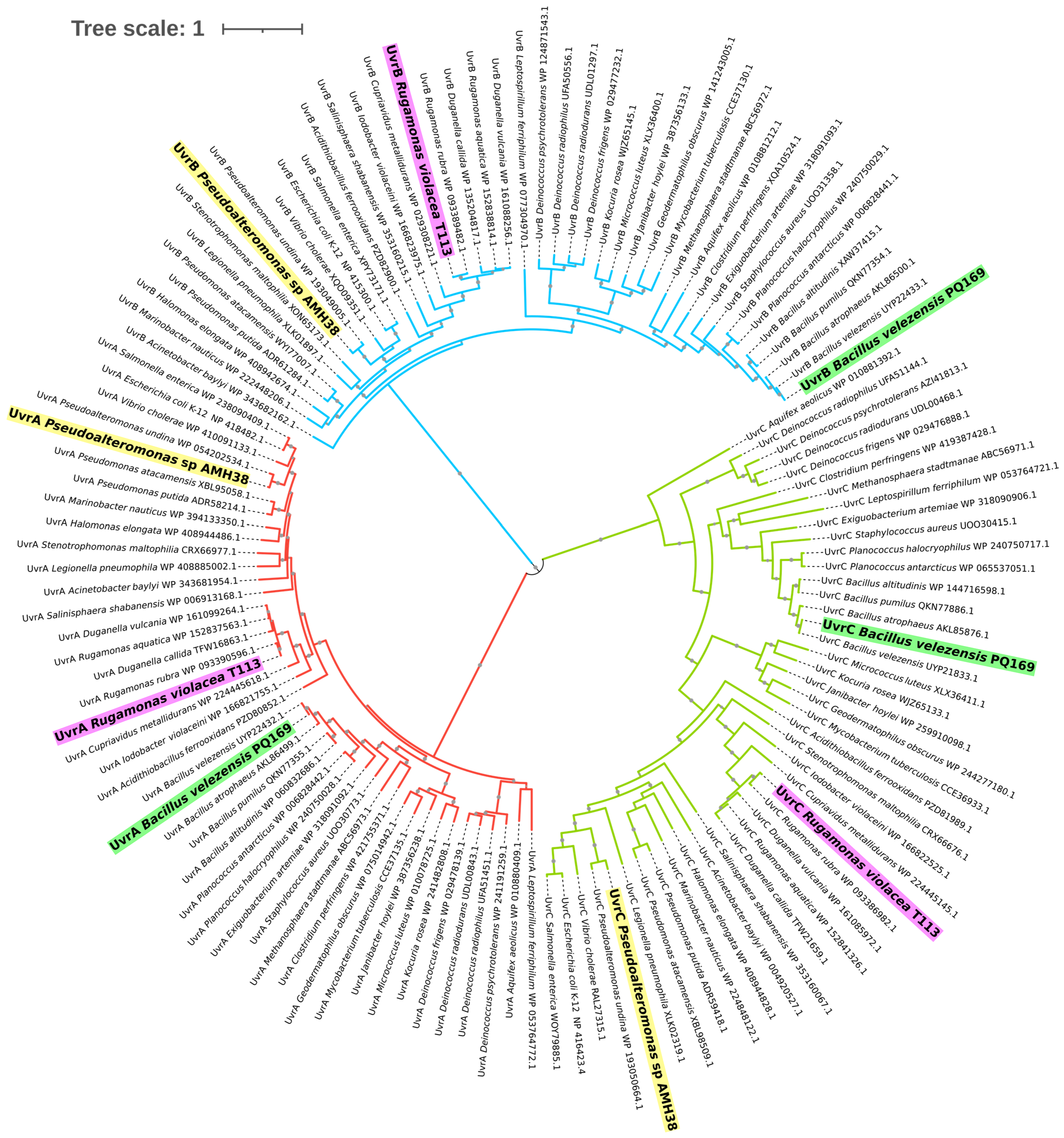
To gain a broader view of the NER system at the sequence level, we expanded our phylogenetic analysis to include amino acid sequences of UvrA, UvrB, and UvrC from a diverse panel of bacteria. UvrA initiates lesion recognition by scanning the DNA for damage, UvrB acts as a helicase verifying the damaged site, and UvrC provides endonuclease activity to excise UV-induced lesions [28]. Including these three components together allows for a more complete assessment of how the excinuclease ABC complex has evolved under different ecological and selective pressures [27,28,39]. Although each gene clusters into its own major branch corresponding to UvrA, UvrB, or UvrC, the same group of bacterial species was considered for each protein, enabling direct comparisons of their evolutionary trajectories under varying environmental pressures (Figure 2).
Across all three proteins, we observed a recurring pattern in which Gram-positive Bacillus species consistently grouped, reflecting strong selective constraints on DNA-repair functionality in taxa that endure desiccation, thermal fluctuations, and high UV exposure [40]. Halotolerant Gram-negative strains, such as Pseudoalteromonas sp. AMH3-8, formed a similarly cohesive set in each subunit’s cluster, suggesting that niche-specific adaptations, particularly those related to osmotic stress and UV tolerance, shape the evolution of the excinuclease system [39]. Pigmented and highly radiation-resistant bacteria, including R. violacea T1-13 and Deinococcus spp., appeared as distinct lineages within each major branch, indicating that protective pigments and robust DNA-repair pathways may coevolve to mitigate oxidative and UV-induced damage across phylogenetically diverse organisms [41,42,43].
Although key functional domains in UvrA, UvrB, and UvrC remain highly conserved, fine-scale sequence variations are apparent in isolates from extreme environments. This divergence is likely the result of convergent evolutionary pressures (e.g., intense solar radiation or extreme temperatures) that necessitate efficient and sometimes specialized DNA-repair mechanisms [39,41,44,45].
The fact that the same sets of strains occupy consistent positions in each Uvr subtree further underscores the importance of the excinuclease ABC system in supporting bacterial survival under harsh conditions. Taken as a whole, our findings demonstrate that while Uvr proteins retain a core functional architecture necessary for nucleotide excision repair, evolutionary modifications in each subunit may reflect fine-tuning to support DNA repair under environmental stressors found in deserts, polar regions, and hypersaline lakes. This comprehensive overview reinforces the value of our three environmental isolates, B. velezensis PQ169, Pseudoalteromonas sp. AMH3-8, and R. violacea T1-13, as comparative models for investigating how bacteria fine-tune the NER pathway to thrive in extreme and highly dynamic ecosystems.
These phylogenetic patterns suggest that, while the functional core of NER proteins is conserved, environmental pressures such as intense UV radiation, desiccation, and osmotic stress may drive adaptive divergence in flexible or regulatory domains. Such changes could affect DNA-damage recognition, protein–protein interactions, or expression dynamics, offering strain-specific optimization under distinct stress regimes. This supports the idea that the NER system, though functionally conserved, remains evolutionarily responsive to ecological challenges across extreme environments.

2.2.2. Photolyases and Structural Insights

Photolyases are flavoproteins that repair UV-induced DNA lesions such as cyclobutane pyrimidine dimers (CPDs) and (6–4) photoproducts, relying on a conserved cryptochrome/photolyase (PHR) domain that binds flavin adenine dinucleotide (FAD) as a cofactor [16,25,46,47]. Absorption of light by FAD promotes an electron transfer reaction that restores the native DNA structure, thus maintaining genome stability [25]. While well-characterized in certain prokaryotic model organisms, photolyases are increasingly recognized across diverse bacterial species [15,48,49].
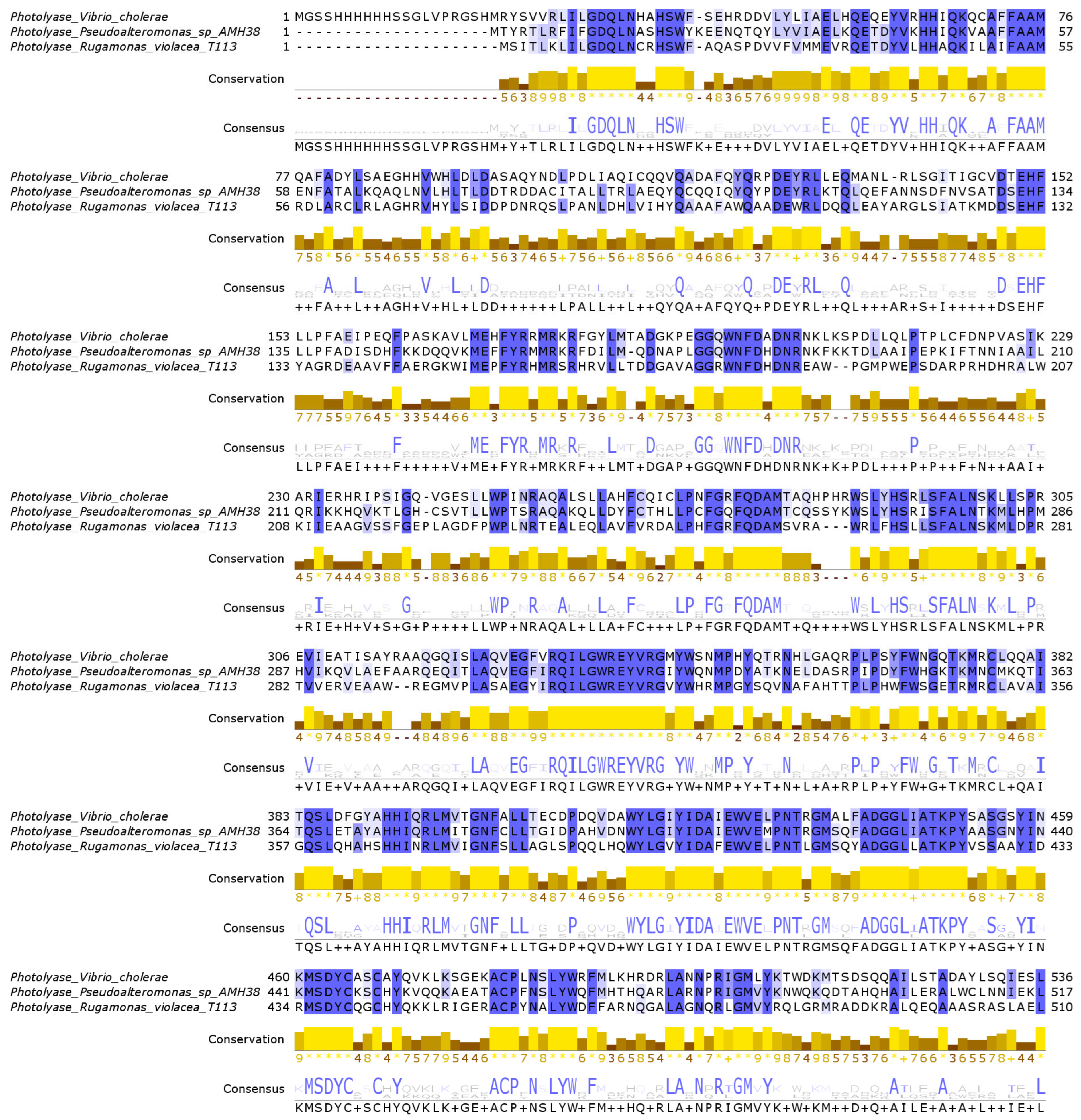
In the genomes of Pseudoalteromonas sp. AMH3-8 and R. violacea T1-13, we identified two genes that potentially encode deoxyribodipyrimidine photolyase-related proteins. To evaluate their sequence-level similarities, we generated a multiple sequence alignment (MSA) with Vibrio cholerae photolyase (PDB ID: 7YKN) as a reference (Figure 3). In this alignment, colored shading marks positions at or above 50% identity among the three sequences, while several sites display complete (100%) identity. These highly conserved residues are distributed across multiple regions of the alignment, reflecting the hallmark motifs of photolyases [50,51].
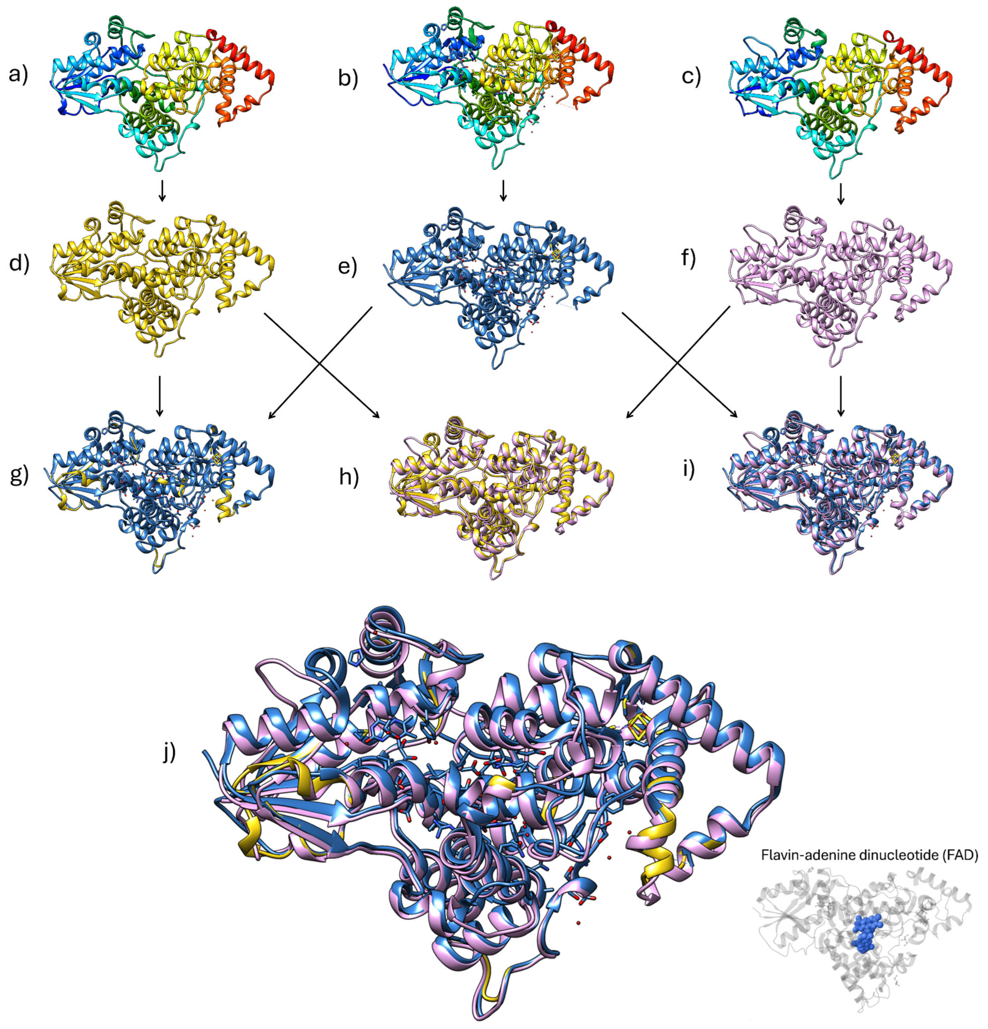
Subsequently, to gain insight into the structural relevance of these conserved residues, we performed three-dimensional (3D) modeling of the Pseudoalteromonas sp. AMH3-8 and Rugamonas violacea T1-13 photolyase-like proteins, using Phyre 2.2 and SWISS-MODEL, and compared the models to the crystal structure of the Vibrio cholerae photolyase (Figure 4). To further quantify the degree of structural similarity, we conducted pairwise Root Mean Square Deviation (RMSD) analysis, which underscores a highly conserved core domain pivotal to their putative biological function [52,53].
The comparison of V. cholerae photolyase and the Pseudoalteromonas sp. AMH3-8 model revealed a remarkably low pruned RMSD of 0.148 Å (492 atom pairs), which increased only slightly to 0.609 Å when all 505 equivalent positions were considered. This finding strongly suggests that Pseudoalteromonas sp. AMH3-8 shares an exceptionally well conserved folding pattern and active site architecture with V. cholerae, implying a near-identical arrangement of key residues crucial for FAD binding and photoreactivation.
In contrast, when V. cholerae was compared to the R. violacea T1-13 model, the pruned RMSD rose to 0.411 Å (432 atom pairs), and the global RMSD reached 2.138 Å (494 atom pairs). Although 0.411 Å still indicates a robust overlap in the core photolyase domain, the higher global RMSD suggests greater divergence in flexible loops or other peripheral regions [53]. A similar trend emerged from the comparison between Pseudoalteromonas sp. AMH3-8 and R. violacea T1-13, with pruned and global RMSDs of 0.444 Å (432 atom pairs) and 2.125 Å (503 atom pairs), respectively. These results imply that, while R. violacea T1-13 and Pseudoalteromonas sp. AMH3-8 maintain a core domain consistent with canonical photolyase structure, they likely differ somewhat in loop length or orientation relative to one another and to V. cholerae.
Despite these variations, all three proteins exhibit low RMSD values in their pruned (core) regions, indicating a high degree of structural conservation critical for the binding of the FAD cofactor and the facilitation of the electron transfer mechanism that underlies photoreactivation. Collectively, these observations reinforce the hypothesis that the Pseudoalteromonas sp. AMH3-8 and R. violacea T1-13 proteins preserve the fundamental fold and active site configuration characteristic of the photolyase family proteins [48,49]. Such close resemblance to a well-studied photolyase (V. cholerae) offers compelling evidence that both novel proteins likely retain the biological function of UV-induced DNA repair [15]. Moreover, given that photolyases have been shown to play pivotal roles in genomic maintenance and UV-stress responses, especially in environments with fluctuating UV exposure [16,48], these findings point to a potentially conserved photoreactivation pathway among these species. Nevertheless, further biochemical analyses, for instance, characterizing UV-induced DNA-repair activity in vitro, will be indispensable to confirm the functional predictions drawn from these in silico models.
Although the predicted structures exhibit high-confidence RMSD values and preserve conserved active site features, it is important to recognize the limitations inherent to in silico modeling. These predictions do not account for dynamic conformational changes, post-translational modifications, or context-dependent interactions in vivo. Therefore, future studies should focus on conducting gene expression analyses of these photolyase-like proteins and evaluating their enzymatic activity through in vitro UV-induced DNA-repair assays in order to experimentally validate their function and structural significance.
Furthermore, while the core architecture and FAD-binding domains of the modeled photolyases are highly conserved, the observed structural differences—particularly in loop regions and peripheral domains—may reflect functional specialization. These variations could modulate substrate affinity, DNA-binding kinetics, or interactions with auxiliary proteins involved in the UV-stress response. Although speculative, such divergence may result in differential repair efficiency or regulatory behavior under varying environmental conditions. Site-directed mutagenesis of these divergent regions, combined with in vitro photoreactivation assays, would be valuable for experimentally assessing the functional impact of the predicted structural variations.

2.3. Pigment-Based Mechanisms

Pigments play a crucial role in protecting cells against UV-induced damage by absorbing harmful wavelengths and scavenging reactive oxygen species, thereby contributing to an enhanced oxidative stress response. In our study, we identified a complete violacein operon in the genome of Rugamonas violacea T1-13, a bacterial isolate from the Antarctic environment. Notably, although we screened the genomes of the other two isolates for pigment-associated genes, we did not find a similarly organized gene cluster. Instead, only scattered genes potentially involved in pigmentation were detected. This suggests that R. violacea T1-13 possesses a specialized and coherent violacein biosynthetic pathway, which may confer enhanced photoprotective properties compared to the other strains. As detailed in Figure 5, a well-conserved gene cluster encodes the enzymes responsible for violacein biosynthesis. This operon structure is consistent with previously characterized violacein gene clusters in other bacteria, suggesting that R. violacea T1-13 can synthesize this pigment [23,54].
The stepwise conversion of L-tryptophan to violacein involves intermediate metabolites and highlights the sequential enzymatic reactions mediated by the vio gene products. The presence of a complete and organized violacein biosynthetic pathway supports the hypothesis that R. violacea T1-13 leverages pigment production as a protective mechanism against high levels of UV radiation commonly encountered in its native Antarctic habitat. Given violacein’s known antioxidant properties and its ability to absorb UV light, it is plausible that this pigment not only contributes to the cellular defense against UV-induced DNA damage but also to mitigating oxidative stress in extreme environments [17]. Collectively, these findings extend our understanding of the multifaceted UV-resistance strategies in extremophilic bacteria by illustrating how pigment biosynthesis integrates with other DNA-repair and stress-response pathways to ensure survival under adverse environmental conditions [17,55,56]. Future studies involving biochemical assays to confirm violacein production and its protective efficacy under UV stress will be critical to fully elucidate its role within the broader context of environmental adaptation in these microorganisms.

2.4. Sporulation as an Adaptive Response

Endospore formation (sporulation) represents one of the most efficient bacterial strategies to cope with severe environmental stresses, including high levels of UV radiation [31]. In our genomic analysis of Bacillus velezensis PQ169, we identified the complete genetic machinery involved in sporulation (Figure 6), which is in line with the prediction that this mechanism may be important for adaptation and survival in the highly irradiated Atacama Desert environment [4].
Sporulation initiation in Bacillus spp. typically occurs upon sensing stressful conditions, such as nutrient limitation or DNA damage induced by UV radiation [57]. In B. velezensis PQ169, this process begins with sensor kinases (KinA, KinB, and KinC) located at the cell membrane, detecting environmental stress signals, illustrated here as UV radiation-induced DNA damage (Figure 6a). These kinases subsequently activate a phosphorelay system composed of Spo0F, Spo0B, and Spo0A, culminating in phosphorylation and activation of Spo0A, the master transcriptional regulator for sporulation.
Active Spo0A induces asymmetric cell division, generating a mother cell and a forespore (Figure 6b). Each compartment initiates a distinct transcriptional program controlled by specific sigma factors (σ). In the forespore, σF drives the early gene expression that is crucial for spore development, followed by σG activity, which regulates the late stages, including cortex and spore coat synthesis. The mother cell sequentially activates σE and σK, coordinating the synthesis of protective layers around the developing spore, ultimately leading to cell lysis and spore release [58,59].
The structural integrity and biochemical resilience of bacterial spores are particularly relevant under UV stress. Spore formation involves the synthesis of UV-absorbing compounds (e.g., dipicolinic acid complexed with calcium ions), protective protein layers, and dehydration of the core, collectively conferring exceptional resistance to UV-induced DNA damage. These layers effectively shield genomic DNA from direct UV exposure, greatly diminishing photoproduct formation and subsequent DNA lesions [29,30].
In addition, recent studies have shown that sporulation in Bacillus is closely linked to RecA-mediated DNA-damage responses. RecA, beyond its canonical role in homologous recombination and SOS induction, plays regulatory and protective functions during sporulation. Activation of the SOS response depends on RecA and occurs in both the mother cell and forespore in response to UV and alkylation-induced DNA lesions, helping maintain genome integrity throughout development [60]. Furthermore, RecA has been implicated in modulating Spo0A phosphorylation levels during sporulation initiation and in repressing DNA replication at later stages, acting as a developmental checkpoint [32]. Importantly, inactivation of RecA or its accessory factors (RecF, RecO, RecR, and RecX) drastically reduces the survival of mature spores exposed to extreme stresses, such as ultrahigh vacuum desiccation and ionizing radiation, which cause single- and double-strand DNA breaks [61]. These findings underscore the essential role of RecA not only in stress-induced DNA repair but also in coordinating damage responses with sporulation progression and spore resilience.
Notably, the extensive genomic investment in sporulation-related genes observed in B. velezensis PQ169 suggests that spore formation is not merely an incidental protective measure, but a fundamental evolutionary adaptation to prolonged and intense UV radiation exposure characteristic of the Atacama Desert. This hypothesis aligns with previous observations in related Bacillus species, where sporulation has been confirmed as a primary adaptive response for survival under multiple environmental stresses, particularly desiccation and radiation [29,30,32,62]. Thus, our findings highlight the significance of the sporulation mechanism as a pivotal strategy in UV-stress resistance in Bacillus velezensis PQ169, underpinning the bacterium’s ecological success and persistence in one of Earth’s harshest environments.
Unlike previous studies that have focused on metagenomic surveys or on a single-model extremophile, this study provides an integrated genome level and structural characterization of UV resistance in three phylogenetically and ecologically distinct bacterial strains. By combining comparative genomics, gene-content profiling, structural modeling, and phylogenetic analyses, we provide new insights into how specific UV-resistance mechanisms may be shaped by environmental pressures such as desiccation, salinity, and temperature extremes. Furthermore, the inclusion of cultivated isolates from underexplored ecosystems such as the Chilean Atacama Desert, Altiplano, and Antarctic soils allows us to propose functional hypotheses grounded in isolate-level data, which can serve as a basis for future experimental validation and broader ecological comparisons.
While this study provides a comprehensive in silico characterization of UV-resistance mechanisms in three extremophilic bacterial strains, it is important to acknowledge its limitations. The predictions presented here, including gene annotations, structural models, and phylogenetic inferences, were derived from computational analyses and are not experimentally validated. Although the methodologies used are well established and supported by cross-validation (e.g., dual annotation pipelines and RMSD comparisons), functional confirmation of the proposed mechanisms remains necessary. Future work should include experimental validation of DNA-repair activity (e.g., through UV survival assays or gene knockouts); biochemical characterization of key proteins, such as photolyases; and transcriptomic analyses under UV stress to evaluate gene expression dynamics. These approaches will help test and refine the hypotheses generated in this study and strengthen our understanding of microbial adaptation to UV radiation in extreme environments.

3. Materials and Methods

3.1. Bacterial Isolates and Genomic DNA Extraction

The strains used in this study were previously isolated by the Microbiology Laboratory at the Pontificia Universidad Católica de Valparaíso. The geographic origins of the isolates were as follows: strain PQ169 from the Atacama Desert (27°6′36″ S, 69°43′37″ W), strain AMH3-8 from the Salar de Huasco (20°17′01″ S; 68°53′21″ W), and strain T1-13 from Antarctica (62°28′54″ S, 59°37′49″ W). Genomic DNA extraction was performed according to the protocol described by Chen et al. [63], with a minor adaptation for strain PQ169. Specifically, the cell pellet was pre-incubated with 25 µL of a 10 mg/mL lysozyme solution at 37 °C for 45 min before resuspension. DNA quality was assessed by agarose gel electrophoresis, and DNA concentration was measured using Qubit Fluorometer (Thermo Fisher Scientific Inc., Waltham, MA, USA).

3.2. Genome Sequencing, Assembly, Annotation, and ANIm-Based Taxonomic Analysis

For genome sequencing, the DNA were sent to TCL Group (Santiago, Chile) for library preparation and sequencing. DNA libraries were prepared using the MGIEasy FS DNA Library Prep Set and the MGIEasy Circularization Module V2.0 (MGI Tech, Tokyo, Japan). Sequencing was performed on the DNBSEQ-G400 platform using the DNBSEQ-G400RS High-Throughput Sequencing Set (MGI), with 150 bp paired-end reads to ensure high-quality, high-depth coverage. Quality controls were performed using FastQC v0.11.9 to assess the integrity and quality of the raw reads. Trimmed reads were generated using Trimmomatic v0.39 software. The genome assembly was then carried out de novo for all three isolates using Velvet v1.2.10. After assembly, genome annotation was performed with Prokka v1.14.6, which provided functional annotation of the assembled genomes. Circular genome maps were generated for each genome using GenoVI v0.2.16. For taxonomic assignment, an AutoMLST https://automlst.ziemertlab.com/ (accessed on 25 May 2025) analysis was performed to define the closest species based on core genome multilocus sequence typing. Next, an in silico hybridization was carried out using TYGS v400 (The Type Strain Genome Server) (https://tygs.dsmz.de/) to compare the genomes against available type strains. Finally, the average nucleotide identity (ANIm) was calculated using JSpeciesWS v4.3.2 (http://jspecies.ribohost.com/jspeciesws/). The ANIm approach that uses MUMmer (NUCmer) was used to align the input sequences. The ANIm graphical representation was generated using Morpheus https://software.broadinstitute.org/morpheus/ (accessed on 25 May 2025), which enabled the visualization and comparison of the ANIm values between the isolates and reference species. Additionally, the annotated genomes were uploaded to the RAST Server (Rapid Annotations using Subsystems Technology https://rast.nmpdr.org/ (accessed on 25 May 2025) to further explore gene functions and metabolic subsystems. This complementary approach allowed for cross-verification of annotation results obtained from Prokka and provided an interactive platform for visualizing and analyzing the predicted pathways and functional categories in each assembled genome.

3.3. Phylogenetic Analysis of UV Resistance-Related Proteins

Protein sequences of NER system (UvrA, UvrB, and UvrC) were retrieved from the National Center for Biotechnology Information (NCBI) database https://www.ncbi.nlm.nih.gov (accessed on 25 May 2025) according to their annotated gene names and accession numbers. The sequences were compiled into FASTA files, and multiple sequence alignments were performed in MEGA 12 (https://www.megasoftware.net), using the MUSCLE algorithm, with the following parameters: gap open = −2.90, gap extend = 0.00, hydrophobicity multiplier = 1.20, and UPGMA as the clustering method. When necessary, additional alignments were carried out on the Phylogeny.fr platform http://www.phylogeny.fr/ (accessed on 25 May 2025) and further refined in Jalview v2.11.4.1 (https://www.jalview.org). Phylogenetic trees were constructed under the maximum likelihood criterion with 1000 bootstrap replicates, with the relative bootstrap values indicating node support. For clarity and improved visualization, the resulting trees were uploaded and edited in the Interactive Tree of Life (iTOL v7) (https://itol.embl.de). Each sequence in the final tree is labeled with its respective protein name (UvrA, UvrB, or UvrC), bacterial species, and NCBI accession number.

3.4. Protein Structure Analysis

Proteins’ structural models (photolyases) were generated using SWISS-MODEL https://swissmodel.expasy.org (accessed on 25 May 2025) and Phyre 2.2 (https://www.sbg.bio.ic.ac.uk/phyre2), referencing the Vibrio cholerae photolyase (PDB ID: 7YKN https://www.ebi.ac.uk/pdbe/entry/pdb/7ykn) to guide template selection. Phyre predictions achieved 100% confidence for over 97% coverage of each query sequence. The predicted three-dimensional structures were visualized in UCSF Chimera v1.19 (https://www.cgl.ucsf.edu/chimera/), and selected models were further validated for structural consistency using the CONFORT tool integrated into the I-TASSER pipeline https://zhanggroup.org/I-TASSER/ (accessed on 25 May 2025). By combining these high-confidence structural predictions with sequence-based phylogenetics, we established a robust framework for examining both the evolutionary relationships and the potential functional implications of the photolyases in our isolates.

4. Conclusions

This study presents a comprehensive analysis of UV-resistance mechanisms in three extremophilic bacteria from distinct Chilean arid environments: highlands, desert, and Antarctica. Genomic and structural data revealed key adaptations, such as robust DNA-repair systems, photolyases, pigment biosynthesis, and sporulation, reflecting diverse strategies that may have been shaped by environmental UV stress. Phylogenetic analyses of NER components showed conserved evolutionary patterns, while structural modeling of photolyases in Pseudoalteromonas sp. AMH3-8 and Rugamonas violacea T1-13, along with the presence of a complete violacein operon in R. violacea T1-13, highlight functional adaptations to UV exposure. In Bacillus velezensis PQ169, a complete sporulation program supports endospore formation as a key survival mechanism in hyper-arid, high-irradiance conditions. Collectively, these findings deepen our understanding of microbial resilience under extreme radiation and reinforce the relevance of extremophiles as models in evolutionary biology and astrobiology.

Author Contributions

Conceptualization, M.N., A.N. and C.Y.; methodology, M.N. and A.N.; validation, M.N., A.N. and C.Y.; formal analysis, M.N., A.N. and C.Y.; investigation, M.N., A.N., F.C., C.R., G.C. and C.Y.; resources, G.C. and C.Y.; data curation, M.N., A.N. and C.Y.; writing—original draft preparation, M.N., A.N. and C.Y.; writing—review and editing, M.N., A.N. and C.Y.; visualization, M.N., A.N. and C.Y.; supervision, M.N., A.N. and C.Y.; project administration, G.C. and C.Y.; funding acquisition, G.C. and C.Y. All authors have read and agreed to the published version of the manuscript.

Funding

We are grateful for financial support from the Corporación Nacional Forestal de Chile (CONAF) through the grant FIBN-CONAF 020/2023 and the Vicerrectoría de Investigación, Creación e Innovación (VINCI) of the Pontificia Universidad Católica de Valparaíso (PUCV), under grant VINCI 122.794/2024. This research was partially supported by HPC OCÉANO (FONDEQUIP No. EQM170214).

Institutional Review Board Statement

No applicable.

Informed Consent Statement

No applicable.

Data Availability Statement

The data presented in this study are available upon request from the corresponding author due to legal restrictions (intellectual property).

Conflicts of Interest

The authors declare no conflicts of interest.

References

  1. Rampelotto, P.H. Extremophiles and extreme environments. Life 2013, 3, 482–485. [Google Scholar] [CrossRef] [PubMed]
  2. Khan, A.; Liu, G.; Zhang, G.; Li, X. Radiation-resistant bacteria in desiccated soil and their potentiality in applied sciences. Front. Microbiol. 2024, 15, 1348758. [Google Scholar] [CrossRef] [PubMed]
  3. Martin-Cuadrado, A.B.; Senel, E.; Martínez-García, M.; Cifuentes, A.; Santos, F.; Almansa, C.; Moreno-Paz, M.; Blanco, Y.; García-Villadangos, M.; García del Cura, M.A.; et al. Prokaryotic and viral community of the sulfate-rich crust from Peñahueca ephemeral lake, an astrobiology analogue. Environ. Microbiol. 2019, 21, 3577–3600. [Google Scholar] [CrossRef] [PubMed]
  4. Cordero, R.R.; Damiani, A.; Jorquera, J.; Sepúlveda, E.; Caballero, M.; Fernandez, S.; Feron, S.; Llanillo, P.J.; Carrasco, J.; Laroze, D.; et al. Ultraviolet radiation in the Atacama desert. Antonie Leeuwenhoek 2018, 111, 1301–1313. [Google Scholar] [CrossRef]
  5. Azua-Bustos, A.; González-Silva, C.; Fairén, A.G. The Atacama Desert in northern Chile as an analog model of Mars. Front. Astron. Space Sci. 2022, 8, 810426. [Google Scholar] [CrossRef]
  6. Azua-Bustos, A.; Urrejola, C.; Vicuña, R. Life at the dry edge: Microorganisms of the Atacama Desert. FEBS Lett. 2012, 586, 2939–2945. [Google Scholar] [CrossRef]
  7. Castro-Severyn, J.; Remonsellez, F.; Valenzuela, S.L.; Salinas, C.; Fortt, J.; Aguilar, P.; Pardo-Esté, C.; Dorador, C.; Quatrini, R.; Molina, F.; et al. Comparative genomics analysis of a new Exiguobacterium strain from Salar de Huasco reveals a repertoire of stress-related genes and arsenic resistance. Front. Microbiol. 2017, 8, 456. [Google Scholar] [CrossRef]
  8. Wierzchos, J.; de los Ríos, A.; Ascaso, C. Microorganisms in desert rocks: The edge of life on earth. Int. Microbiol. 2012, 15, 173–183. [Google Scholar] [CrossRef]
  9. Castro-Severyn, J.; Pardo-Esté, C.; Mendez, K.N.; Fortt, J.; Marquez, S.; Molina, F.; Castro-Nallar, E.; Remonsellez, F.; Saavedra, C.P. Living to the high extreme: Unraveling the composition, structure, and functional insights of bacterial communities thriving in the arsenic-rich Salar de Huasco altiplanic ecosystem. Microbiol. Spectr. 2021, 9, e0044421. [Google Scholar] [CrossRef]
  10. Hernández, K.L.; Yannicelli, B.; Olsen, L.M.; Dorador, C.; Menschel, E.J.; Molina, V.; Remonsellez, F.; Hengst, M.B.; Jeffrey, W.H. Microbial activity response to solar radiation across contrasting environmental conditions in Salar de Huasco, northern Chilean Altiplano. Front. Microbiol. 2016, 7, 1857. [Google Scholar] [CrossRef]
  11. Pérez, V.; Dorador, C.; Molina, V.; Yáñez, C.; Hengst, M. Rhodobacter sp. Rb3, an aerobic anoxygenic phototroph which thrives in the polyextreme ecosystem of the Salar de Huasco, in the Chilean Altiplano. Antonie Leeuwenhoek 2018, 111, 1449–1465. [Google Scholar] [CrossRef] [PubMed]
  12. Salwan, R.; Sharma, V. Genomics of prokaryotic extremophiles to unfold the mystery of survival in extreme environments. Microbiol. Res. 2022, 264, 127156. [Google Scholar] [CrossRef] [PubMed]
  13. Angelakis, G.N.; Psarologaki, C.; Pirintsos, S.; Kotzabasis, K. Extremophiles and extremophilic behaviour-new insights and perspectives. Life 2024, 14, 1425. [Google Scholar] [CrossRef] [PubMed]
  14. Cordero, R.R.; Feron, S.; Damiani, A.; Redondas, A.; Carrasco, J.; Sepúlveda, E.; Jorquera, J.; Fernandoy, F.; Llanillo, P.; Rowe, P.M.; et al. Persistent extreme ultraviolet irradiance in Antarctica despite the ozone recovery onset. Sci. Rep. 2022, 12, 1266. [Google Scholar] [CrossRef]
  15. He, Y.; Qu, C.; Zhang, L.; Miao, J. DNA photolyase from Antarctic marine bacterium Rhodococcus sp. NJ-530 can repair DNA damage caused by ultraviolet. 3 Biotech 2021, 11, 102. [Google Scholar] [CrossRef]
  16. Marizcurrena, J.J.; Morel, M.A.; Braña, V.; Morales, D.; Martinez-López, W.; Castro-Sowinski, S. Searching for novel photolyases in UVC-resistant Antarctic bacteria. Extremophiles 2017, 21, 409–418. [Google Scholar] [CrossRef]
  17. Silva, T.R.E.; Silva, L.C.F.J.; de Queiroz, A.C.; Alexandre Moreira, M.S.; de Carvalho Fraga, C.A.; de Menezes, G.C.A.; Rosa, L.H.; Bicas, J.; de Oliveira, V.M.; Duarte, A.W.F. Pigments from Antarctic bacteria and their biotechnological applications. Crit. Rev. Biotechnol. 2021, 41, 809–826. [Google Scholar] [CrossRef]
  18. Orellana, R.; Macaya, C.; Bravo, G.; Dorochesi, F.; Cumsille, A.; Valencia, R.; Rojas, C.; Seeger, M. Living at the frontiers of life: Extremophiles in Chile and their potential for bioremediation. Front. Microbiol. 2018, 9, 2309. [Google Scholar] [CrossRef]
  19. Meslier, V.; DiRuggiero, J. Endolithic microbial communities as model systems for ecology and astrobiology. In Model Ecosystems in Extreme Environments; Acdemic Press (Elsevier): Cambridge, MA, USA, 2019; pp. 145–168. [Google Scholar] [CrossRef]
  20. Dunlap, C.A.; Kim, S.-J.; Kwon, S.-W.; Rooney, A.P. Bacillus velezensis is not a later heterotypic synonym of Bacillus amyloliquefaciens; Bacillus methylotrophicus, Bacillus amyloliquefaciens subsp. plantarum and ‘Bacillus oryzicola’ are later heterotypic synonyms of Bacillus velezensis based on phylogenomics. Int. J. Syst. Evol. Microbiol. 2016, 66, 1212–1217. [Google Scholar] [CrossRef]
  21. Fan, B.; Wang, C.; Song, X.; Ding, X.; Wu, L.; Wu, H.; Gao, X.; Borriss, R. Bacillus velezensis FZB42 in 2018: The Gram-positive model strain for plant growth promotion and biocontrol. Front. Microbiol. 2018, 9, 2491. [Google Scholar] [CrossRef]
  22. Parrilli, E.; Tedesco, P.; Fondi, M.; Tutino, M.L.; Lo Giudice, A.; de Pascale, D.; Fani, R. The art of adapting to extreme environments: The model system Pseudoalteromonas. Phys. Life Rev. 2021, 36, 137–161. [Google Scholar] [CrossRef] [PubMed]
  23. Sedláček, I.; Holochová, P.; Sobotka, R.; Busse, H.-J.; Švec, P.; Králová, S.; Šedo, O.; Pilný, J.; Staňková, E.; Koublová, V.; et al. Classification of a violacein-producing psychrophilic group of isolates associated with freshwater in Antarctica and description of Rugamonas violacea sp. Nov. Microbiol. Spectr. 2021, 9, e0045221. [Google Scholar] [CrossRef] [PubMed]
  24. Vechtomova, Y.L.; Telegina, T.A.; Buglak, A.A.; Kritsky, M.S. UV radiation in DNA damage and repair involving DNA-photolyases and cryptochromes. Biomedicines 2021, 9, 1564. [Google Scholar] [CrossRef]
  25. Goosen, N.; Moolenaar, G.F. Repair of UV damage in bacteria. DNA Repair 2008, 7, 353–379. [Google Scholar] [CrossRef]
  26. Morita, R.; Nakane, S.; Shimada, A.; Inoue, M.; Iino, H.; Wakamatsu, T.; Fukui, K.; Nakagawa, N.; Masui, R.; Kuramitsu, S. Molecular mechanisms of the whole DNA repair system: A comparison of bacterial and eukaryotic systems. J. Nucleic Acids 2010, 2010, 179594. [Google Scholar] [CrossRef]
  27. Van Houten, B.; Kad, N. Investigation of bacterial nucleotide excision repair using single-molecule techniques. DNA Repair 2014, 20, 41–48. [Google Scholar] [CrossRef]
  28. Truglio, J.J.; Croteau, D.L.; Van Houten, B.; Kisker, C. Prokaryotic nucleotide excision repair: The UvrABC system. Chem. Rev. 2006, 106, 233–252. [Google Scholar] [CrossRef]
  29. Riesenman, P.J.; Nicholson, W.L. Role of the spore coat layers in Bacillus subtilis spore resistance to hydrogen peroxide, artificial UV-C, UV-b, and solar UV radiation. Appl. Environ. Microbiol. 2000, 66, 620–626. [Google Scholar] [CrossRef]
  30. Davis, G.M.; Greenhill, A.R.; Munro, T.W.; Horner, J.; Marsden, S.C.; Carter, B.D. Partial protection of Bacillus subtilis spores on simulated martian media enhances survival against UV radiation. Discov. Space 2025, 129, 3. [Google Scholar] [CrossRef]
  31. Mao, Y.; Yang, Y.; Lin, F.; Chu, H.; Zhou, L.; Han, J.; Zhou, J.; Su, X. Functional analysis of stress resistance of Bacillus cereus SCL10 strain based on whole-genome sequencing. Microorganisms 2024, 12, 1168. [Google Scholar] [CrossRef]
  32. Suárez, V.P.; Martínez, L.E.; Leyva-Sánchez, H.C.; Valenzuela-García, L.I.; Lara-Martínez, R.; Jiménez-García, L.F.; Ramírez-Ramírez, N.; Obregon-Herrera, A.; Cuéllar-Cruz, M.; Robleto, E.A.; et al. Transcriptional coupling and repair of 8-OxoG activate a RecA-dependent checkpoint that controls the onset of sporulation in Bacillus subtilis. Sci. Rep. 2021, 11, 2513. [Google Scholar] [CrossRef] [PubMed]
  33. Gaasbeek, E.J.; van der Wal, F.J.; van Putten, J.P.M.; de Boer, P.; van der Graaf-van Bloois, L.; de Boer, A.G.; Vermaning, B.J.; Wagenaar, J.A. Functional characterization of excision repair and RecA-dependent recombinational DNA repair in Campylobacter jejuni. J. Bacteriol. 2009, 191, 3785–3793. [Google Scholar] [CrossRef] [PubMed]
  34. Briaud, P.; Gautier, T.; Rong, V.; Mereghetti, L.; Lanotte, P.; Hiron, A. The Streptococcus agalactiae exonuclease ExoVII is required for resistance to exogenous DNA-damaging agents. J. Bacteriol. 2023, 205, e0002423. [Google Scholar] [CrossRef] [PubMed]
  35. On, Y.Y.; Welch, M. The methylation-independent mismatch repair machinery in Pseudomonas aeruginosa. Microbiology 2021, 167, 001120. [Google Scholar] [CrossRef]
  36. Blanchard, L.; de Groot, A. Coexistence of SOS-dependent and SOS-independent regulation of DNA repair genes in radiation-resistant Deinococcus bacteria. Cells 2021, 10, 924. [Google Scholar] [CrossRef]
  37. de Groot, A.; Blanchard, L. DNA repair and oxidative stress defense systems in radiation-resistant Deinococcus murrayi. Can. J. Microbiol. 2023, 69, 416–431. [Google Scholar] [CrossRef]
  38. Kim, S.R.; Maenhaut-Michel, G.; Yamada, M.; Yamamoto, Y.; Matsui, K.; Sofuni, T.; Nohmi, T.; Ohmori, H. Multiple pathways for SOS-induced mutagenesis in Escherichia coli: An overexpression of dinB/dinP results in strongly enhancing mutagenesis in the absence of any exogenous treatment to damage DNA. Proc. Natl. Acad. Sci. USA 1997, 94, 13792–13797. [Google Scholar] [CrossRef]
  39. Crowley, D.J.; Boubriak, I.; Berquist, B.R.; Clark, M.; Richard, E.; Sullivan, L.; DasSarma, S.; McCready, S. The uvrA, uvrB and uvrC genes are required for repair of ultraviolet light induced DNA photoproducts in Halobacterium sp. NRC-1. Saline Syst. 2006, 2, 11. [Google Scholar] [CrossRef]
  40. Au, N.; Kuester-Schoeck, E.; Mandava, V.; Bothwell, L.E.; Canny, S.P.; Chachu, K.; Colavito, S.A.; Fuller, S.N.; Groban, E.S.; Hensley, L.A.; et al. Genetic composition of the Bacillus subtilis SOS system. J. Bacteriol. 2005, 187, 7655–7666. [Google Scholar] [CrossRef]
  41. Subramani, G.; Srinivasan, S. Involvement of nucleotide excision repair and Rec-dependent pathway genes for UV radiation resistance in Deinococcus irradiatisoli 17bor-2. Genes 2023, 14, 1803. [Google Scholar] [CrossRef]
  42. Kurth, D.; Belfiore, C.; Gorriti, M.F.; Cortez, N.; Farias, M.E.; Albarracín, V.H. Genomic and proteomic evidences unravel the UV-resistome of the poly-extremophile Acinetobacter sp. Ver3. Front. Microbiol. 2015, 6, 328. [Google Scholar] [CrossRef] [PubMed]
  43. Dos Santos, A.; Schultz, J.; Almeida Trapp, M.; Modolon, F.; Romanenko, A.; Kumar Jaiswal, A.; Gomes, L.; Rodrigues-Filho, E.; Rosado, A.S. Investigating polyextremophilic bacteria in Al Wahbah crater, Saudi Arabia: A terrestrial model for life on Saturn’s moon Enceladus. Astrobiology 2024, 24, 824–838. [Google Scholar] [CrossRef] [PubMed]
  44. Kraithong, T.; Hartley, S.; Jeruzalmi, D.; Pakotiprapha, D. A peek inside the machines of bacterial nucleotide excision repair. Int. J. Mol. Sci. 2021, 22, 952. [Google Scholar] [CrossRef]
  45. Ellington, A.J.; Schult, T.J.; Reisch, C.R.; Christner, B.C. The genetic determinants of extreme UV radiation and desiccation tolerance in a bacterium recovered from the stratosphere. Microorganisms 2025, 13, 756. [Google Scholar] [CrossRef]
  46. Ramírez-Gamboa, D.; Díaz-Zamorano, A.L.; Meléndez-Sánchez, E.R.; Reyes-Pardo, H.; Villaseñor-Zepeda, K.R.; López-Arellanes, M.E.; Sosa-Hernández, J.E.; Coronado-Apodaca, K.G.; Gámez-Méndez, A.; Afewerki, S.; et al. Photolyase production and current applications: A review. Molecules 2022, 27, 5998. [Google Scholar] [CrossRef]
  47. Sancar, A. Structure and function of DNA photolyase. Biochemistry 1994, 33, 2–9. [Google Scholar] [CrossRef]
  48. Portero, L.R.; Alonso-Reyes, D.G.; Zannier, F.; Vazquez, M.P.; Farías, M.E.; Gärtner, W.; Albarracín, V.H. Photolyases and cryptochromes in UV-resistant bacteria from high-altitude Andean lakes. Photochem. Photobiol. 2019, 95, 315–330. [Google Scholar] [CrossRef]
  49. Walker, E.L.; Bose, J.L.; Stabb, E.V. Photolyase confers resistance to UV light but does not contribute to the symbiotic benefit of bioluminescence in Vibrio fischeri ES114. Appl. Environ. Microbiol. 2006, 72, 6600–6606. [Google Scholar] [CrossRef]
  50. Balaji, S.; Srinivasan, N. Comparison of sequence-based and structure-based phylogenetic trees of homologous proteins: Inferences on protein evolution. J. Biosci. 2007, 32, 83–96. [Google Scholar] [CrossRef]
  51. Pellegrini, M.; Marcotte, E.M.; Thompson, M.J.; Eisenberg, D.; Yeates, T.O. Assigning protein functions by comparative genome analysis: Protein phylogenetic profiles. Proc. Natl. Acad. Sci. USA 1999, 96, 4285–4288. [Google Scholar] [CrossRef]
  52. Damm, K.L.; Carlson, H.A. Gaussian-weighted RMSD superposition of proteins: A structural comparison for flexible proteins and predicted protein structures. Biophys. J. 2006, 90, 4558–4573. [Google Scholar] [CrossRef] [PubMed]
  53. Magala, P.; Klevit, R.E.; Thomas, W.E.; Sokurenko, E.V.; Stenkamp, R.E. RMSD analysis of structures of the bacterial protein FimH identifies five conformations of its lectin domain. Proteins 2020, 88, 593–603. [Google Scholar] [CrossRef] [PubMed]
  54. Kothari, V.; Sharma, S.; Padia, D. Recent research advances on Chromobacterium violaceum. Asian Pac. J. Trop. Med. 2017, 10, 744–752. [Google Scholar] [CrossRef] [PubMed]
  55. Órdenes-Aenishanslins, N.; Anziani-Ostuni, G.; Vargas-Reyes, M.; Alarcón, J.; Tello, A.; Pérez-Donoso, J.M. Pigments from UV-resistant Antarctic bacteria as photosensitizers in dye sensitized solar cells. J. Photochem. Photobiol. B 2016, 162, 707–714. [Google Scholar] [CrossRef]
  56. Reis-Mansur, M.C.P.P.; Cardoso-Rurr, J.S.; Silva, J.V.M.A.; de Souza, G.R.; Cardoso, V.d.S.; Mansoldo, F.R.P.; Pinheiro, Y.; Schultz, J.; Lopez Balottin, L.B.; da Silva, A.J.R.; et al. Carotenoids from UV-resistant Antarctic Microbacterium sp. LEMMJ01. Sci. Rep. 2019, 9, 9554. [Google Scholar] [CrossRef]
  57. Guerrero M., G.G. Sporulation, structure assembly, and germination in the soil bacterium Bacillus thuringiensis: Survival and success in the environment and the insect host. Microbiol. Res. 2023, 14, 466–491. [Google Scholar] [CrossRef]
  58. Al-Hinai, M.A.; Jones, S.W.; Papoutsakis, E.T. The Clostridium sporulation programs: Diversity and preservation of endospore differentiation. Microbiol. Mol. Biol. Rev. 2015, 79, 19–37. [Google Scholar] [CrossRef]
  59. Fisher, P.R. Microbial development. In Encyclopedia of Molecular Cell Biology and Molecular Medicine; Wiley-VCH Verlag GmbH & Co. KGaA: Weinheim, Germany, 2006. [Google Scholar] [CrossRef]
  60. Ramírez-Guadiana, F.H.; Barajas-Ornelas, R.d.C.; Corona-Bautista, S.U.; Setlow, P.; Pedraza-Reyes, M. The RecA-dependent SOS response is active and required for processing of DNA damage during Bacillus subtilis sporulation. PLoS ONE 2016, 11, e0150348. [Google Scholar] [CrossRef]
  61. Vlašić, I.; Mertens, R.; Seco, E.M.; Carrasco, B.; Ayora, S.; Reitz, G.; Commichau, F.M.; Alonso, J.C.; Moeller, R. Bacillus subtilis RecA and its accessory factors, RecF, RecO, RecR and RecX, are required for spore resistance to DNA double-strand break. Nucleic Acids Res. 2014, 42, 2295–2307. [Google Scholar] [CrossRef]
  62. Richardson, E.A.; Abruzzo, N.O.; Taylor, C.E.; Stevens, B.R.; Cuda, J.P.; Weeks, E.N.I. Combining DL-methionine and Bacillus thuringiensis subspecies israelensis: Prospects for a mosquito larvicide. Insects 2020, 11, 880. [Google Scholar] [CrossRef]
  63. Chen, W.P.; Kuo, T.T. A simple and rapid method for the preparation of gram-negative bacterial genomic DNA. Nucleic Acids Res. 1993, 21, 2260. [Google Scholar] [CrossRef]
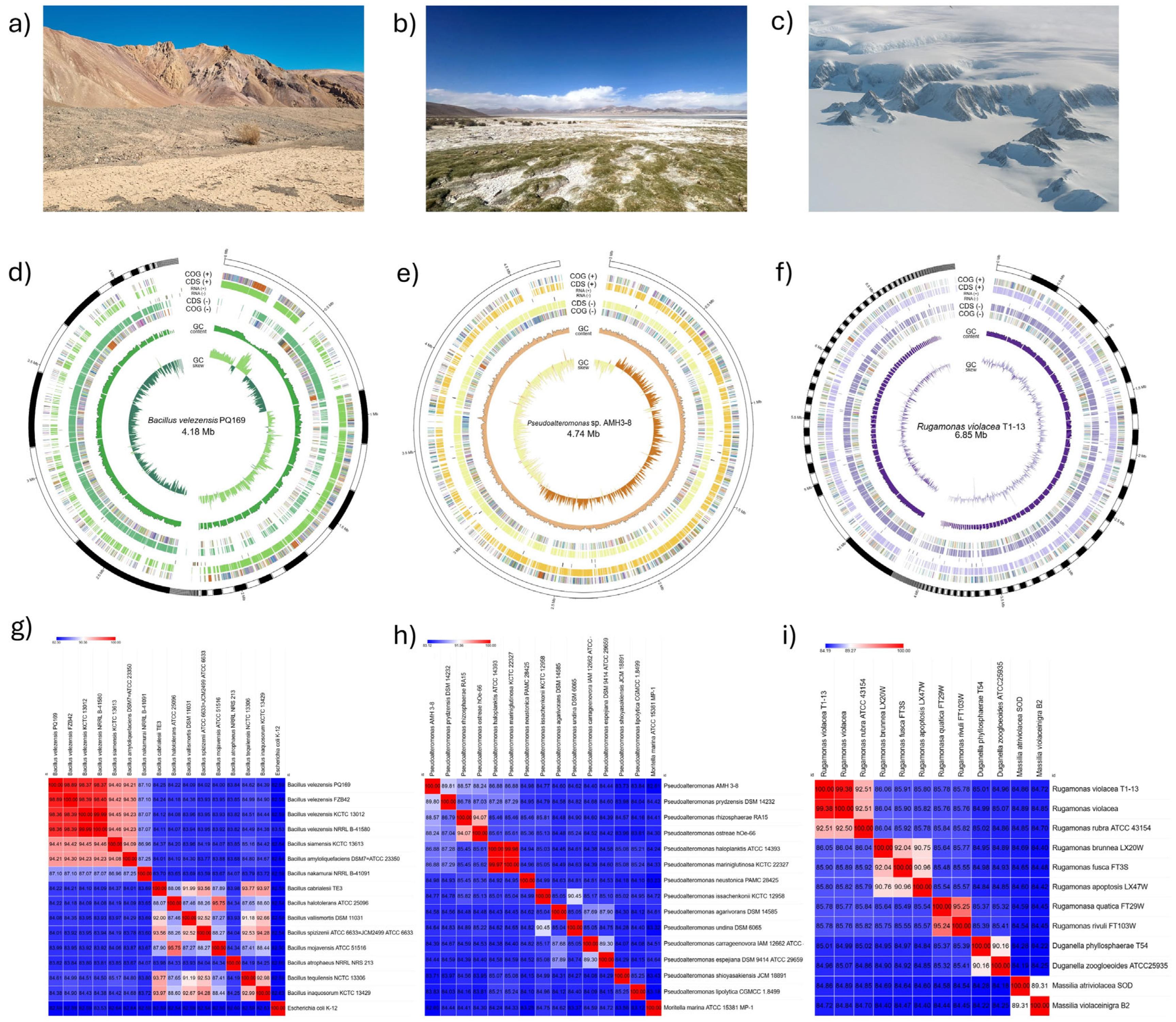
Figure 1. Isolation sites, genomic assembly characteristics, and ANI-based taxonomic assignment of three extremophilic bacterial isolates. (ac) Representative sampling sites of bacterial isolates: (a) Atacama Desert, (b) Salar de Huasco, and (c) Antarctic soil. (df) Circular genome representations illustrating variability in genome size, contiguity, and genetic content: (d) Bacillus velezensis PQ169 (76 contigs, 4.18 Mb), (e) Pseudoalteromonas sp. AMH3-8 (single contig, 4.74 Mb), and (f) Rugamonas violacea T1-13 (171 contigs, 6.85 Mb). Rings (outer to inner) represent predicted CDSs, RNA genes (rRNA, tRNA, and tmRNA), GC content, and GC skew. (gi) Heatmaps depicting average nucleotide identity based on MUMmer (ANIm) analysis between each isolate and closely related reference strains, supporting definitive taxonomic assignment: (g) Bacillus velezensis PQ169, (h) Pseudoalteromonas sp. AMH3-8, and (i) Rugamonas violacea T1-13. Color gradients indicate similarity percentages, with red color representing higher ANI values, and blue lower values.
Ijms 26 05842 g001
Figure 2. Phylogenetic tree based on the amino acid sequences of UvrA, UvrB, and UvrC proteins (NER system). Multiple sequence alignment was performed using MUSCLE on the Phylogeny.fr platform, followed by tree construction with PhyML and visualization using TreeDyn. The resulting tree was further arranged and refined using the Interactive Tree of Life (iTOL v7) tool. The maximum likelihood method was employed with 1000 bootstrap replicates, where the node sizes reflect the level of bootstrap support. Each label includes the gene name, the bacterial species, and the corresponding NCBI accession number. Three environmental strains are highlighted: Bacillus velezensis PQ-169 (green), Pseudoalteromonas sp. AMH3-8 (yellow), and Rugamonas violacea T1-13 (purple). These strains, along with other extremophilic, UV-resistant, and pathogenic bacteria, were included to compare UvrA (red clade), UvrB (blue clade), and UvrC (green clade) variations under extreme environmental conditions.
Ijms 26 05842 g002
Figure 3. Multiple sequence alignment between the photolyase of Vibrio cholerae and photolyase-related proteins from Pseudoalteromonas sp. AMH3-8 and Rugamonas violacea T1-13. The alignment was generated in MEGA12, using the MUSCLE algorithm, and visualized in Jalview, where blue shading indicates residues with over 50% conservation. The consensus sequence (bottom line) and the associated histogram (in yellow/brown) further highlight highly conserved regions, supporting the structural and functional similarities observed among these photolyase-like proteins.
Ijms 26 05842 g003
Figure 4. Comparative structural analysis of photolyase-related proteins from Pseudoalteromonas sp. AMH3-8, Vibrio cholerae, and Rugamonas violacea T1-13. (a) Three-dimensional (3D) model of the putative deoxyribodipyrimidine photolyase-related protein from Pseudoalteromonas sp. AMH3-8. (b) X-ray structure of the Vibrio cholerae photolyase (PDB ID: 7YKN), used as a reference in the structural comparisons. (c) Three-dimensional model of the photolyase-related protein from R. violacea T1-13. The recoloring (df) facilitates the identification of each model’s overall topology. (g) Superposition of the Pseudoalteromonas sp. AMH3-8 model and the V. cholerae photolyase, illustrating the high overall structural similarity. (h) Superposition of the Pseudoalteromonas sp. AMH3-8 and R. violacea T1-13 models, showing conserved folds. (i) Superposition of the V. cholerae photolyase and the R. violacea T1-13 model. (j) Combined overlay of all three structures (Pseudoalteromonas sp. AMH3-8, V. cholerae, and R. violacea T1-13), highlighting the putative FAD-binding pocket. These comparisons reveal a shared photolyase-like architecture among the three proteins, suggesting a conserved role in UV-induced DNA repair.
Ijms 26 05842 g004
Figure 5. Genetic organization and biosynthetic pathway of violacein in Rugamonas violacea T1-13. (a) Genomic arrangement of the violacein operon in R. violacea T1-13, showing the sequential organization of key biosynthetic genes (e.g., vioA, vioB, vioC, vioD, and vioE) within a ~6 kb region. Different colored arrows indicate individual genes and their transcriptional orientation. (b) Proposed biosynthetic pathway for violacein production, starting from L-tryptophan and progressing through various intermediates (such as IPA imine and protodeoxyviolaceinic intermediates) to yield the final product, violacein. The pathway diagram also identifies the cytoplasmic localization of pigment biosynthesis in this Gram-negative bacterium.
Ijms 26 05842 g005
Figure 6. Spore-formation cycle in Bacillus due to UV damage. (a) Activation of the UV damage-response pathway at the membrane, where kinases KinA, KinB, and KinC are activated, leading to the phosphorylation of Spo0A. This process triggers the differentiation of the mother cell and the forespore. (b) Stages of spore formation, including asymmetric division; engulfment of the forespore by the mother cell; and the roles of σF, σE, σG, and σK in spore maturation and the eventual lysis of the mother cell, leading to spore release and germination.
Ijms 26 05842 g006
Table 1. Presence of key DNA-repair components related to UV-induced damage in the genome of three environmental bacterial strains: Bacillus velezensis PQ169, Pseudoalteromonas sp. AMH3-8, and Rugamonas violacea T1-13. Components are categorized according to their role in nucleotide excision repair (NER), base excision repair (BER), mismatch repair (MMR), direct photoreactivation, oxidative damage prevention, or the SOS response. A checkmark (✓) indicates the presence of the protein based on genome annotation, and a dash (–) denotes its absence or undetected annotation.
DNA-Repair ComponentsFunctionB. velezensis PQ169Pseudoalteromonas sp. AMH3-8R. violacea T1-13
UvrAInitiates nucleotide excision repair (NER) by recognizing DNA damage.
UvrBParticipates in NER by verifying DNA damage and forming the incision complex.
UvrCMakes incisions on both sides of the DNA lesion in NER.
UvrDDNA helicase that removes the damaged DNA strand during NER.
PhrDNA photolyase that directly reverses UV-induced pyrimidine dimers using light energy.
RecAPromotes homologous recombination and SOS response by facilitating LexA autocleavage.
LexARepressor of SOS response; inactivated by RecA to induce DNA-repair genes.
MutSMismatch recognition protein involved in mismatch repair (MMR).
MutLActs as a matchmaker in MMR by coordinating repair proteins.
MutTPrevents incorporation of oxidized guanine (8-oxo-dGTP) into DNA.
MutMRemoves 8-oxoguanine from DNA via base excision repair (BER).
MutYRemoves adenines misincorporated opposite 8-oxoguanine (G:A mismatch repair).
PolBDNA polymerase II involved in translesion synthesis and repair.
DinBDNA polymerase IV, error-prone translesion polymerase involved in bypassing UV lesions.
Disclaimer/Publisher’s Note: The statements, opinions and data contained in all publications are solely those of the individual author(s) and contributor(s) and not of MDPI and/or the editor(s). MDPI and/or the editor(s) disclaim responsibility for any injury to people or property resulting from any ideas, methods, instructions or products referred to in the content.