Article

Yamabushitake Mushroom (Hericium erinaceus (Bull.) Pers. 1797) Mycelium Improves Reproductive System Dysfunction in Male Rats Induced by Polystyrene Microplastics

1
Department of Food Science, National Taiwan Ocean University, Keelung 20224, Taiwan
2
Department of Fisheries Product Technology, Faculty of Agriculture, Universitas Sriwijaya, Indralaya 30662, Indonesia
3
Institute of Marine Environment and Ecology, National Taiwan Ocean University, Keelung 20224, Taiwan
*
Authors to whom correspondence should be addressed.
Int. J. Mol. Sci. 2025, 26(12), 5735; https://doi.org/10.3390/ijms26125735
Submission received: 8 May 2025 / Revised: 10 June 2025 / Accepted: 13 June 2025 / Published: 15 June 2025
(This article belongs to the Section Molecular Pharmacology)

Abstract

The use of plastic products has increased, leading to higher levels of plastic pollution, and it is becoming a major public health concern. Health risks—especially those related to reproductive system dysfunction caused by polystyrene microplastics (PS-MPs)—are emerging issues that require urgent attention. This study aimed to investigate the effects of erinacine A-enriched Hericium erinaceus mycelium (HE) on high-fat-diet- and PS-MP-induced reproductive system dysfunction in male rats. Reproductive dysfunction was induced by administering a high-fat diet followed by exposure to PS-MPs for six weeks. The results showed that HE treatment significantly reduced nitric oxide levels and enhanced glutathione peroxidase activity. Furthermore, HE supplementation significantly downregulated pro-inflammatory cytokines such as interleukin (IL)-6 and IL-1β. Additionally, HE treatment significantly increased Kiss1 concentration, upregulated follicle-stimulating hormone and testosterone levels, reduced the area of the seminiferous tubule lumen, and prevented a reduction in epithelial thickness. HE treatment also significantly increased sperm count and reduced sperm abnormalities. Based on these findings, HE supplementation helps prevent reproductive system dysfunction by reducing oxidative stress and pro-inflammatory cytokines. Therefore, erinacine A-enriched H. erinaceus mycelium could be considered a potential food supplement or functional food ingredient for the treatment of reproductive or testicular dysfunction.

Graphical Abstract

1. Introduction

As modern lifestyles evolve, the consumption of plastic products has increased, resulting in higher levels of plastic pollution. Plastic pollution, particularly in the form of microplastics, has become a significant environmental and public health issue [1]. Microplastics (MPs) are small fragments of plastic, ranging from 1 μm to 5 mm in size. Key sources of microplastics include polyethylene bags, packaging materials, cosmetics, plastic containers, electrical devices, glass, and various other products [2]. Humans can be exposed to microplastics through the consumption of contaminated water and food or by inhaling particles present in the air. Once ingested or inhaled, microplastics can lead to a range of harmful effects, including immune system disruptions, an increased risk of reproductive toxicity, digestive issues, liver toxicity, and cancer [3,4,5].
Polystyrene microplastics (PS-MPs) are a type of microplastic commonly found alongside other major plastics such as polypropylene, polyvinyl chloride, and polyethylene [6]. Animal studies have shown that PS-MPs induce the production of pro-inflammatory factors, including interleukins (IL)-6, IL-1β, and tumor necrosis factor (TNF)-α, leading to abnormalities in sperm quality in mice [2,7]. Polystyrene microplastics have also been linked to male reproductive system dysfunctions [4,8,9]. A previous study reported that PS-MPs decreased testosterone levels in mouse models [10,11]. Additionally, previous research has reported that PS-MPs cause disruptions in spermatogenesis, impair sperm quality, and trigger testicular tissue inflammation in mice [6,12]. Moreover, PS-MPs can induce oxidative stress, which further decreases sperm quality and disrupts the blood–testis barrier [13]. Consequently, the health risks, particularly reproductive damage associated with PS-MPs exposure, are emerging concerns that require urgent attention.
Hericium erinaceus (Bull.) Pers. 1797 is an edible mushroom that has been recognized as a traditional medicine, especially in the East Asian region. This mushroom has traditionally been used as both food and medicine in China, Japan, and Korea [14]. It is also known as Houtougu in China, Yamabushitake in Japan, and Norugungdengi in Korea. It belongs to the Hericiaceae family in the Basidiomycetes class [15,16]. This mushroom contains numerous bioactive metabolites, including high-molecular-weight substances such as polysaccharides and polyphenols, as well as low-molecular-weight substances such as erinacines and hericenones [17]. A previous study has reported that around 253 metabolites are known from Hericium species, including cyathane diterpenes, steroids, benzofurans, alkaloids, phenols, chromenes, pyrones, and other miscellaneous metabolites [18]. Erinacine A is a bioactive cyathane diterpenoid found in the H. erinaceus mycelia [19]. Hericium erinaceus extracts reduce oxidative stress and regulates inflammatory cytokines, such as inducible nitric oxide synthase (iNOS), interleukin (IL)-1β, IL-6, and tumor necrosis factor (TNF)-α, in both cell and animal models [20,21]. It also exhibits a protective effect against hydrogen peroxide-induced apoptosis [22]. Additionally, the diterpenoids erinacine A, B, and C in H. erinaceus mycelium have shown potential therapeutic benefits [23,24]. Moreover, erinacine A can pass through the blood–brain barrier in rats to support the development of H. erinaceus as a functional food for neurohealth improvement [25]. Based on these previous studies, we hypothesized that H. erinaceus mycelium could also improve reproductive system dysfunction in a male rat model. However, no such study has been reported in the literature. Therefore, this study aimed to investigate the protective effects of erinacine A-enriched Hericium erinaceus mycelium on reproductive system dysfunction induced by a high-fat diet and polystyrene microplastics in male rats. This study provides the first in vivo evidence of its protective role against PS-MP-induced male reproductive dysfunction. It also aimed to evaluate the effects of erinacine A-enriched H. erinaceus mycelium on oxidative stress, inflammatory cytokines, reproductive hormones, and sperm morphology and quality.

2. Results

2.1. Effects of Erinacine A-Enriched H. erinaceus Mycelium (HE) on the Organ Weights of the Rats

The effects of erinacine A-enriched H. erinaceus mycelium (HE) on the weight of the kidney, spleen, abdominal fat, epididymal fat, and testes in rats are shown in Table 1. No significant differences (p > 0.05) were observed among the groups after 6 weeks of HE treatment.

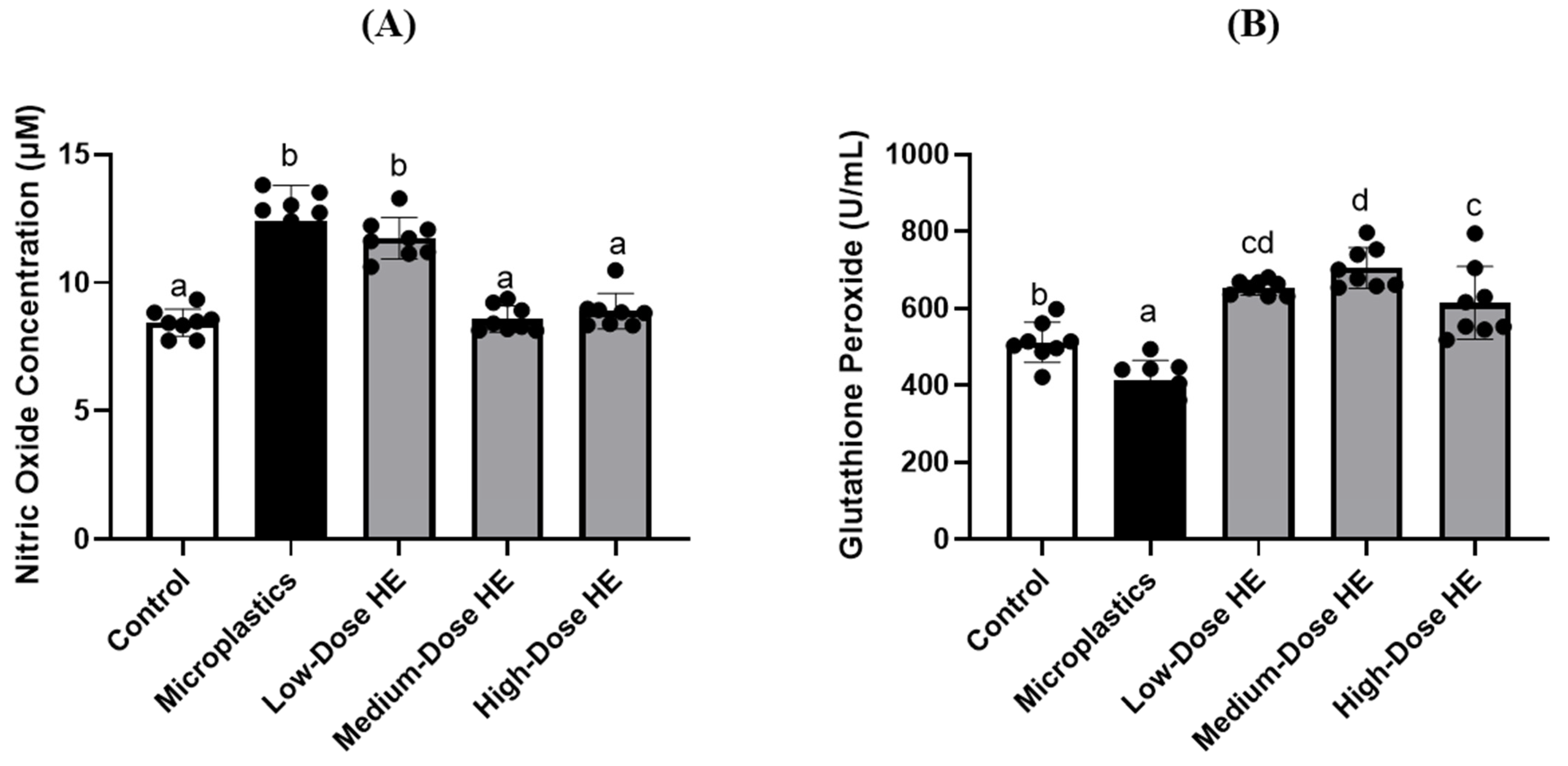
2.2. HE Supplementation Regulates Plasma Nitric Oxide Concentration and Glutathione Peroxidase Activity

The nitric oxide (NO) level in the microplastics group was significantly increased (p < 0.05) compared to the control and treated groups (Figure 1). Conversely, glutathione peroxidase (GPx) activity was significantly decreased (p < 0.05) in the microplastics group compared to the control and treated groups. After 6 weeks of treatment with HE, NO levels were significantly reduced (p < 0.05), particularly with medium and high doses of HE, compared to the microplastics group. In contrast, GPx activity was significantly increased (p < 0.05).

2.3. HE Reduces the Levels of Interleukin-6 and Interleukin-1β in Rat Plasma

The interleukin (IL)-6 level significantly (p < 0.05) increased in the microplastics group compared to the control and treated groups (Figure 2). However, this level significantly (p < 0.05) decreased after treatment with different doses of HE for 6 weeks. As shown in Figure 2, the level of IL-1β also significantly (p < 0.05) increased in the microplastics group compared to the control group. However, treatment with the medium dose of HE significantly (p < 0.05) decreased the IL-1β level.

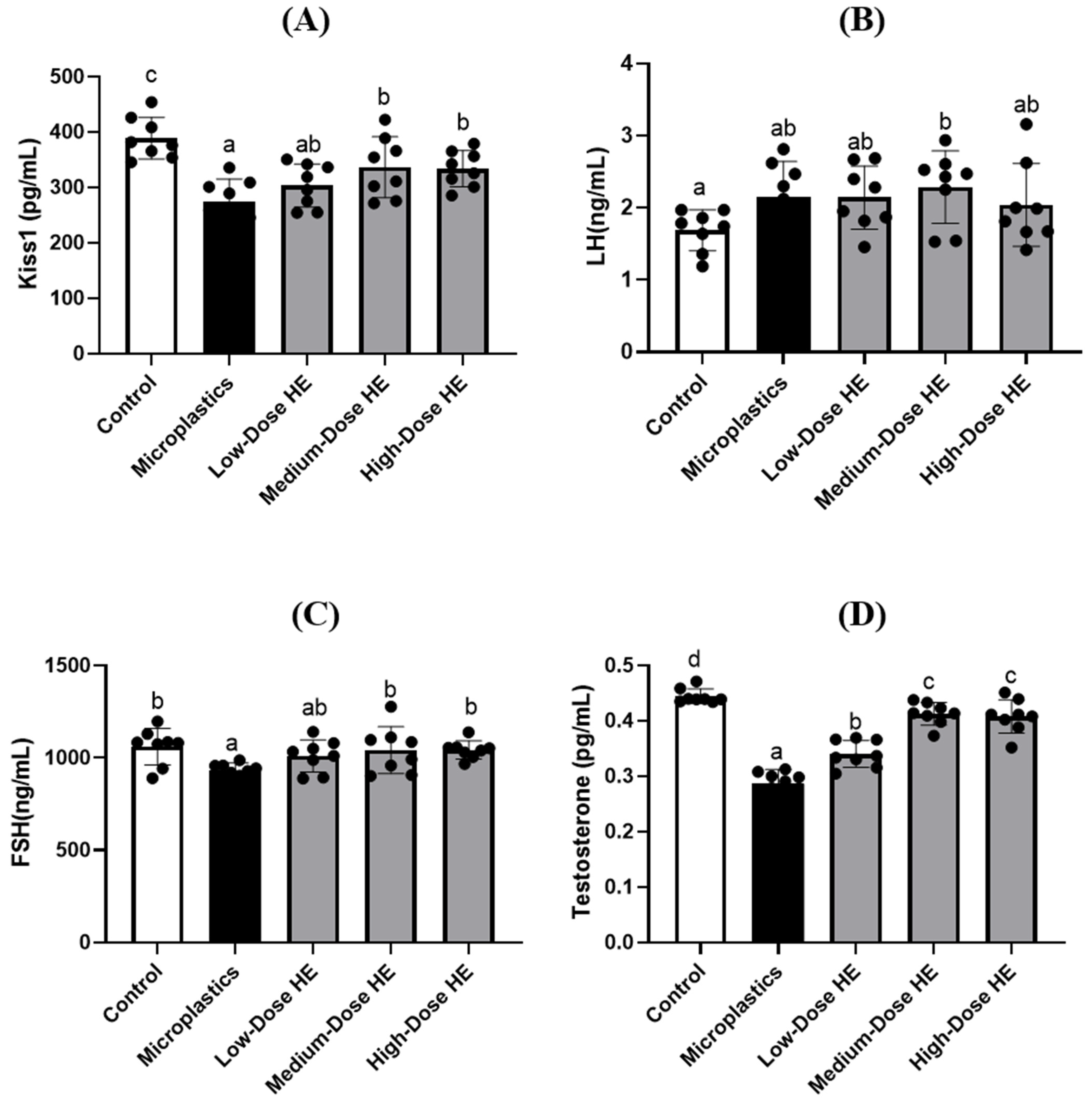
2.4. HE Regulates Kiss1 and Reproductive Hormone Concentrations

Kiss1 level was significantly (p < 0.05) reduced in the microplastics group compared to the control group, as shown in Figure 3. Treatment with HE for 6 weeks significantly (p < 0.05) increased Kiss1 levels, especially for the medium and high doses of HE. The reproductive hormones, such as luteinizing hormone (LH), follicle-stimulating hormone (FSH), and testosterone, were measured at the end of the experiment (Figure 3). FSH and testosterone levels were significantly different (p < 0.05) in the microplastics group compared to the control group. However, these levels significantly (p < 0.05) improved after treatment with medium and high doses of HE for 6 weeks. On the other hand, there were no significant differences (p > 0.05) in LH levels between the microplastics and control groups, as well as the treated groups.

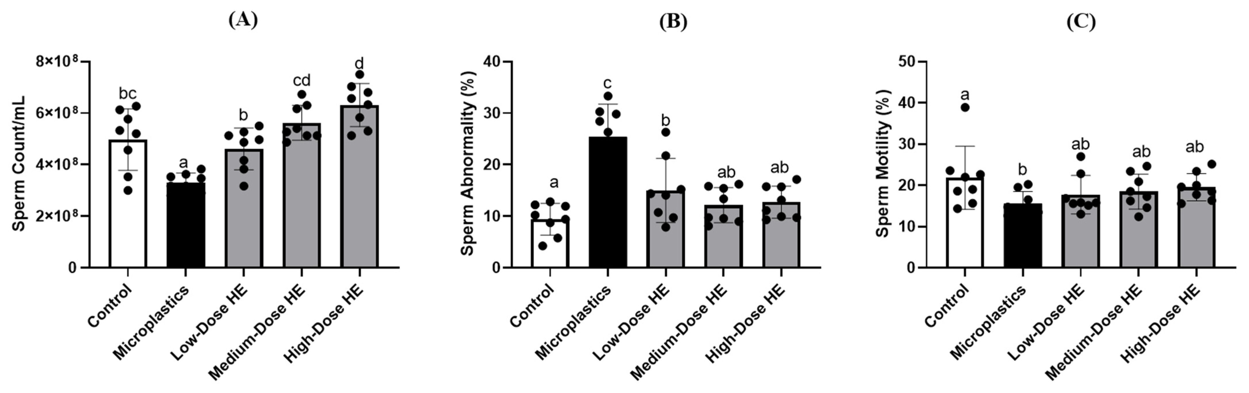
2.5. Effects of HE on the Sperm Count, Abnormality, and Motility

The rat sperm count in the microplastics group was significantly reduced (p < 0.05) compared to the control and treated groups (Figure 4). In contrast, sperm abnormalities were significantly increased (p < 0.05) in the microplastics group compared to the control and treated groups. These sperm properties were significantly (p < 0.05) improved after treatment with HE for 6 weeks. Additionally, there was no significant difference (p > 0.05) in sperm motility between the microplastics and treated groups.

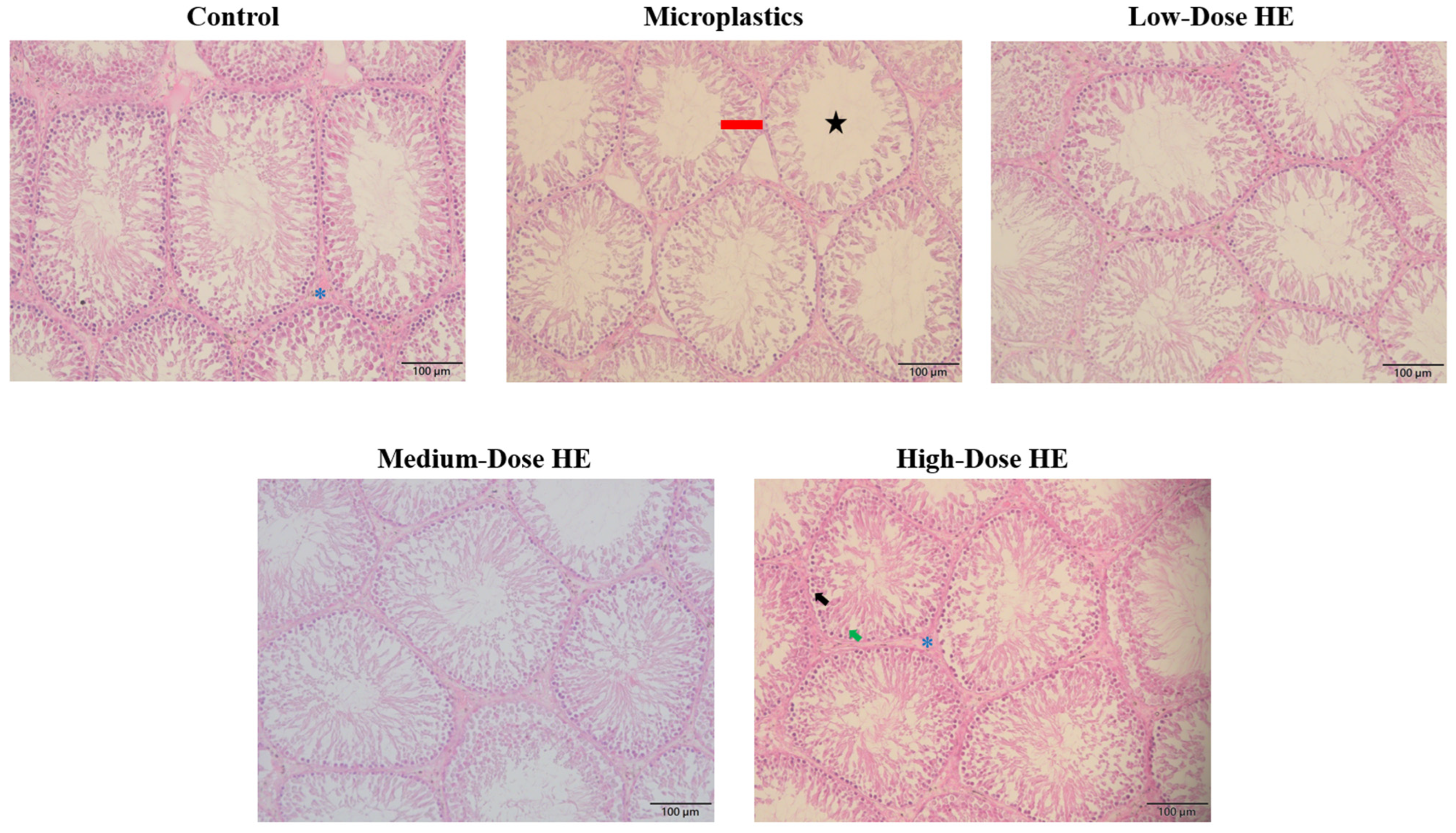
2.6. Effects of the HE on the Seminiferous Tubule’s Lumen Area and Thickness of the Epithelium

The morphology of the seminiferous tubule lumen and epithelium in rats after treatment with erinacine A-enriched H. erinaceus mycelium (HE) is shown in Figure 5, while the seminiferous tubules lumen area and epithelium thickness are presented in Table 2. These data indicate a significant (p < 0.05) increase in seminiferous tubule area in the microplastics group. Consequently, the epithelium thickness significantly (p < 0.05) decreased in this group. However, the values of the seminiferous tubule area and the thickness of the epithelium significantly (p < 0.05) improved after treatment with HE.

3. Discussion

This study presents the first in vivo evidence of the protective effects of erinacine A-enriched Hericium erinaceus mycelium against male reproductive dysfunction induced by polystyrene microplastics. However, it has some limitations, such as the absence of a positive control and a lack of primary data on the underlying molecular mechanisms, including no direct evidence of the modulation of specific molecular pathways.
In this present study, the microplastic-induced and erinacine A-enriched H. erinaceus mycelium (HE) had no effect on organ weights, including the kidney, spleen, abdominal fat, epididymal fat, and testis (Table 1). However, a reduction in glutathione peroxidase (GPx) activity was observed in the microplastics group, which resulted in an increase in nitric oxide (NO) levels in this group (Figure 1). A previous study also reported that polystyrene microplastics induce oxidative stress by increasing nitric oxide (NO) and malondialdehyde (MDA) levels, while reducing enzymatic antioxidant activity, including GPx [8]. Glutathione peroxidase is an enzymatic antioxidant that plays a role in regulating oxidative stress in the body. This enzyme converts and reduces hydrogen peroxide to water, thereby decreasing its adverse effects [26]. The GPx activity increased after treatment with HE for 6 weeks, accompanied by a decrease in nitric oxide (NO) concentration (Figure 1). A previous study reported that GPx catalyzes the breakdown of various oxidative species, including peroxynitrite (ONOO) [27]. In a mouse model, erinacine A-enriched H. erinaceus mycelium also reduced iNOS levels and regulated oxidative stress [28]. The present study indicates that HE effectively decreases oxidative stress in the rat model. Additionally, erinacine A-enriched H. erinaceus mycelium enhanced the expression of antioxidant enzymes and Nrf2 nuclear proteins [19].
Figure 2 shows an increase in some pro-inflammatory cytokines, such as interleukin (IL)-6 and IL-1β, in the microplastics groups. A previous study also reported that microplastics significantly induced plasma IL-6 and tumor necrosis factor (TNF)-α in a rat model [8]. In a mouse study, polystyrene microplastics also elevated pro-inflammatory cytokines, such as TNF-α, IL-6, and IL-1β [7] The level of IL-6 was successfully reduced after treatment with various doses of HE. A previous study reported that erinacine A-enriched H. erinaceus mycelium also decreased IL-6 and TNF-α, pro-inflammatory cytokines [29]. This condition indicates that HE exhibits anti-inflammatory activity. We hypothesize that HE acts as an anti-inflammatory agent by inhibiting the translocation of the nuclear factor (NF)-κB transcription factor. As reported by a previous study, HE could block the phosphorylation of NF-κB and its translocation into the nucleus, resulting in a reduction in pro-inflammatory mediators [29,30].
The level of Kiss1 is reduced in the microplastic group (Figure 3). Kisspeptin is a neuropeptide encoded by the kisspeptin 1 (KISS1/Kiss1) gene. This peptide plays an important role in reproductive function, including in the release of reproductive hormones by stimulating gonadotropin-releasing hormone (GnRH) [31,32]. On the other hand, a decrease in some reproductive hormones, such as follicle-stimulating hormone (FSH) and testosterone, is observed in the microplastic group (Figure 3). This condition is due to the lower Kiss1 concentration observed in the present study. A previous study also reported that polystyrene microplastics reduce testosterone levels in a rat model [8]. In the present study, the level of Kiss1 increased after treatment with HE for 6 weeks, resulting in the improvement of some reproductive hormones, including FSH and testosterone.
The sperm count is reduced in the microplastics group, whereas sperm abnormalities are increased (Figure 4). This condition is due to the lower levels of reproductive hormones in this group. A previous study also reported that lower testosterone levels induced poor sperm quality [33]. Following improvements in FSH and testosterone levels, an increase in sperm count and a reduction in sperm abnormalities were observed in the treated group (Figure 4). A previous study reported that at intermediate and high levels of FSH, the total sperm count was successfully improved [34]. Testosterone is the testicular androgen necessary for the initiation and maintenance of spermatogenesis [35]. Therefore, the improvement of sperm abnormality is also observed after treatment with HE for 6 weeks. As reported by previous studies, testosterone regulates the sperm morphology [33,36]. We hypothesized that the improvement in sperm quality is also associated with the reduction in oxidative stress by HE. In the present study, HE successfully enhanced GPx activity and reduced oxidative stress. Oxidative stress, particularly in sperm, causes adverse effects by damaging sperm DNA and triggering apoptosis in sperm cells [37]. A previous study reported that increasing antioxidant activity protects tissues from oxidative stress, resulting in improvements in sperm count and function [38,39]. Additionally, erinacine A-enriched H. erinaceus mycelium exhibits free radical scavenging activity [40]. Together with the reduction in oxidative stress, HE also improves the sperm quality through its anti-inflammatory activity. A previous study reported that the negative effects on spermatogenesis are caused by oxidative stress, primarily resulting from elevated levels of ROS and pro-inflammatory cytokines [41].
Figure 5 shows the increase in the seminiferous tubules’ lumen area/space in the microplastics group, as well as the shrinkage of the seminiferous tubule epithelium. This condition indicates a low concentration of elongated spermatids and sperm cells. As reported by previous studies, the seminiferous tubule lumen is the site where developing sperm cells are located [42,43]. The morphology of the seminiferous tubules improved with treatment using HE for 6 weeks. This condition confirms that HE successfully protects the testis from the adverse effects of polystyrene microplastics.
In this study, erinacine A-enriched H. erinaceus mycelium successfully improved the reproductive system of the rats. This effect was due to the reduction in oxidative stress and certain pro-inflammatory cytokines by the erinacine A-enriched H. erinaceus mycelium. A previous study reported that oxidative stress and inflammation are important factors in the pathophysiology of infertility [44]. The pro-inflammatory cytokines and oxidative stress also led to a reduction in kisspeptin, resulting in decreased reproductive hormones [45]. Therefore, the inhibition of oxidative stress and pro-inflammatory cytokines by various doses of erinacine A-enriched H. erinaceus mycelium resulted in an improved reproductive system. A previous study reported that erinacine A-enriched H. erinaceus mycelium activates the BDNF/TrkB/PI3K/Akt/GSK-3β pathways and inhibits nuclear factor (NF)-κB transcription in an animal model [29]. Furthermore, NF-κB is a transcription factor that regulates the expression of pro-inflammatory cytokines [46].

4. Materials and Methods

4.1. Materials

Erinacine A-enriched Hericium erinaceus (Bull.) Pers. 1797 mycelium was provided by Grape King Biotechnology Co., Ltd. (Taoyuan City, Taiwan). The presence of erinacine A in the H. erinaceus mycelium has been confirmed according to previous studies [47,48]. The polystyrene microplastics (PS-MPs) were acquired from the Cell-Bio Biotechnology Co., Ltd. (New Taipei City, Taiwan) with a particle size ranging from 0.4 to 0.6 μm and a concentration of 10% (w/v) (Cat. No. DNM-P004). Roswell Park Memorial Institute (RPMI) medium, interleukin (IL)-1β (Cat. No. BMS630), IL-6 (Cat. No. ERA31RB), and tumor necrosis factor (TNF)-α (Cat. No. KRC3011) ELISA kits were purchased from Thermo Fisher Scientific (Waltham, MA, USA). Glutathione peroxidase (GPx) commercial kit was purchased from Randox Laboratories (Crumlin, County Antrim, UK). Luteinizing hormone (LH, Cat. No. ER1123), follicle-stimulating hormone (FSH, Cat. No. ER0960), testosterone (Cat. No. ER1462), and Kiss1 (Cat. No. ER0628) ELISA kits were purchased from Wuhan Fine Biotech (Wuhan, China).

4.2. Polystyrene Microplastics Solution Preparation
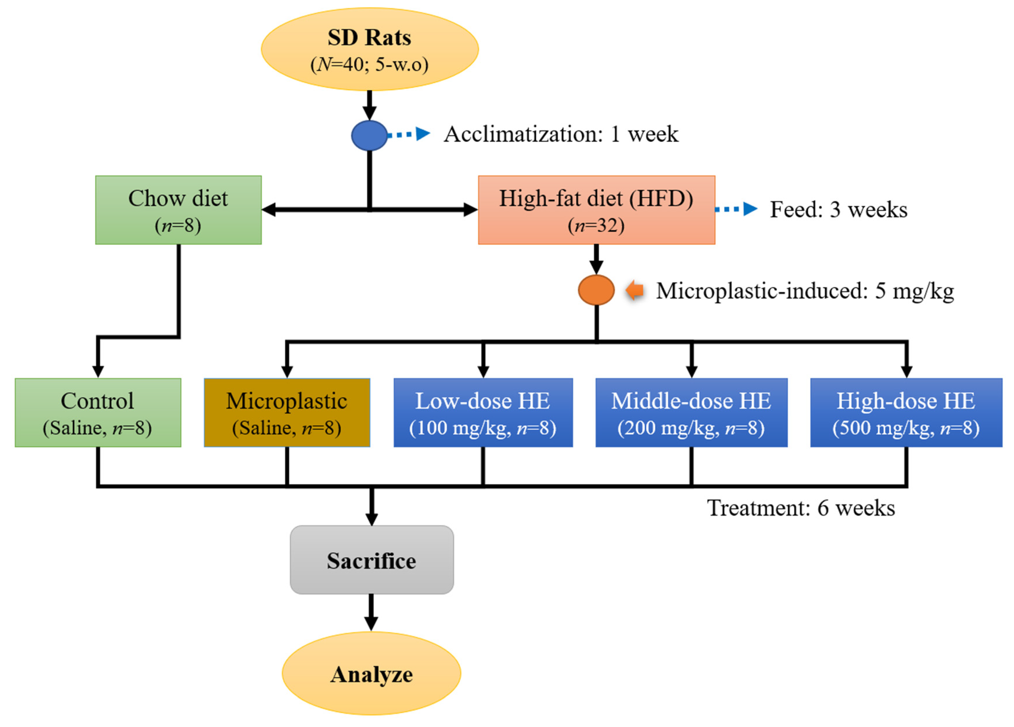
A 10% (w/v) solution of polystyrene microplastics (PS-MPs) was prepared in deionized water and treated with ultrasonic vibration for 30 min to ensure uniform dispersion of the plastic particles. Fresh suspensions were prepared daily to maintain consistency and prevent aggregation [49].

4.3. Animal Study

4.3.1. Animal Treatment

Forty male Sprague Dawley (SD) rats (5 weeks old, N = 40)) were housed individually and provided with water ad libitum and a standard chow diet (LabDiet 5001). The rats were maintained under standard laboratory conditions (temperature 23 ± 1 °C, humidity 40–60%, 12-h light/dark cycle). The rats were acclimatized for a week. The Institutional Animal Care and Use Committee (IACUC Approval No. 111046, Date: 1 August 2023) of the National Taiwan Ocean University reviewed and approved all protocols. After the acclimatization phase, the rats were randomly divided into two main groups (Figure 6). A group continued to be fed by chow diet (control, n = 8) and another group was fed with a high-fat diet (HFD) for 3 weeks (n = 32). After that, the HFD group was randomly divided into 4 groups. These group were daily induced with 5 mg/kg (oral gavage) polystyrene microplastics (PS-MPs) and the dose of PS-MPs was adapted from a previous study [8]. Then, these groups were either treated with various doses of erinacine A-enriched H. erinaceus mycelium (HE) or none; a group without HE treatment (Microplastic group, n = 8) and three groups were either treated by daily oral gavage with 100 mg/kg body weight of HE (Low-dose HE, n = 8), 200 mg/kg (Medium-dose HE, n = 8), or 500 mg/kg (High-dose HE, n = 8). The doses used were adapted from previously published methods [50,51]. The control and microplastic groups were administered daily with a saline solution. The rats were sacrificed after treatment for 6 weeks. The rats fasted prior to sacrifice for 12 h. They were sacrificed in an empty chamber through exposure to CO2 until euthanasia [52]. Blood was collected for further analysis and the organs (kidney, spleen, testes) as well as abdominal and epididymis fat were weighed at the day of sacrifice. One of the testes was soaked in 4% formaldehyde solution for histopathological analysis.

4.3.2. Blood Sample Collection

The blood was collected from the rats on the day of sacrifice. The blood was collected from the rats’ abdominal aorta. Then, it was centrifuged at 3000 rpm for 15 min at 4 C to separate the plasma. The plasma (supernatant) was collected and stored at freezing temperatures (−25 °C) for subsequent analysis [53].

4.3.3. Nitric Oxide and Glutathione Peroxidase Analysis

The nitric oxide (NO) content was determined using the Griess reagent, which consists of 1% (w/v) sulphanilamide in 5% (v/v) phosphoric acid and 0.1% (w/v) N-(1-naphthyl) ethylenediamine-HCl, following a previously described method [54]. In brief, 100 μL of rat plasma from each group and 100 μL of serially diluted standards were transferred to a new 96-well plate, then mixed with an equal volume of Griess reagent and incubated for 10 min. Absorbance was measured at 540 nm using a microplate spectrophotometer. Plasma enzymatic antioxidant activities, such as glutathione peroxidase (GPx), were analyzed according to the manufacturer’s protocol for the Randox Laboratories kit.

4.3.4. Pro-Inflammatory Cytokines, Kiss1, and Reproductive Hormones Analysis

The plasma pro-inflammatory cytokines, such as interleukin (IL)-6 and IL-1β, as well as Kiss1 concentration and reproductive hormones such as luteinizing hormone (LH), follicle-stimulating hormone (FSH), and testosterone concentrations, were analyzed using enzyme-linked immunosorbent assay (ELISA) kits performed according to the manufacturer’s protocols.

4.3.5. Sperm Count, Abnormality, and Motility Analysis

The swim-up method was used to collect sperm from the epididymis. Epididymis was cut into 3 pieces, immersed in 8 mL Roswell Park Memorial Institute (RPMI) medium and shaken in an orbital shaker at 100 rpm for 10 min. Epididymis with RPMI medium was centrifuged at 100× g for 5 min and incubated at 37 °C in a 5% CO2 incubator for 30 min. Finally, the sperms were collected from the supernatant and observed under the microscope to determine the sperm count, abnormality, and motility [55].

4.3.6. Testis Histopathology Analysis

The testis was cut into 5 mm sections from the middle part using a scalpel. The group was marked on the lid and then it was soaked in a 4% formaldehyde solution at room temperature for paraffin embedding and hematoxylin and eosin (H&E) staining. The staining was performed by Rapid Science Co., Ltd. (Taichung, Taiwan). The testicular tissue section images were used ImageJ (ver. 1.54p) microscope image processing software to determine the seminiferous tubules’ lumen area and thickness of the epithelium to evaluate the impact on seminiferous tubules by polystyrene microplastics [56].

4.4. Statistical Analysis

The data were presented as mean ± standard deviation (SD) and analyzed by one-way analysis of variance (one-way ANOVA) followed by Duncan’s multiple comparison tests; p < 0.05 was considered significantly different. Statistical analysis was performed with Statistical Product and Service Solution (SPSS) 22.0 software (IBM Corporation, Armonk, NY, USA).

5. Conclusions

Polystyrene microplastics increase oxidative stress and pro-inflammatory cytokines, while also reducing sperm quality and altering seminiferous tubule morphology. However, treatment with erinacine A-enriched Hericium erinaceus mycelium effectively enhances glutathione peroxidase activity, reduces pro-inflammatory cytokines, and increases Kiss1, follicle-stimulating hormone, and testosterone levels. These improvements lead to enhanced sperm quality and seminiferous tubule structure. Overall, erinacine A-enriched H. erinaceus mycelium exerts protective effects against reproductive dysfunction by regulating oxidative stress and inflammatory cytokine levels. Therefore, it holds potential as a source of food supplements or functional food ingredients for the treatment of reproductive system dysfunctions. However, further investigation is needed to assess its bioavailability, stability, and consumer acceptance.

Author Contributions

Conceptualization, D.-F.H. and Z.-L.K.; Formal analysis, Y.-Y.H. and E.-Y.W.; Supervision, D.-F.H. and Z.-L.K.; Writing—original draft, Y.-Y.H. and S.S.; Writing—review and editing, S.S., R.-F.S. and Z.-L.K. All authors have read and agreed to the published version of the manuscript.

Funding

This research received no external funding.

Institutional Review Board Statement

The animal study protocol was approved by the Institutional Review Board (or Ethics Committee) of National Taiwan Ocean University (IACUC Approval No. 111046, date: 1 August 2023).

Informed Consent Statement

Not applicable.

Data Availability Statement

The original contributions presented in this study are included in the article. Further inquiries can be directed to the corresponding author.

Conflicts of Interest

The authors declare no conflicts of interest.

Abbreviations

The following abbreviations are used in this manuscript:
ELISAEnzyme-linked immunosorbent assays
FSHFollicle-stimulating hormone
GPxGlutathione peroxidase
HEErinacine A-enriched H. erinaceus mycelium
ILInterleukin
LHLuteinizing hormone
MDAMalondialdehyde
NONitric oxide
PS-MPsPolystyrene microplastics
ROSReactive oxygen species
RPMIRoswell Park Memorial Institute
TNFTumor necrosis factor

References

  1. Ghosh, S.; Sinha, J.K.; Ghosh, S.; Vashisth, K.; Han, S.; Bhaskar, R. Microplastics as an emerging threat to the global environment and human health. Sustainability 2023, 15, 10821. [Google Scholar] [CrossRef]
  2. Wang, W.; Guan, J.; Feng, Y.; Nie, L.; Xu, Y.; Xu, H.; Fu, F. Polystyrene microplastics induced nephrotoxicity associated with oxidative stress, inflammation, and endoplasmic reticulum stress in juvenile rats. Front. Nutr. 2023, 9, 1059660. [Google Scholar] [CrossRef]
  3. Mamun, A.A.; Prasetya, T.A.E.; Dewi, I.R.; Ahmad, M. Microplastics in human food chains: Food becoming a threat to health safety. Sci. Total Environ. 2023, 858, 159834. [Google Scholar] [CrossRef]
  4. Wang, M.; Wu, Y.; Li, G.; Xiong, Y.; Zhang, Y.; Zhang, M. The hidden threat: Unraveling the impact of microplastics on reproductive health. Sci. Total Environ. 2024, 935, 173177. [Google Scholar] [CrossRef]
  5. Dubey, I.; Khan, S.; Kushwaha, S. Developmental and reproductive toxic effects of exposure to microplastics: A review of associated signaling pathways. Front. Toxicol. 2022, 4, 901798. [Google Scholar] [CrossRef] [PubMed]
  6. Ijaz, M.U.; Najam, S.; Hamza, A.; Azmat, R.; Ashraf, A.; Unuofin, J.O.; Lebelo, S.L.; Simal-Gandara, J. Pinostrobin alleviates testicular and spermatological damage induced by polystyrene microplastics in adult albino rats. Biomed. Pharmacother. 2023, 162, 114686. [Google Scholar] [CrossRef]
  7. Hou, B.; Wang, F.; Liu, T.; Wang, Z. Reproductive toxicity of polystyrene microplastics: In vivo experimental study on testicular toxicity in mice. J. Hazard. Mater. 2021, 405, 124028. [Google Scholar] [CrossRef]
  8. Hwang, Y.-Y.; Sudirman, S.; Wei, E.-Y.; Kong, Z.-L.; Hwang, D.-F. Fucoidan from cladosiphon okamuranus enhances antioxidant activity and prevents reproductive dysfunction in polystyrene microplastic-induced male rats. Biomed. Pharmacother. 2024, 170, 115912. [Google Scholar] [CrossRef] [PubMed]
  9. Zhang, C.; Chen, J.; Ma, S.; Sun, Z.; Wang, Z. Microplastics may be a significant cause of male infertility. Am. J. Men’s Health 2022, 16, 15579883221096549. [Google Scholar] [CrossRef]
  10. Jin, H.; Yan, M.; Pan, C.; Liu, Z.; Sha, X.; Jiang, C.; Li, L.; Pan, M.; Li, D.; Han, X.; et al. Chronic exposure to polystyrene microplastics induced male reproductive toxicity and decreased testosterone levels via the lh-mediated lhr/camp/pka/star pathway. Part. Fibre Toxicol. 2022, 19, 13. [Google Scholar] [CrossRef]
  11. Li, Y.; Liu, Y.; Chen, Y.; Yao, C.; Yu, S.; Qu, J.; Chen, G.; Wei, H. Combined effects of polystyrene nanoplastics and lipopolysaccharide on testosterone biosynthesis and inflammation in mouse testis. Ecotoxicol. Environ. Saf. 2024, 273, 116180. [Google Scholar] [CrossRef] [PubMed]
  12. Zhang, Y.; Hou, B.; Liu, T.; Wu, Y.; Wang, Z. Probiotics improve polystyrene microplastics-induced male reproductive toxicity in mice by alleviating inflammatory response. Ecotoxicol. Environ. Saf. 2023, 263, 115248. [Google Scholar] [CrossRef] [PubMed]
  13. Xie, X.; Deng, T.; Duan, J.; Xie, J.; Yuan, J.; Chen, M. Exposure to polystyrene microplastics causes reproductive toxicity through oxidative stress and activation of the p38 mapk signaling pathway. Ecotoxicol. Environ. Saf. 2020, 190, 110133. [Google Scholar] [CrossRef] [PubMed]
  14. Qi, J.-Z. Hericium erinaceus: The enchanting medicinal-culinary mushroom of east asian tradition. Integr. Med. Discov. 2024, 8, e24013. [Google Scholar] [CrossRef]
  15. Chutimanukul, P.; Phatthanamas, W.; Thepsilvisut, O.; Chantarachot, T.; Thongtip, A.; Chutimanukul, P. Commercial scale production of yamabushitake mushroom (hericium erinaceus (bull.) pers. 1797) using rubber and bamboo sawdust substrates in tropical regions. Sci. Rep. 2023, 13, 13316. [Google Scholar] [CrossRef]
  16. Gravina, A.G.; Pellegrino, R.; Auletta, S.; Palladino, G.; Brandimarte, G.; D’Onofrio, R.; Arboretto, G.; Imperio, G.; Ventura, A.; Cipullo, M.; et al. Hericium erinaceus, a medicinal fungus with a centuries-old history: Evidence in gastrointestinal diseases. World J. Gastroenterol. 2023, 29, 3048–3065. [Google Scholar] [CrossRef]
  17. Szućko-Kociuba, I.; Trzeciak-Ryczek, A.; Kupnicka, P.; Chlubek, D. Neurotrophic and neuroprotective effects of hericium erinaceus. Int. J. Mol. Sci. 2023, 24, 15960. [Google Scholar] [CrossRef] [PubMed]
  18. Qi, J.; Wu, J.; Kang, S.; Gao, J.; Hirokazu, K.; Liu, H.; Liu, C. The chemical structures, biosynthesis, and biological activities of secondary metabolites from the culinary-medicinal mushrooms of the genus hericium: A review. Chin. J. Nat. Med. 2024, 22, 676–698. [Google Scholar] [CrossRef]
  19. Wu, Y.-L.; Sun, H.-L.; Chang, J.-C.; Lin, W.-Y.; Chen, P.-Y.; Chen, C.-C.; Lee, L.-Y.; Li, C.-C.; Hsieh, M.; Chen, H.-W.; et al. Erinacine a-enriched hericium erinaceus mycelium ethanol extract lessens cellular damage in cell and drosophila models of spinocerebellar ataxia type 3 by improvement of nrf2 activation. Antioxidants 2024, 13, 1495. [Google Scholar] [CrossRef]
  20. Wang, L.-Y.; Huang, C.-S.; Chen, Y.-H.; Chen, C.-C.; Chen, C.-C.; Chuang, C.-H. Anti-inflammatory effect of erinacine c on no production through down-regulation of nf-κb and activation of nrf2-mediated ho-1 in bv2 microglial cells treated with lps. Molecules 2019, 24, 3317. [Google Scholar] [CrossRef]
  21. Lee, K.-F.; Chen, J.-H.; Teng, C.-C.; Shen, C.-H.; Hsieh, M.-C.; Lu, C.-C.; Lee, K.-C.; Lee, L.-Y.; Chen, W.-P.; Chen, C.-C.; et al. Protective effects of hericium erinaceus mycelium and its isolated erinacine a against ischemia-injury-induced neuronal cell death via the inhibition of inos/p38 mapk and nitrotyrosine. Int. J. Mol. Sci. 2014, 15, 15073–15089. [Google Scholar] [CrossRef] [PubMed]
  22. Wei, J.; Lin, C.; Wang, Z.-X.; Zhang, Y.-L.; Li, S.-X.; Hu, X.-S.; Hui, H.-P.; Wang, Z.; Zhao, Y.-Q.; Wang, X.-J.; et al. Metabolites of culinary-medicinal mushroom hericium erinaceus showed anti-neuroinflammatory activity in bv2 cells and anti-h2o2-induced oxidative stress activity in sh-sy5y cells. Nat. Prod. J. 2025, 15, e030624230635. [Google Scholar] [CrossRef]
  23. Qiu, Y.; Lin, G.; Liu, W.; Zhang, F.; Linhardt, R.J.; Wang, X.; Zhang, A. Bioactive compounds in hericium erinaceus and their biological properties: A review. Food Sci. Hum. Wellness 2024, 13, 1825–1844. [Google Scholar] [CrossRef]
  24. Zhang, Y.; Liu, L.; Bao, L.; Yang, Y.; Ma, K.; Liu, H. Three new cyathane diterpenes with neurotrophic activity from the liquid cultures of hericium erinaceus. J. Antibiot. 2018, 71, 818–821. [Google Scholar] [CrossRef]
  25. Chen, Y.-Y.; Lin, T.-W.; Li, I.C.; Tsung, L.; Hou, C.-H.; Yang, C.-Y.; Li, T.-J.; Chen, C.-C. A pilot pharmacokinetic and metabolite identification study of erinacine a in a single landrace pig model. Heliyon 2024, 10, e37850. [Google Scholar] [CrossRef] [PubMed]
  26. Lubos, E.; Loscalzo, J.; Handy, D.E. Glutathione peroxidase-1 in health and disease: From molecular mechanisms to therapeutic opportunities. Antioxid. Redox Signal. 2011, 15, 1957–1997. [Google Scholar] [CrossRef]
  27. Panday, S.; Talreja, R.; Kavdia, M. The role of glutathione and glutathione peroxidase in regulating cellular level of reactive oxygen and nitrogen species. Microvasc. Res. 2020, 131, 104010. [Google Scholar] [CrossRef]
  28. Lee, L.-Y.; Chou, W.; Chen, W.-P.; Wang, M.-F.; Chen, Y.-J.; Chen, C.-C.; Tung, K.-C. Erinacine a-enriched hericium erinaceus mycelium delays progression of age-related cognitive decline in senescence accelerated mouse prone 8 (samp8) mice. Nutrients 2021, 13, 3659. [Google Scholar] [CrossRef]
  29. Chiu, C.-H.; Chyau, C.-C.; Chen, C.-C.; Lee, L.-Y.; Chen, W.-P.; Liu, J.-L.; Lin, W.-H.; Mong, M.-C. Erinacine a-enriched hericium erinaceus mycelium produces antidepressant-like effects through modulating bdnf/pi3k/akt/gsk-3β signaling in mice. Int. J. Mol. Sci. 2018, 19, 341. [Google Scholar] [CrossRef]
  30. Lee, S.-L.; Hsu, J.-Y.; Chen, T.-C.; Huang, C.-C.; Wu, T.-Y.; Chin, T.-Y. Erinacine a prevents lipopolysaccharide-mediated glial cell activation to protect dopaminergic neurons against inflammatory factor-induced cell death in vitro and in vivo. Int. J. Mol. Sci. 2022, 23, 810. [Google Scholar] [CrossRef]
  31. Ogawa, S.; Parhar, I.S. Biological significance of kisspeptin–kiss 1 receptor signaling in the habenula of teleost species. Front. Endocrinol. 2018, 9, 222. [Google Scholar] [CrossRef] [PubMed]
  32. Skorupskaite, K.; George, J.T.; Anderson, R.A. The kisspeptin-gnrh pathway in human reproductive health and disease. Hum. Reprod. Update 2014, 20, 485–500. [Google Scholar] [CrossRef] [PubMed]
  33. Trussell, J.C.; Coward, R.M.; Santoro, N.; Stetter, C.; Kunselman, A.; Diamond, M.P.; Hansen, K.R.; Krawetz, S.A.; Legro, R.S.; Heisenleder, D.; et al. Association between testosterone, semen parameters, and live birth in men with unexplained infertility in an intrauterine insemination population. Fertil. Steril. 2019, 111, 1129–1134. [Google Scholar] [CrossRef]
  34. Cannarella, R.; La Vignera, S.; Condorelli, R.; Mongioì, L.; Calogero, A. Fsh dosage effect on conventional sperm parameters: A meta-analysis of randomized controlled studies. Asian J. Androl. 2020, 22, 309–316. [Google Scholar] [CrossRef]
  35. Grande, G.; Barrachina, F.; Soler-Ventura, A.; Jodar, M.; Mancini, F.; Marana, R.; Chiloiro, S.; Pontecorvi, A.; Oliva, R.; Milardi, D. The role of testosterone in spermatogenesis: Lessons from proteome profiling of human spermatozoa in testosterone deficiency. Front. Endocrinol. 2022, 13, 852661. [Google Scholar] [CrossRef]
  36. Guardo, F.D.; Vloeberghs, V.; Bardhi, E.; Blockeel, C.; Verheyen, G.; Tournaye, H.; Drakopoulos, P. Low testosterone and semen parameters in male partners of infertile couples undergoing ivf with a total sperm count greater than 5 million. J. Clin. Med. 2020, 9, 3824. [Google Scholar] [CrossRef] [PubMed]
  37. Azenabor, A.; Ekun, A.O.; Akinloye, O. Impact of inflammation on male reproductive tract. J. Reprod. Infertil. 2015, 16, 123–129. [Google Scholar]
  38. Walke, G.; Gaurkar, S.S.; Prasad, R.; Lohakare, T.; Wanjari, M. The impact of oxidative stress on male reproductive function: Exploring the role of antioxidant supplementation. Cureus 2023, 17, e42583. [Google Scholar] [CrossRef]
  39. Walczak–Jedrzejowska, R.; Wolski, J.K.; Slowikowska–Hilczer, J. The role of oxidative stress and antioxidants in male fertility. Cent. Eur. J. Urol. 2013, 65, 60–67. [Google Scholar] [CrossRef]
  40. Khan, M.A.; Tania, M.; Liu, R.; Rahman, M.M. Hericium erinaceus: An edible mushroom with medicinal values. J. Complement. Integr. Med. 2013, 10, 253–258. [Google Scholar] [CrossRef]
  41. Hasan, H.; Bhushan, S.; Fijak, M.; Meinhardt, A. Mechanism of inflammatory associated impairment of sperm function, spermatogenesis and steroidogenesis. Front. Endocrinol. 2022, 13, 897029. [Google Scholar] [CrossRef]
  42. Owembabazi, E.; Nkomozepi, P.; Calvey, T.; Mbajiorgu, E.F. Co-administration of alcohol and combination antiretroviral therapy (cart) in male sprague dawley rats: A study on testicular morphology, oxidative and cytokines perturbations. Anat. Cell Biol. 2023, 56, 236–251. [Google Scholar] [CrossRef] [PubMed]
  43. Kuchakulla, M.; Narasimman, M.; Khodamoradi, K.; Khosravizadeh, Z.; Ramasamy, R. How defective spermatogenesis affects sperm DNA integrity. Andrologia 2020, 53, e13615. [Google Scholar] [CrossRef]
  44. Ojo, O.A.; Nwafor-Ezeh, P.I.; Rotimi, D.E.; Iyobhebhe, M.; Ogunlakin, A.D.; Ojo, A.B. Apoptosis, inflammation, and oxidative stress in infertility: A mini review. Toxicol. Rep. 2023, 10, 448–462. [Google Scholar] [CrossRef] [PubMed]
  45. Magata, F.; Tsukamura, H.; Matsuda, F. The impact of inflammatory stress on hypothalamic kisspeptin neurons: Mechanisms underlying inflammation-associated infertility in humans and domestic animals. Peptides 2023, 162, 170958. [Google Scholar] [CrossRef]
  46. Liu, T.; Zhang, L.; Joo, D.; Sun, S.-C. Nf-κb signaling in inflammation. Signal Transduct. Target. Ther. 2017, 2, e17023. [Google Scholar] [CrossRef]
  47. Tsai, P.-C.; Wu, Y.-K.; Hu, J.-H.; Li, I.C.; Lin, T.-W.; Chen, C.-C.; Kuo, C.-F. Preclinical bioavailability, tissue distribution, and protein binding studies of erinacine a, a bioactive compound from hericium erinaceus mycelia using validated lc-ms/ms method. Molecules 2021, 26, 4510. [Google Scholar] [CrossRef]
  48. Tsai-Teng, T.; Chin-Chu, C.; Li-Ya, L.; Wan-Ping, C.; Chung-Kuang, L.; Chien-Chang, S.; Chi-Ying, H.F.; Chien-Chih, C.; Shiao, Y.-J. Erinacine a-enriched hericium erinaceus mycelium ameliorates alzheimer’s disease-related pathologies in appswe/ps1de9 transgenic mice. J. Biomed. Sci. 2016, 23, 49. [Google Scholar] [CrossRef] [PubMed]
  49. Shiu, R.-F.; Vazquez, C.I.; Chiang, C.-Y.; Chiu, M.-H.; Chen, C.-S.; Ni, C.-W.; Gong, G.-C.; Quigg, A.; Santschi, P.H.; Chin, W.-C. Nano- and microplastics trigger secretion of protein-rich extracellular polymeric substances from phytoplankton. Sci. Total Environ. 2020, 748, 141469. [Google Scholar] [CrossRef]
  50. Chen, S.N.; Chang, C.S.; Yang, M.F.; Chen, S.; Soni, M.; Mahadevan, B. Subchronic toxicity and genotoxicity studies of hericium erinaceus β-glucan extract preparation. Curr. Res. Toxicol. 2022, 3, 100068. [Google Scholar] [CrossRef]
  51. Li, I.C.; Chen, Y.-L.; Lee, L.-Y.; Chen, W.-P.; Tsai, Y.-T.; Chen, C.-C.; Chen, C.-S. Evaluation of the toxicological safety of erinacine a-enriched hericium erinaceus in a 28-day oral feeding study in sprague–dawley rats. Food Chem. Toxicol. 2014, 70, 61–67. [Google Scholar] [CrossRef] [PubMed]
  52. Sudirman, S.; Su, C.-Y.; Tsou, D.; Lee, M.-C.; Kong, Z.-L. Hippocampus kuda protein hydrolysate improves male reproductive dysfunction in diabetic rats. Biomed. Pharmacother. 2021, 140, 111760. [Google Scholar] [CrossRef] [PubMed]
  53. Mussbacher, M.; Schrottmaier, W.C.; Salzmann, M.; Brostjan, C.; Schmid, J.A.; Starlinger, P.; Assinger, A. Optimized plasma preparation is essential to monitor platelet-stored molecules in humans. PLoS ONE 2017, 12, e0188921. [Google Scholar] [CrossRef] [PubMed]
  54. Baek, S.-H.; Park, T.; Kang, M.-G.; Park, D. Anti-inflammatory activity and ros regulation effect of sinapaldehyde in lps-stimulated raw 264.7 macrophages. Molecules 2020, 25, 4089. [Google Scholar] [CrossRef]
  55. Collodel, G.; Federico, M.G.; Geminiani, M.; Martini, S.; Bonechi, C.; Rossi, C.; Figura, N.; Moretti, E. Effect of trans-resveratrol on induced oxidative stress in human sperm and in rat germinal cells. Reprod. Toxicol. 2011, 31, 239–246. [Google Scholar] [CrossRef]
  56. Wang, W.-Y.; Meng, L.-J.; Xu, Y.-J.; Gong, T.; Yang, Y. Effects of 4% paraformaldehyde and modified davidson’s fluid on the morphology and immunohistochemistry of xiang pig testes. J. Toxicol. Pathol. 2020, 33, 97–104. [Google Scholar] [CrossRef]
Figure 1. Effects of erinacine A-enriched Hericium erinaceus mycelium on (A) nitric oxide concentration and (B) glutathione peroxidase activity in plasma of rats after 6 weeks of treatment. Values with different letters (a–d) indicate significant differences (p < 0.05), as analyzed by Duncan’s multiple range test. Data are presented as mean ± SD (n = 8 rats/group). The circles (•) in each bar indicate individual data points; HE, Erinacine A-enriched Hericium erinaceus mycelium.
Ijms 26 05735 g001
Figure 2. Effects of erinacine A-enriched Hericium erinaceus mycelium extract on (A) IL-6 and (B) IL-1β levels in plasma of rats after 6 weeks of treatment. Values with different letters (a–c) indicate significant differences (p < 0.05), as analyzed by Duncan’s multiple range test. Data are presented as mean ± SD (n = 8 rats/group). The circles (•) in each bar indicate individual data points; HE, Erinacine A-enriched Hericium erinaceus mycelium extract; IL, interleukin.
Ijms 26 05735 g002
Figure 3. Effects of erinacine A-enriched Hericium erinaceus mycelium extract on (A) Kiss1 concentration, (B) LH, (C) FSH, and (D) testosterone hormones in plasma of rats after 6 weeks of treatment. Values with different letters (a–d) indicate significant differences (p < 0.05), as analyzed by Duncan’s multiple range test. Data are presented as mean ± SD (n = 8 rats/group). The circles (•) in each bar indicate individual data points; HE, Erinacine A-enriched Hericium erinaceus mycelium extract; LH, luteinizing hormone; FSH, follicle-stimulating hormone.
Ijms 26 05735 g003
Figure 4. Effects of erinacine A-enriched Hericium erinaceus mycelium extract on the sperm (A) count, (B) abnormality, and (C) motility of the rats after 6 weeks of treatment. The values with different letters (a–d) represent significant difference (p < 0.05) as analyzed by Duncan’s multiple range test. Data are shown as mean ± SD (n = 8 rats/group). The circles (•) in each bar indicate individual data points; HE, Erinacine A-enriched Hericium erinaceus mycelium.
Ijms 26 05735 g004
Figure 5. The representative morphology of seminiferous tubules in each rat group after 6 weeks of treatment with HE. HE, Erinacine A-enriched Hericium erinaceus mycelium. Figure legends: Blue asterisk, interstitial tissue containing Leydig cell; black star, seminiferous lumen; red bar, seminiferous epithelium; green arrow, Sertoli cell; black arrow, spermatogonia.
Ijms 26 05735 g005
Figure 6. Flowchart of high-fat-diet- and microplastic-induced rats treated with Hericium erinaceus mycelium (HE) for 6 weeks. HE, Erinacine A-enriched H. erinaceus mycelium; HFD, high-fat diet; SD, Sprague Dawley.
Ijms 26 05735 g006
Table 1. Effect of erinacine A-enriched Hericium erinaceus mycelium (HE) on organ weights (% of body weight) in rats.
GroupKidneySpleenAbdominal FatTestesEpididymis Fat
Control4.23 ± 0.350.83 ± 0.188.83 ± 1.263.89 ± 0.898.02 ± 1.26
Microplastics3.98 ± 0.370.74 ± 0.087.18 ± 1.983.74 ± 0.786.41 ± 1.75
Low-Dose HE4.08 ± 0.480.70 ± 0.108.76 ± 2.454.43 ± 1.387.07 ± 2.24
Medium-Dose HE4.26 ± 0.270.74 ± 0.107.93 ± 2.753.84 ± 0.937.40 ± 2.16
High-Dose HE4.08 ± 0.550.76 ± 0.179.17 ± 2.343.79 ± 0.846.82 ± 2.29
Data are shown as the mean ± SD (n = 8 rats/group). HE, erinacine A-enriched Hericium erinaceus mycelium.
Table 2. Effects of erinacine A-enriched Hericium erinaceus mycelium extract on the seminiferous tubule lumen area and epithelium thickness in rats after 6 weeks of treatment.
GroupsSeminiferous Tubule Area (%)Thickness of Epithelium (µm)
Control17.55 ± 2.22 a110.23 ± 31.89 b
Microplastics41.74 ± 8.54 b62.03 ± 9.19 a
Low-Dose HE20.77 ± 8.03 a91.91 ± 13.72 b
Medium-Dose HE12.86 ± 0.61 a104.25 ± 13.93 b
High-Dose HE13.14 ± 1.57 a109.64 ± 14.06 b
The values with different letters (a,b) in a column represent significant difference (p < 0.05) as analyzed by Duncan’s multiple range test. Data are shown as the mean ± SD (n = 8 rats/group). HE, Erinacine A-enriched Hericium erinaceus mycelium.
Disclaimer/Publisher’s Note: The statements, opinions and data contained in all publications are solely those of the individual author(s) and contributor(s) and not of MDPI and/or the editor(s). MDPI and/or the editor(s) disclaim responsibility for any injury to people or property resulting from any ideas, methods, instructions or products referred to in the content.

