Article

Nanocarbon-Driven Recovery of Mechano-Kinetic Properties of Injured Rat Gastrocnemius Muscle

1
ESC “Institute of Biology and Medicine”, Taras Shevchenko National University of Kyiv, Volodymyrska Str. 64, 01601 Kyiv, Ukraine
2
State Institution “Institute of Traumatology and Orthopedics”, National Academy of Medical Science of Ukraine, Bulvarno-Kudriavska Str. 27, 01054 Kyiv, Ukraine
3
Institute of Chemistry and Biotechnology, Technical University of Ilmenau, Weimarer Str. 25, 98693 Ilmenau, Germany
4
Department of Physical Education, Gdansk University of Physical Education and Sport, Kazimierza Gorskiego Str. 1, 80-336 Gdansk, Poland
5
Department of Movement Physiology, Bogomoletz Institute of Physiology NAS of Ukraine, Bogomoletz Str. 4, 01024 Kyiv, Ukraine
*
Author to whom correspondence should be addressed.
Int. J. Mol. Sci. 2025, 26(12), 5511; https://doi.org/10.3390/ijms26125511
Submission received: 10 May 2025 / Revised: 3 June 2025 / Accepted: 8 June 2025 / Published: 9 June 2025
(This article belongs to the Special Issue Nanoparticles in Nanobiotechnology and Nanomedicine: 2nd Edition)

Abstract

Traumatic muscle injuries often lead to prolonged functional impairments due to oxidative stress, metabolic disturbances, and structural damage. This study investigates the therapeutic potential of chronic administration of C60 fullerene aqueous (C60FAS) solutions in enhancing muscle recovery post-injury. Rats with experimentally induced gastrocnemius muscle trauma received C60FAS orally at a daily dose of 1 mg/kg body weight. Functional assessments included measurements of maximal force generation and time to peak contraction. Biochemical analyses evaluated lactate, superoxide dismutase (SOD), and catalase (CAT) levels, while histological examinations assessed muscle fiber integrity and collagen deposition. Results demonstrated significant improvements in muscle function, with a 35–40% increase in maximal force and a 27–38% acceleration in contraction time. Biochemical analysis revealed a 25% decrease in lactate concentration, potentially indicating improved metabolic function. This change is in line with normalized SOD and CAT activities, suggesting enhanced redox balance following treatment. Histological analyses revealed preserved myofibrillar architecture and reduced fibrosis in treated muscles. These findings suggest that C60 fullerene facilitates muscle recovery through antioxidant protection, metabolic support, and structural preservation, highlighting its potential as a therapeutic agent for muscle injuries.

1. Introduction

Muscle injuries represent a pervasive clinical challenge, affecting athletes, military personnel, and aging populations alike. Despite their high incidence, treatment strategies remain largely palliative, hampered by the complex interplay of inflammation, oxidative stress, and fibrosis that hinders effective regeneration [1]. The resultant chronic inflammation–fibrosis axis leads to contractures and functional deficits, with current therapies failing to restore preinjury mechano-kinetic properties [2,3]. Animal models of partial-thickness muscle incision (e.g., gastrocnemius) recapitulate this pathology, revealing persistent deficits in tetanic force and fatigue resistance due to collagenous scarring [4,5]. Conventional treatments, such as non-steroidal anti-inflammatory drugs and corticosteroids, primarily offer symptomatic relief and may not adequately address the underlying pathophysiological mechanisms, i.e., excessive reactive oxygen species (ROS) and dysregulated immune responses, sometimes leading to adverse side effects [6]. This therapeutic gap has spurred interest in nanobiotechnology, where engineered nanoparticles (NPs) offer precision modulation of pathological processes at the molecular level [7]. Among these, carbon-based nanomaterials, particularly C60 fullerenes, stand out for their unique physicochemical properties, including exceptional electron-accepting capacity, and the ability to ROS scavenge C60’s ability to scavenge multiple free radicals—up to six electrons per molecule—exceeds the stoichiometric limits of conventional antioxidants (e.g., vitamin E or N-acetylcysteine), enabling sustained ROS neutralization even at low doses [8,9]. This attribute is particularly beneficial in the context of muscle injuries, where oxidative stress plays a pivotal role in secondary tissue damage. Studies have demonstrated that C60 fullerenes can mitigate oxidative damage, reduce inflammation, and promote tissue regeneration [5,10]. Furthermore, C60 fullerenes have been shown to preserve mitochondrial function, inhibit profibrotic signaling pathways such as TGF-β/Smad, modulate immune responses by attenuating NLRP3 inflammasome activation [11], and even increase the force productivity of skeletal muscles after glyphosate poisoning [12]. These multifaceted actions position C60 fullerenes as promising candidates for enhancing muscle repair and functional recovery following injury. Therefore, gaining an in-depth insight into the molecular and cellular processes that drive skeletal muscle adaptation in response to various stressors can enhance existing knowledge in muscle physiology. This understanding may also pave the way for novel therapeutic strategies aimed at promoting tissue regeneration and recovery [13].
In our previous study [5,10], we utilized a traumatic muscle injury model to examine the effects of the C60 fullerene aqueous solution (C60FAS), administered chronically over a two-week period, on key time-dependent mechano-kinetic parameters of muscle function. These parameters included the duration of sustained maximal muscle contraction, the relaxation time required for the muscle to return to its baseline state after stimulation, and the difference between initial and peak (maximal vs. minimal) muscle contraction forces during specific stimulation patterns. Animals treated with the fullerene solution exhibited significant improvements across all contraction parameters. We hypothesized that C60 fullerene, acting as an antioxidant agent, would enhance post-injury muscle recovery, particularly in contraction dynamics critical for precise movement control, such as the time to reach maximal and minimal contractile force. Furthermore, we propose that extended treatment (up to 45 days) could optimize the restoration of muscle function to near pre-injury levels.
Therefore, the aim of this study was to investigate the therapeutic potential of aqueous C60FAS in facilitating the recovery of posttraumatic mechano-kinetic properties, specifically focusing on the maximum force developed and the time to reach maximum contraction force by the muscle during certain patterns of electrical stimulation of the gastrocnemius muscle in a rat model of muscle injury. The experimental design involved inducing controlled transverse injuries to the muscle, followed by treatment with C60 fullerenes. Recovery was assessed through tensometric measurements, biochemical assays evaluating oxidative stress markers (superoxide dismutase (SOD), catalase (CAT), and lactate), and histological analyses over intervals of 15, 30, and 45 days.

2. Results

2.1. Control Muscle Performance

Control measurements of maximal muscle force in control animals (Groups 1–3: intact; Groups 4–6: sham-treated with C60FAS) demonstrated stable values throughout the experimental period (15 days: 6.4 ± 0.05 N; 30 days: 7.8 ± 0.06 N; 45 days: 8.9 ± 0.07 N), with no statistically significant differences between these groups at any timepoint (p > 0.05). This consistency allowed us to combine these groups as a unified control for subsequent comparative analyses (Figure 1).

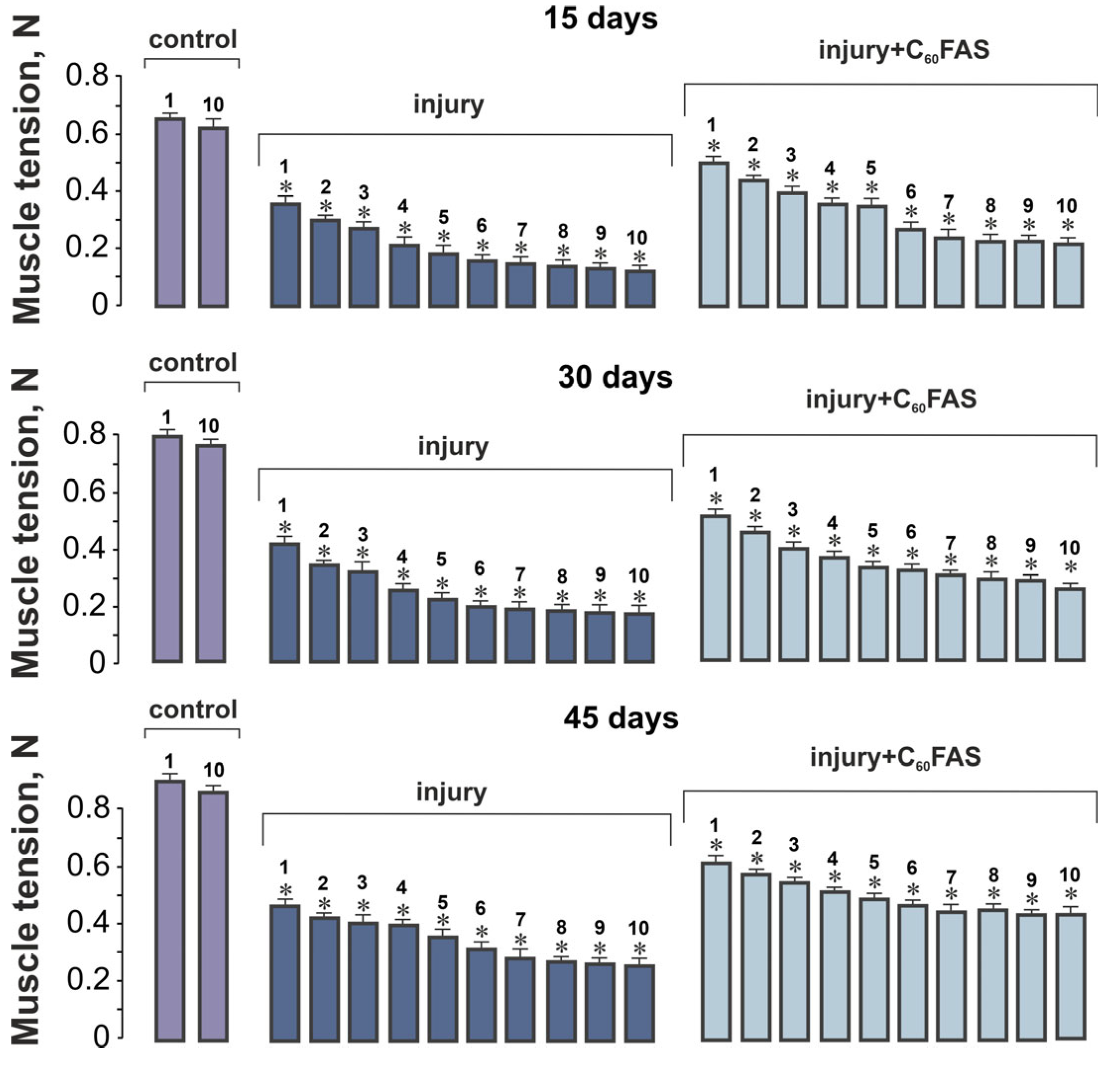
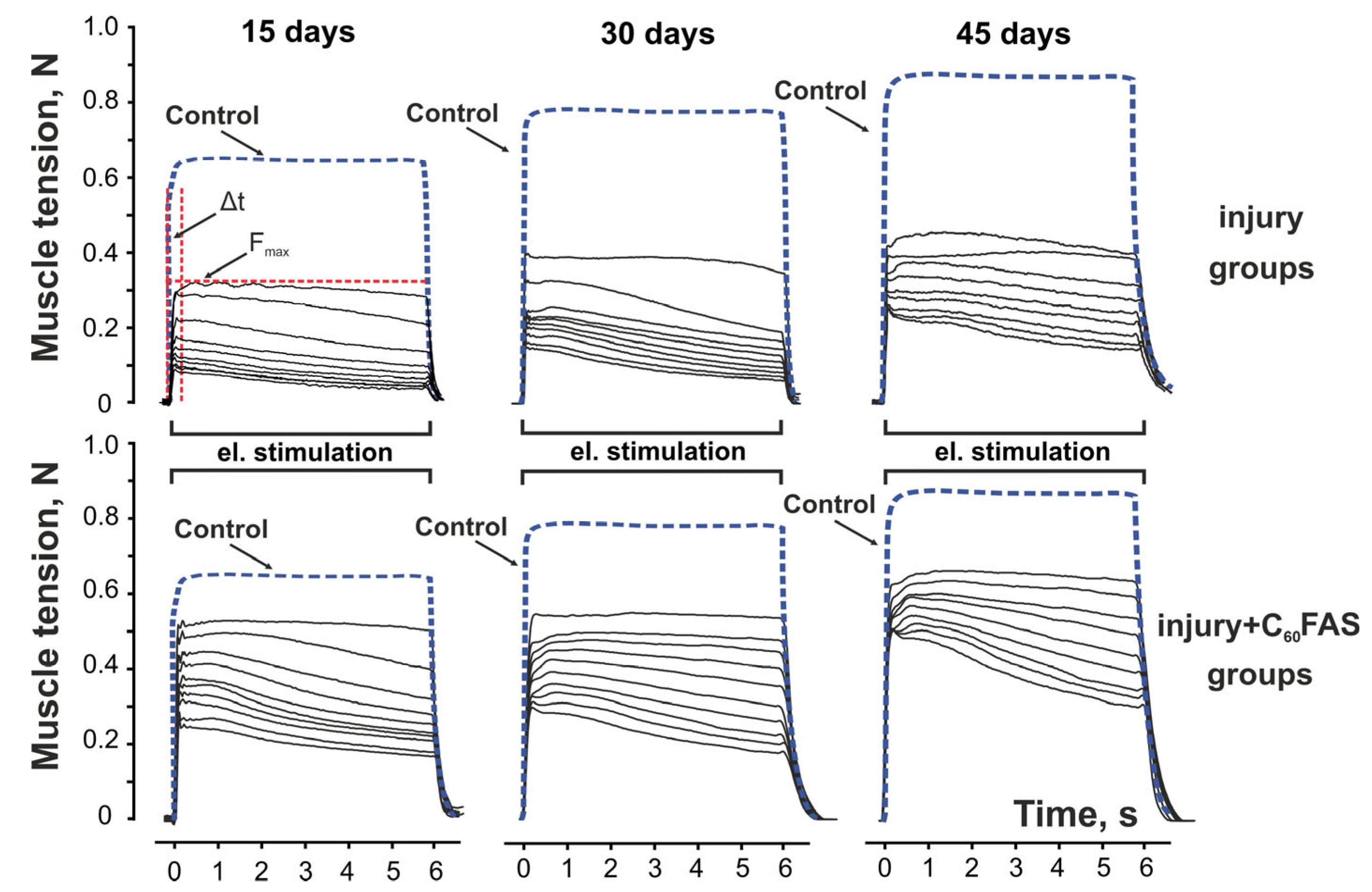
2.2. Muscle Contraction Force Generation

The impact of muscle injury became evident when examining force generation patterns. At 15 days post-injury, injured animals exhibited severely compromised contractile performance, generating only 58 ± 3% of control force values during the first stimulation series, with further decline to 38 ± 2% by the tenth series. Remarkably, daily administration of aqueous C60FAS significantly ameliorated these deficits, restoring force generation to 91 ± 4% and 62 ± 3% of control levels for the respective stimulation series (p < 0.05 for both comparisons). This therapeutic effect showed progressive enhancement with longer treatment duration. By day 30, injured, untreated muscles reached only 43 ± 2% of control force capacity, while C60FAS-treated animals achieved 83 ± 4% recovery. Similarly, at 45 days post-injury, the injury-only group remained at 37 ± 2% of control values, whereas C60FAS treatment enabled 52 ± 2% functional recovery (Figure 1).

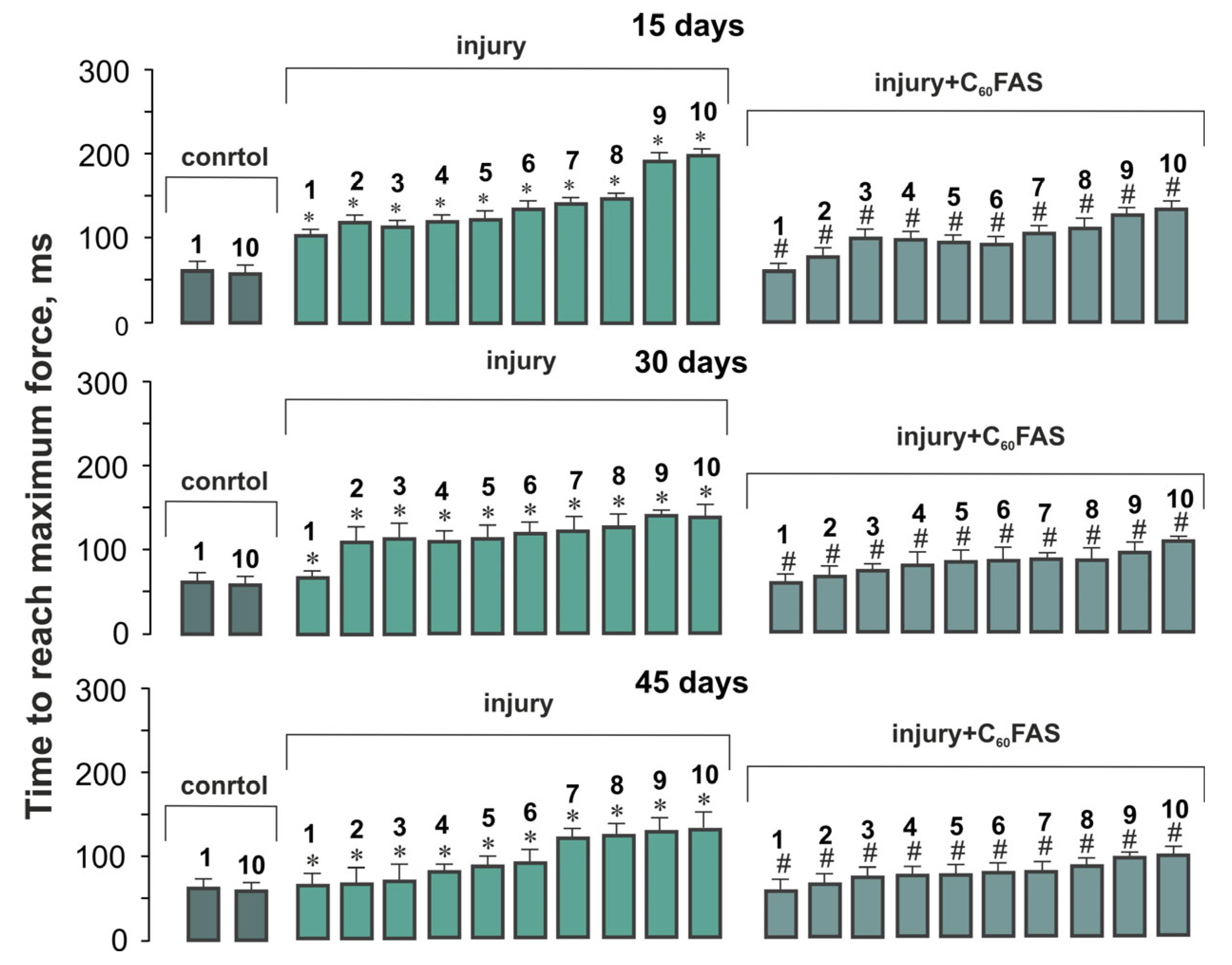
2.3. Temporal Dynamics of Muscle Contraction

The temporal characteristics of muscle contraction revealed another dimension of C60’s therapeutic action. In control animals, the time required to reach maximal contraction force remained constant at approximately 60 ms throughout all stimulation series. Muscle injury substantially disrupted this parameter, particularly at 15 days post-trauma, where the activation time increased to 107 ms for the first contraction and reached 199 ms by the tenth series, representing 1.8- and 3.4-fold prolongations, respectively. C60FAS treatment effectively mitigated these delays, reducing the values to 62 ± 5 ms (first contraction) and 110 ± 10 ms (tenth contraction), corresponding to 31–38% improvements compared to untreated injured animals (Figure 2). These findings suggest that C60FAS not only enhances force generation capacity but also improves the speed of neuromuscular activation following injury (especially noticeable on day 45 of the experiment).

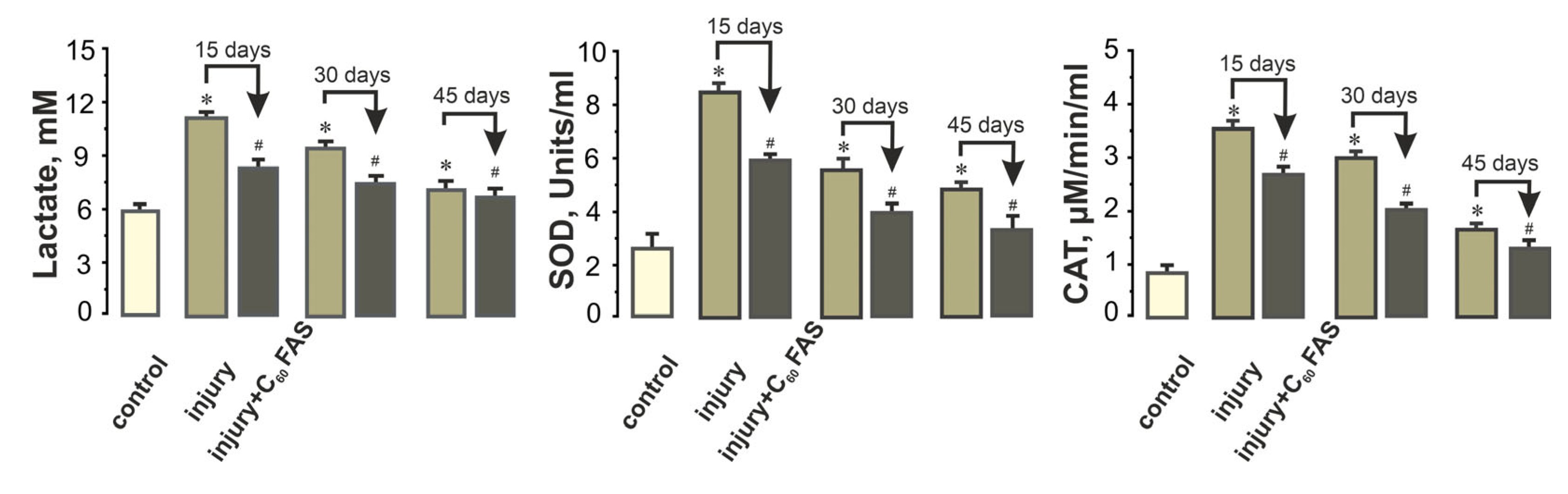
2.4. Systemic Biochemical Markers of Muscle Recovery

To elucidate the mechanisms underlying the observed functional improvements, we conducted comprehensive analyses of plasma biomarkers associated with muscle injury and oxidative stress.
Lactate dynamics similarly demonstrated treatment benefits. While control animals maintained lactate at 5.9 ± 0.50. mM, injury caused an immediate increase to 11.3 ± 0.7 mM at 15 days, with subsequent decline to 7.4 ± 0.8 mM by 45 days. C60FAS-treated animals showed a significant decrease in lactate clearance, achieving values of 8.5 ± 0.5 mM (15 days), 7.3 ± 0.4 mM (30 days), and 6.9 ± 0.5 mM (45 days)—reflecting approximately 25% greater oxidation capacity compared to their untreated counterparts (Figure 3).
Oxidative stress markers provided further mechanistic insights:
SOD activity surged to 8.1 ± 0.2 U/mL (15 days post-injury) vs. 2.6 ± 0.4 U/mL in controls, indicating substantial oxidative challenge. C60FAS treatment reduced this elevation by 35–40% across all timepoints (15 days: 5.9 ± 0.2 U/mL; p < 0.05).
CAT activity followed a similar pattern, increasing from 0.9 ± 0.1 µM/min (control) to 3.4 ± 0.1 µM/min post-injury (15 days), with C60FAS treatment producing 25–35% reductions (15 days: 2.4 ± 0.2 µM/min; Figure 3) and a decrease in the following days.
These biochemical findings collectively demonstrate that C60 fullerene exerts significant antioxidant effects, attenuating injury-induced oxidative stress and facilitating metabolic recovery.

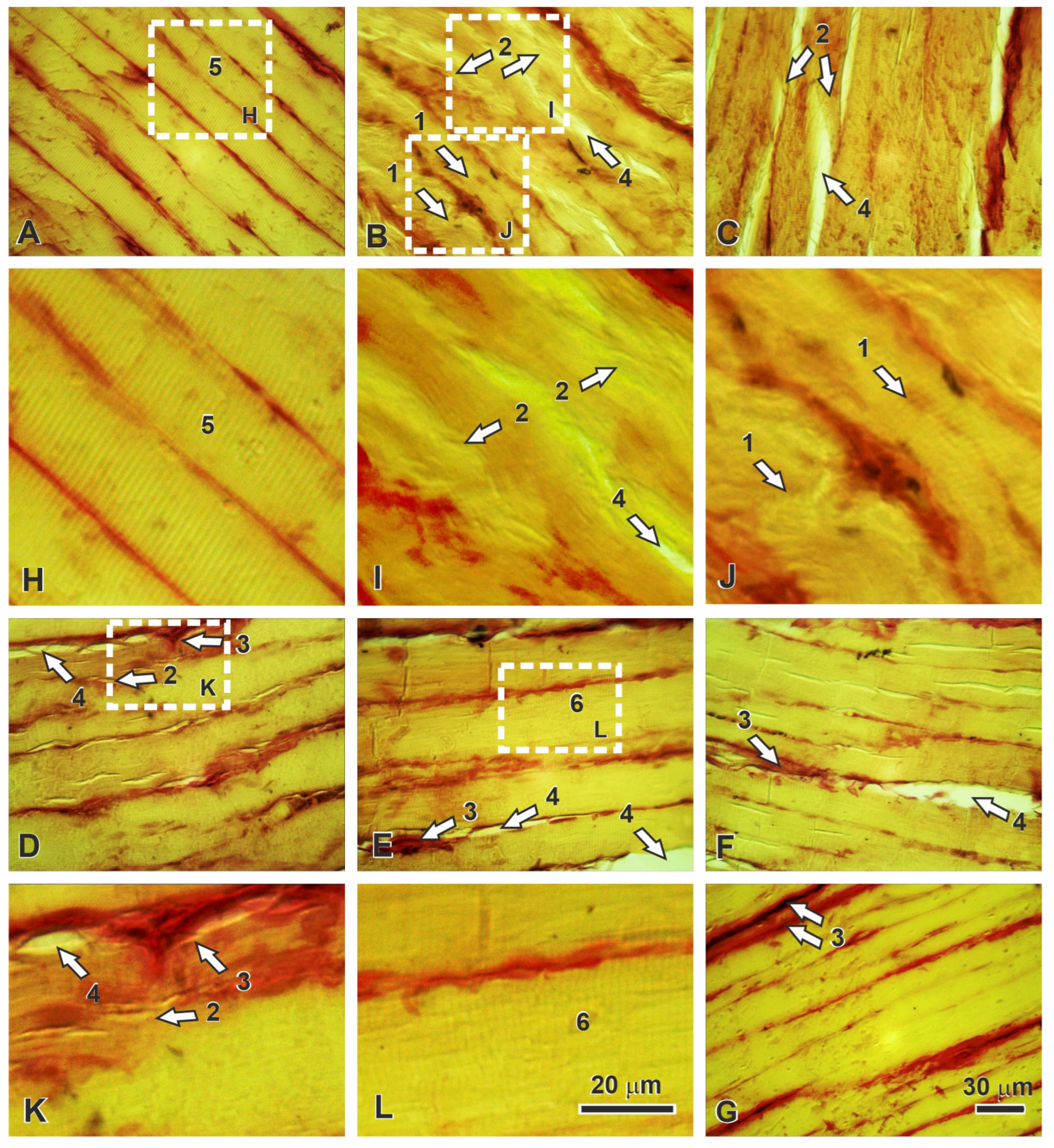
2.5. Histopathological Correlates of Functional Recovery

Histological examination provided structural confirmation of the functional and biochemical improvements observed with C60FAS treatment. Control muscles displayed characteristic polygonal fibers with well-defined transverse striation and minimal endomysial connective tissue (Figure 4A).
The temporal progression of injury and recovery revealed distinct treatment effects:
Fifteen days post-injury: Untreated muscles showed extensive fiber destruction, loss of striation, and marked endomysial edema (Figure 4B). C60FAS-treated specimens exhibited significantly better preservation of fiber architecture, with maintained striation patterns and reduced connective tissue infiltration (Figure 4C).
Thirty days: While untreated muscles displayed persistent fiber tortuosity and developing fibrosis (Figure 4D), C60FAS-treated samples showed more organized regeneration with fewer pathological fibers (Figure 4E).
Forty-five days: The regeneration process in untreated animals remained incomplete, with numerous thin fibers and substantial collagen deposition (Figure 4F). In contrast, C60FAS-treated muscles approached near-normal histology, with only minimal residual fibrosis (Figure 4G).
This histological progression correlates precisely with the functional recovery timeline, demonstrating that C60FAS not only accelerates structural regeneration but also enhances the quality of muscle repair.

3. Discussion

This study demonstrates that chronic administration of aqueous C60FAS solutions significantly enhances functional recovery following traumatic muscle injury through multimodal mechanisms involving antioxidant protection, metabolic modulation, and structural preservation.
The observed improvements in muscle function and redox balance following C60FAS administration may be attributed to several interrelated mechanisms such as direct ROS scavenging, mitochondrial protection, and anti-inflammatory effects. The molecular mechanism underlying the antioxidant reactions of C60 compounds is that electron-deficient regions of C60 cooperate with the attached malonyl groups to electrostatically guide and stabilize superoxide, thereby promoting its decomposition [14,15]. Additionally, C60 nanoparticles have been shown to be selectively internalized by cerebral endothelial cells damaged by oxidative stress, significantly inhibiting apoptosis induced by ROS. This protective effect is associated with the modulation of the c-Jun N-terminal kinase signaling pathway [16]. Furthermore, some studies have demonstrated that fullerenes can significantly alleviate ROS-dependent neuronal injury induced by NMDA or potassium deficiency in vitro [17,18]. The neuroprotective effects of fullerenes have also been validated in models of cortical infarction induced by ischemia–reperfusion [15,19]. These findings suggest that the structure of C60 enables efficient electron acceptance, allowing it to neutralize multiple ROS, which aligns with our observed reductions in oxidative stress markers, such as SOD and CAT activity. Moreover, the strong electron affinity of fullerene materials supports their role in preventing excess electrons in the electron transport chain by functioning as a capacitor-like electron sink. Their electrophilic nature stimulates the Nrf2/ARE pathways, leading to an antioxidant response and mitochondrial biogenesis, thereby helping to maintain mitochondrial homeostasis [20]. This mechanism may explain the improved lactate metabolism observed in our study, as preserved mitochondrial function shifts energy production toward oxidative phosphorylation. Additionally, C60 has been reported to indirectly inhibit NF-κB activation, potentially reducing the production of pro-inflammatory cytokines and demonstrating anti-inflammatory properties in diabetic complications [21,22].
Our results align with and substantially extend previous reports on fullerene-based therapies in musculoskeletal disorders [5,10], while providing novel insights into their time-dependent effects on mechanokinetic parameters and oxidative stress dynamics. Our findings demonstrate that C60FAS treatment significantly improves two critical aspects of muscle function: maximal force generation (35–40% enhancement) and time to peak contraction (27–38% acceleration). These parameters are fundamental to the excitation-contraction-relaxation cycle. Precise coordination between motoneuron pool activation and muscle fiber recruitment is essential for optimal muscle function [23]. Injury-induced oxidative stress can disrupt this coordination by damaging voltage-gated sodium channels, thereby delaying action potential propagation [24]. Oxidative stress impairs mitochondrial ATP production and increases the generation of ROS [25]. Inflammatory processes following muscle injury are a source of ROS and contribute to the intensification of lipid peroxidation processes [26]. Therefore, in the present study, we evaluated changes in the blood of experimental animals in biochemical parameters.
The 25% lactate reduction may reflect either improved metabolic efficiency or attenuated inflammation, consistent with our observed decreases in SOD/CAT activity. It can also be suggested that a reduction in lactate levels may improve glycolytic ATP production, which is crucial for the detachment of myosin heads during rapid muscle contractions. This observation aligns with studies indicating that fullerenes stabilize mitochondrial membrane potential and reduce intracellular ROS production [27,28]. Such effects are clinically relevant for joint positioning accuracy, especially in fine motor tasks such as hand grasping, where millisecond delays in force generation can compromise movement precision [29,30,31,32].
The antioxidant defense system plays a central role in maintaining muscle homeostasis, particularly during post-traumatic recovery. SOD stands as the first line of protection against oxidative damage by catalyzing the conversion of superoxide radicals to hydrogen peroxide. Restoration of SOD activity achieved through fullerene C60 intervention demonstrates the compound’s capacity to disrupt the cycle of oxidative stress and mitochondrial dysfunction. Similarly, CAT works in concert with SOD by decomposing hydrogen peroxide into water and molecular oxygen, thereby preventing the formation of highly reactive hydroxyl radicals [33]. The normalization of CAT activity by 30–35% following fullerene C60 treatment indicates successful modulation of the intracellular redox environment.
The observed modulation of CAT activity following muscle injury and subsequent C60FAS treatment underscores the compound’s ability to restore redox equilibrium in damaged tissues. In the acute phase of muscle trauma, the increase in ROS activity triggers a compensatory upregulation of CAT. However, prolonged oxidative stress can exhaust this adaptive response [34]. Our experimental data align with these biochemical principles, as the initial post-injury CAT elevation was followed by a gradual decline. The 30–35% reduction in CAT activity suggests C60FAS partially supplants endogenous antioxidants by decreasing the H2O2 level through its electron-accepting carbon cage structure [26]. This dual action likely prevents the destructive feedback loop wherein H2O2 accumulation further inhibits CAT, exacerbating oxidative damage to lipids and proteins.
Morphological analyses provided structural correlates to these biochemical improvements. Injured muscle sections typically exhibit hallmark features of oxidative pathology [35]. These changes reflect the direct impact of ROS on cellular integrity. Samples from C60FAS-treated animals demonstrated markedly preserved myofibrillar architecture, with fewer necrotic fibers and reduced collagen deposition. This suggests that by maintaining CAT activity and reducing overall oxidative stress, the treatment safeguarded not only contractile proteins but also the extracellular matrix remodeling processes essential for functional recovery [36]. These morphological improvements further explain the biomechanical enhancements observed, such as increased maximum force developed and the time to reach maximum contraction, as intact sarcomeres and reduced fibrosis directly translate to better force transmission. Collectively, these findings position C60FAS as a modulator of both molecular and structural repair pathways in muscle tissue.

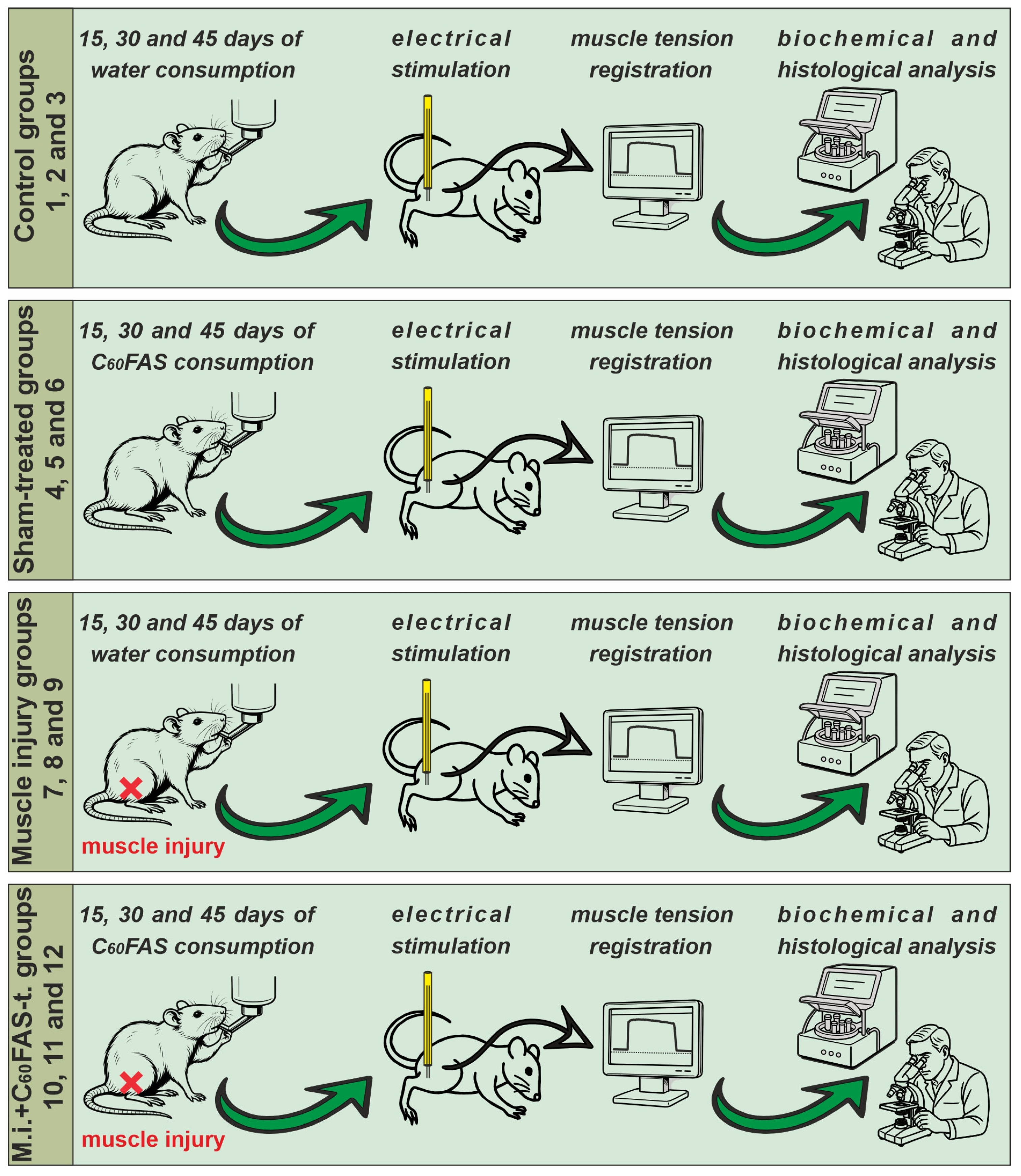
4. Materials and Methods

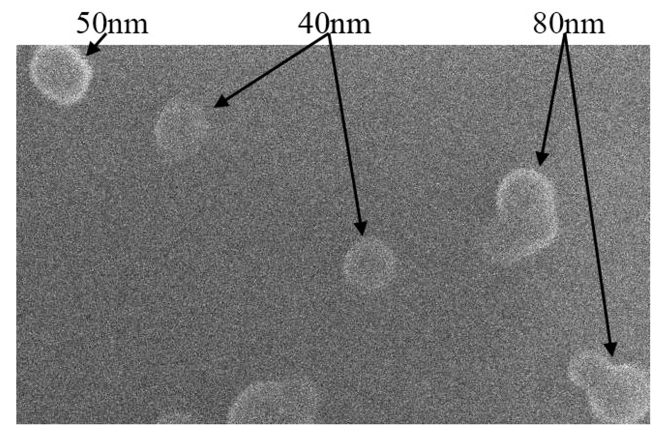
4.1. Material Preparation and Characterization

C60FAS was prepared with high purity exceeding 99.96% using an established ultrasonic-assisted phase transfer method from organic to aqueous media, as previously described [37]. The solution purity was rigorously verified through HPLC and GC/MS analyses to confirm the absence of residual impurities, including carbon black and organic solvents [38]. Comprehensive characterization of the C60FAS was performed using atomic force microscopy (AFM Solver Pro M system, NT-MDT, Optophase, Lyon, France) and dynamic light scattering (DLS, Zetasizer Nano-ZS90, Malvern Instruments, Malvern, UK). AFM imaging revealed two distinct molecular arrangements: individual C60 molecules appearing as discrete nanoscale features approximately 0.7 nm in height, and small nanoclusters measuring 1.3–2 nm in height, representing oligomeric aggregates. DLS measurements further confirmed the colloidal properties, showing a mean hydrodynamic diameter of 82 nm and a zeta potential of −23.9 mV, both indicating excellent solution stability. Finally, the scanning electron microscopy (SEM, FEI/Philips XL30 ESEM, Hillsboro, OR, USA) image of such nanoobjects (Figure 5) indicates their almost spherical shape. The maximum achievable concentration of stable C60 fullerene nanoparticles in aqueous medium using this preparation method was determined to be 0.15 mg/mL. This thorough characterization protocol ensures the reproducibility and quality of the C60FAS for subsequent biological applications.

4.2. Procedure and Experimental Animals

Male Wistar rats (100–120 g, 1-month-old) were used in the study. The animals were obtained from the vivarium of the ESC “Institute of Biology and Medicine” of Taras Shevchenko National University of Kyiv. The rats were housed in Plexiglas cages and kept in an air-filtered, temperature-controlled room (20–22 °C) under a 12-h light/12-h dark cycle. They received a standard pellet diet and water ad libitum. All animal procedures were approved by the Biomedical Ethics Committee of the ESC “Institute of Biology and Medicine” (Protocol #2, 2 September 2022) and conducted in accordance with the European Union Directive 2010/63/EU for the protection of animals used for scientific purposes. The study complied with ARRIVE guidelines.
The animals were randomly divided into 12 groups (n = 5 per group):
Groups 1–3: Control group (intact animals without muscle injury and/or C60FAS treatment), evaluated at 15, 30, and 45 days of the experiment, respectively.
Groups 4–6: Sham-treated group (animals administered C60FAS but not subjected to muscle injury), evaluated at 15, 30, and 45 days.
Groups 7–9: Muscle injury group (rats with injured gastrocnemius muscle), evaluated at 15, 30, and 45 days.
Groups 10–12: Muscle injury + C60FAS-treated group, evaluated at 15, 30, and 45 days.
Animals in Groups 7–9 and 10–12 received oral C60FAS at a dose of 1 mg/kg daily after injury induction. This dose was selected based on its demonstrated efficacy in previous studies [5]. Muscle trauma was induced on day 0. We used independent animal cohorts for each time point (15, 30, and 45 days). Following the electrophysiological assessments, all animals underwent tensometric, biochemical, and histological analyses (see schematic representation of the study, Figure 6).

4.3. Operations and Electrical Stimulation Protocols

Animals were deeply anesthetized via intraperitoneal injection of Zoletil (40 mg/kg, Virbac, Carros, France). The skin over the right hindlimb was carefully dissected to expose the gastrocnemius muscle. The muscle was subjected to transverse incisions (1 mm depth) at three equidistant points. The skin wound was then sutured using synthetic absorbable copolymer suture material. Throughout the procedure and subsequent experiment, heart rate and body temperature were monitored.
At 15, 30, and 45 days post-injury, rats were reanesthetized (using the same protocol) for terminal experiments. The right gastrocnemius muscle was isolated from surrounding tissues, and its tendon was detached at the distal insertion point. The sciatic nerve was carefully separated and transected proximally, with all branches except those innervating the gastrocnemius muscle being cut. The animals were then secured to a platform, and the nerve was placed on a bipolar platinum wire electrode for electrical stimulation. The exposed muscle and nerve were immersed in paraffin oil within a skin-flap pool.
The gastrocnemius muscle was connected via the Achilles tendon to a servo-controlled muscle puller. Muscle tension was measured using semiconductor strain gauge resistors affixed to a stiff steel beam mounted on the linear motor’s moving part. The puller’s stiffness exceeded 0.06 N/mm, with length transient time constants ≤60 ms.
Muscle contraction was induced using 10 intermittent high-frequency electrical stimulation series (6 s duration, separated by 4 s rest intervals) [5]. Each series consisted of 0.2-ms rectangular pulses at 50 s−1. Stimulus current was set to 1.3–1.4 times the motor threshold using a High-Power Bi-Phase Current Stimulator (Aurora Scientific Inc., Aurora, Canada). Examples of muscle tension patterns induced by above mentioned stimulations have been shown in Figure 7. Tensometric signals were digitized at 10 kHz and recorded using a Force Transducer System (Aurora Scientific Inc., Aurora, ON, Canada). Following the experiment, animals were euthanized via Nembutal overdose.

4.4. Biomechanical, Biochemical, and Histological Analyses

Muscle contraction parameters and blood biochemical markers were assessed at 15, 30, and 45 days post-injury.
The biomechanical parameters evaluated included:
Maximal contraction force generation: This global indicator of muscular dysfunction reflects impairments in both neural and myotonic components. Pathological reductions may result from disrupted synaptic currents in motoneurons, leading to impaired summation of transmembrane potentials and consequent alterations in action potential sequences that trigger contraction.
Time to reach maximum force response: This parameter reflects the muscle’s ability to generate rapid, ballistic movements. Changes in this metric indicate physiological dysfunction during maximal force generation tasks.
Biochemical analyses measured concentrations of lactate and oxidative stress markers (CAT, SOD) using clinical diagnostic analyzers (RNL-200 and JN-1101-TR2, Amsterdam, Netherlands).
For histological evaluation, gastrocnemius muscle samples were fixed in 10% formalin, paraffin-embedded, and sectioned at 5 µm. Sections were stained with hematoxylin and van Gieson picrofuchsin [39]. Digital micrographs were acquired at ×400 magnification using an Olympus BX41 microscope equipped with a C-5050 Zoom digital camera. Pathohistological profiles were assessed via light microscopy.

4.5. Statistical Analysis

Biomechanical data are presented as mean ± S.E.M. and were analyzed using two-way ANOVA with time and C60FAS treatment as factors. Between-group differences were assessed via Bonferroni post hoc tests (significance threshold: p < 0.05).
Biochemical data (mean ± S.E.M.) were also compared across time points and effects of C60FAS using two-way ANOVA followed by Bonferroni’s multiple comparison test (p < 0.05 considered significant). Data normality was verified using the Shapiro–Wilk test, and variance homogeneity was assessed via Levene’s test. All statistical analyses were performed using Origin 9.4 software (Origin Lab Corp., Northampton, MA, USA).
C60FAS treatment yielded large effect sizes in functional recovery (Cohen’s d = 1.0 for Fₘₐₓ) and moderate-to-large effects in oxidative stress reduction (d = 0.9 for CAT activity), suggesting clinically meaningful benefits. The large effect sizes observed in biomechanical parameters (e.g., Δt reduction) underscore C60FAS’s capacity to restore neuromuscular function. The 30–35% improvement in antioxidant markers (d > 0.8) further supports its role as a potent nanotherapeutic.

5. Conclusions

The present study provides evidence that C60FAS administration leads to notable improvements in neuromuscular function and redox balance in a rat model of muscle injury. Specifically, enhancements were observed across biomechanical, biochemical, and histological parameters, with improvements ranging from 35% to 40% compared to untreated injured animals. These findings suggest that C60FAS may have potential applications in mitigating muscle trauma effects, particularly in contexts requiring precise motor control, such as sports injuries or post-surgical rehabilitation. Given the preclinical nature of this research, further studies are necessary to fully elucidate the underlying mechanisms of action and to assess the efficacy and safety of C60FAS in clinical settings. Future investigations should focus on exploring the long-term effects, optimal dosing strategies, and potential interactions with other therapeutic agents. Additionally, expanding the scope to include diverse models of muscle injury could provide a more comprehensive understanding of C60FAS’s potential benefits. Overall, while these initial findings are promising, translating them into clinical practice will require rigorous validation through extensive research.

Author Contributions

Conceptualization, D.N. and Y.P.; methodology, D.N., U.R. and I.V.; software, O.S., Y.K. and T.A.; validation, O.S., Y.K., U.R. and Y.P.; formal analysis, D.N., A.M., I.V., U.R. and I.S.; investigation, D.N., I.V., O.S., Y.K., I.S. and U.R.; writing—original draft preparation, D.N. and A.M.; writing—review and editing, A.M.; visualization, O.S., Y.K. and T.A.; supervision, Y.P.; project administration, T.A.; funding acquisition, I.S. All authors have read and agreed to the published version of the manuscript.

Funding

This research received no external funding.

Institutional Review Board Statement

The use of the laboratory animals was approved by the Biomedical Ethics Committee of the ESC “Institute of Biology and Medicine” of Taras Shevchenko National University of Kyiv (protocol #2, 2 September 2022) and performed in accordance with the European Union Directive of 22 September 2010 (2010/63/EU) for the protection of animals used for scientific purposes.

Informed Consent Statement

Not applicable.

Data Availability Statement

The datasets used and/or analyzed during the current study are available from the corresponding author on reasonable request.

Conflicts of Interest

The authors declare no conflicts of interest.

Abbreviations

The following abbreviations are used in this manuscript:
AFMAtomic force microscopy
ANOVAAnalysis of variance
CATCatalase
C60FASC60 fullerene aqueous solutions
DLSDynamic light scattering
GC/MSGas Chromatography/Mass Spectrometry
HPLCHigh-Performance Liquid Chromatography
NLRP3NOD-like receptor protein 3
NMDAN-methyl-D-aspartate
NPsNanoparticles
ROSReactive oxygen species
S.E.M.Standard error mean
SEMScanning electron microscopy
SODSuperoxide dismutase

References

  1. Järvinen, T.A.; Järvinen, T.L.; Kääriäinen, M.; Äärimaa, V.; Vaittinen, S.; Kalimo, H.; Järvinen, M. Muscle injuries: Optimising recovery. Best Pract. Res. Clin. Rheumatol. 2007, 21, 317–331. [Google Scholar] [CrossRef] [PubMed]
  2. Henderson, N.C.; Rieder, F.; Wynn, T.A. Fibrosis: From mechanisms to medicines. Nature 2020, 587, 555–566. [Google Scholar] [CrossRef] [PubMed]
  3. Prylutskyy, Y.; Nozdrenko, D.; Motuziuk, O.; Prylutska, S.; Bogutska, K.; Abramchuk, O.; Morenko, A.; Franskevych, D.; Scharff, P.; Ritter, U. C60 fullerene reduces the development of post-traumatic dysfunction in rat soleus muscle. Int. J. Mol. Sci. 2024, 25, 12206. [Google Scholar] [CrossRef]
  4. Greising, S.M.; Warren, G.L.; Southern, W.M.; Nichenko, A.S.; Qualls, A.E.; Corona, B.T.; Call, J.A. Early rehabilitation for volumetric muscle loss injury augments endogenous regenerative aspects of muscle strength and oxidative capacity. BMC Musculoskelet. Disord. 2018, 19, 173. [Google Scholar] [CrossRef]
  5. Prylutskyy, Y.; Nozdrenko, D.; Motuziuk, O.; Prylutska, S.; Vareniuk, I.; Nurishchenko, N.; Franskevych, D.; Soroca, V.; Bogutska, K.; Ritter, U. C60 fullerene promotes post-traumatic recovery of the rat muscle gastrocnemius. Nanomedicine 2025, 20, 571–584. [Google Scholar] [CrossRef]
  6. Duchesne, E.; Dufresne, S.S.; Dumont, N.A. Impact of inflammation and anti-inflammatory modalities on skeletal muscle healing: From fundamental research to the clinic. Phys. Ther. 2017, 97, 807–817. [Google Scholar] [CrossRef]
  7. Pelaz, B.; Alexiou, C.; Alvarez-Puebla, R.A.; Alves, F.; Andrews, A.M.; Ashraf, S.; Balogh, L.P.; Ballerini, L.; Bestetti, A.; Brendel, C.; et al. Diverse applications of nanomedicine. ACS Nano 2017, 11, 2313–2381. [Google Scholar] [CrossRef]
  8. Gharbi, N.; Pressac, M.; Hadchouel, M.; Szwarc, H.; Wilson, S.R.; Moussa, F. [60] fullerene is a powerful antioxidant in vivo with no acute or subacute toxicity. Nano Lett. 2005, 5, 2578–2585. [Google Scholar] [CrossRef] [PubMed]
  9. Dodd, S.; Dean, O.; Copolov, D.L.; Malhi, G.S.; Berk, M. N-acetylcysteine for antioxidant therapy: Pharmacology and clinical utility. Expert Opin. Biol. Ther. 2008, 8, 1955–1962. [Google Scholar] [CrossRef]
  10. Prylutskyy, Y.; Nozdrenko, D.; Motuziuk, O.; Prylutska, S.; Nurishchenko, N.; Franskevych, D.; Cherepanov, V.; Kalinin, I.; Korzhyk, O.; Ritter, U. C60 fullerene improves the contractile activity of the injured rat muscle gastrocnemius. Nanotechnology 2025, 36, 125101. [Google Scholar] [CrossRef]
  11. Jin, L.; Ding, M.; Oklopcic, A.; Aghdasi, B.; Xiao, L.; Li, Z.; Jevtovic-Todorovic, V.; Li, X. Nanoparticle fullerol alleviates radiculopathy via NLRP3 inflammasome and neuropeptides. Nanomedicine 2017, 13, 2049–2059. [Google Scholar] [CrossRef] [PubMed]
  12. Nozdrenko, D.; Abramchuk, O.; Prylutska, S.; Vygovska, O.; Soroca, V.; Bogutska, K.; Khrapatyi, S.; Prylutskyy, Y.; Scharff, P.; Ritter, U. Analysis of biomechanical parameters of muscle soleus contraction and blood biochemical parameters in rat with chronic glyphosate intoxication and therapeutic use of C60 fullerene. Int. J. Mol. Sci. 2021, 22, 4977. [Google Scholar] [CrossRef]
  13. Forcina, L.; Cosentino, M.; Musarò, A. Mechanisms regulating muscle regeneration: Insights into the interrelated and time-dependent phases of tissue healing. Cells 2020, 9, 1297. [Google Scholar] [CrossRef]
  14. Markovic, Z.; Trajkovic, V. Biomedical potential of the reactive oxygen species generation and quenching by fullerenes (C60). Biomaterials 2008, 29, 3561–3573. [Google Scholar] [CrossRef]
  15. Li, C.W.; Li, L.L.; Chen, S.; Zhang, J.X.; Lu, W.L. Antioxidant Nanotherapies for the Treatment of Inflammatory Diseases. Front. Bioeng. Biotechnol. 2020, 8, 200. [Google Scholar] [CrossRef]
  16. Lao, F.; Chen, L.; Li, W.; Ge, C.; Qu, Y.; Sun, Q.; Zhao, Y.; Han, D.; Chen, C. Fullerene nanoparticles selectively enter oxidation-damaged cerebral microvessel endothelial cells and inhibit jnk-related apoptosis. ACS Nano 2009, 3, 3358–3368. [Google Scholar] [CrossRef] [PubMed]
  17. Dugan, L.L.; Turetsky, D.M.; Du, C.; Lobner, D.; Wheeler, M.; Almli, C.R.; Shen, C.K.; Luh, T.Y.; Choi, D.W.; Lin, T.S. Carboxyfullerenes as neuroprotective agents. Proc. Natl. Acad. Sci. USA 1997, 94, 9434–9439. [Google Scholar] [CrossRef]
  18. Ali, S.S.; Hardt, J.I.; Dugan, L.L. Sod activity of carboxyfullerenes predicts their neuroprotective efficacy: A structure-activity study. Nanomed. Nanotechnol. 2008, 4, 283–294. [Google Scholar] [CrossRef] [PubMed]
  19. Lin, M.Y.; Su-Feng, F.; Shin-Zong, L.; Cheng-Kong, C.; Tieng-Yau, L.; Low-Tone, H. Local carboxyfullerene protects cortical infarction in rat brain. Neurosci. Res. 2002, 43, 317–321. [Google Scholar] [CrossRef]
  20. Bourassa, D.J. An alternate view of fullerene material’s antioxidant activity (NOLF 4 in series: “will nanocarbon onion-like fullerenes (NOLFs) play a decisive role in the future of molecular medicine?”). J. Nanomed. Res. 2020, 8, 1–5. [Google Scholar]
  21. Hou, W.; Shen, L.; Zhu, Y.; Wang, X.; Du, T.; Yang, F.; Zhu, Y. Fullerene Derivatives for Tumor Treatment: Mechanisms and Application. Int. J. Nanomed. 2024, 2024, 9771–9797. [Google Scholar] [CrossRef] [PubMed]
  22. Nedzvetsky, V.S.; Sukharenko, E.V.; Baydas, G.; Andrievsky, G.V. Water-soluble C60 fullerene ameliorates astroglial reactivity and TNFa production in retina of diabetic rats. Regul. Mech. Biosyst. 2019, 10, 513–519. [Google Scholar] [CrossRef]
  23. Burke, R. Spinal motoneurons. In Neuroscience in the 21st Century: From Basic to Clinical; Springer International Publishing: Cham, Switzerland, 2022; pp. 1437–1473. [Google Scholar]
  24. Mahapatra, C.; Thakkar, R.; Kumar, R. Modulatory impact of oxidative stress on action potentials in pathophysiological states: A comprehensive review. Antioxidants 2024, 13, 1172. [Google Scholar] [CrossRef]
  25. Ye, S.; Zhou, T.; Cheng, K.; Chen, M.; Wang, Y.; Jiang, Y.; Yang, P. Carboxylic Acid Fullerene (C60) derivatives attenuated neuroinflammatory responses by modulating mitochondrial dynamics. Nanoscale Res. Lett. 2015, 10, 953. [Google Scholar] [CrossRef]
  26. Yin, J.J.; Lao, F.; Fu, P.P.; Wamer, W.G.; Zhao, Y.; Wang, P.C.; Qiu, Y.; Sun, B.; Xing, G.; Dong, J.; et al. The scavenging of reactive oxygen species and the potential for cell protection by functionalized fullerene materials. Biomaterials 2009, 30, 611–621. [Google Scholar] [CrossRef]
  27. Li, X.; Yang, Y.; Zhang, B.; Lin, X.; Fu, X.; An, Y.; Zou, Y.; Wang, J.X.; Wang, Z.; Yu, T. Lactate metabolism in human health and disease. Signal Transduct. Target. Ther. 2022, 7, 305. [Google Scholar] [CrossRef] [PubMed]
  28. Juel, C.; Halestrap, A.P. Lactate transport in skeletal muscle—Role and regulation of the monocarboxylate transporter. J. Physiol. 1999, 517 Pt 3, 633–642. [Google Scholar] [CrossRef]
  29. Cuzzocrea, S.; Riley, D.P.; Caputi, A.P.; Salvemini, D. Antioxidant therapy: A new pharmacological approach in shock, inflammation, and ischemia/reperfusion injury. Pharmacol. Rev. 2001, 53, 135–159. [Google Scholar] [CrossRef]
  30. Crevecoeur, F.; Thonnard, J.L.; Lefèvre, P.; Scott, S.H. Long-latency feedback coordinates upper-limb and hand muscles during object manipulation Tasks. eNeuro. 2016, 3, ENEURO.0129-15.2016. [Google Scholar] [CrossRef]
  31. Vlasenko, O.V.; Pilyavskii, A.I.; Maiskii, V.A.; Maznichenko, A. Fos immunoreactivity and NADPH-d reactivity in the brain cortex of rats realizing motivated stereotyped movements by the forelimb. Neurophysiology 2008, 40, 295–303. [Google Scholar] [CrossRef]
  32. Tal’nov, A.N.; Tomiak, T.; Maznychenko, A.V.; Dovgalets, G.V.; Kostyukov, A.I. Firing patterns of human biceps brachii motor units during isotorque ramp-and-hold movements in the elbow joint. Neurophysiology 2014, 46, 212–220. [Google Scholar] [CrossRef]
  33. Mao, G.D.; Thomas, P.D.; Lopaschuk, G.D.; Poznansky, M.J. Superoxide dismutase (SOD)-catalase conjugates. Role of hydrogen peroxide and the Fenton reaction in SOD toxicity. J. Biol. Chem. 1993, 268, 416–420. [Google Scholar] [CrossRef] [PubMed]
  34. Gebicka, L.; Krych-Madej, J. The role of catalases in the prevention/promotion of oxidative stress. J. Inorg. Biochem. 2019, 197, 110699. [Google Scholar] [CrossRef] [PubMed]
  35. Li, J.; Tang, M.; Yang, G.; Wang, L.; Gao, Q.; Zhang, H. Muscle injury associated elevated oxidative stress and abnormal myogenesis in patients with idiopathic scoliosis. Int. J. Biol. Sci. 2019, 15, 2584–2595. [Google Scholar] [CrossRef]
  36. Wight, T.N.; Potter-Perigo, S. The extracellular matrix: An active or passive player in fibrosis? Am. J. Physiol. Gastrointest. Liver Physiol. 2011, 301, G950–G955. [Google Scholar] [CrossRef]
  37. Ritter, U.; Prylutskyy, Y.u.I.; Evstigneev, M.P.; Davidenko, N.A.; Cherepanov, V.V.; Senenko, A.I.; Marchenko, O.A.; Naumovets, A.G. Structural features of highly stable reproducible C60 fullerene aqueous colloid solution probed by various techniques. Fuller. Nanotubes Carbon Nanostruct. 2015, 23, 530–534. [Google Scholar] [CrossRef]
  38. Keykhosravi, S.; Rietveld, I.B.; Couto, D.; Tamarit, J.L.; Barrio, M.; Céolin, R.; Moussa, F. [60] Fullerene for medicinal purposes, a purity criterion towards regulatory considerations. Materials 2019, 12, 2571. [Google Scholar] [CrossRef]
  39. Suvarna, K.S.; Layton, C.; Bancroft, J.D. Bancroft’s Theory and Practice of Histological Techniques, 8th ed.; Elsevier: Amsterdam, The Netherlands, 2018; p. 584. ISBN 9780702068645. [Google Scholar]
Figure 1. Muscle tension of rat gastrocnemius muscle following trauma and C60FAS treatment. Graph shows mean ± S.E.M. values for maximal contraction force generation (N, Newton) across 10 consecutive tetanic contractions (contractions 1–10 shown). Control data display values for the 1st and 10th contractions only. Experimental groups include injury (untreated trauma) and injury + C60FAS (trauma animals receiving daily oral administration of 1 mg/mL C60FAS). Time points represent 15, 30, and 45 days post-injury. Statistical significance (* p < 0.05 vs. control).
Ijms 26 05511 g001
Figure 2. Temporal dynamics of contraction kinetics in rat gastrocnemius muscle following trauma and C60FAS treatment. Graph shows mean ± S.E.M. values for time to peak force (ms) across 10 consecutive tetanic contractions (contractions 1–10 shown). Control data display values for the 1st and 10th contractions only. Experimental groups include injury (untreated trauma) and injury + C60FAS (trauma animals receiving daily oral administration of 1 mg/mL C60FAS). Time points represent 15, 30, and 45 days post-injury. Statistical significance (* p < 0.05 vs. control; # p < 0.05 vs. injury group).
Ijms 26 05511 g002
Figure 3. Concentrations of lactate and indicators of pro- and antioxidant balance (SOD, CAT) in the plasma of injured rats and rats receiving C60FAS after injury. Control, injury, and injury + C60FAS—indices of control group, groups of injured rats, and rats who daily consumed orally C60FAS at a dose of 1 mg/mL after the initiation of injury; 15, 30, and 45 days—days after the initiation of open muscle injury, respectively; * p < 0.05 relative to control group; # p < 0.05 relative to injury group.
Ijms 26 05511 g003
Figure 4. Representative micrographs of gastrocnemius muscle: (A)—control; (B)—15 days after trauma; (C)—15 days after trauma and C60FAS-treatments; (D)—30 days after trauma; (E)—30 days after trauma and C60FAS-treatments; (F)—45 days after trauma; (G)—45 days after trauma and C60FAS-treatments. The rectangular regions enclosed by dashed lines in (A,B,D,E) are shown in (H–L) at higher magnification. 1—tortuous muscle fibers; 2—fibrilization of muscle fibers; 3—expansion of collagen fibers; 4—ground substance of connective tissue; 5—normal cross-striation; 6—poorly visible cross-striation. Hematoxylin and picrofuchsin staining by van Gieson. Scale bars for sections (AG)—30 μm, for (HL)—20 μm.
Ijms 26 05511 g004
Figure 5. SEM image of C60 fullerene nanoparticles formed by thermal evaporation of the dispersant from the volume of a C60FAS (0.15 mg/mL) microdroplet at room temperature.
Ijms 26 05511 g005
Figure 6. Schematic representation of the study. Groups 1–3: controls (water); 4–6: sham (C60FAS); 7–9: injury (water); 10–12: injury + C60FAS-treated (1 mg/kg/day). Evaluated at 15, 30, and 45 days. Protocol: oral administration → electrophysiology → biochemical/histological analyses. Parts of the figure were generated using DALL-E.
Ijms 26 05511 g006
Figure 7. Representative recordings of 10 consecutive tetanic contractions in rat gastrocnemius muscle from different experimental groups. Data from one animal per group are shown as individual mechanograms, while control group data (blue line) represent averaged values across all control animals. Experimental groups include trauma (untreated injury) and trauma + C60FAS (injured animals treated with water-soluble C60 fullerene). Key parameters are indicated: Δt (time from stimulation onset to peak force), Fmax (maximal force amplitude), and time points (15, 30, 45 days post-injury). The Δt and Fmax parameters are indicated by vertical and horizontal red dashed lines, respectively. N—muscle tension (Newton).
Ijms 26 05511 g007
Disclaimer/Publisher’s Note: The statements, opinions and data contained in all publications are solely those of the individual author(s) and contributor(s) and not of MDPI and/or the editor(s). MDPI and/or the editor(s) disclaim responsibility for any injury to people or property resulting from any ideas, methods, instructions or products referred to in the content.

