Review

Effect of KLF15-Mediated Circadian Rhythm on Myocardial Infarction: A Narrative Review

Acupuncture and Tuina School, Chengdu University of Traditional Chinese Medicine, Chengdu 610075, China
*
Authors to whom correspondence should be addressed.
Int. J. Mol. Sci. 2025, 26(10), 4831; https://doi.org/10.3390/ijms26104831
Submission received: 17 April 2025 / Revised: 15 May 2025 / Accepted: 16 May 2025 / Published: 18 May 2025
(This article belongs to the Section Biochemistry)

Abstract

Normal circadian rhythms are essential for organisms to adapt to diurnal changes and maintain an optimal state of physiological function. Disturbances in circadian rhythms such as shift work and working at night increase the risk of cardiovascular disease. Myocardial infarction exhibits a marked circadian rhythm, usually peaking in the early morning. Krüppel-like factor 15 (KLF15), a transcription factor with a circadian rhythm, plays an important role in cardiac physiopathology. It has a protective effect against myocardial injury after myocardial infarction by regulating energy metabolism and inflammatory factors, among other pathways. Currently, the association between circadian rhythm, KLF15, and myocardial infarction is unclear, thus this paper reviews how circadian rhythm influences the role of KLF15 in myocardial infarction, aiming to reveal the association between circadian rhythm, KLF15, and myocardial infarction, and to explore the underlying mechanisms, to provide new theoretical insights and therapeutic strategies for the clinical treatment of myocardial infarction.

1. Introduction

Myocardial infarction is a critical disease resulting from acute occlusion of coronary arteries leading to myocardial ischemia, hypoxia, and necrosis, which in turn severely affects cardiac function. As one of the leading causes of death and disability worldwide, its morbidity and mortality rates continue to rise, posing a major threat to public health [1,2]. The high cost of treatment not only aggravates the economic burden of patients’ families, but also creates a heavy social medical burden, which makes the study of myocardial infarction particularly urgent. Clinical symptoms of myocardial infarction include chest pain from the left arm to the neck, shortness of breath, sweating, nausea and vomiting, abnormal heartbeat, anxiety and depression, and weakness [3,4]. A definitive diagnosis of myocardial infarction is made when a patient presents with an increase or decrease in cardiac biomarkers (e.g., troponin) with at least one value higher than 99% of the reference value, accompanied by at least one symptom of myocardial ischemia.
The occurrence of myocardial infarction is closely related to a number of risk factors, the traditional risk factors include advanced age, hypertension, diabetes, and obesity. However, it is worth noting that the incidence of myocardial infarction in young people under the age of 45 has been on the rise in recent years. Unlike older patients whose morbidity is mostly due to underlying diseases, the main risk factors for younger patients are more inclined to poor lifestyle (e.g., lack of exercise, prolonged staying up late) and inappropriate stress coping styles (including smoking, alcohol abuse, and drug abuse). In particular, staying up late is one of the most important factors in the development of myocardial infarction in young people [5]. This is associated with the involvement of circadian rhythm-related genes in regulating the physiological activities of the heart [6,7], such as heart rate, blood pressure, and myocardial contractility which fluctuate throughout the day, peaking in the afternoon and troughing at night [8]. These changes are regulated by the autonomic nervous system in addition to the central biological clock, which, by influencing the vasculature and peripheral biological clocks in the myocardial cells, ensures that the heart’s rhythm matches that of the whole organism [9,10]. The cardiac biological clock consists of circadian oscillators, circadian input/output pathways, and circadian regulators. Rhythm oscillators are the core components that influence cyclic changes in gene expression and protein levels to regulate physiological processes within the organism [11]. The core oscillator, in turn, is composed of multiple transcriptional-translational feedback loops of genes and proteins [11] that are tightly coupled to systemic signals (e.g., hormones, autonomic inputs) and local factors (e.g., metabolic demands) that fine-tune cardiac physiology throughout the 24-h cycle. Circadian input pathways regulate and reset the biological clock by receiving signals from the outside world [12], the most important of which is light. Natural light is detected by cells in the retina’s specialized photoreceptors, and the signals are transmitted to the hypothalamus’s suprachiasmatic nucleus (SCN), which then transmits the signals to other areas of the brain and peripheral tissues including the heart [13]. In the cardiovascular system, the SCN transmits circadian signals to the heart through hormonal signaling (e.g., glucocorticoid or melatonin-mediated signaling) and the autonomic nervous system (sympathetic and parasympathetic nerves) [14,15,16], which ensures that the cardiac peripheral bioclocks’ rhythmic activity is synchronized with the external light-dark cycle, thus optimizing the cardiac electrophysiologic properties metabolic activity and contractile function to adapt to the demands of daily activity patterns [17]. A disturbance in this cascade of regulation (e.g., suprachiasmatic nucleus with the peripheral biological clock) can lead to cardiac rhythmic dysregulation and increase the risk of cardiovascular diseases such as arrhythmias and heart failure.
KLF15, an important rhythm gene in the heart [18], whose expression is regulated by core clock genes such as Bmal1, exhibits a clear pattern of circadian oscillations [19]. It has been found [19] that KLF15 regulates biphasic transcriptional oscillations of genes in the heart: one phase corresponds to the active phase in rodents, dominated by maximization of energy supply, and the other phase corresponds to the resting phase, dominated by cardiac remodeling and repair. However, in the case of normal core clock gene rhythms, this phase assignment becomes disordered when the rhythm of KLF15 is disrupted, this shows that KLF15 is the node between the heart and biological clock rhythms. And it has been found [20,21] that the onset of myocardial infarction is characterized by a significant circadian rhythm, with its incidence peaking in the early morning hours. Is there a link between the onset of myocardial infarction and KLF15 and circadian rhythm? Therefore, the purpose of this paper is to explore the relationship between the three and their potential mechanisms, with a view to providing new perspectives for the prevention and treatment of myocardial infarction.
Objectives: This review systematically investigated (1) the circadian rhythmicity of KLF15, (2) the role of KLF15 in the physiology and pathology of myocardial infarction, and (3) the therapeutic potential of modulating KLF15.

2. Methodology

2.1. Literature Search Strategy

The literature search for this review encompassed comprehensive coverage of international authoritative databases, including PubMed, Web of Science, and Embase, as well as Chinese databases, such as China National Knowledge Infrastructure (CNKI), Wanfang, and China Science and Technology Journal Database (VIP), to ensure a thorough retrieval of both English and Chinese publications. The search strategy was developed using the core terms “KLF15”, “circadian rhythm”, and “myocardial infarction” derived from the Medical Subject Headings (MeSHs) thesaurus, with no time restrictions applied to fully integrate historical foundations and recent advancements in the field. The literature screening prioritized thematic relevance, emphasizing the inclusion of highly cited original studies, reviews, and methodological papers. Manual searches of high-impact journals within the field were additionally performed. Although this narrative review did not adhere to the standardized search protocols of systematic reviews, cross-verification across multiple databases and iterative keyword optimization ensured the representativeness of the selected literature in elucidating the mechanistic links between KLF15, circadian rhythms, and myocardial infarction. This approach established a robust evidence base to support subsequent theoretical explorations.

2.2. Key References Table

The key references for this review are listed in Table 1.

3. Effect of KLF15-Mediated Circadian Rhythm on Myocardial Infarction

3.1. Myocardial Infarction Is Characterized by a Significant Circadian Rhythm

Epidemiological investigations have shown that the occurrence of sudden cardiac death, myocardial infarction, and stroke have circadian rhythms, in which myocardial infarction is mostly concentrated in the early morning from 6–9 a.m., and some scholars believe that neural awakening and endogenous risk factors in the early morning hours are one of the reasons for the high prevalence of myocardial infarction [22]; moreover, elevated blood pressure, heart rate, coagulation activity, and vascular tone underlie the high incidence of this disease, and the blood pressure is often higher in the daytime than at night [48], and the overall trend is higher in the morning and lower in the evening, but this rhythm disappeared in mice knocked out of the Bmal1 or CLOCK genes, suggesting that changes in blood pressure have a circadian rhythm; as a cardioprotective agent, natriuretic peptide also has a circadian rhythm, which makes it able to offset the cardiac damage produced with the circadian changes in blood pressure [23]. The vascular endothelium, which maintains blood flow to vital organs, is regulated by endothelin-1 [24] and oxidative stress [25] and both biomarkers also have significant circadian rhythms, with increases in both biomarkers in the morning increasing the risk of cardiovascular disease; coagulation activity is also enhanced in the morning, as evidenced by elevated levels of prothrombotic fibrinogen activator inhibitor-1 [26] and platelet surface activity GPIIb-IIIa (the last part of the platelet aggregation pathway) [27], which may increase the likelihood of thrombosis and the risk of myocardial infarction by the appearance of a morning peak on waking up in the morning [49].

3.2. Effect of Circadian Rhythm Disturbances on Myocardial Infarction

Long late nights and shift work have become the norm in today’s society, significantly increasing the risk of myocardial infarction in young and middle-aged people, and possibly even leading to sudden death [28]. This is due to the fact that circadian rhythm disruption contributes to elevated basal reactive oxygen species (ROS), elevated levels of thiobarbituric acid reactive substances (TBARS), and hydrogen peroxide in the body. At the same time, it leads to a decrease in antioxidant capacity, prompting the body’s oxidative stress to be more intense, and cardiomyocytes are more prone to ischemic stress and hypoxia-reoxygenation injury, increasing myocardial cell death [29,50], therefore, it is more likely to suffer from heart disease than the general population [51,52]. In an earlier experiment, TBARS and Glutathione peroxidase (GPx) were observed to oscillate throughout the day in the rat heart, with GPx concentrations decreasing and TBARS concentrations increasing in the evening [53]. This phenomenon also suggests that the mismatch between high ROS production and low antioxidant capacity may reduce the heart’s response to oxidative stress in a time-dependent manner, thereby impairing cardiac adaptation to oxidative stress and causing damage.
Circadian rhythm disturbances also have an impact on the prognostic outcome of patients with myocardial infarction. The prognosis of mice recovering from experimentally induced myocardial ischemia is disturbed by rapidly changing light/dark cycles. After myocardial infarction occurs, the body repairs the heart in order to remove dead cells and debris by triggering an inflammatory response that activates cardiomyocyte fibrosis to form a scar [54,55,56]. The circadian mechanism plays a key role in the process of recruiting immune cells [30]. When the biological clock gene is impaired, oxidative stress, mitochondrial dysfunction, and ultimately cardiomyocyte death occurs. However, when gene expression is restored, it improves cardiac function and cellular activity by upregulating cellular autophagy [31]. A similar phenomenon can also affect the recovery of hospitalized patients [32]. Data have also shown that in mice knocked out of the cardiac-specific Bmal1 gene, the level of genes involved in oxidative stress, including GPx, superoxide dismutase, and oxygen transport proteins, is enhanced, suggesting that disruption of the genetic mechanisms of circadian rhythms triggers oxidative stress in the heart [33], and that when circadian rhythms are disrupted, it worsens the response to myocardial infarction [42,57].

3.3. KLF15 Gene: A Central Regulator of Cardiac Circadian Rhythm

KLF15, as the only recognized peripheral biological clock in the heart, is essential for the regulation of cardiac physiological functions. Darwin Jeyaraj et al. conducted experiments on mice with light-dark cycle rotation every 12 h and found that there are rhythmic oscillations of KLF15 in peripheral tissues and organs, such as the heart, liver, and skeletal muscle, and that KLF15 is able to regulate the rhythmic expression of about 75% of the genes occurring in the heart, which is needed to maintain normal physiological function of the heart [19,34]. Chip-seq found that Bmal1 is rhythmically expressed at the promoter of KLF15, indicating that KLF15 expression is regulated by the Endogenous Core Clock Mechanism (ECM) [35], suggesting that KLF15 is characterized by endogenous circadian oscillations. In addition, KLF15 is involved in regulating the expression of cardiac ion channels and endogenous circadian rhythm of the QT interval, thus maintaining the relationship between normal ventricular rhythm and circadian rhythm. Thus, when KLF15 expression is abnormal, it leads to disruption or loss of rhythmic oscillations of most genes in the heart, which, in turn, affects the cardiac ion channel expression and the endogenous circadian rhythm of the QT interval, inducing the appearance of ventricular arrhythmia. It has also been noted that arrhythmias are associated with a higher incidence of myocardial infarction in the early morning period [18]. It has been found that when KLF15 is knocked out or the rhythm expression is disturbed when the core clock genes are normally expressed, the orderly gene oscillations presented in the heart are disrupted, proving that KLF15 is the hub between the biological clock rhythms and the heart. This shows that the external circadian rhythm can affect the rhythm of KLF15 through the core clock gene, and the circadian rhythm of KLF15 can affect the incidence of myocardial infarction, so there is a correlation between the three.

3.4. The Role of KLF15 in the Pathologic Process of Myocardial Infarction

3.4.1. Modulation of the Inflammatory Response

It has been found that when myocardial infarction occurs, pro-inflammatory factors in cardiomyocytes are increased, such as IL-6, TNF-α, IL-1β, signaling that the NF-κB signaling pathway may be activated and involved in myocardial inflammatory response [36]. KLF15 can inhibit NF-κB transcriptional activation and attenuate inflammatory response by inhibiting p300-mediated acetylation [58]. KLF15 can also inhibit MAPK transcriptional activation by inhibiting P38 and ERK1/2 [37]. Furthermore, WWP1 can catalyze K48-linked polyubiquitination and degradation by targeting KLF15, triggering excessive cardiomyocyte inflammation after myocardial infarction. Twist2 acts as a transcription factor with anti-inflammatory and attenuating endoplasmic reticulum stress, ameliorating mitochondrial content and oxidative stress functions, KLF15 can directly regulate the promoter of Twist2 and enhance transcription, thereby exerting its function to reduce myocardial injury [38]. In addition, KLF15 can also alleviate post-ischemic reperfusion injury by regulating CTRP12 [39]. KLF15 is also a direct target of miRNA-223-3p and miRNA-137-3p, both of which can reduce the expression of KLF15 by targeting inhibition of KLF15, increase cardiomyocyte apoptosis and oxidative stress, and exacerbate ischemia-reperfusion injury [59,60]. In contrast, KLF15 expression can be increased by inhibiting the above two RNAs, thus playing a therapeutic role. Thus, KLF15 can regulate the inflammatory response after myocardial infarction through various pathways such as NF-κB, MAPK, Twist2, and CTRP12 [61].

3.4.2. Inhibition of Apoptosis

Previous studies have confirmed that KLF15 is involved in the regulation of apoptosis in a variety of diseases. For example, KLF15 overexpression promotes the proliferation and metastasis of H/R-induced HTR8/SVneo cells by mediating the PI3K-AKTpathway, and inhibits oxidative stress and apoptosis [40]. In cellular experimental studies of a gastric cancer model, overexpression of KLF15 promotes apoptosis and autophagy through PI3K/Akt/ mTOR promoted apoptosis and autophagy, and inhibited the proliferation and invasion of gastric cancer cells [62]. KLF15 was able to attenuate lipopolysaccharide-induced apoptosis and inflammatory response in renal tubular epithelial cells by interacting with PPARδ [63].
KLF15 is widely expressed in cardiomyocytes and fibroblasts of the heart [64]. A 2020 study found [34] that mice knocked down for KLF15 had a larger infarcted area of the heart after modeling compared to normal mice, suggesting the importance of KLF15 for cardioprotection. MiRNA-223-3p negatively regulated the expression of KLF15, leading to increased apoptosis in cardiomyocytes, whereas silencing miR-223-3p could upregulate KLF15 expression to attenuate oxidative stress damage to H9c2 cells. At the same time, this process also prevents cardiomyocyte apoptosis and protects cardiomyocytes from hypoxic injury by decreasing the expression of Bax and C-caspase3 and increasing the expression of Bcl-2 [65]. Xialu’s study [66] confirms that the inhibition of KLF15 ubiquitylation can effectively alleviate the apoptosis induced by the increase of WWP1 expression after myocardial infarction.

3.4.3. Regulation of Energy Metabolism

Studies have shown that KLF15 regulates nutrient fluxes to maintain energy homeostasis in fasting or exercising organisms under normal circadian rhythms [35,41,65,66]. KLF15 is closely related to several key metabolic pathways [67,68], such as glucose metabolism, fatty acid metabolism, and amino acid metabolism, and is involved in the regulation of myocardial ischemia, cardiomyocyte hypertrophy, and fibrosis, and plays an important role in cardiovascular system development, ischemic adaptation, and cardiac remodeling [69].
Glucose Metabolism and Cardiac Energy Supply
KLF15, as an important transcription factor that activates gluconeogenesis-related genes, is able to maintain the body in a normal glycemic state under fasting. KLF15 releases alanine and other amino acids into the circulation for hepatic uptake through the catabolism of skeletal muscle branched-chain amino acids (BCAAs) and up-regulates key enzymes of BCAA catabolism to inhibit lipogenesis and promote gluconeogenesis, thereby providing the liver with a substrate for gluconeogenesis [70]. Myocardial glucose uptake is mainly carried out under Glucose Transporter Type 4 GLUT4, which can promote myocardial glucose uptake when the body is in ischemia, exercise stimulation or insulin secretion is insufficient. It has been shown that KLF15 can promote the entry of glucose into cardiomyocytes by regulating insulin secretion [42], thus providing energy for the physiological activities of the heart. Some studies have found that glucocorticoids can promote glucose metabolism in the heart by activating KLF15, and glucocorticoid (GC) is the pathway of circadian signaling from SCN to the heart, and its own circadian rhythm can mimic the effect of the external circadian rhythm on KLF15, which shows that circadian rhythm can affect the regulation of glucose metabolism by KLF15 [43].
Homeostatic Maintenance of Lipid Metabolism
KLF15 is a key regulator of cardiac lipid metabolism. From birth, KLF15 accumulates in the heart and decreases in heart failure, a trend similar to the pattern of lipid oxidation. KLF15 activates its direct transcriptional targets by interacting with p300, which in turn affects lipid flux and metabolic homeostasis [41]. In patients with heart failure, the heart’s ability to metabolize lipids is reduced, leading to excessive lipid deposition within the heart. KLF15 negatively regulates cardiac lipid deposition and participates in the process of cardiac functional recovery after unloading [44]. Muscle-specific KLF15 knockout mice were found to have significantly reduced lipid utilization and were more prone to obesity [66,71]. PPAR-δ and PPAR-γ, as members of the Peroxisome Proliferator-Activated Receptor (PPAR) family, can regulate adipocyte differentiation and lipid metabolism, and it has been demonstrated that KLF15 directly affects the regulation of lipid metabolism in muscle tissues by PPAR-δ [72], and that KLF15 can regulate the expression of PPAR-γ to inhibit adipogenesis [45,73].
A large part of the energy supply of the adult heart relies on fatty acid oxidation supply, especially in the fasting state, KLF15 can affect fatty acid oxidative catabolism for energy support by regulating the oscillatory expression of cardiac metabolic genes [74,75]. In summary, KLF15 can regulate lipid metabolism in the organism and cardiac tissues, and when KLF15 is absent or dysrhythmic, the ordered rhythmic oscillations are abnormal, thus affecting the energy supply to the body and heart.
Circadian Rhythmicity of Branched-Chain Amino Acid Metabolism
Branched-chain amino acids (BCAAs) play an important role in muscle energy metabolism, and it has been found that KLF15 is able to drive the activation of BCAA to accelerate proteolysis [76], whereas GC is able to prompt KLF15 to exacerbate the activation of BCAA, further accelerating proteolysis, suggesting that, at the peak of the rhythmic expression of GC, it is able to increase the metabolic rate by increasing the expression of KLF15. It has also been shown that, when KLF15 levels decline, BCAA key metabolic enzymes are also absent, and the decline or absence of both of the above, in a mouse model of heart failure, leads to a large accumulation of branched-chain keto acids (BCKAs), which in turn negatively affects mitochondrial function [46]. These indirectly demonstrate that circadian rhythmic expression of KLF15 likewise affects the degree of activation of BCAA metabolism. In addition, a study in Chinese sea bass found that fasting led to a disturbance in the rhythmic expression of KLF15 [47]. Whereas the circadian rhythmicity of BCAA metabolism exists in a KLF15-dependent manner [77]. KLF15 can also act as a rhythmic oscillatory regulator of nitrogen flux and homeostasis, mammals need to remove toxic metabolic by-products by converting ammonia to urea, and it has been found that the expression of genes involved in the metabolism of amino acids and the urea cycle has a rhythmicity [78,79], whereas KLF15 is able to regulate nitrogen homeostasis in mice with a circadian rhythm, and when KLF15 is absent and the rhythm is disturbed, the total amino acid and urea levels in plasma are altered [35], and the concentration of BCAA is elevated, which proves that KLF15 and its circadian rhythmicity are able to influence the metabolic changes of BCAA and nitrogen homeostasis in vivo.
In summary, under physiological conditions, KLF15 can influence multiple energy metabolic pathways in the body and the heart through its own circadian rhythmic characteristics, to maintain energy demand during the active period, and, at the same time, during the resting period, to alleviate the inflammatory response and apoptosis, to repair daily damage to the heart. However, when KLF15 is absent or the rhythm is disturbed, it will result in the heart not being able to repair the damage caused by inflammation and apoptosis in a timely manner, leading to both an increased risk of myocardial infarction and loss of restraint and repair of pathologic damage after myocardial infarction. In addition, it also affects the energy metabolism of the heart, leading to an inadequate supply of energy metabolism and exacerbating myocardial infarction damage [Table 2].

4. Discussion

KLF15 is downstream of the core clock gene Bmal1 and is influenced by it, making its own expression characterized by a significant circadian rhythm. As a hub between the biological clock and cardiac genes, its own rhythmic expression can influence the normal circadian rhythm of most gene expression in the heart under physiological conditions. The unique biphasic clustering of gene functions it generates within the heart allows the heart to obtain an adequate supply of energy during physical activity and to repair cardiac damage at rest [19], ensuring the health of the heart. However, when the circadian rhythm is disturbed or KLF15 is deficient, the ion channels of the heart become dysregulated, leading to the occurrence of arrhythmia and laying a hidden danger for the occurrence of myocardial infarction. In addition, the abnormality of the two will cause the normal energy metabolism to be disrupted, resulting in insufficient energy supply to the heart, unable to cope with the energy crisis after myocardial infarction, exacerbating the death of cardiac myocytes, resulting in more serious post-infarction damage. At the same time, the inflammatory response and oxidative stress, which were previously under the rhythmic control of KLF15, were also abnormally aggravated, causing more serious myocardial damage and accelerating the process of cardiac failure after myocardial infarction. In the course of myocardial infarction treatment, whether the circadian rhythm is normal or not, and whether the KLF15 expression is increased or not, play an indispensable role in the prognosis of the patients. It has been found that patients with myocardial infarction disrupt their own circadian rhythm after the onset of the disease, and the disorganization of the circadian rhythm will result in a poor prognosis of the patients [80]. Furthermore, upregulation of KLF15 expression after myocardial infarction has been shown to improve the damage [61]. The perspective of KLF15 and circadian rhythm provides new research direction and therapeutic ideas for the treatment of myocardial infarction [Figure 1].
Upon receiving light signals, the SCN modulates KLF15-regulated cardiac rhythms through both autonomic neural pathways and hormonal signaling pathways. KLF15 orchestrates energy metabolism during the active phase and facilitates damage repair during the resting phase. The morning peak incidence of myocardial infarction results from coordinated effects of arrhythmias, morning blood pressure surge, platelet activation, and inhibited fibrinolysis—all jointly regulated by core clock genes and KLF15.

5. Limitations and Future Research Directions

Since this review primarily utilizes rodent models to elucidate the role of KLF15-mediated circadian rhythms in myocardial infarction (MI), while rodent models have provided valuable mechanistic insights—such as KLF15’s regulation of cardiac metabolism and inflammation—their applicability to human pathophysiology is constrained by critical differences. Therefore, it is essential to acknowledge significant translational gaps. From the perspective of circadian physiology, humans exhibit unique circadian behaviors (e.g., sleep-wake cycles, hormonal fluctuations) and environmental exposures (e.g., artificial light, shift work), which cannot be fully replicated in rodents. For instance, the morning peak in MI incidence in humans may involve complex interactions between neural activation and systemic stress factors (e.g., abrupt blood pressure surges), whereas these pathways have not been closely examined in rodent models. Additionally, although KLF15 is conserved across species, its rhythmic expression and downstream targets in human cardiomyocytes remain poorly characterized. Human-based studies are scarce, and existing data mainly rely on postmortem tissues or in vitro models, which may fail to capture dynamic circadian interactions. Furthermore, rodent models typically focus on a single disease condition, neglecting systemic states, which differs from humans who often present with comorbidities. Metabolic disorders, in particular, may disrupt KLF15’s circadian regulation, thereby altering its cardioprotective effects.
To enhance clinical relevance, future studies should incorporate time-dependent sampling of patient blood or tissues to correlate KLF15 expression rhythms with MI timing and patient outcomes. Furthermore, human induced pluripotent stem cell (iPSC)-derived cardiomyocyte models could be integrated with microfluidic chip technology to better mimic human physiological conditions. Given that existing KLF15-targeting drugs may have off-target effects and gene therapy remains underdeveloped, non-pharmacological interventions such as acupuncture warrant exploration. Emerging evidence suggests that acupuncture can modulate biological clock genes and enhance drug efficacy [81], with a favorable safety profile. If rigorously validated in clinical trials, acupuncture may emerge as a breakthrough adjuvant therapy for synchronizing KLF15 rhythms in MI secondary prevention. KLF15, as a Therapeutic Target for Circadian-Based Cardiac Repair and a circadian checkpoint in the heart, KLF15 maintains rhythmic gene expression critical for cardiac health, influencing MI progression and recovery. Therefore, KLF15 represents a promising therapeutic target, with potential strategies including:
(1)
Restoring KLF15 Oscillation: Reprogramming downstream gene networks by reinstating KLF15’s circadian rhythm to facilitate myocardial repair.
(2)
Harnessing KLF15’s Multifunctional Effects: Leveraging its anti-inflammatory, antioxidant, and metabolic regulatory properties to mitigate inflammation, reduce cardiomyocyte death, limit infarct expansion, and prevent further cardiac dysfunction.
(3)
Timed Intervention Strategies: Prolonging KLF15 expression during its peak or trough phases to induce compensatory upregulation and prevent infarction.
These chronotherapeutic approaches not only deepen our understanding of cardiac disease mechanisms but also provide a theoretical and practical framework for time-precise treatments. Further validation in murine models is essential to advance clinical translation, offering novel strategies to address critical public health challenges in cardiovascular disease.

6. Conclusions

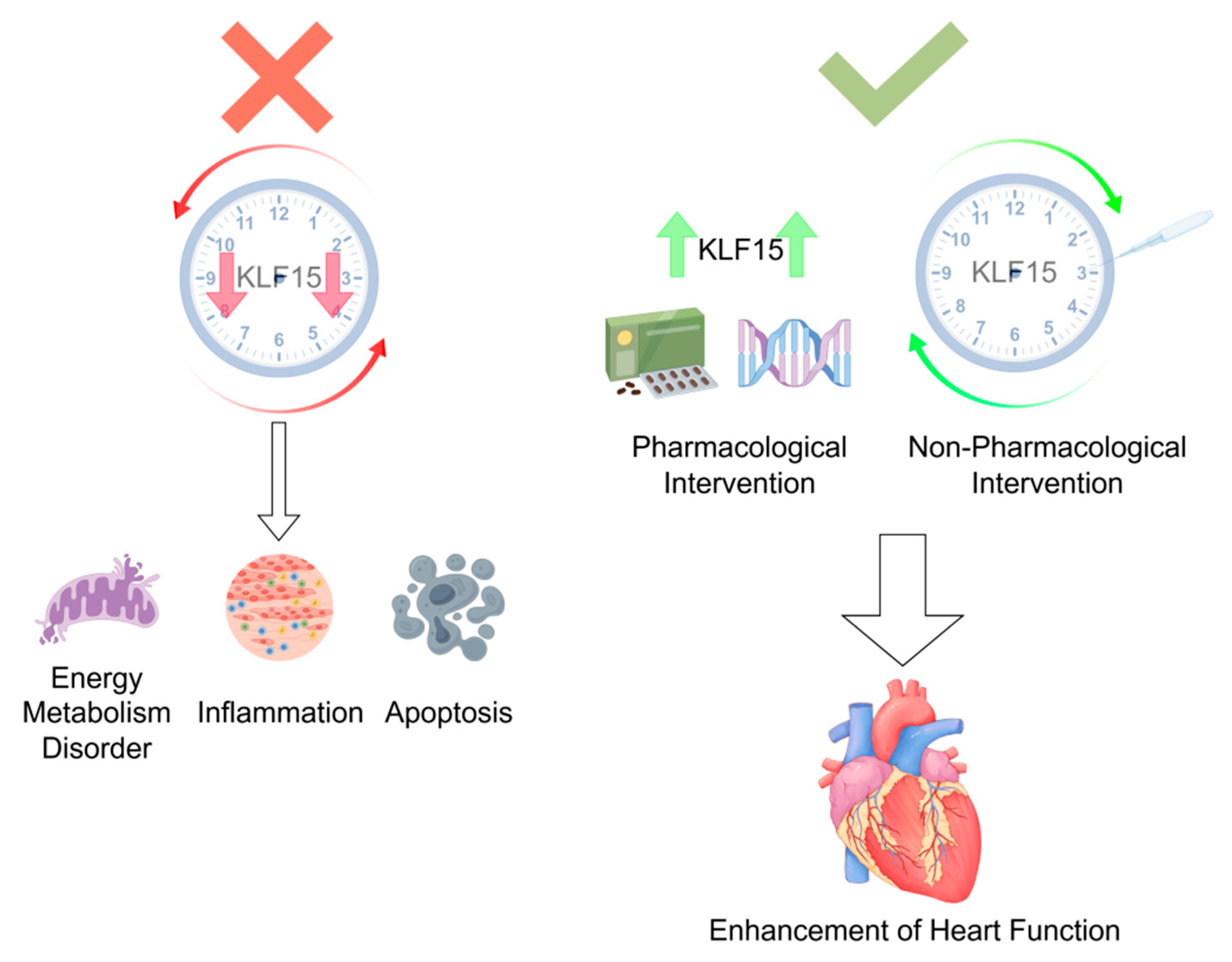
The pivotal role of KLF15 in mediating circadian regulation of myocardial infarction (MI) underscores the profound influence of biological rhythms on cardiovascular health and disease. KLF15 dysfunction disrupts cardiac metabolic homeostasis, exacerbates inflammatory injury, and impairs post-infarction repair, while its rhythmic restoration offers significant therapeutic promise. As mechanistic insights into KLF15’s temporal regulation deepen, this transcription factor may emerge as a key target for MI prevention and treatment. Future research should employ advanced translational approaches to bridge the gap between preclinical findings and clinical applications. Multivariate analyses could determine whether KLF15 expression patterns or genetic variants serve as prognostic biomarkers for MI susceptibility or recovery. Moreover, therapeutic strategies should capitalize on KLF15’s circadian properties, such as timed pharmacological interventions or lifestyle modifications that synchronize its rhythmic activity. By integrating chronobiological principles with cardiovascular medicine, KLF15-centered therapies may revolutionize MI management—transforming circadian biology from a conceptual framework into actionable clinical strategies. These innovations hold the potential to improve patient outcomes and pave the way for personalized, time-sensitive cardiovascular care [Figure 2].
When KLF15 rhythm is disturbed or its expression is reduced, it will cause more serious abnormalities of energy metabolism, inflammation and apoptosis. Up-regulation of KLF15 concentration by pharmacologic or non-pharmacologic therapies corrects KLF15 rhythm disturbances and improves cardiac function after myocardial infarction.

Author Contributions

J.Z.: Conceptualization, Methodology, Visualization, Writing—original draft. Z.C.: Validation, Formal Analysis. J.Y.: Investigation, Data Curation. L.D.: Supervision. H.Y.: Visualization. D.C.: Conceptualization, Supervision. Z.Z.: Writing—review and editing. All authors have read and agreed to the published version of the manuscript.

Funding

This study was supported by a grant from the National Natural Science Foundation of China (82274653). Study sponsors were not involved in study design, data interpretation, writing the article, or the decision to submit the article for publication.

Data Availability Statement

This review article is based on previously published studies. All data analyzed in this work are publicly available through the cited references. No new datasets were generated or analyzed during this review. Links to the original sources are provided in the reference list.

Acknowledgments

The authors express their gratitude to the referenced studies for providing open-access datasets that were instrumental in facilitating the analysis. The schematic figures in this study were created using Figdraw (www.figdraw.com).

Conflicts of Interest

The authors declare that they have no competing interests.

References

  1. Liu, M.; He, X.; Wang, Z.; Hu, S. Executive Summary of the China Cardiovascular Health and Disease Report 2023 (Epidemiology and Interventional Diagnosis/Treatment Status of Cardiovascular Diseases). Chin. J. Intervent. Cardiol. 2024, 32, 541–550. [Google Scholar]
  2. Xu, F.; Wang, G.; Ye, N.; Bian, W.; Yang, L.; Ma, C.; Zhao, D.; Liu, J.; Hao, Y.; Liu, J.; et al. Mild renal insufficiency and attributable risk of adverse In-hospital outcomes in patients with Acute Coronary Syndrome from the improving care for Cardiovascular Disease in China (CCC) project. BMC Nephrol. 2022, 23, 29. [Google Scholar] [CrossRef] [PubMed]
  3. Thygesen, K.; Alpert, J.S.; White, H.D.; Jaffe, A.S.; Apple, F.S.; Galvani, M.; Katus, H.A.; Newby, L.K.; Ravkilde, J.; Chaitman, B.; et al. Universal definition of myocardial infarction. Circulation 2007, 116, 2634–2653. [Google Scholar] [CrossRef]
  4. Lu, L.; Liu, M.; Sun, R.; Zheng, Y.; Zhang, P. Myocardial Infarction: Symptoms and Treatments. Cell Biochem. Biophys. 2015, 72, 865–867. [Google Scholar] [CrossRef]
  5. Sagris, M.; Antonopoulos, A.S.; Theofilis, P.; Oikonomou, E.; Siasos, G.; Tsalamandris, S.; Antoniades, C.; Brilakis, E.S.; Kaski, J.C.; Tousoulis, D. Risk factors profile of young and older patients with myocardial infarction. Cardiovasc. Res. 2022, 118, 2281–2292. [Google Scholar] [CrossRef] [PubMed]
  6. Rabinovich-Nikitin, I.; Kirshenbaum, E.; Kirshenbaum, L.A. Autophagy, Clock Genes, and Cardiovascular Disease. Can. J. Cardiol. 2023, 39, 1772–1780. [Google Scholar] [CrossRef]
  7. Delisle, B.P.; Prabhat, A.; Burgess, D.E.; Stumpf, I.G.; McCarthy, J.J.; Procopio, S.B.; Zhang, X.; Esser, K.A.; Schroder, E.A. Circadian influences on sudden cardiac death and cardiac electrophysiology. J. Mol. Cell. Cardiol. 2025, 200, 93–112. [Google Scholar] [CrossRef]
  8. Hou, T.; Guo, Z.; Gong, M.C. Circadian variations of vasoconstriction and blood pressure in physiology and diabetes. Curr. Opin. Pharmacol. 2021, 57, 125–131. [Google Scholar] [CrossRef]
  9. Crnko, S.; Du Pré, B.C.; Sluijter, J.P.G.; Van Laake, L.W. Circadian rhythms and the molecular clock in cardiovascular biology and disease. Nat. Rev. Cardiol. 2019, 16, 437–447. [Google Scholar] [CrossRef]
  10. Wu, X.; Liu, Z.; Shi, G.; Xing, L.; Wang, X.; Gu, X.; Qu, Z.; Dong, Z.; Xiong, J.; Gao, X.; et al. The circadian clock influences heart performance. J. Biol. Rhythm. 2011, 26, 402–411. [Google Scholar] [CrossRef]
  11. Hurley, J.M.; Loros, J.J.; Dunlap, J.C. Circadian Oscillators: Around the Transcription-Translation Feedback Loop and on to Output. Trends Biochem. Sci. 2016, 41, 834–846. [Google Scholar] [CrossRef] [PubMed]
  12. Koronowski, K.B.; Sassone-Corsi, P. Communicating clocks shape circadian homeostasis. Science 2021, 371, eabd0951. [Google Scholar] [CrossRef] [PubMed]
  13. Lu, Q.; Kim, J.Y. Mammalian circadian networks mediated by the suprachiasmatic nucleus. FEBS J. 2022, 289, 6589–6604. [Google Scholar] [CrossRef]
  14. Le Minh, N.; Damiola, F.; Tronche, F.; Schütz, G.; Schibler, U. Glucocorticoid hormones inhibit food-induced phase-shifting of peripheral circadian oscillators. EMBO J. 2001, 20, 7128–7136. [Google Scholar] [CrossRef]
  15. Cailotto, C.; Lei, J.; van der Vliet, J.; van Heijningen, C.; van Eden, C.G.; Kalsbeek, A.; Pévet, P.; Buijs, R.M. Effects of nocturnal light on (clock) gene expression in peripheral organs: A role for the autonomic innervation of the liver. PLoS ONE 2009, 4, e5650. [Google Scholar] [CrossRef] [PubMed]
  16. D’Souza, A.; Wang, Y.; Anderson, C.; Bucchi, A.; Baruscotti, M.; Olieslagers, S.; Mesirca, P.; Johnsen, A.B.; Mastitskaya, S.; Ni, H.; et al. A circadian clock in the sinus node mediates day-night rhythms in Hcn4 and heart rate. Heart Rhythm. 2021, 18, 801–810. [Google Scholar] [CrossRef]
  17. Zhu, C.; Li, S.; Zhang, H. Heart Failure and Arrhythmias: Circadian and Epigenetic Interplay in Myocardial Electrophysiology. Int. J. Mol. Sci. 2025, 26, 96–99. [Google Scholar] [CrossRef]
  18. Jeyaraj, D.; Haldar, S.M.; Wan, X.; McCauley, M.D.; Ripperger, J.A.; Hu, K.; Lu, Y.; Eapen, B.L.; Sharma, N.; Ficker, E.; et al. Circadian rhythms govern cardiac repolarization and arrhythmogenesis. Nature 2012, 483, 96–99. [Google Scholar] [CrossRef]
  19. Zhang, L.; Prosdocimo, D.A.; Bai, X.; Fu, C.; Zhang, R.; Campbell, F.; Liao, X.; Coller, J.; Jain, M.K. KLF15 Establishes the Landscape of Diurnal Expression in the Heart. Cell Rep. 2015, 13, 2368–2375. [Google Scholar] [CrossRef]
  20. Abdel-Rahman, E.A.; Hosseiny, S.; Aaliya, A.; Adel, M.; Yasseen, B.; Al-Okda, A.; Radwan, Y.; Saber, S.H.; Elkholy, N.; Elhanafy, E.; et al. Sleep/wake calcium dynamics, respiratory function, and ROS production in cardiac mitochondria. J. Adv. Res. 2021, 31, 35–47. [Google Scholar] [CrossRef]
  21. Molnar, J.; Zhang, F.; Weiss, J.; Ehlert, F.A.; Rosenthal, J.E. Diurnal pattern of QTc interval: How long is prolonged? Possible relation to circadian triggers of cardiovascular events. J. Am. Coll. Cardiol. 1996, 27, 76–83. [Google Scholar] [CrossRef]
  22. Culić, V. Acute risk factors for myocardial infarction. Int. J. Cardiol. 2007, 117, 260–269. [Google Scholar] [CrossRef]
  23. Takeda, N.; Maemura, K. Circadian clock and the onset of cardiovascular events. Hypertens. Res. 2016, 39, 383–390. [Google Scholar] [CrossRef] [PubMed]
  24. Kurihara, H.; Yamaoki, K.; Nagai, R.; Yoshizumi, M.; Takaku, F.; Satoh, H.; Inui, J.; Yazaki, Y. Endothelin: A potent vasoconstrictor associated with coronary vasospasm. Life Sci. 1989, 44, 1937–1943. [Google Scholar] [CrossRef] [PubMed]
  25. Förstermann, U.; Münzel, T. Endothelial nitric oxide synthase in vascular disease: From marvel to menace. Circulation 2006, 113, 1708–1714. [Google Scholar] [CrossRef] [PubMed]
  26. Scheer, F.A.; Shea, S.A. Human circadian system causes a morning peak in prothrombotic plasminogen activator inhibitor-1 (PAI-1) independent of the sleep/wake cycle. Blood 2014, 123, 590–593. [Google Scholar] [CrossRef]
  27. Scheer, F.A.; Michelson, A.D.; Frelinger, A.L., 3rd; Evoniuk, H.; Kelly, E.E.; McCarthy, M.; Doamekpor, L.A.; Barnard, M.R.; Shea, S.A. The human endogenous circadian system causes greatest platelet activation during the biological morning independent of behaviors. PLoS ONE 2011, 6, e24549. [Google Scholar] [CrossRef]
  28. Knutsson, A. Increased risk of ischaemic heart disease in shift workers. Occup. Med. 1995, 45, 55. [Google Scholar] [CrossRef]
  29. Kirshenbaum, L.A.; Hill, M.; Singal, P.K. Endogenous antioxidants in isolated hypertrophied cardiac myocytes and hypoxia-reoxygenation injury. J. Mol. Cell. Cardiol. 1995, 27, 263–272. [Google Scholar] [CrossRef]
  30. Mann, D.L. The emerging role of innate immunity in the heart and vascular system: For whom the cell tolls. Circ. Res. 2011, 108, 1133–1145. [Google Scholar] [CrossRef]
  31. Rabinovich-Nikitin, I.; Rasouli, M.; Reitz, C.J.; Posen, I.; Margulets, V.; Dhingra, R.; Khatua, T.N.; Thliveris, J.A.; Martino, T.A.; Kirshenbaum, L.A. Mitochondrial autophagy and cell survival is regulated by the circadian Clock gene in cardiac myocytes during ischemic stress. Autophagy 2021, 17, 3794–3812. [Google Scholar] [CrossRef] [PubMed]
  32. Alibhai, F.J.; Tsimakouridze, E.V.; Chinnappareddy, N.; Wright, D.C.; Billia, F.; O’Sullivan, M.L.; Pyle, W.G.; Sole, M.J.; Martino, T.A. Short-term disruption of diurnal rhythms after murine myocardial infarction adversely affects long-term myocardial structure and function. Circ. Res. 2014, 114, 1713–1722. [Google Scholar] [CrossRef] [PubMed]
  33. Kohsaka, A.; Das, P.; Hashimoto, I.; Nakao, T.; Deguchi, Y.; Gouraud, S.S.; Waki, H.; Muragaki, Y.; Maeda, M. The circadian clock maintains cardiac function by regulating mitochondrial metabolism in mice. PLoS ONE 2014, 9, e112811. [Google Scholar] [CrossRef]
  34. Li, L.; Li, H.; Tien, C.L.; Jain, M.K.; Zhang, L. Kruppel-Like Factor 15 Regulates the Circadian Susceptibility to Ischemia Reperfusion Injury in the Heart. Circulation 2020, 141, 1427–1429. [Google Scholar] [CrossRef]
  35. Jeyaraj, D.; Scheer, F.A.; Ripperger, J.A.; Haldar, S.M.; Lu, Y.; Prosdocimo, D.A.; Eapen, S.J.; Eapen, B.L.; Cui, Y.; Mahabeleshwar, G.H.; et al. KLF15 orchestrates circadian nitrogen homeostasis. Cell Metab. 2012, 15, 311–323. [Google Scholar] [CrossRef] [PubMed]
  36. He, S.; Lu, Y.; Guo, Y.; Li, S.; Lu, X.; Shao, S.; Zhou, H.; Wang, R.; Wang, J.; Gao, P.; et al. Krüppel-Like Factor 15 Modulates CXCL1/CXCR2 Signaling-Mediated Inflammatory Response Contributing to Angiotensin II-Induced Cardiac Remodeling. Front. Cell Dev. Biol. 2021, 9, 644954. [Google Scholar] [CrossRef] [PubMed]
  37. Wang, B.; Xu, H.; Kong, J.; Liu, D.; Qin, W.; Bai, W. Krüppel-Like Factor 15 Reduces Ischemia-Induced Apoptosis Involving Regulation of p38/MAPK Signaling. Hum. Gene Ther. 2021, 32, 1471–1480. [Google Scholar] [CrossRef]
  38. Zhou, L.; Li, Q.; Chen, A.; Liu, N.; Chen, N.; Chen, X.; Zhu, L.; Xia, B.; Gong, Y.; Chen, X. KLF15-activating Twist2 ameliorated hepatic steatosis by inhibiting inflammation and improving mitochondrial dysfunction via NF-κB-FGF21 or SREBP1c-FGF21 pathway. FASEB J. 2019, 33, 14254–14269. [Google Scholar] [CrossRef]
  39. Liao, B.; Tian, X. CTRP12 alleviates cardiomyocyte ischemia-reperfusion injury via regulation of KLF15. Mol. Med. Rep. 2022, 26, 247. [Google Scholar] [CrossRef]
  40. Zheng, H.; Ye, W.; Huang, K.; Chen, Q.; Yang, J.; Luo, L. KLF15 alleviates oxidative stress and apoptosis of H/R-induced trophoblast cells to improve invasion and migration capacity via the activation of IGF1R. Tissue Cell 2024, 90, 102485. [Google Scholar] [CrossRef]
  41. Prosdocimo, D.A.; Anand, P.; Liao, X.; Zhu, H.; Shelkay, S.; Artero-Calderon, P.; Zhang, L.; Kirsh, J.; Moore, D.; Rosca, M.G.; et al. Kruppel-like factor 15 is a critical regulator of cardiac lipid metabolism. J. Biol. Chem. 2014, 289, 5914–5924. [Google Scholar] [CrossRef] [PubMed]
  42. Nagare, T.; Sakaue, H.; Matsumoto, M.; Cao, Y.; Inagaki, K.; Sakai, M.; Takashima, Y.; Nakamura, K.; Mori, T.; Okada, Y.; et al. Overexpression of KLF15 transcription factor in adipocytes of mice results in down-regulation of SCD1 protein expression in adipocytes and consequent enhancement of glucose-induced insulin secretion. J. Biol. Chem. 2011, 286, 37458–37469. [Google Scholar] [CrossRef]
  43. Durumutla, H.B.; Prabakaran, A.D.; El Abdellaoui Soussi, F.; Akinborewa, O.; Latimer, H.; McFarland, K.; Piczer, K.; Werbrich, C.; Jain, M.K.; Haldar, S.M.; et al. Glucocorticoid chronopharmacology promotes glucose metabolism in heart through a cardiomyocyte-autonomous transactivation program. JCI Insight 2024, 9, e182599. [Google Scholar] [CrossRef] [PubMed]
  44. Jiao, J.; Zou, S.; Jian, C.; Tang, F.; Xiao, Y.; Chen, L. Regulatory Effects of Krüppel-like Factor 15 on Lipid Deposition and Cardiac Function in Rats Following Pressure Overload/Unloading. J. Third Mil. Med. Univ. 2015, 37, 1813–1817. [Google Scholar] [CrossRef]
  45. Hu, Y.; Xu, J.; Gao, R.; Xu, Y.; Huangfu, B.; Asakiya, C.; Huang, X.; Zhang, F.; Huang, K.; He, X.; et al. Diallyl Trisulfide Prevents Adipogenesis and Lipogenesis by Regulating the Transcriptional Activation Function of KLF15 on PPARγ to Ameliorate Obesity. Mol. Nutr. Food Res. 2022, 66, e2200173. [Google Scholar] [CrossRef] [PubMed]
  46. Sun, H.; Olson, K.C.; Gao, C.; Prosdocimo, D.A.; Zhou, M.; Wang, Z.; Jeyaraj, D.; Youn, J.Y.; Ren, S.; Liu, Y.; et al. Catabolic Defect of Branched-Chain Amino Acids Promotes Heart Failure. Circulation 2016, 133, 2038–2049. [Google Scholar] [CrossRef]
  47. Zhu, X.; Liu, J.; Cai, M.; Bao, L.; Pan, Y.; Wu, P.; Chu, W.; Zhang, J. The circadian rhythm regulates branched-chain amino acids metabolism in fast muscle of Chinese perch (Siniperca chuatsi) during short-term fasting by Clock-KLF15-Bcat2 pathway. Br. J. Nutr. 2023, 130, 604–615. [Google Scholar] [CrossRef]
  48. Millar-Craig, M.W.; Bishop, C.N.; Raftery, E.B. Circadian variation in blood-pressure. Lancet 1978, 1, 1210–1211. [Google Scholar]
  49. Thosar, S.S.; Shea, S.A. Circadian control of human cardiovascular function. Curr. Opin. Pharmacol. 2021, 57, 89–97. [Google Scholar] [CrossRef]
  50. Kirshenbaum, L.A.; Singal, P.K. Increase in endogenous antioxidant enzymes protects hearts against reperfusion injury. Am. J. Physiol. 1993, 265, H484–H493. [Google Scholar] [CrossRef]
  51. Teixeira, K.R.C.; Dos Santos, C.P.; de Medeiros, L.A.; Mendes, J.A.; Cunha, T.M.; De Angelis, K.; Penha-Silva, N.; de Oliveira, E.P.; Crispim, C.A. Night workers have lower levels of antioxidant defenses and higher levels of oxidative stress damage when compared to day workers. Sci. Rep. 2019, 9, 4455. [Google Scholar] [CrossRef] [PubMed]
  52. Zhao, Y.; Lu, X.; Wan, F.; Gao, L.; Lin, N.; He, J.; Wei, L.; Dong, J.; Qin, Z.; Zhong, F.; et al. Disruption of Circadian Rhythms by Shift Work Exacerbates Reperfusion Injury in Myocardial Infarction. J. Am. Coll. Cardiol. 2022, 79, 2097–2115. [Google Scholar] [CrossRef] [PubMed]
  53. Lapenna, D.; De Gioia, S.; Mezzetti, A.; Porreca, E.; Ciofani, G.; Marzio, L.; Capani, F.; Di Ilio, C.; Cuccurullo, F. Circadian variations in antioxidant defences and lipid peroxidation in the rat heart. Free Radic. Res. Commun. 1992, 17, 187–194. [Google Scholar] [CrossRef] [PubMed]
  54. Frangogiannis, N.G. The inflammatory response in myocardial injury, repair, and remodelling. Nat. Rev. Cardiol. 2014, 11, 255–265. [Google Scholar] [CrossRef]
  55. Frangogiannis, N.G.; Smith, C.W.; Entman, M.L. The inflammatory response in myocardial infarction. Cardiovasc. Res. 2002, 53, 31–47. [Google Scholar] [CrossRef]
  56. Nian, M.; Lee, P.; Khaper, N.; Liu, P. Inflammatory cytokines and postmyocardial infarction remodeling. Circ. Res. 2004, 94, 1543–1553. [Google Scholar] [CrossRef]
  57. Alibhai, F.J.; Tsimakouridze, E.V.; Reitz, C.J.; Pyle, W.G.; Martino, T.A. Consequences of Circadian and Sleep Disturbances for the Cardiovascular System. Can. J. Cardiol. 2015, 31, 860–872. [Google Scholar] [CrossRef]
  58. Huang, B.; Yang, X.D.; Lamb, A.; Chen, L.F. Posttranslational modifications of NF-kappaB: Another layer of regulation for NF-kappaB signaling pathway. Cell Signal 2010, 22, 1282–1290. [Google Scholar] [CrossRef]
  59. Zhao, T.; Qiu, Z.; Gao, Y. MiR-137-3p exacerbates the ischemia-reperfusion injured cardiomyocyte apoptosis by targeting KLF15. Naunyn Schmiedebergs Arch. Pharmacol. 2020, 393, 1013–1024. [Google Scholar] [CrossRef]
  60. Tang, Q.; Li, M.Y.; Su, Y.F.; Fu, J.; Zou, Z.Y.; Wang, Y.; Li, S.N. Absence of miR-223-3p ameliorates hypoxia-induced injury through repressing cardiomyocyte apoptosis and oxidative stress by targeting KLF15. Eur. J. Pharmacol. 2018, 841, 67–74. [Google Scholar] [CrossRef]
  61. Lu, X.; Yang, B.; Qi, R.; Xie, Q.; Li, T.; Yang, J.; Tong, T.; Niu, K.; Li, M.; Pan, W.; et al. Targeting WWP1 ameliorates cardiac ischemic injury by suppressing KLF15-ubiquitination mediated myocardial inflammation. Theranostics 2023, 13, 417–437. [Google Scholar] [CrossRef] [PubMed]
  62. Wang, X.; Liu, J.; Xi, S.; Pan, X.; Fang, X. Exploring the Mechanism of KLF15 on the Biological Activity and Autophagy of Gastric Cancer Cells based on PI3K/Akt/Mtor Signaling Pathway. Comb. Chem. High Throughput Screen. 2024, 27, 1515–1523. [Google Scholar] [CrossRef]
  63. Shao, Y.; Li, X.; Zhou, W.; Qian, S.; Wang, L.; Fang, X. KLF15 attenuates lipopolysaccharide-induced apoptosis and inflammatory response in renal tubular epithelial cells via pparδ. Shock 2024, 62, 574–581. [Google Scholar] [CrossRef]
  64. Prosdocimo, D.A.; Sabeh, M.K.; Jain, M.K. Kruppel-like factors in muscle health and disease. Trends Cardiovasc. Med. 2015, 25, 278–287. [Google Scholar] [CrossRef] [PubMed]
  65. Gray, S.; Wang, B.; Orihuela, Y.; Hong, E.G.; Fisch, S.; Haldar, S.; Cline, G.W.; Kim, J.K.; Peroni, O.D.; Kahn, B.B.; et al. Regulation of gluconeogenesis by Krüppel-like factor 15. Cell Metab. 2007, 5, 305–312. [Google Scholar] [CrossRef] [PubMed]
  66. Haldar, S.M.; Jeyaraj, D.; Anand, P.; Zhu, H.; Lu, Y.; Prosdocimo, D.A.; Eapen, B.; Kawanami, D.; Okutsu, M.; Brotto, L.; et al. Kruppel-like factor 15 regulates skeletal muscle lipid flux and exercise adaptation. Proc. Natl. Acad. Sci. USA 2012, 109, 6739–6744. [Google Scholar] [CrossRef]
  67. Li, H.; An, X.; Bao, L.; Li, Y.; Pan, Y.; He, J.; Liu, L.; Zhu, X.; Zhang, J.; Cheng, J.; et al. MiR-125a-3p-KLF15-BCAA Regulates the Skeletal Muscle Branched-Chain Amino Acid Metabolism in Nile Tilapia (Oreochromis niloticus) During Starvation. Front. Genet. 2020, 11, 852. [Google Scholar] [CrossRef]
  68. Han, S.; Ray, J.W.; Pathak, P.; Sweet, D.R.; Zhang, R.; Gao, H.; Jain, N.; Koritzinsky, E.H.; Matoba, K.; Xu, W.; et al. KLF15 regulates endobiotic and xenobiotic metabolism. Nat. Metab. 2019, 1, 422–430. [Google Scholar] [CrossRef]
  69. Zhao, Y.; Song, W.; Wang, L.; Rane, M.J.; Han, F.; Cai, L. Multiple roles of KLF15 in the heart: Underlying mechanisms and therapeutic implications. J. Mol. Cell. Cardiol. 2019, 129, 193–196. [Google Scholar] [CrossRef]
  70. Fan, L.; Hsieh, P.N.; Sweet, D.R.; Jain, M.K. Krüppel-like factor 15: Regulator of BCAA metabolism and circadian protein rhythmicity. Pharmacol. Res. 2018, 130, 123–126. [Google Scholar] [CrossRef]
  71. Fan, L.; Sweet, D.R.; Prosdocimo, D.A.; Vinayachandran, V.; Chan, E.R.; Zhang, R.; Ilkayeva, O.; Lu, Y.; Keerthy, K.S.; Booth, C.E.; et al. Muscle Krüppel-like factor 15 regulates lipid flux and systemic metabolic homeostasis. J. Clin. Investig. 2021, 131, e139496. [Google Scholar] [CrossRef] [PubMed]
  72. Fan, L.; Sweet, D.R.; Fan, E.K.; Prosdocimo, D.A.; Madera, A.; Jiang, Z.; Padmanabhan, R.; Haldar, S.M.; Vinayachandran, V.; Jain, M.K. Transcription factors KLF15 and PPARδ cooperatively orchestrate genome-wide regulation of lipid metabolism in skeletal muscle. J. Biol. Chem. 2022, 298, 101926. [Google Scholar] [CrossRef] [PubMed]
  73. Gire, D.; Acharya, J.; Malik, S.; Inamdar, S.; Ghaskadbi, S. Molecular mechanism of anti-adipogenic effect of vitexin in differentiating hMSCs. Phytother. Res. 2021, 35, 6462–6471. [Google Scholar] [CrossRef]
  74. Suzuki, J.; Shen, W.J.; Nelson, B.D.; Selwood, S.P.; Murphy, G.M., Jr.; Kanehara, H.; Takahashi, S.; Oida, K.; Miyamori, I.; Kraemer, F.B. Cardiac gene expression profile and lipid accumulation in response to starvation. Am. J. Physiol. Endocrinol. Metab. 2002, 283, E94–E102. [Google Scholar] [CrossRef] [PubMed]
  75. Sugi, K.; Hsieh, P.N.; Ilkayeva, O.; Shelkay, S.; Moroney, B.; Baadh, P.; Haynes, B.; Pophal, M.; Fan, L.; Newgard, C.B.; et al. Kruppel-like factor 15 is required for the cardiac adaptive response to fasting. PLoS ONE 2018, 13, e0192376. [Google Scholar] [CrossRef]
  76. Walter, L.M.; Deguise, M.O.; Meijboom, K.E.; Betts, C.A.; Ahlskog, N.; van Westering, T.L.E.; Hazell, G.; McFall, E.; Kordala, A.; Hammond, S.M.; et al. Interventions Targeting Glucocorticoid-Krüppel-like Factor 15-Branched-Chain Amino Acid Signaling Improve Disease Phenotypes in Spinal Muscular Atrophy Mice. EBioMedicine 2018, 31, 226–242. [Google Scholar] [CrossRef]
  77. Hsieh, P.N.; Zhang, L.; Jain, M.K. Coordination of cardiac rhythmic output and circadian metabolic regulation in the heart. Cell. Mol. Life Sci. 2018, 75, 403–416. [Google Scholar] [CrossRef]
  78. Feigin, R.D.; Klainer, A.S.; Beisel, W.R. Circadian periodicity of blood amino-acids in adult men. Nature 1967, 215, 512–514. [Google Scholar] [CrossRef]
  79. Reddy, A.B.; Karp, N.A.; Maywood, E.S.; Sage, E.A.; Deery, M.; O’Neill, J.S.; Wong, G.K.; Chesham, J.; Odell, M.; Lilley, K.S.; et al. Circadian orchestration of the hepatic proteome. Curr. Biol. 2006, 16, 1107–1115. [Google Scholar] [CrossRef]
  80. Chin, W.C.; Chu, P.H.; Wu, L.S.; Lee, K.T.; Lin, C.; Ho, C.T.; Yang, W.S.; Chung, I.H.; Huang, Y.S. The Prognostic Significance of Sleep and Circadian Rhythm for Myocardial Infarction Outcomes: Case-Control Study. J. Med. Internet Res. 2025, 27, e63897. [Google Scholar] [CrossRef]
  81. Yao, L.; Liu, Y.; Li, M.; Zheng, H.; Sun, M.; He, M.; Zhong, Z.; Ma, S.; Huang, H.; Wang, H. The central regulatory effects of acupuncture in treating primary insomnia: A review. Front. Neurol. 2024, 15, 1406485. [Google Scholar] [CrossRef] [PubMed]
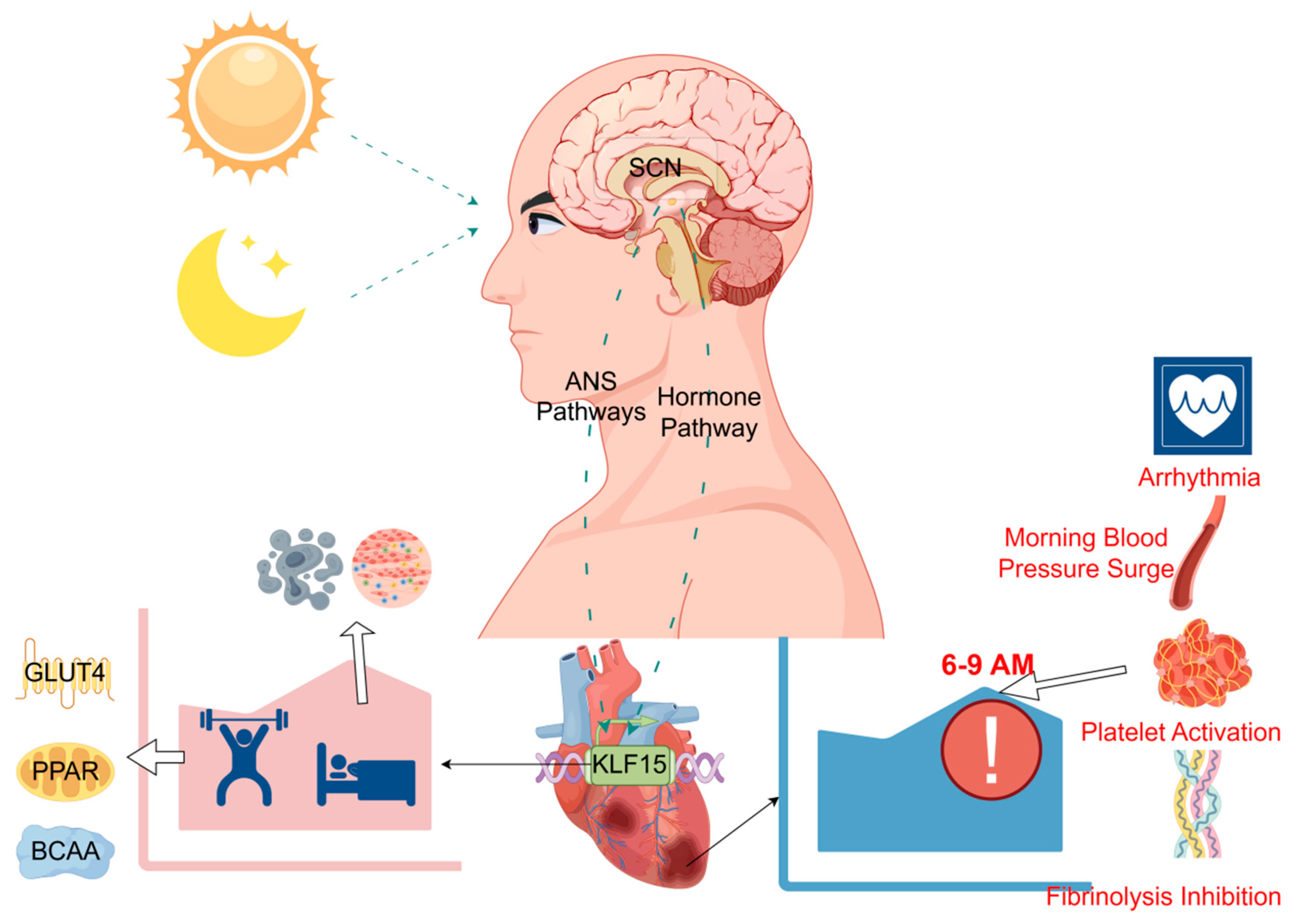
Figure 1. KLF15-mediated regulation of cardiac circadian rhythm and risk of myocardial infarction.
Ijms 26 04831 g001
Figure 2. KLF15’s regulatory role in the heart. By Figdraw.
Ijms 26 04831 g002
Table 1. Key References in the Review. MI (myocardial infarction); NF-κB (Nuclear Factor kappa-light-chain-enhancer of activated B cells); MAPK (Mitogen-Activated Protein Kinase); PI3K-AKT (Phosphoinositide 3-Kinase-Protein Kinase B Signaling Pathway); PPARγ (Peroxisome Proliferator-Activated Receptor Gamma); BCAA (Branched-chain amino acids); BCKA (Branched-chain keto acids).
ReferenceYearAuthorKey Findings
[1]2024Liu, M.; He, X.; Wang, Z.; Hu, S.Provides data on the epidemiology of cardiovascular diseases and the current status of interventional diagnosis and treatment in China.
[3]2007Thygesen, K.; Alpert, J. S.; White, H. D.; Jaffe, A. S.; Apple, F. S.; Galvani, M.; Katus, H. A.; Newby, L. K.; Ravkilde, J.; Chaitman, B.; et al.International diagnostic criteria for myocardial infarction.
[5]2022Sagris, M.; Antonopoulos, A. S.; Theofilis, P.; Oikonomou, E.; Siasos, G.; Tsalamandris, S.; Antoniades, C.; Brilakis, E. S.; Kaski, J. C.; Tousoulis, D. Adverse lifestyle behaviors, including chronic sleep deprivation, markedly elevate the incidence of acute myocardial infarction among young adults.
[6]2023Rabinovich-Nikitin, I.; Kirshenbaum, E.; Kirshenbaum, L. A.Reveals the critical role of the biological clock-autophagy axis in cardiovascular diseases, providing a theoretical foundation for chronotherapy.
[7]2025Delisle, B. P.; Prabhat, A.; Burgess, D. E.; Stumpf, I. G.; McCarthy, J. J.; Procopio, S. B.; Zhang, X.; Esser, K. A.; Schroder, E. A.elucidates the molecular mechanism by which the biological clock regulates ion channels via KLF15, thereby explaining the morning peak incidence of myocardial infarction and cardiac arrhythmias.
[10]2011Wu, X.; Liu, Z.; Shi, G.; Xing, L.; Wang, X.; Gu, X.; Qu, Z.; Dong, Z.; Xiong, J.; Gao, X.; et al. This study provides direct evidence in animal models that biological clock genes dynamically regulate cardiac function in real time.
[19]2015Zhang, L.; Prosdocimo, D. A.; Bai, X.; Fu, C.; Zhang, R.; Campbell, F.; Liao, X.; Coller, J.; Jain, M. K.KLF15 orchestrates cardiac metabolism and repair through a biphasic regulatory mode, thereby providing mechanistic evidence for the circadian nature of myocardial infarction.
[21]1996Molnar, J.; Zhang, F.; Weiss, J.; Ehlert, F. A.; Rosenthal, J. E.This study establishes a clinically relevant link between QTc rhythm variations and temporal patterns of cardiovascular events, providing evidence for the dual regulation of cardiac electrophysiology by both the autonomic nervous system and biological clock.
[22]2007Culić, V. Myocardial infarction exhibits circadian rhythmicity, correlating with neural activation and blood pressure fluctuations.
[23]2016Takeda, N.; Maemura, K.The Bmal1/CLOCK genes regulate circadian blood pressure rhythms, and their deficiency causes rhythm disruption, thereby increasing myocardial infarction risk.
[24]1989Kurihara, H.; Yamaoki, K.; Nagai, R.; Yoshizumi, M.; Takaku, F.; Satoh, H.; Inui, J.; Yazaki, Y.Endothelin-1 levels exhibit a morning surge, which may contribute to the higher morning incidence of myocardial infarction.
[25]2006Förstermann, U.; Münzel, T.Oxidative stress reduces endothelial nitric oxide synthase (eNOS) activity, leading to increased vascular tone in the morning and consequently exacerbating cardiovascular risk.
[26]2014Scheer, F. A.; Shea, S. A.The morning peak of plasminogen activator inhibitor-1 (PAI-1) increases thrombotic risk and may underlie the morning predominance of myocardial infarction.
[27]2011Scheer, F. A.; Michelson, A. D.; Frelinger, A. L., 3rd; Evoniuk, H.; Kelly, E. E.; McCarthy, M.; Doamekpor, L. A.; Barnard, M. R.; Shea, S. A.The surface activity of platelet GPIIb-IIIa complex peaks during circadian morning hours, enhancing thrombus formation and potentially explaining the morning peak incidence of myocardial infarction.
[28]1995Knutsson, A. Circadian rhythm disruption elevates myocardial infarction risk.
[29]1995Kirshenbaum, L. A.; Hill, M.; Singal, P. K. Circadian disruption exacerbates oxidative stress, rendering cardiomyocytes more vulnerable to injury.
[30]2011Mann, D. L. Circadian regulation of immune responses affects post-MI repair.
[31]2021Rabinovich-Nikitin, I.; Rasouli, M.; Reitz, C. J.; Posen, I.; Margulets, V.; Dhingra, R.; Khatua, T. N.; Thliveris, J. A.; Martino, T. A.; Kirshenbaum, L. A. The Bmal1 gene enhances mitophagy to protect ischemic cardiomyocytes and improve survival rates.
[32]2014Alibhai, F. J.; Tsimakouridze, E. V.; Chinnappareddy, N.; Wright, D. C.; Billia, F.; O’Sullivan, M. L.; Pyle, W. G.; Sole, M. J.; Martino, T. A.Light/dark cycle disruption exacerbates post-MI cardiac remodeling and impairs functional recovery in mice.
[33]2014Kohsaka, A.; Das, P.; Hashimoto, I.; Nakao, T.; Deguchi, Y.; Gouraud, S. S.; Waki, H.; Muragaki, Y.; Maeda, M. Bmal1 deficiency dysregulates cardiac oxidative stress-related gene expression and exacerbates myocardial infarction injury.
[34]2020Li, L.; Li, H.; Tien, C. L.; Jain, M. K.; Zhang, L. KLF15 maintains normal ventricular rhythm by regulating the circadian oscillation of cardiac ion channels and QT interval.
[35]2012Jeyaraj, D.; Scheer, F. A.; Ripperger, J. A.; Haldar, S. M.; Lu, Y.; Prosdocimo, D. A.; Eapen, S. J.; Eapen, B. L.; Cui, Y.; Mahabeleshwar, G. H.; et al.KLF15 serves as a pivotal link between circadian rhythms and cardiac function.
[36]2021He, S.; Lu, Y.; Guo, Y.; Li, S.; Lu, X.; Shao, S.; Zhou, H.; Wang, R.; Wang, J.; Gao, P.; et al. KLF15 exerts anti-inflammatory effects via NF-κB/MAPK inhibition.
[37]2021Wang, B.; Xu, H.; Kong, J.; Liu, D.; Qin, W.; Bai, W. KLF15 attenuates ischemic apoptosis via p38/MAPK inhibition.
[38]2019Zhou, L.; Li, Q.; Chen, A.; Liu, N.; Chen, N.; Chen, X.; Zhu, L.; Xia, B.; Gong, Y.; Chen, X.KLF15-Twist2 axis inhibits inflammation and enhances mitochondrial homeostasis.
[39]2022Liao, B.; Tian, X.KLF15 alleviates ischemia-reperfusion injury by modulating CTRP12 expression.
[40]2024Zheng, H.; Ye, W.; Huang, K.; Chen, Q.; Yang, J.; Luo, L.KLF15 activates PI3K-AKT to enhance cytoprotection against oxidative damage.
[41]2014Prosdocimo, D. A.; Anand, P.; Liao, X.; Zhu, H.; Shelkay, S.; Artero-Calderon, P.; Zhang, L.; Kirsh, J.; Moore, D.; Rosca, M. G.; et al.KLF15-p300 axis attenuates cardiac lipotoxicity by reprogramming lipid metabolism.
[42]2011Nagare, T.; Sakaue, H.; Matsumoto, M.; Cao, Y.; Inagaki, K.; Sakai, M.; Takashima, Y.; Nakamura, K.; Mori, T.; Okada, Y.; et al.KLF15 optimizes cardiac energetics via insulin-GLUT4 axis.
[43]2024Durumutla, H. B.; Prabakaran, A. D.; El Abdellaoui Soussi, F.; Akinborewa, O.; Latimer, H.; McFarland, K.; Piczer, K.; Werbrich, C.; Jain, M. K.; Haldar, S. M.; et al.Glucocorticoid-KLF15 circadian axis coordinates cardiac glucose utilization.
[44]2015Jiao, J.; Zou, S.; Jian, C.; Tang, F.; Xiao, Y.; Chen, L. KLF15 enhances post-unloading cardiac recovery by suppressing lipid accumulation.
[45]2022Hu, Y.; Xu, J.; Gao, R.; Xu, Y.; Huangfu, B.; Asakiya, C.; Huang, X.; Zhang, F.; Huang, K.; He, X.; et al. KLF15 attenuates lipid accumulation via PPARγ downregulation.
[46]2016Sun, H.; Olson, K. C.; Gao, C.; Prosdocimo, D. A.; Zhou, M.; Wang, Z.; Jeyaraj, D.; Youn, J. Y.; Ren, S.; Liu, Y.; et al. KLF15 deficiency disrupts BCAA catabolism, causing BCKA-induced mitochondrial impairment in heart failure.
[47]2023Zhu, X.; Liu, J.; Cai, M.; Bao, L.; Pan, Y.; Wu, P.; Chu, W.; Zhang, J. Fasting-induced Clock-KLF15-Bcat2 axis dysregulation impairs BCAA metabolism in fish muscle.
Table 2. The multiple roles of KLF15.
Key PointReference(s)Observed Effect on MI
KLF15-InflammationsNF-κBHe, S et al. (2021) [36]KLF15 is able to attenuate inflammatory responses by inhibiting NF-κB pathway activation
Huang, B et al. (2010) [58]
MAPKWang, B et al. (2021) [37]KLF15 is able to attenuate inflammatory responses by inhibiting MAPK pathway activation
Twist2Zhou, L et al. (2019) [38]KLF15 ameliorates postmyocardial infarction injury by regulating Twist2
CTRP12Liao, B et al. (2022) [39]KLF15 can alleviate post-ischemic reperfusion injury by regulating CTRP1250
KLF15-ApoptosisPI3K-AKTZheng, H et al. (2024) [40]KLF15 ameliorates apoptosis through the PI3K-AKT pathway
KLF15-EnergyMetabolismGlucose Metabolism Nagare, T et al. (2011) [42]KLF15 promotes the entry of glucose into cardiomyocytes for cardiac energy supply by regulating insulin secretion
Lipid MetabolismFan, L et al. (2022) [72]KLF15 maintains lipid homeostasis in the heart by regulating PPAR-δ and PPAR-γ
Gire, D et al. (2021) [73]
Hu, Y et al. (2022) [45]
Branched-Chain AminoAcid MetabolismSun, H et al. (2016) [46]KLF15 improves cardiac function by regulating BCKAs
Jeyaraj, D et al. (2012) [35]
Disclaimer/Publisher’s Note: The statements, opinions and data contained in all publications are solely those of the individual author(s) and contributor(s) and not of MDPI and/or the editor(s). MDPI and/or the editor(s) disclaim responsibility for any injury to people or property resulting from any ideas, methods, instructions or products referred to in the content.