Review

Biomarkers as Beacons: Illuminating Sepsis-Associated Hepato-Renal Injury

by
Maria-Antoanela Pasare
1,2,
Cristian Sorin Prepeliuc
1,3,
Maria Gabriela Grigoriu
2,
Ionela-Larisa Miftode
2,4,* and
Egidia Gabriela Miftode
2,4
1
Doctoral School, “Grigore T. Popa” University of Medicine and Pharmacy, 700115 Iasi, Romania
2
“Sf Parascheva” Clinical Hospital of Infectious Diseases, 700116 Iasi, Romania
3
Emergency Hospital “Mavromati”, 710221 Botosani, Romania
4
Department of Infectious Diseases, Faculty of Medicine, “Grigore T. Popa” University of Medicine and Pharmacy, 700115 Iasi, Romania
*
Author to whom correspondence should be addressed.
Int. J. Mol. Sci. 2025, 26(10), 4825; https://doi.org/10.3390/ijms26104825
Submission received: 31 March 2025 / Revised: 13 May 2025 / Accepted: 16 May 2025 / Published: 18 May 2025
(This article belongs to the Special Issue Molecular Mechanisms and Pathophysiology of Sepsis)

Abstract

Sepsis, defined as a dysregulated host response to infection, is one of the leading causes of mortality worldwide. It unleashes in the organism a cascade of molecules, cytokines, and proteins which leads to an inflammatory storm. If this response to infection is uncontrolled, any organ is susceptible to damage. Acute kidney injury (AKI) is one of the most frequent organ dysfunctions in septic patients, and while it can be reversible, its presence leads to a higher burden of morbidity and mortality. While serum creatinine is essential in evaluating kidney function, the pathophysiology of AKI is not completely elucidated, and a plethora of novel biomarkers have been studied in the hope of an early diagnosis and fast treatment. While the liver is not as affected by sepsis, it plays an important role as a guardian by providing acute-phase proteins, activating neutrophils, and controlling iron balance. Acute liver failure (ALF) could impair the organism’s capacity to contain and eliminate pathogens. Some molecules have been associated with either AKI or ALF, although biomarkers specific for organ dysfunction are difficult to validate. The aim of this review is to understand the role of several molecules in the pathophysiology of sepsis and their clinical ability for diagnosing or predicting sepsis-induced hepato-renal dysfunction.

1. Introduction

To our best understanding today, sepsis represents a life-threatening organ dysfunction that is caused by a dysregulated host response to infection [1]. The task force that was appointed to define sepsis recommends using the SOFA (sequential organ failure assessment) scoring system to evaluate organ dysfunction in a patient with a suspected or confirmed infection [2].
Sepsis is one of the major contributors to the global health burden, alongside stroke, heart disease, and kidney disease [3,4]. Recent data indicate that sepsis incidence rates vary between 500 and 1500 cases per 100,000 people. Mortality rates are lower in high-income countries, between 15 and 25%, and can reach up to 40% in low- and middle-income countries [5]. Further epidemiological studies will elucidate sepsis’s incidence and health impact through the coronavirus disease (COVID-19) pandemic years, as some have categorized sepsis while referring to viral sepsis and some as a bacterial infection associated with COVID-19 [6].
On a cellular level, sepsis starts with the recognition of pathogen-derived molecular patterns (PAMPs) and damage-associated molecular patterns (DAMPs), which bind to receptors of antigen-presenting cells. Immune cells release a plethora of cytokines, both pro-inflammatory: such as interleukin (IL)-1, IL-6, IL-12, IL-18, and tumor necrosis factor alpha (TNF-α), and anti-inflammatory: IL-10, IL-27, and transforming growth factor beta 1 (TGFβ1). Inflammation leads to the release of acute-phase proteins by the liver, which in turn determines immune cell recruitment, activation of the complement system, the coagulation pathway, and iron regulation. Following the response from the innate immune system, the adaptive immune system comes into play as the organism tries to resolve the disease [7,8]. Part of the pathophysiology of sepsis and the interplay of various molecules is synthesized in Figure 1.
The delicate balance of inflammation and anti-inflammation is disrupted in sepsis, and while both pathways are upregulated, the resulting inflammation triggers progressive tissue damage and multi-organ dysfunction [7]. The kidneys and the liver are highly susceptible to microcirculatory dysfunction, and sepsis complicated with either hepatic or renal injury leads to high mortality rates, from 38% to almost 50% [8,9]. Hepato-renal dysfunction can exacerbate injury to other organs and can lead to an influx of toxins into the central nervous system. Additionally, coagulopathy can produce cerebral ischemia or hemorrhage [10]. Therefore, it is imperative to recognize sepsis-associated acute kidney injury (SA-AKI) and sepsis-associated liver injury (SALI) in a timely manner. Classical methods to detect kidney injury, such as creatinine and urine output, are not sufficiently sensitive, and their changes do not occur early enough to detect declines in renal function [11]. Liver biomarkers, such as bilirubin, aminotransferases, and alkaline phosphatase, are not satisfactory in their ability to reflect liver dysfunction, to differentiate acute from chronic injury, and to predict mortality [12].
As a result, in the last decades, research has been focused on finding biomarkers that aid in predicting diagnosis and prognosis. Although many molecules have been proposed, only a few are used in practice today. Biomarkers are objective indicators that characterize normal to pathogenic processes or pharmacological responses to therapeutic intervention. An ideal biomarker must be highly sensitive and specific to the provided data, reproducible, and cost-effective [13].
The scope of this review is to examine certain interleukins and molecules that show promising diagnostic or prognostic values related to sepsis-specific hepatic or renal injury, but whose clinical utility is still under investigation. We have excluded biomarkers that have been extensively reviewed in recent years, such as IL-6, interferons (IFNs), neutrophil gelatinase-associated lipocalin (NGAL), kidney injury molecule 1 (KIM-1), and presepsin.

2. Sepsis Phenotypes and Endotypes

Given that sepsis is a highly heterogeneous syndrome, the concepts of sepsis phenotypes and endotypes have emerged. Various phenotype categories divide sepsis based on temperature, hemodynamics, and organ dysfunction. Temperature profiling identified three different groups: hypothermic, normothermic, and hyperthermic, with the highest mortality among those with a greater deviation from normal temperature, either below or above. As a distinction, patients in the hypothermic group exhibit an inclination towards a tempered anti-inflammatory response compared to febrile patients, who exhibit higher plasma concentrations of both pro- and anti-inflammatory cytokines. (IL-6, TNF-α, granulocyte colony-stimulating factor, IL-10, IL-27). Hemodynamic profiling distinguishes the highest mortality among patients with a mean systolic blood pressure (SBP) around 80 mmHg. For organ dysfunction, multiple research groups have identified distinct phenotypes, with the highest mortality among patients with cardiogenic dysfunction/shock associated with either renal impairment or altered mental status [14,15,16]. One study on a substantially large cohort categorized patients into: “α phenotype” with less organ dysfunction and lower median values compared to the entire study group for IL-10, IL-6, IL-8, procalcitonin, and TNF-α; “β phenotype” with renal injury and greater comorbidities; “γ phenotype” with associated cardiovascular, hematologic, and inflammatory abnormalities, and “δ phenotype”, linked to cardiovascular and hepatic injury and which associated the highest values for IL-10, IL-6, IL-8, procalcitonin, D-dimer, and plasminogen activator inhibitor 1 (PAI-1) [17].
Concerning endotypes, multiple research groups have defined various categories, without any scholarly consensus. One proposed classification denotes the following endotypes: neutrophilic-suppressive (upregulation of innate system pathways); inflammatory (increased inflammatory response); innate host defense (moderate regulation of interleukin signaling); interferon (high expression of interferons); and adaptive (upregulation of adaptive immune pathways) [18]. Another classification indicates two extremes: macrophage activation-like syndrome (MALS) and immuno-paralysis. In MALS, a high concentration of pro-inflammatory cytokines leads to liver dysfunction, disseminated intravascular coagulation (DIC), and pancytopenia. Ferritin levels exceed 4420 nanograms/milliliter (ng/mL) and are associated with increased concentrations of IL-6, IL-18, and soluble cluster of differentiation 163 (sCD163), and a decreased ratio of IL-10/TNF-α. Immuno-paralysis is defined by a low percentage of monocytes that express human leucocyte antigen (HLA)-DR and therefore present a weak ability to initiate adaptive immune responses. Patients who did not meet either criterion were considered to fall between the two groups [19,20].

3. The Liver and Kidney in Sepsis

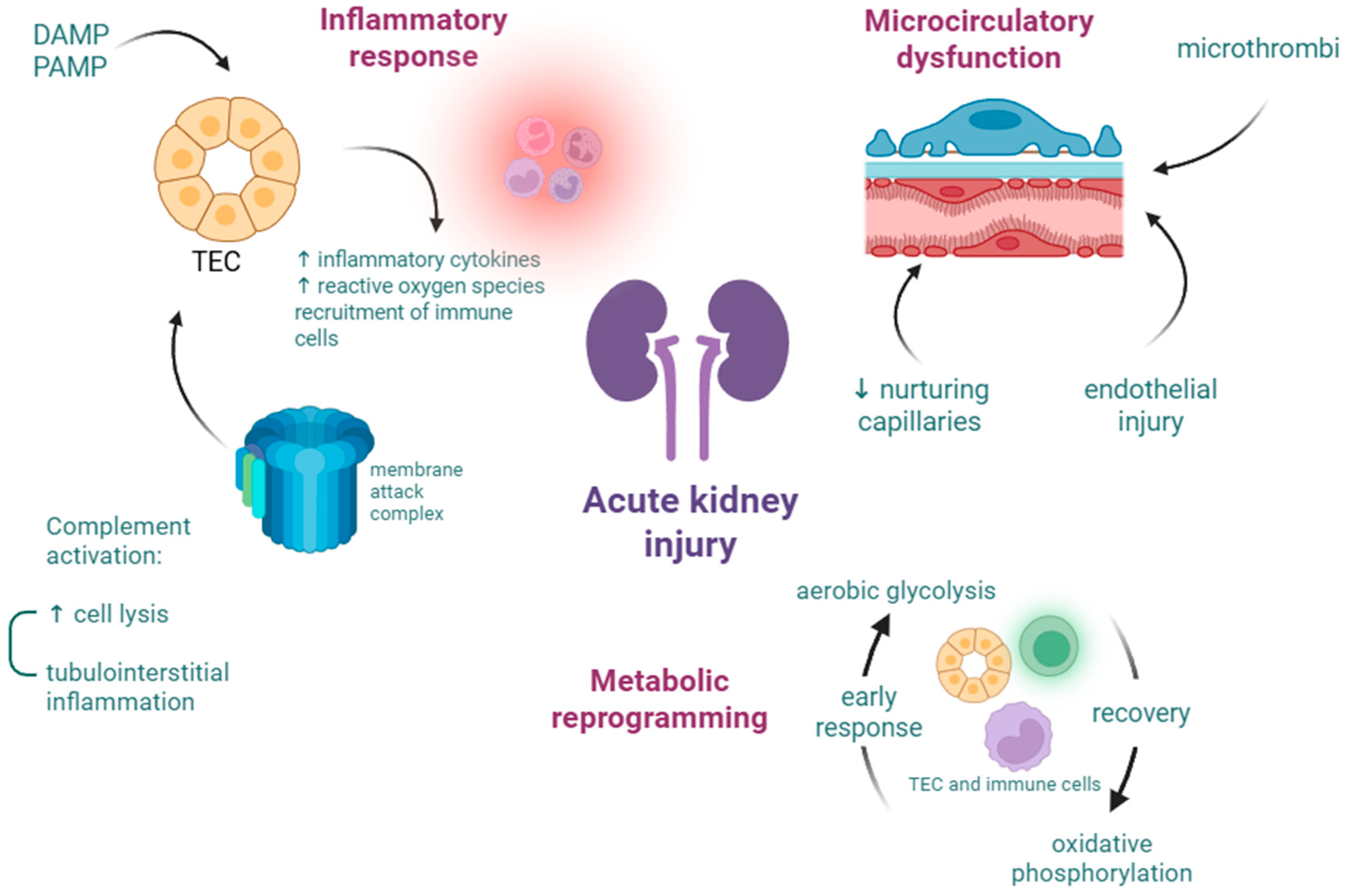
3.1. Acute Kidney Injury

Acute kidney injury is increasingly frequent in patients with sepsis or septic shock, contributing to worse outcomes and longer hospitalization [21]. AKI is defined by KDIGO criteria, which rely on creatinine or urine output, and in the absence of baseline information, clinicians use eGFR [9]. Crucial roles of the kidneys are maintaining fluid and electrolyte balance and filtering waste, such as exogenous drugs. Administration of intravenous fluids and antibiotics can contribute to adverse renal outcomes due to fluid overload and toxicity [22]. Sepsis-associated AKI pathogenesis (Figure 2) is not completely elucidated, but the most relevant theories are related to circulatory dysfunction, immune response, and metabolic changes. Microcirculatory dysfunction determines the loss of nurturing capillaries, microthrombi formation, and endothelial injury due to shedding of the glycocalyx and hyperpermeability. The inflammatory response in the kidneys is managed by renal tubular epithelial cells (TECs) that bind to DAMPs and PAMPs, triggering the expression of pro-inflammatory cytokines and immune cell recruitment. The complement system is mainly composed of C1–C9 components and their subunits. The activation of the complement cascade induces the formation of the membrane attack complex (C5b-C9), causing cell lysis [23,24].

3.2. Acute Liver Failure and Liver Dysfunction

Although the liver is less affected by sepsis, with an incidence of 11–12% sepsis-associated liver failure [12,25], its importance in pathophysiology is greater. Some of the mechanisms by which the liver protects the organism are related but not limited to: Kupffer cells that release pro-inflammatory cytokines and prevent the entrance of bacteria in the circulatory system, the recruitment and activation of neutrophils, the synthesis of complement factors, and acute-phase proteins [26]. Controlling iron metabolism is also part of liver defenses, as many microbes require iron. Sepsis-associated liver dysfunction can arise from either hypoxic hepatitis (hypoxic cell death leads to a serum increase in aminotransferases) or cholestasis (inflammation of bile ducts leads to impaired bile flow). Liver dysfunction in sepsis can progress to acute liver failure [8,26].
Another way the liver acts as a guardian is by producing clotting factors to help limit pathogens in a fibrin network. Sepsis-induced coagulopathy (SIC) represents the disruption in the fine balance of the coagulation pathway, leading to hypercoagulability in the early stages, followed by the depletion of clotting factors. After immune cells are activated by pathogens, and cytokines are released, monocytes and endothelial cells respond by releasing tissue factor (TF). TF activates the extrinsic coagulation pathway, and as sepsis progresses, anticoagulants decrease, leading to their inability to control clot formation. Fibrinolytic activity is also impaired due to elevated levels of PAI-1, which accentuates the formation of microthrombi [26,27]. SIC is an important contributor to organ injury through microthrombi, and in later stages, through the risk of bleeding due to consumption of clotting factors. Around 29% of critically ill septic patients are diagnosed with SIC [7].

4. Biomarkers That Pertain to AKI or ALF
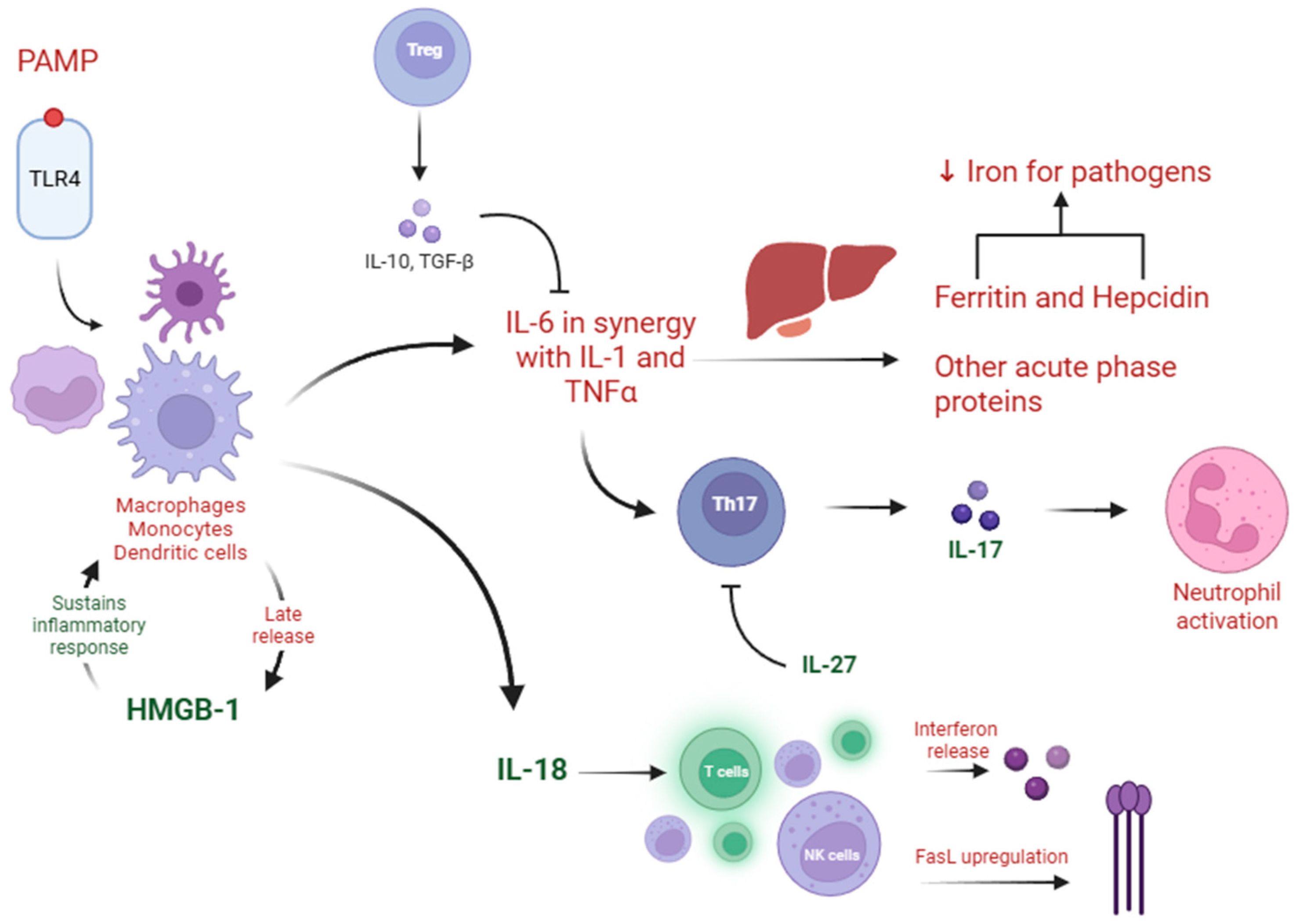
4.1. IL-18

Interleukin-18 is part of the interleukin-1 family, and its precursor is found in Kupffer cells and other hematopoietic and non-hematopoietic cells. As a cytokine, it plays different roles depending on the receptor or other interleukins. Combined with interleukin-12, it helps T cells and natural killer cells to produce IFN-gamma. Without IL-12 or IL-15, it can lead to the differentiation of naive T cells into T helper-2 cells. Similar to other cytokines, it increases nitric oxide (NO) synthesis and cell adhesion molecules, but one specific property is Fas ligand induction, which leads to cell apoptosis. In order to minimize the immune response, IL-18 binding protein (IL-18BP) serves as the main regulatory mechanism, reducing the plasmatic levels of IL-18 [28,29].
Animal studies concluded that IL-18 not only increases cytokine levels but also helps promote serum immunoglobulin M (IgM) levels. Research has demonstrated that mice injected with Escherichia coli have increased serum IgM levels after multiple IL-18 injections compared to control mice, but a single shot of IL-18 only increases IFN-gamma levels [30]. Similar results were found in a study with mice that sustained burn injuries and were injected with Pseudomonas aeruginosa. IL-18 injections increased the mice survival rate, and serum IgM levels were higher [31].
Regarding its clinical significance, IL-18 has been shown to play a role in various diseases that display an inflammatory component. High concentrations of IL-18 have been found in patients with Crohn’s disease, asthma, chronic obstructive pulmonary disease, systemic juvenile idiopathic arthritis, and Still’s disease [28]. Given that sepsis and inflammatory disorders could clinically overlap, evidence suggests significant differences in IL-18 plasma levels between a small cohort of patients diagnosed with Still’s disease and septic patients, as the molecule displayed higher values in patients with active inflammatory disease [32].
The value of IL-18 as a mortality predictor in sepsis is disputed, as there are varying results from clinical and in vitro studies. In a study involving 40 sepsis and septic shock patients, the authors found that although IL-18 was higher at all study times in septic patients compared to healthy volunteers, there was no difference between survivors and non-survivors [33]. However, in another study involving 65 septic patients, the authors showed that patients with elevated IL-18 levels have about a 90% higher risk of death [34].
Concerning AKI, a study involving approximately 1400 patients from intensive care units found that IL-18 performed poorly to moderately regarding its ability to predict new AKI or progression of kidney injury. However, only 6.2% of those patients were diagnosed with sepsis [35]. Given that interleukin-18 is expressed in renal tubular cells, one meta-analysis shows that in patients with cirrhosis, it can differentiate between acute tubular necrosis and hepatorenal syndrome with high specificity and sensitivity [36]. SA-AKI has a complicated pathophysiology, and it is believed that it is characterized by reversible injury of the tubular cells and not by necrosis [37]. One study showed that IL-18, alongside KIM-1 and the renal resistive index, are good predictors for developing AKI in patients with sepsis, with IL-18 demonstrating the highest sensitivity [38]. The renal resistive index, an ultrasound-derived parameter, provides real-time perfusion data of the kidneys and can predict AKI [39]. Another retrospective study, which included patients with both bacterial and viral sepsis, concluded that IL-18 can be found in a wide range of elevated plasma concentrations. Mortality, AKI, and acute respiratory distress syndrome (ARDS) were correlated with IL-18 levels in both groups of sepsis [40]. Interleukin-18 serum levels rise after 6 h in the ICU, showing a significant difference between AKI and non-AKI septic patients. Nevertheless, the combination of IL-18 and NGAL performs better for AKI diagnosis, with a sensitivity of 77.9% and specificity of 94.6% [41]. Similar results were observed regarding IL-18 and the NLRP3 inflammasome, which were upregulated in septic patients compared to the control group and further increased in AKI patients versus non-AKI [42]. A study conducted on pregnant women, of which only 44% had a positive quick SOFA score on admission, concluded that IL-18 was not a good predictor for developing AKI [43]. Therefore, IL-18 could be a potential biomarker for predicting sepsis-associated AKI but not for other related causes.
Serum concentrations of IL-18 alongside IL-1β rise in a time-dependent manner in ALF models. After inhibiting pyroptosis proteins, survival rates greatly improved, liver damage was attenuated, and IL-18 concentration decreased significantly [44]. In patients with acute-on-chronic liver failure (ACLF) complicated with sepsis, serum IL-18 is significantly raised when compared to patients with ACLF with or without systemic inflammation. The authors of this study also noted that the five patients with signs of systemic inflammation and increasing IL-18 concentrations within 24 h of admission also developed sepsis [45]. IL-18 is a reliable predictor for the occurrence of hepato-biliary dysfunction (HBD) and DIC in septic patients, with an area under the curve of 0.78. A multiparametric approach, which also included IL-18BP, sCD163, soluble CD25 (sCD25), ferritin, IL-10, and other biomarkers, leads to an increase in the area under the curve to 0.93 [46]. The main key findings related to IL-18, as well as hepcidin and ferritin, can be found in Table 1.

4.2. Hepcidin

Hepcidin is a key molecule in iron homeostasis, primarily produced by the liver and in smaller quantities by the heart, kidneys, macrophages, and other cells. There are three isoforms, of which only hepcidin-25 is considered actively involved in iron metabolism, while hepcidin-22 and hepcidin-20 are regarded as products of degradation. Hepcidin binds with ferroportin to limit iron efflux, resulting in both anemia and reduced availability of iron for bacteria. Liver production of hepcidin is downregulated by increased erythropoiesis, while conditions of inflammation and infection promote its synthesis, especially through interleukin-6. Elevated serum hepcidin has been reported in various cancers, inflammatory disorders, and sepsis [47].
One study, which included a limited number of patients with septic shock, reported that median hepcidin values were elevated compared to normal values, with maximal concentration registered at the time of inclusion. Additionally, in five patients who developed secondary complications such as pleural effusion and pneumonia, a further increase in hepcidin levels was noted [48]. In a follow-up study, the authors concluded that hepcidin values were significantly higher in the sepsis group than in the non-sepsis control group. Hepcidin levels decreased in the subsequent days and did not correlate with 28-day mortality [49]. One paper reported that hepcidin has the potential to discriminate between survivors and non-survivors in cancer patients who develop sepsis [50]. Another research group examined hepcidin and ferritin in patients with septic shock, COVID-19, and those who had undergone surgery. Although plasma hepcidin levels did not differ substantially among these groups, the hepcidin-to-ferritin ratio was significantly lower in non-survivors compared to survivors, only in the septic shock cohort [51].
In mouse sepsis models, hepcidin exhibits a protective role against organ damage. Hepcidin knock-out animals displayed greater mortality rates and more severe organ damage. Hepatic injury was assessed by area of necrosis and transaminase levels, which were significantly higher in knock-out mice than in controls [52]. The protective role of hepcidin is further supported by findings in another preclinical study. The authors showed that hepcidin pretreatment in mice polymicrobial sepsis models is associated with better preserved renal function and reduced tubular injury [53]. One retrospective study involving 807 participants with severe AKI (of which 64% were diagnosed with sepsis) highlighted that low plasma concentrations of hepcidin are correlated with 60-day mortality. Further research is needed to establish if hepcidin could provide therapeutic benefits for critically ill patients [54].
Data from one study reveal that hepcidin is an independent predictor of persistent AKI in septic patients [55]. The same authors showed that serum hepcidin can be a good predictor of AKI occurrence within 7 days of admission for septic patients. Combining hepcidin with urinary NGAL leads to a sensitivity of 79.6% and a specificity of 78.3% for AKI occurrence in sepsis patients [56]. Contrasting results obtained by another research group indicated no correlation between hepcidin concentrations and the development of stage 2–3 AKI for hospitalized patients with either sepsis or non-septic conditions [57]. While hepcidin has been linked to safeguarding the organism from sepsis-associated organ damage, further clinical trials are required to establish hepcidin as a biomarker for SA-AKI.

4.3. Ferritin

Pertaining to iron homeostasis, ferritin is a ubiquitous protein that is pivotal in the storage of intracellular iron and can also act as an iron carrier. Serum concentrations of ferritin act as a tool to examine iron storage. In acute and chronic inflammation, serum ferritin is usually elevated; however, iron is sequestered as a defensive mechanism. Therefore, ferritin has been widely researched as a marker in various states of inflammation related to malignancies, chronic kidney disease, autoimmune disorders, and acute infections [58].
Serum ferritin has been demonstrated to have a good prognostic value in children hospitalized for sepsis or septic shock. Patients with median values between 200 and 500 ng/mL had the best outcome, while patients with concentrations exceeding 500 ng/mL had the highest mortality [59,60,61]. A large retrospective study demonstrated that for every 1000 ng/mL increase in serum ferritin values, there is a non-linear increase in mortality between 28 days and one year [62].
As a protein, ferritin is composed of heavy chains (FtH) and light chains (FtL), with its circulatory form mainly attributed to light chains. In deficient FtH mice, survival rates are significantly improved in cecal ligation puncture (CLP) models, possibly due to a compensatory increase in serum ferritin concentrations. In these mice, renal function was better preserved, and hepatic injury was limited [63]. However, light chain-deficient mice exhibit the same levels of AKI when compared to control mice; therefore, an overexpression of ferritin could protect from organ damage in sepsis, while the loss of light chains does not influence the extension of the disease [64].
Ferritin has been linked to renal recovery in patients with AKI; however, patients with sepsis-associated AKI were excluded from the study [65]. In a large retrospective cohort study, using the MIMIC-IV database, higher ferritin was associated with a longer hospital stay and higher mortality, higher SOFA, and an increased risk of AKI development. The authors noted that even in the normal range, an increase in serum ferritin is still linked with an enhanced risk of AKI compared with the low ferritin group [66]. Another study on the MIMIC-IV.2.2 database extrapolated only patients with SA-AKI. Patients with ferritin over 1056.2 ng/mL had the highest values for heart rate, temperature, creatinine, SOFA score, and in-hospital mortality [67].
Given that ferritin can be released from damaged hepatocytes, its role as a biomarker in liver injury has been investigated. Mice deficient in FtH have higher serum ferritin concentrations. Sepsis models in these mice have revealed better preservation of renal function and less damage to the liver and lungs [63]. One investigation revealed that the FtH protein could establish disease tolerance to sepsis by regulating glucose metabolism. The authors showed that both FtH-deficient mice and control mice had low blood glucose after CLP; however, the control group restored the blood glucose the following days and the FtH-deficient mice did not [68]. In clinical studies, patients with sepsis-induced liver injury displayed significantly higher levels of serum ferritin, iron, and total-iron binding capacity compared to septic patients without hepatic injury [69].
More recently, levels of ferritin have been associated with mortality in COVID-19 patients, with median values almost three times higher for non-survivors compared to survivors [70,71]. Although sepsis was present in all non-survivor cases, ferritin was not directly correlated with this outcome [70]. Ferritin can also be used as a diagnostic tool for hepatic injury in COVID-19 patients, and it has been correlated with viral clearance and hospital stay but not antibiotic use [72,73].

4.4. IL-27

Interleukin-27 is a cytokine with a complex role in the immune system, expressed primarily by macrophages, monocytes, dendritic cells, and other endothelial or epithelial cells. Through its receptor, it was initially believed to have a pro-inflammatory role, as it induces interferon-gamma production; however, it has been proven to also produce an anti-inflammatory response [74]. Studies on mice have demonstrated that blocking the IL-27 receptor (IL-27R) leads to a decrease in mortality in sepsis models [75]. Alveolar macrophage killing capacity is also higher in mice lacking IL-27R, as the inflammatory response in these mice offers better protection against Pseudomonas aeruginosa infection [76]. For human studies, a meta-analysis showed that IL-27 is a good diagnostic biomarker for sepsis both in pediatric and adult settings. With a sensitivity of 0.84 and a specificity of 0.71, it has similar accuracy to procalcitonin and presepsin [77].
Serum concentration of IL-27 has also been shown to be higher in patients with sepsis-related acute hepatic injury compared to septic patients without liver injury. The increase in IL-27 was not associated with ICU stay [78]. The same authors confirmed that IL-27R-deficient mice had less severe histological hepatic damage, therefore confirming its role as a pro-inflammatory cytokine in the process of sepsis-induced ALF [78]. Further research indicates that in mouse models with CLP-induced infection, IL-27 exacerbates liver injury. Both serum levels and hepatic expression of IL-27 are increased after LPS stimulation. Gadolinium chloride (which inhibits Kupffer cells) pretreatment can lead to reduced concentrations of IL-27, attenuating liver damage but not injury to other organs, such as the lungs and kidneys [79]. Similar results were obtained in a follow-up study, with IL-27 knock-out mice presenting fewer inflammatory factors and attenuated hepatic injury. The authors determined that IL-27 promotes macrophage pyroptosis, leading to aggravated ALF [80].
Mouse models with induced AKI demonstrate that IL-27 treatment can minimize kidney damage, expressed by less tubular injury, increased production of anti-inflammatory cytokines, and decreased production of TNF-alpha, IL-6, and IL-17A [81]. In the previous study, AKI was induced by ischemia reperfusion, which can be a pathophysiological mechanism by which sepsis causes AKI [22]. Serum concentrations are also higher in sepsis-associated myocardial dysfunction compared to the control group. However, mouse studies showed that for the myocardium, IL-27 plays an anti-inflammatory role, as pretreatment with IL-27 decreases myocardial injury biomarkers [82]. The main key findings related to IL-27, as well as the following biomarkers: IL-17A, IL-10, IL-33/ST2, high-mobility group box-1, proenkephalin (PENK), C-X-C Motif Chemokine Ligand 9 (CXCL9), soluble CD163, soluble CD25, can be found in Table 2.

4.5. IL-17A

The Interleukin-17 family is a group of molecules that play a role in the inflammatory response against infection. They have the ability to stimulate macrophages, dendritic cells, and others to produce chemokines, cytokines, and matrix metalloproteinases. The most common protein from this group is IL-17A, mainly produced by a subset of CD4 T cells called Th17 cells [83].
Considering the newer hypothesis regarding SA-AKI pathogenesis, IL-17A has been studied in septic mice to observe its role in renal function. Data show that IL-17A knock-out mice have a better survival rate than wild-type mice under sepsis conditions. Histological changes pertain to ameliorated tissue damage and a lower tubular injury score [84]. Therefore, IL-17A could become a novel target for managing SA-AKI therapy. Another study on mice divided into a multiple organ dysfunction (MODS) group, LPS-challenged group, and sham-operated group, indicated that IL-17 levels decreased in the MODS group, gradually over time, possibly being consumed by the kidney. The same study included patients hospitalized with sepsis, and results expressed that IL-17 levels are higher in SA-AKI compared to sepsis associated with chronic renal failure [85].
One paper highlights the importance of the Th17 ratio to regulatory T cells in septic patients with and without AKI. The authors found that the Th17/Treg ratio could be used as a predictive factor for SA-AKI development and that IL-17 concentrations were higher in the group with acute kidney injury [86]. Similar results were obtained by another research team, where IL-17A levels were proportionately higher as the AKI stage was worse [87].
In patients with ACLF complicated with sepsis, IL-17 serum concentrations are highest when compared to patients with ACLF or liver cirrhosis. There is a significant difference between patients with ACLF and patients with cirrhosis, which could indicate that IL-17 plays a role in acute liver injury [88].

4.6. IL-10

The interleukin-10 molecule is part of the IL-10 superfamily, mainly produced by monocytes, macrophages, and T cells, with other immune cells capable of synthesizing this cytokine. Although many cytokines have pro-inflammatory roles, IL-10 is one of the most potent anti-inflammatory and immunosuppressive molecules. It can decrease or inhibit antigen presentation, phagocytosis, the expression of major histocompatibility complex (MHC) class II, and the production of IL-1 or TNF-alpha [89]. IL-10 can suppress IL-6 secretion from Kupffer cells when the hepatic cells are stimulated with endotoxin [90]. IL-10 plays a substantial role in the cytokine storm during sepsis. IL-10-deficient mice exhibit prolonged signs of illness and higher mortality after undergoing CLP. In septic patients, NK cells increase their IL-10 production [91].
As mentioned in Chapter 4.1, IL-10 levels are elevated in patients with liver impairment (Figure 3). In the subset of septic patients with HBP and DIC, IL-10 correlated with ferritin and IL-18, in contrast, with septic patients without HBD and DIC [46]. In the previously noted study regarding patients with ACLF and sepsis, the serum concentration of IL-10 was significantly increased compared to patients with ACLF or liver cirrhosis [88]. In pediatric septic patients, IL-10 could be used to differentiate children with organ dysfunction from those with a hyperinflammatory state. The sensitivity of diagnosis for IL-10 was 89.6% for concentrations >18.4 picograms (pg)/mL. The most common types of organ dysfunction in this study were low platelets and high bilirubin, indicating liver impairment [92].
In mice, IL-10 plays a crucial role in the development of the anti-inflammatory response in septic AKI. In the first hours after inducing sepsis conditions, there is an increase in pro-inflammatory response, and at 24 h, there is an increase in IL-10. For ischemic AKI, cytokine levels were lower, and there was no peak in IL-10 levels [93]. Findings highlight that IL-10 levels are higher in patients with septic AKI compared to sepsis patients without AKI, and the receiver operating characteristic curve (ROC) analysis for predicting AKI was nearly as good as NGAL [94].

4.7. IL-33 and ST2

Interleukin-33 is part of the IL-1 superfamily, and it binds to Interleukin 1 receptor-like 1 (ST2), also part of the IL-1 receptor superfamily. IL-33 is produced mainly by endothelial and epithelial cells, stromal cells such as fibroblasts, and smooth muscle cells. When tissue damage happens, IL-33 is released into the extracellular space and acts as an alarmin, alerting immune cells [95,96]. ST2 has been especially studied as a marker for myocardial fibrosis with good predictive value for mortality in heart failure patients. However, it could also play a role in distinguishing the presence or absence of sepsis [97,98].
In septic mice, IL-33 treatment reduces mortality compared to control group mice. In the same experiment, pretreated mice showed a marked increase in neutrophil migration to the infection site, which led to an improvement in bacterial clearance [99]. IL-33 also promotes NK cells’ activation in septic mice, and IL-33 treatment has a beneficial role in the early stages of sepsis but not in the late phases [100].
One study aimed to investigate the differences in the cytokine profile between septic patients with and without metabolic liver disease. Patients with liver disease had higher concentrations of IL-17 and IL-33 both on day 1 and day 5 of hospitalization. Patients in the metabolic disease group also had a significant increase in serum IL-33 between admission and day 5 and a decrease in IL-10. In the same category, IL-33 had a negative correlation with sepsis severity [101].
IL-33 plays a role in AKI, as its inhibition through ST2 can provide protection against functional and histological changes in mice induced with cisplatin [102]. In a hospital setting, patients with AKI who survived showed lower serum ST2 concentrations; therefore, the IL-33 receptor could act as a prognostic marker. Although the most common cause of AKI was sepsis, ST-2 could not distinguish between the different etiologies of AKI [103].

4.8. HMGB1

High-mobility group box-1 is part of a group of chromosomal proteins that play an essential role in sustaining life. As a nuclear protein, it binds deoxyribonucleic acid (DNA) and enhances transcription, replication, and repair. However, when released in the extracellular space, it acts as a potent pro-inflammatory mediator [104]. Due to its dual intra- and extracellular activity, HMGB1 has been studied in relation to different types of cancers, correlating to poor prognosis, and to sepsis. In murine polymicrobial sepsis models, HMGB1 levels usually peak late, associating with death [105]. Early clinical studies concluded that in sepsis patients, HMGB1 also peaks late and associates with the severity of organ dysfunction, but it cannot distinguish between survivors and non-survivors [106,107]. Nevertheless, academics revealed that late peaks of HMGB1 associated with inflammatory disease in infectious patients act as a strong risk factor for mortality [108].
HMGB1 sustains a pro-inflammatory loop in mice with induced acute liver failure by LPS and D-galactosamine hydrochloride (D-gal). When antibodies were used to neutralize HMGB1, levels of alanine transaminase were back to normal, and liver histology was similar to the control group [109]. HMGB1 accentuates liver injury by pyroptosis of macrophages in murine CLP models [110]. Comparable results were obtained by another group of authors on ALF and AKI animal models. After inhibiting the TNF-alpha pathway, serum concentrations of HMGB1, IL-1, and IL-18 decreased, and liver and renal function were drastically improved [111].
In CLP models, HMGB1 can also localize in renal tubular epithelial cells and pass into urine, unlike in healthy settings. In these conditions, HMGB1 can interact with TECs and promote active secretion of IL-1 and IL-6, which can aggravate sepsis and SA-AKI [112].

4.9. Proenkephalin

Proenkephalin and other biologically active enkephalins are a result of preproenkephalin A cleavage. This family of molecules act as endogenous opioids. PENK has gained interest due to the fact that the second highest density of opioid receptors is found in the kidney. Depending on the type of receptor, enkephalins can influence diuresis and natriuresis, and reduce kidney inflammation. PENK has been mostly studied in relation to AKI and septic AKI, as it can be freely filtered through the renal glomerulus [113,114].
In one study (preprint) that included 529 patients with sepsis or septic shock, AKI was diagnosed in 44% of subjects. PENK levels on admission were correlated with AKI severity, resulting in 72% sensitivity and 83% specificity to diagnose AKI in the first 48 h [115]. In the ALBIOS trial, 28% of patients developed AKI within 48 h, from a total of 895 patients. PENK concentrations were notably elevated in patients with AKI than in those without. Among those with AKI, PENK was lower in patients who improved renal function than in those who did not; therefore, PENK could be a candidate for predicting renal function recovery [116]. One meta-analysis, which included 11 studies, concluded that PENK shows an area under the curve of 0.77 for early AKI diagnosis. From the included studies, only four comprised sepsis-associated AKI [117].
PENK also shows promising value for predicting sepsis severity and mortality, as concentrations were significantly higher in non-survivors compared to survivors [118,119].

4.10. C-X-C Motif Chemokine Ligand 9

Chemokines are key drivers for chemotaxis, differentiation, and multiplication of leukocytes. C-X-C motif chemokine ligand 9, alongside CXCL10 and -11, determines the recruitment of cytotoxic lymphocytes, natural killers, and macrophages. Immune cells release CXCL9 in response to IFNγ [120]. One group of authors proposed IFN-driven sepsis (IDS) as a new sepsis endotype, besides immuno-paralysis and MALS. They demonstrated that almost 20% of sepsis patients, from a cohort of 5503, display IDS characteristics. As IFNγ is the sole trigger for CXCL9, this marker had higher values in the IDS category compared to MALS. Although the greatest mortality rate was registered in the MALS category, IDS is independently associated with 28-day mortality. Serum ferritin and IL-18 displayed the highest concentration in the MALS group, followed by the IDS and immuno-paralysis groups [121].
CXCL9 has been proven to distinguish between acute interstitial nephritis and other causes of AKI, but none were related to sepsis. It has also been associated with tubular inflammation, and low urinary levels can exclude kidney injury related to infection [122,123]. To the best of our knowledge, CXCL9 has not yet been linked to SA-AKI.
C-X-C motif chemokine receptor 3 (CXCR3) is the ligand for CXCL9, and in response to IFN, hepatocytes secrete CXCR3. In ischemia reperfusion models in mice, levels of CXCL9 are increased, leading to T cell infiltration. Blocking the interaction between CXCL9 and CXCR3 increases survival, and hepatocellular damage is reduced [124].

4.11. Soluble CD163

CD163 acts as a receptor for the hemoglobin–haptoglobin complexes, and it is expressed on the surface of macrophages, monocytes, and lymphocytes. After pathogen recognition, monocyte activation determines an increase in sCD163, with the role of depleting iron sources for bacteria. In the later phases of sepsis, sCD163 has been shown to lead to the release of IL-10 [125]. In small clinical settings, sCD163 levels were higher in septic patients compared to controls and correlated with IL-10. sCD163 was also statistically significantly higher in non-survivors, as patients with better outcomes displayed gradually declining serum levels [126,127,128]. Another group of authors showed that sCD163 is markedly increased in septic shock, and the sCD163/IL-18 ratio in septic and septic shock patients is significantly lower than in controls. For every 1 unit increase in this ratio, there was a 3.9-fold increase in the odds of death, but the results were not statistically significant [129].
Urinary sCD163 has been proposed as a diagnostic tool for SA-AKI, with a sensitivity of 80% and a specificity of 56%. The authors correlated urine sCD163 levels with serum sCD163 and discovered good concordance [130]. sCD163 concentrations have been linked to mortality in patients with acute liver failure of different etiologies [131]. In cirrhotic patients, median sCD163 levels are significantly elevated in those with acute decompensation and infection compared to those without infection [132].

4.12. Soluble CD25

The IL-2/IL-2 receptor (IL-2R) pathway plays a complex role in the immune system, establishing immune tolerance. IL-2R is composed of multiple chains, of which the α chain (CD25) is shed from T cells upon activation. Soluble CD25 has been researched as an indicator of immune-mediated disorders [133].
As a sepsis biomarker, sCD25 has been shown to display higher serum concentrations compared to controls, alongside sCD163 and IL-18; however, the author did not perform any multiparameter analysis [134].
Concerning SA-AKI, sCD25 has been investigated together with IL-10 and NGAL. Results showed that sCD25 was significantly elevated in septic AKI compared to both sepsis without AKI and AKI of other causes. In this context, sCD25 performed closely to NGAL regarding SA-AKI diagnosis, and their combination had the highest performance [94].

5. Future Directions and Study Limitations

Future directions regarding the integration of biomarkers in sepsis classification, diagnosis, and prognosis relate to the use of machine learning, a multiparametric approach, and personalized therapy. Machine learning utilizes large data sets to generate prediction algorithms and uncover hidden patterns. Data may take the form of biomarkers, metabolomics, and gene expression, which help create sepsis profiles, assess mortality prognosis, and support treatment decisions [135]. Specificity and sensitivity for sepsis-related organ injury can be significantly enhanced by using multi-biomarker panels, as they represent various aspects of the immune response. However, this approach faces limitations due to its cost, availability, and lack of standardization [136]. In addition to diagnosis and prognosis, biomarkers may also be used for personalized therapy in the form of agonists or antagonists. For example, hepcidin agonists could maintain renal function in septic conditions, and using antibodies against HMGB1 prevents ALF [53,109].
This review has several limitations. First, we limited our search to articles published in the English language, which may have led to the exclusion of pertinent studies in other languages. Second, the studies included in our review displayed considerable heterogeneity regarding study design and patient demographics, limiting our ability to draw uniform conclusions. Third, much of the presented evidence is derived from small-scale studies, which hinders the predictive validity of biomarkers. Fourth, we found limited data evaluating multiparametric approaches, which could improve diagnostic accuracy.

6. Conclusions

Sepsis is not only a heavy burden on the individual but on the medical society as well. While every organ can be gravely impacted by sepsis, acute kidney injury remains the most frequent organ dysfunction, increasing mortality risk, and liver damage can vastly influence the inflammatory response and modulate injury in other organs. While consecrated molecules such as C-reactive protein, procalcitonin, and presepsin perform well in practice, there is still a need for more specific diagnostic and predictive biomarkers for sepsis and related organ dysfunction. The biomarkers described in this review have multiple functions and could potentially be used in either AKI or ALF settings. IL-18, hepcidin, PENK, and sCD25 are good candidates for SA-AKI diagnosis. The sensitivity and specificity of IL-18 and hepcidin are enhanced by combining with NGAL. PENK also displays prognostic value for renal recovery, and ST-2 and IL-17 could be predictive of AKI severity and mortality. Liver injury is assessed by ferritin, IL-27, HMGB1, and CXCL9, mainly in preclinical studies, which leaves a gap in the knowledge regarding their abilities to act as biomarkers for SALI in clinical environments. We consider that biomarkers hold great potential for characterizing the complexity of sepsis syndrome, as various phenotypes display different biomarker serum levels and associate with different types of organ dysfunction. Further research is needed to establish the utility and practicality of novel biomarkers in clinical settings, with the goal of enhancing diagnostic accuracy and improving therapeutic choices.

Author Contributions

Conceptualization, M.-A.P. and I.-L.M.; writing—original draft preparation, M.-A.P., C.S.P. and M.G.G.; writing—review and editing, I.-L.M. and E.G.M.; supervision, E.G.M. All authors have read and agreed to the published version of the manuscript.

Funding

This research received no external funding.

Conflicts of Interest

The authors declare no conflicts of interest.

Abbreviations

ACLF: Acute-On-Chronic Liver Failure; AKI: Acute Kidney Injury; ALF: Acute Liver Failure; ARDS: Acute Respiratory Distress Syndrome; CD: Cluster Of Differentiation; COVID-19: Coronavirus disease; CLP: Cecal Ligation Puncture; CXCL: C-X-C Motif Chemokine Ligand; CXCR: C-X-C Motif Chemokine Receptor; DAMPs: damage-associated molecular patterns; D-Gal: D-Galactosamine Hydrochloride; DIC: Disseminated Intravascular Coagulation; DNA: Deoxyribonucleic Acid; eGFR: Estimated Glomerular Filtration Rate; FtH: Ferritin Heavy Chain; FtL: Ferritin Light Chain; HLA: Human Leucocyte Antigen; HBD: Hepato-Biliary Dysfunction; HMGB1: high-mobility group box-1; ICU: Intensive Care Unit; Ig: Immunoglobulin; IL: Interleukin; IL-18BP: IL-18 Binding Protein; IL-2R: IL-2 receptor; IL-27R: IL-27 receptor; IFN: Interferon; IDS: IFN-driven sepsis; KDIGO: Kidney Disease: Improving Global Outcomes; KIM-1: Kidney Injury Molecule 1; LPS: Lipopolysaccharide; MALS: Macrophage Activation-Like Syndrome; ml: milliliter; MHC: Major Histocompatibility Complex; MODS: Multiple Organ Dysfunction Syndrome; ng: nanograms; NGAL: Neutrophil Gelatinase-Associated Lipocalin; NK: Natural Killer; NLRP3: NOD-, LRR-, and Pyrin Domain-Containing Protein 3; NO: Nitric Oxide; PAI-1: Plasminogen Activator Inhibitor 1; PAMPs: pathogen-derived molecular patterns; PENK: Proenkephalin; pg: picograms; SA-AKI: Sepsis-Associated Acute Kidney Injury; SALI: Sepsis-Associated Liver Injury; SIC: Sepsis-Induced Coagulopathy; SBP: Systolic Blood Pressure; SOFA: Sequential Organ Failure Assessment; sCD25: soluble CD25; sCD163: soluble CD163; TECs: renal tubular epithelial cells; ROC: receiver operating characteristic curve; TF: Tissue Factor; TGFβ-1: transforming growth factor beta 1; Th: T helper; TNF: tumor necrosis factor.

References

  1. Singer, M.; Deutschman, C.S.; Seymour, C.W.; Shankar-Hari, M.; Annane, D.; Bauer, M.; Bellomo, R.; Bernard, G.R.; Chiche, J.-D.; Coopersmith, C.M.; et al. The Third International Consensus Definitions for Sepsis and Septic Shock (Sepsis-3). JAMA 2016, 315, 801. [Google Scholar] [CrossRef]
  2. Lambden, S.; Laterre, P.F.; Levy, M.M.; Francois, B. The SOFA score—Development, utility and challenges of accurate assessment in clinical trials. Crit. Care 2019, 23, 374. [Google Scholar] [CrossRef] [PubMed]
  3. Rudd, K.E.; Kissoon, N.; Limmathurotsakul, D.; Bory, S.; Mutahunga, B.; Seymour, C.W.; Angus, D.C.; West, T.E. The global burden of sepsis: Barriers and potential solutions. Crit. Care 2018, 22, 232. [Google Scholar] [CrossRef] [PubMed]
  4. Miftode, R.-S.; Costache, I.-I.; Constantinescu, D.; Mitu, O.; Timpau, A.-S.; Hancianu, M.; Leca, D.-A.; Miftode, I.-L.; Jigoranu, R.-A.; Oancea, A.-F.; et al. Syndecan-1: From a Promising Novel Cardiac Biomarker to a Surrogate Early Predictor of Kidney and Liver Injury in Patients with Acute Heart Failure. Life 2023, 13, 898. [Google Scholar] [CrossRef] [PubMed]
  5. La Via, L.; Sangiorgio, G.; Stefani, S.; Marino, A.; Nunnari, G.; Cocuzza, S.; La Mantia, I.; Cacopardo, B.; Stracquadanio, S.; Spampinato, S.; et al. The Global Burden of Sepsis and Septic Shock. Epidemiologia 2024, 5, 456–478. [Google Scholar] [CrossRef]
  6. Karakike, E.; Giamarellos-Bourboulis, E.J.; Kyprianou, M.; Fleischmann-Struzek, C.; Pletz, M.W.; Netea, M.G.; Reinhart, K.; Kyriazopoulou, E. Coronavirus Disease 2019 as Cause of Viral Sepsis: A Systematic Review and Meta-Analysis*. Crit. Care Med. 2021, 49, 2042–2057. [Google Scholar] [CrossRef]
  7. Jarczak, D.; Kluge, S.; Nierhaus, A. Sepsis—Pathophysiology and Therapeutic Concepts. Front. Med. 2021, 8, 628302. [Google Scholar] [CrossRef]
  8. Strnad, P.; Tacke, F.; Koch, A.; Trautwein, C. Liver—guardian, modifier and target of sepsis. Nat. Rev. Gastroenterol. Hepatol. 2017, 14, 55–66. [Google Scholar] [CrossRef]
  9. Bellomo, R.; Kellum, J.A.; Ronco, C.; Wald, R.; Martensson, J.; Maiden, M.; Bagshaw, S.M.; Glassford, N.J.; Lankadeva, Y.; Vaara, S.T.; et al. Acute kidney injury in sepsis. Intensive Care Med. 2017, 43, 816–828. [Google Scholar] [CrossRef]
  10. Gotts, J.E.; Matthay, M.A. Sepsis: Pathophysiology and clinical management. BMJ 2016, 353, i1585. [Google Scholar] [CrossRef]
  11. Poston, J.T.; Koyner, J.L. Sepsis associated acute kidney injury. BMJ 2019, 364, k4891. [Google Scholar] [CrossRef] [PubMed]
  12. Nesseler, N.; Launey, Y.; Aninat, C.; Morel, F.; Mallédant, Y.; Seguin, P. Clinical review: The liver in sepsis. Crit. Care 2012, 16, 235. [Google Scholar] [CrossRef]
  13. Saxena, J.; Das, S.; Kumar, A.; Sharma, A.; Sharma, L.; Kaushik, S.; Kumar Srivastava, V.; Jamal Siddiqui, A.; Jyoti, A. Biomarkers in sepsis. Clin. Chim. Acta 2024, 562, 119891. [Google Scholar] [CrossRef] [PubMed]
  14. Bhavani, S.V.; Wolfe, K.S.; Hrusch, C.L.; Greenberg, J.A.; Krishack, P.A.; Lin, J.; Lecompte-Osorio, P.; Carey, K.A.; Kress, J.P.; Coopersmith, C.M.; et al. Temperature Trajectory Subphenotypes Correlate With Immune Responses in Patients With Sepsis. Crit. Care Med. 2020, 48, 1645–1653. [Google Scholar] [CrossRef]
  15. Leijte, G.P.; Kox, M.; Pickkers, P. Fever in Sepsis: Still a Hot Topic. Am. J. Respir. Crit. Care Med. 2019, 200, 263–264. [Google Scholar] [CrossRef]
  16. Papathanakos, G.; Andrianopoulos, I.; Xenikakis, M.; Papathanasiou, A.; Koulenti, D.; Blot, S.; Koulouras, V. Clinical Sepsis Phenotypes in Critically Ill Patients. Microorganisms 2023, 11, 2165. [Google Scholar] [CrossRef] [PubMed]
  17. Seymour, C.W.; Kennedy, J.N.; Wang, S.; Chang, C.-C.H.; Elliott, C.F.; Xu, Z.; Berry, S.; Clermont, G.; Cooper, G.; Gomez, H.; et al. Derivation, Validation, and Potential Treatment Implications of Novel Clinical Phenotypes for Sepsis. JAMA 2019, 321, 2003. [Google Scholar] [CrossRef]
  18. Baghela, A.; Pena, O.M.; Lee, A.H.; Baquir, B.; Falsafi, R.; An, A.; Farmer, S.W.; Hurlburt, A.; Mondragon-Cardona, A.; Rivera, J.D.; et al. Predicting sepsis severity at first clinical presentation: The role of endotypes and mechanistic signatures. eBioMedicine 2022, 75, 103776. [Google Scholar] [CrossRef]
  19. Leventogiannis, K.; Kyriazopoulou, E.; Antonakos, N.; Kotsaki, A.; Tsangaris, I.; Markopoulou, D.; Grondman, I.; Rovina, N.; Theodorou, V.; Antoniadou, E.; et al. Toward personalized immunotherapy in sepsis: The PROVIDE randomized clinical trial. Cell Rep. Med. 2022, 3, 100817. [Google Scholar] [CrossRef]
  20. Kyriazopoulou, E.; Leventogiannis, K.; Norrby-Teglund, A.; Dimopoulos, G.; Pantazi, A.; Orfanos, S.E.; Rovina, N.; Tsangaris, I.; Gkavogianni, T.; Botsa, E.; et al. Macrophage activation-like syndrome: An immunological entity associated with rapid progression to death in sepsis. BMC Med. 2017, 15, 172. [Google Scholar] [CrossRef]
  21. Umbro, I.; Gentile, G.; Tinti, F.; Muiesan, P.; Mitterhofer, A.P. Recent advances in pathophysiology and biomarkers of sepsis-induced acute kidney injury. J. Infect. 2016, 72, 131–142. [Google Scholar] [CrossRef]
  22. Peerapornratana, S.; Manrique-Caballero, C.L.; Gómez, H.; Kellum, J.A. Acute kidney injury from sepsis: Current concepts, epidemiology, pathophysiology, prevention and treatment. Kidney Int. 2019, 96, 1083–1099. [Google Scholar] [CrossRef] [PubMed]
  23. Manrique-Caballero, C.L.; Del Rio-Pertuz, G.; Gomez, H. Sepsis-Associated Acute Kidney Injury. Crit. Care Clin. 2021, 37, 279–301. [Google Scholar] [CrossRef]
  24. Fattahi, F.; Zetoune, F.S.; Ward, P.A. Complement as a Major Inducer of Harmful Events in Infectious Sepsis. Shock 2020, 54, 595–605. [Google Scholar] [CrossRef] [PubMed]
  25. Kramer, L.; Jordan, B.; Druml, W.; Bauer, P.; Metnitz, P.G.H. Incidence and prognosis of early hepatic dysfunction in critically ill patients—A prospective multicenter study. Crit. Care Med. 2007, 35, 1099-e7. [Google Scholar] [CrossRef]
  26. Yao, Y.; Wang, D.; Yin, Y. Advances in sepsis-associated liver dysfunction. Burn. Trauma 2014, 2, 97. [Google Scholar] [CrossRef] [PubMed]
  27. Tsantes, A.G.; Parastatidou, S.; Tsantes, E.A.; Bonova, E.; Tsante, K.A.; Mantzios, P.G.; Vaiopoulos, A.G.; Tsalas, S.; Konstantinidi, A.; Houhoula, D.; et al. Sepsis-Induced Coagulopathy: An Update on Pathophysiology, Biomarkers, and Current Guidelines. Life 2023, 13, 350. [Google Scholar] [CrossRef]
  28. Kaplanski, G. Interleukin-18: Biological properties and role in disease pathogenesis. Immunol. Rev. 2018, 281, 138–153. [Google Scholar] [CrossRef]
  29. Yasuda, K.; Nakanishi, K.; Tsutsui, H. Interleukin-18 in Health and Disease. Int. J. Mol. Sci. 2019, 20, 649. [Google Scholar] [CrossRef]
  30. Kinoshita, M.; Kuranaga, N.; Matsumoto, A.; Ono, S.; Shinomiya, N.; Hiraide, H.; Seki, S. Multiple interleukin-18 injections promote both mouse Th1 and Th2 responses after sublethal Escherichia coli infection. Clin. Exp. Immunol. 2005, 143, 41–49. [Google Scholar] [CrossRef]
  31. Kinoshita, M.; Shinomiya, N.; Ono, S.; Tsujimoto, H.; Kawabata, T.; Matsumoto, A.; Hiraide, H.; Seki, S. Restoration of Natural IgM Production from Liver B Cells by Exogenous IL-18 Improves the Survival of Burn-Injured Mice Infected with Pseudomonas aeruginosa. J. Immunol. 2006, 177, 4627–4635. [Google Scholar] [CrossRef]
  32. Priori, R.; Colafrancesco, S.; Alessandri, C.; Minniti, A.; Perricone, C.; Iaiani, G.; Palazzo, D.; Valesini, G. Interleukin 18: A Biomarker for Differential Diagnosis Between Adult-onset Still’s Disease and Sepsis. J. Rheumatol. 2014, 41, 1118–1123. [Google Scholar] [CrossRef]
  33. Mierzchala-Pasierb, M.; Krzystek-Korpacka, M.; Lesnik, P.; Adamik, B.; Placzkowska, S.; Serek, P.; Gamian, A.; Lipinska-Gediga, M. Interleukin-18 serum levels in sepsis: Correlation with disease severity and inflammatory markers. Cytokine 2019, 120, 22–27. [Google Scholar] [CrossRef] [PubMed]
  34. Eidt, M.V.; Nunes, F.B.; Pedrazza, L.; Caeran, G.; Pellegrin, G.; Melo, D.A.S.; Possuelo, L.; Jost, R.T.; Dias, H.B.; Donadio, M.V.F.; et al. Biochemical and inflammatory aspects in patients with severe sepsis and septic shock: The predictive role of IL-18 in mortality. Clin. Chim. Acta 2016, 453, 100–106. [Google Scholar] [CrossRef] [PubMed]
  35. Nisula, S.; Yang, R.; Poukkanen, M.; Vaara, S.T.; Kaukonen, K.M.; Tallgren, M.; Haapio, M.; Tenhunen, J.; Korhonen, A.M.; Pettilä, V.; et al. Predictive value of urine interleukin-18 in the evolution and outcome of acute kidney injury in critically ill adult patients. Br. J. Anaesth. 2015, 114, 460–468. [Google Scholar] [CrossRef]
  36. Puthumana, J.; Ariza, X.; Belcher, J.M.; Graupera, I.; Ginès, P.; Parikh, C.R. Urine Interleukin 18 and Lipocalin 2 Are Biomarkers of Acute Tubular Necrosis in Patients With Cirrhosis: A Systematic Review and Meta-analysis. Clin. Gastroenterol. Hepatol. 2017, 15, 1003–1013.e3. [Google Scholar] [CrossRef] [PubMed]
  37. Zarbock, A.; Gomez, H.; Kellum, J.A. Sepsis-induced acute kidney injury revisited: Pathophysiology, prevention and future therapies. Curr. Opin. Crit. Care 2014, 20, 588–595. [Google Scholar] [CrossRef]
  38. Shaker, A.M.; Mohamed, M.F.; Thabet, K.K.; Ramzy, T.; Abdelhamid, Y.M. Serum Interleukin-18, Kidney Injury Molecule-1, and the Renal Resistive Index for Predicating Acute Kidney Injury in Critically Ill Patients with Sepsis. Saudi J. Kidney Dis. Transplant. 2023, 34, S153–S160. [Google Scholar] [CrossRef]
  39. Cuttone, G.; Geraci, G.; La Via, L.; Sorbello, M.; Pappalardo, F.; Carollo, C. Exploring the Utility of Renal Resistive Index in Critical Care: Insights into ARDS and Cardiac Failure. Biomedicines 2025, 13, 519. [Google Scholar] [CrossRef]
  40. Qu, H.-Q.; Snyder, J.; Connolly, J.; Glessner, J.; Kao, C.; Sleiman, P.; Hakonarson, H. Circulating LIGHT (TNFSF14) and Interleukin-18 Levels in Sepsis-Induced Multi-Organ Injuries. Biomedicines 2022, 10, 264. [Google Scholar] [CrossRef]
  41. Liu, Y.; Li, C.; Yang, X.; Guo, S.; Cui, Z.; Kang, H.; Ma, Z.; Wang, H. Neutrophil Gelatinase-Associated Lipocalin and Interleukin-18 in the Prediction of Acute Kidney Injury in Sepsis Patients. Int. J. Gen. Med. 2024, 17, 6335–6341. [Google Scholar] [CrossRef] [PubMed]
  42. Zhou, J.; Ye, Y.; Chen, Z.; Liu, Y.; Wu, B.; Huang, H. Upregulation of Peripheral Blood NLRP3 and IL-18 in Patients With Acute Kidney Injury in Sepsis and Its Clinical Significance. Immun. Inflamm. Dis. 2024, 12, e70113. [Google Scholar] [CrossRef] [PubMed]
  43. Naqvi, R.; Hossain, N.; Butt, S.; Bhellar, Z.; Fatima, E.; Imtiaz, S.; Ghulam Moosa, P.; Abbas, K.; Jafri, S.B.; Khan, S. Efficacy of multiple Biomarkers: NGAL, KIM1, Cystatin C and IL18 in predicting pregnancy related acute kidney injury. Pak. J. Med. Sci. 2022, 39, 34. [Google Scholar] [CrossRef]
  44. Chen, Y.-L.; Xu, G.; Liang, X.; Wei, J.; Luo, J.; Chen, G.-N.; Yan, X.-D.; Zhong, M.; Lv, X. Inhibition of hepatic cells pyroptosis attenuates CLP-induced acute liver injury. Am. J. Transl. Res. 2016, 8, 5685. [Google Scholar] [PubMed]
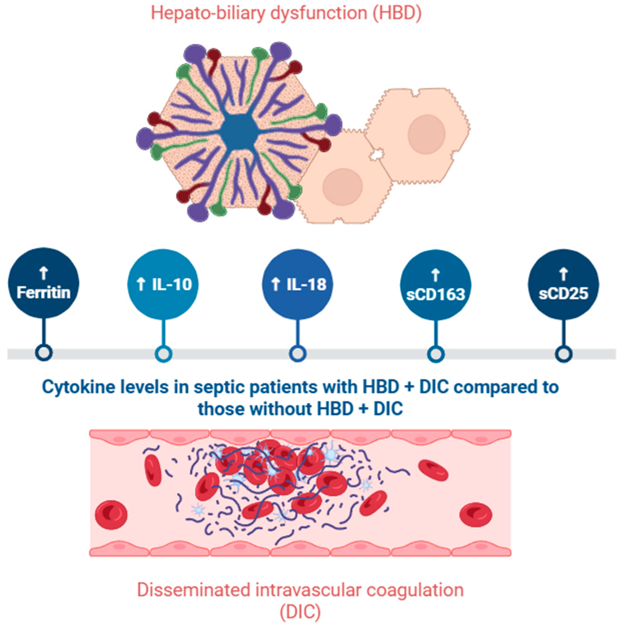
  45. Yadav, P.; Trehanpati, N.; Maiwall, R.; Sehgal, R.; Singh, R.; Islam, M.; Jagdish, R.K.; Vijayaraghavan, R.; Maheshwari, D.; Bhat, S.; et al. Soluble factors and suppressive monocytes can predict early development of sepsis in acute-on-chronic liver failure. Hepatol. Commun. 2022, 6, 2105–2120. [Google Scholar] [CrossRef]
  46. Anderko, R.R.; Gómez, H.; Canna, S.W.; Shakoory, B.; Angus, D.C.; Yealy, D.M.; Huang, D.T.; Kellum, J.A.; Carcillo, J.A. Sepsis with liver dysfunction and coagulopathy predicts an inflammatory pattern of macrophage activation. Intensive Care Med. Exp. 2022, 10, 6. [Google Scholar] [CrossRef]
  47. Kali, A.; Charles, M.V.; Seetharam, R.S. Hepcidin—A novel biomarker with changing trends. Pharmacogn. Rev. 2015, 9, 35. [Google Scholar] [CrossRef]
  48. Olinder, J.; Ehinger, D.; Liljenborg, E.; Herwald, H.; Rydén, C. Plasma Levels of Hepcidin and Reticulocyte Haemoglobin during Septic Shock. J. Innate Immun. 2020, 12, 448–460. [Google Scholar] [CrossRef]
  49. Olinder, J.; Börjesson, A.; Norrman, J.; West, T.; Carlström, J.; Gustafsson, A.; Annborn, M.; Herwald, H.; Rydén, C. Hepcidin discriminates sepsis from other critical illness at admission to intensive care. Sci. Rep. 2022, 12, 14857. [Google Scholar] [CrossRef]
  50. Suastika, N.K.W.; Suega, K. The Role of Hepcidin Level as a Predictor for Mortality in Cancer Patients with Sepsis. Open Access Maced. J. Med. Sci. 2022, 10, 2599–2602. [Google Scholar] [CrossRef]
  51. Hortová-Kohoutková, M.; Skotáková, M.; Onyango, I.G.; Slezáková, M.; Panovský, R.; Opatřil, L.; Slanina, P.; De Zuani, M.; Mrkva, O.; Andrejčinová, I.; et al. Hepcidin and ferritin levels as markers of immune cell activation during septic shock, severe COVID-19 and sterile inflammation. Front. Immunol. 2023, 14, 1110540. [Google Scholar] [CrossRef] [PubMed]
  52. Zeng, C.; Chen, Q.; Zhang, K.; Chen, Q.; Song, S.; Fang, X. Hepatic Hepcidin Protects against Polymicrobial Sepsis in Mice by Regulating Host Iron Status. Anesthesiology 2015, 122, 374–386. [Google Scholar] [CrossRef] [PubMed]
  53. Scindia, Y.; Wlazlo, E.; Leeds, J.; Loi, V.; Ledesma, J.; Cechova, S.; Ghias, E.; Swaminathan, S. Protective Role of Hepcidin in Polymicrobial Sepsis and Acute Kidney Injury. Front. Pharmacol. 2019, 10, 615. [Google Scholar] [CrossRef] [PubMed]
  54. Leaf, D.E.; Rajapurkar, M.; Lele, S.S.; Mukhopadhyay, B.; Boerger, E.A.S.; Mc Causland, F.R.; Eisenga, M.F.; Singh, K.; Babitt, J.L.; Kellum, J.A.; et al. Iron, Hepcidin, and Death in Human AKI. J. Am. Soc. Nephrol. 2019, 30, 493–504. [Google Scholar] [CrossRef]
  55. Qiu, Z.-L.; Yan, B.-Q.; Xu, D.-W.; Zhao, R.; Shen, K.; Lu, S.-Q. Mortality and serum hepcidin are associated with persistent and transient acute kidney injury in septic patients. Clin. Nephrol. 2021, 95, 303–311. [Google Scholar] [CrossRef]
  56. Qiu, Z.-L.; Yan, B.-Q.; Zhao, R.; Xu, D.-W.; Shen, K.; Deng, X.; Lu, S.-Q. Combination of hepcidin with neutrophil gelatinase-associated lipocalin for prediction of the development of sepsis-induced acute kidney injury. Clin. Chim. Acta 2021, 523, 38–44. [Google Scholar] [CrossRef]
  57. Olinder, J.; Stjernqvist, M.J.; Lindén, A.; Salomonsson, E.T.; Annborn, M.; Herwald, H.; Rydén, C. Hepcidin, in contrast to heparin binding protein, does not portend acute kidney injury in patients with community acquired septic shock. PLoS ONE 2024, 19, e0299257. [Google Scholar] [CrossRef]
  58. Wang, W.; Knovich, M.A.; Coffman, L.G.; Torti, F.M.; Torti, S.V. Serum ferritin: Past, present and future. Biochim. Biophys. Acta (BBA)-Gen. Subj. 2010, 1800, 760–769. [Google Scholar] [CrossRef]
  59. Garcia, P.C.R.; Longhi, F.; Branco, R.G.; Piva, J.P.; Lacks, D.; Tasker, R.C. Ferritin levels in children with severe sepsis and septic shock. Acta Paediatr. 2007, 96, 1829–1831. [Google Scholar] [CrossRef]
  60. Tonial, C.T.; Costa, C.A.D.; Andrades, G.R.H.; Crestani, F.; Einloft, P.R.; Bruno, F.; Miranda, A.P.; Fiori, H.H.; Garcia, P.C.R. Prediction of Poor Outcomes for Septic Children According to Ferritin Levels in a Middle-Income Setting*. Pediatr. Crit. Care Med. 2020, 21, e259–e266. [Google Scholar] [CrossRef]
  61. Sarkar, M.; Roychowdhury, S.; Uz Zaman, M.; Raut, S.; Bhakta, S.; Nandy, M. Can serum ferritin be employed as prognostic marker of pediatric septic shock and severe sepsis? J. Pediatr. Crit. Care 2021, 8, 20. [Google Scholar] [CrossRef]
  62. He, L.; Guo, C.; Su, Y.; Ding, N. The relationship between serum ferritin level and clinical outcomes in sepsis based on a large public database. Sci. Rep. 2023, 13, 8677. [Google Scholar] [CrossRef] [PubMed]
  63. Zarjou, A.; Black, L.M.; McCullough, K.R.; Hull, T.D.; Esman, S.K.; Boddu, R.; Varambally, S.; Chandrashekar, D.S.; Feng, W.; Arosio, P.; et al. Ferritin Light Chain Confers Protection Against Sepsis-Induced Inflammation and Organ Injury. Front. Immunol. 2019, 10, 131. [Google Scholar] [CrossRef] [PubMed]
  64. Odum, J.D.; Akhter, J.; Verma, V.; Vollmer, G.; Davidson, A.; Hyndman, K.A.; Bolisetty, S. Myeloid-specific ferritin light chain deletion does not exacerbate sepsis-associated AKI. Am. J. Physiol.-Ren. Physiol. 2024, 327, F171–F183. [Google Scholar] [CrossRef]
  65. Dimitrijevic, Z.M.; Salinger-Martinovic, S.S.; Jankovic, R.J.; Mitic, B.P. Elevated Serum Ferritin Levels Are Predictive of Renal Function Recovery among Patients with Acute Kidney Injury. Tohoku J. Exp. Med. 2019, 248, 63–71. [Google Scholar] [CrossRef]
  66. Fang, Y.-P.; Zhang, H.-J.; Guo, Z.; Ren, C.-H.; Zhang, Y.-F.; Liu, Q.; Wang, Z.; Zhang, X. Effect of Serum Ferritin on the Prognosis of Patients with Sepsis: Data from the MIMIC-IV Database. Emerg. Med. Int. 2022, 2022, 2104755. [Google Scholar] [CrossRef] [PubMed]
  67. Zhang, X.; Luo, T.; Luo, Y.; Lu, M.; Xie, Y. Association Between Serum Ferritin and In-hospital Mortality in Critical Ills with Sepsis-Associated Acute Kidney Injury: A Retrospective Cohort Study. Mod. Health Sci. 2025, 8, p58. [Google Scholar] [CrossRef]
  68. Weis, S.; Carlos, A.R.; Moita, M.R.; Singh, S.; Blankenhaus, B.; Cardoso, S.; Larsen, R.; Rebelo, S.; Schäuble, S.; Del Barrio, L.; et al. Metabolic Adaptation Establishes Disease Tolerance to Sepsis. Cell 2017, 169, 1263–1275.e14. [Google Scholar] [CrossRef]
  69. Wang, T.-W.; Zhou, L.-L.; Yuan, J.; Zhou, W.-X.; Wang, H.-R.; Yu, T.-T.; Zhai, J.-C.; Tang, C.-B.; Jiang, W.; Yu, J.-Q.; et al. Study of the relationship between iron metabolism disorders and sepsis-associated liver injury: A prospective observational study. World J. Gastroenterol. 2025, 31, 104584. [Google Scholar] [CrossRef]
  70. Zhou, F.; Yu, T.; Du, R.; Fan, G.; Liu, Y.; Liu, Z.; Xiang, J.; Wang, Y.; Song, B.; Gu, X.; et al. Clinical course and risk factors for mortality of adult inpatients with COVID-19 in Wuhan, China: A retrospective cohort study. Lancet 2020, 395, 1054–1062. [Google Scholar] [CrossRef]
  71. Timpau, A.-S.; Miftode, R.-S.; Petris, A.O.; Costache, I.-I.; Miftode, I.-L.; Rosu, F.M.; Anton-Paduraru, D.-T.; Leca, D.; Miftode, E.G. Mortality Predictors in Severe COVID-19 Patients from an East European Tertiary Center: A Never-Ending Challenge for a No Happy Ending Pandemic. J. Clin. Med. 2021, 11, 58. [Google Scholar] [CrossRef] [PubMed]
  72. Cao, P.; Wu, Y.; Wu, S.; Wu, T.; Zhang, Q.; Zhang, R.; Wang, Z.; Zhang, Y. Elevated serum ferritin level effectively discriminates severity illness and liver injury of coronavirus disease 2019 pneumonia. Biomarkers 2021, 26, 207–212. [Google Scholar] [CrossRef]
  73. Timpau, A.-S.; Miftode, R.-S.; Costache, I.-I.; Petris, A.O.; Miftode, I.-L.; Gheorghe, L.; Timpau, R.; Miftode, I.D.; Prepeliuc, C.S.; Coman, I.; et al. An Overview of the Impact of Bacterial Infections and the Associated Mortality Predictors in Patients with COVID-19 Admitted to a Tertiary Center from Eastern Europe. Antibiotics 2023, 12, 144. [Google Scholar] [CrossRef] [PubMed]
  74. Yoshida, H.; Hunter, C.A. The Immunobiology of Interleukin-27. Annu. Rev. Immunol. 2015, 33, 417–443. [Google Scholar] [CrossRef]
  75. Wirtz, S.; Tubbe, I.; Galle, P.R.; Schild, H.J.; Birkenbach, M.; Blumberg, R.S.; Neurath, M.F. Protection from lethal septic peritonitis by neutralizing the biological function of interleukin 27. J. Exp. Med. 2006, 203, 1875–1881. [Google Scholar] [CrossRef]
  76. Cao, J.; Xu, F.; Lin, S.; Song, Z.; Zhang, L.; Luo, P.; Xu, H.; Li, D.; Zheng, K.; Ren, G.; et al. IL-27 controls sepsis-induced impairment of lung antibacterial host defence. Thorax 2014, 69, 926–937. [Google Scholar] [CrossRef]
  77. Wang, Y.; Zhao, J.; Yao, Y.; Zhao, D.; Liu, S. Interleukin-27 as a Diagnostic Biomarker for Patients with Sepsis: A Meta-Analysis. BioMed Res. Int. 2021, 2021, 5516940. [Google Scholar] [CrossRef] [PubMed]
  78. Fan, J.; Zhang, Y.; Zheng, D.; Zhang, M.; Liu, H.; He, M.; Wu, Z. IL-27 is elevated in sepsis with acute hepatic injury and promotes hepatic damage and inflammation in the CLP model. Cytokine 2020, 127, 154936. [Google Scholar] [CrossRef]
  79. Fan, J.; He, M.; Wang, C.-J.; Zhang, M. Gadolinium Chloride Inhibits the Production of Liver Interleukin-27 and Mitigates Liver Injury in the CLP Mouse Model. Mediat. Inflamm. 2021, 2021, 2605973. [Google Scholar] [CrossRef]
  80. Ye, L.; Wang, L.; Kuang, G.; Zhang, Z.; Peng, Q.; He, M.; Fan, J. IL-27 aggravates acute hepatic injury by promoting macrophage M1 polarization to induce Caspase-11 mediated Pyroptosis in vitro and in vivo. Cytokine 2025, 188, 156881. [Google Scholar] [CrossRef]
  81. Zhou, P.; Deng, B.; Wu, M.; Ding, F.; Wang, L. Interleukin-27 Ameliorates Renal Ischemia-Reperfusion Injury through Signal Transducers and Activators of Transcription 3 Signaling Pathway. Kidney Blood Press. Res. 2019, 44, 1453–1464. [Google Scholar] [CrossRef] [PubMed]
  82. Gao, F.; Yang, Y.; Feng, X.; Fan, T.; Jiang, L.; Guo, R.; Liu, Q. Interleukin-27 is elevated in sepsis-induced myocardial dysfunction and mediates inflammation. Cytokine 2016, 88, 1–11. [Google Scholar] [CrossRef] [PubMed]
  83. Wang, Y.; Zhang, Y.; Shou, S.; Jin, H. The role of IL-17 in acute kidney injury. Int. Immunopharmacol. 2023, 119, 110307. [Google Scholar] [CrossRef]
  84. Luo, C.; Luo, F.; Zhang, L.; Xu, Y.; Cai, G.; Fu, B.; Feng, Z.; Sun, X.; Chen, X. Knockout of interleukin-17A protects against sepsis-associated acute kidney injury. Ann. Intensive Care 2016, 6, 56. [Google Scholar] [CrossRef] [PubMed]
  85. Maravitsa, P.; Adamopoulou, M.; Pistiki, A.; Netea, M.G.; Louis, K.; Giamarellos-Bourboulis, E.J. Systemic over-release of interleukin-17 in acute kidney injury after septic shock: Clinical and experimental evidence. Immunol. Lett. 2016, 178, 68–76. [Google Scholar] [CrossRef] [PubMed]
  86. Zhou, X.; Yao, J.; Lin, J.; Liu, J.; Dong, L.; Duan, M. Th17/Regulatory T-Cell Imbalance and Acute Kidney Injury in Patients with Sepsis. J. Clin. Med. 2022, 11, 4027. [Google Scholar] [CrossRef]
  87. Jin, H.; Wei, W.; Zhao, Y.; Ma, A.; Sun, K.; Lin, X.; Liu, Q.; Shou, S.; Zhang, Y. The roles of interleukin-17A in risk stratification and prognosis of patients with sepsis-associated acute kidney injury. Kidney Res. Clin. Pract. 2023, 42, 742–750. [Google Scholar] [CrossRef]
  88. Li, L.; Chen, L.; Lin, F.; Mu, J.; Wang, D.; Zhang, W.; Mi, L.; Qin, Y.; Zhou, Y. Study of the Expression of Inflammatory Factors IL-4, IL-6, IL-10, and IL-17 in Liver Failure Complicated by Coagulation Dysfunction and Sepsis. J. Inflamm. Res. 2021, 14, 1447–1453. [Google Scholar] [CrossRef]
  89. Sabat, R. IL-10 family of cytokines. Cytokine Growth Factor Rev. 2010, 21, 315–324. [Google Scholar] [CrossRef]
  90. Knolle, P.A.; Löser, E.; Protzer, U.; Duchmann, R.; Schmitt, E.; Zum Büschenfelde, K.-H.M.; Rose-John, S.; Gerken, G. Regulation of endotoxin-induced IL-6 production in liver sinusoidal endothelial cells and Kupffer cells by IL-10. Clin. Exp. Immunol. 2003, 107, 555–561. [Google Scholar] [CrossRef]
  91. Jensen, I.J.; McGonagill, P.W.; Butler, N.S.; Harty, J.T.; Griffith, T.S.; Badovinac, V.P. NK Cell–Derived IL-10 Supports Host Survival during Sepsis. J. Immunol. 2021, 206, 1171–1180. [Google Scholar] [CrossRef] [PubMed]
  92. Zeng, G.; Chen, D.; Zhou, R.; Zhao, X.; Ye, C.; Tao, H.; Sheng, W.; Wu, Y. Combination of C-reactive protein, procalcitonin, IL -6, IL -8, and IL -10 for early diagnosis of hyperinflammatory state and organ dysfunction in pediatric sepsis. Clin. Lab. Anal. 2022, 36, e24505. [Google Scholar] [CrossRef]
  93. Lee, S.-Y.; Lee, Y.-S.; Choi, H.-M.; Ko, Y.-S.; Lee, H.-Y.; Jo, S.-K.; Cho, W.-Y.; Kim, H.-K. Distinct pathophysiologic mechanisms of septic acute kidney injury: Role of immune suppression and renal tubular cell apoptosis in murine model of septic acute kidney injury. Crit. Care Med. 2012, 40, 2997–3006. [Google Scholar] [CrossRef] [PubMed]
  94. Cho, E.; Lee, J.H.; Lim, H.J.; Oh, S.W.; Jo, S.K.; Cho, W.Y.; Kim, H.; Lee, S.Y. Soluble CD25 is increased in patients with sepsis-induced acute kidney injury. Nephrology 2014, 19, 318–324. [Google Scholar] [CrossRef] [PubMed]
  95. Xu, H.; Turnquist, H.R.; Hoffman, R.; Billiar, T.R. Role of the IL-33-ST2 axis in sepsis. Mil. Med. Res. 2017, 4, 3. [Google Scholar] [CrossRef]
  96. Zhou, Y.; Xu, Z.; Liu, Z. Role of IL-33-ST2 pathway in regulating inflammation: Current evidence and future perspectives. J. Transl. Med. 2023, 21, 902. [Google Scholar] [CrossRef]
  97. Hur, M.; Kim, H.; Kim, H.J.; Yang, H.S.; Magrini, L.; Marino, R.; Cardelli, P.; Somma, S.D.; Network, O.B.O.G. Soluble ST2 Has a Prognostic Role in Patients With Suspected Sepsis. Ann. Lab. Med. 2015, 35, 570–577. [Google Scholar] [CrossRef]
  98. Cepoi, M.-R.; Duca, S.T.; Chetran, A.; Costache, A.D.; Spiridon, M.R.; Afrăsânie, I.; Leancă, S.A.; Dmour, B.-A.; Matei, I.T.; Miftode, R.S.; et al. Chronic Kidney Disease Associated with Ischemic Heart Disease: To What Extent Do Biomarkers Help? Life 2023, 14, 34. [Google Scholar] [CrossRef]
  99. Alves-Filho, J.C.; Sônego, F.; Souto, F.O.; Freitas, A.; Verri, W.A.; Auxiliadora-Martins, M.; Basile-Filho, A.; McKenzie, A.N.; Xu, D.; Cunha, F.Q.; et al. Interleukin-33 attenuates sepsis by enhancing neutrophil influx to the site of infection. Nat. Med. 2010, 16, 708–712. [Google Scholar] [CrossRef]
  100. Bao, Q.; Lv, R.; Lei, M. IL-33 attenuates mortality by promoting IFN-γ production in sepsis. Inflamm. Res. 2018, 67, 531–538. [Google Scholar] [CrossRef]
  101. Vrsaljko, N.; Radmanic Matotek, L.; Zidovec-Lepej, S.; Vince, A.; Papic, N. The Impact of Steatotic Liver Disease on Cytokine and Chemokine Kinetics During Sepsis. Int. J. Mol. Sci. 2025, 26, 2226. [Google Scholar] [CrossRef] [PubMed]
  102. Akcay, A.; Nguyen, Q.; He, Z.; Turkmen, K.; Won Lee, D.; Hernando, A.A.; Altmann, C.; Toker, A.; Pacic, A.; Ljubanovic, D.G.; et al. IL-33 Exacerbates Acute Kidney Injury. J. Am. Soc. Nephrol. 2011, 22, 2057–2067. [Google Scholar] [CrossRef]
  103. Erfurt, S.; Hoffmeister, M.; Oess, S.; Asmus, K.; Patschan, S.; Ritter, O.; Patschan, D. Soluble IL-33 receptor predicts survival in acute kidney injury. J. Circ. Biomark. 2022, 11, 28–35. [Google Scholar] [CrossRef] [PubMed]
  104. Huang, W.; Tang, Y.; Li, L. HMGB1, a potent proinflammatory cytokine in sepsis. Cytokine 2010, 51, 119–126. [Google Scholar] [CrossRef]
  105. Diener, K.R.; Al-Dasooqi, N.; Lousberg, E.L.; Hayball, J.D. The multifunctional alarmin HMGB1 with roles in the pathophysiology of sepsis and cancer. Immunol. Cell Biol. 2013, 91, 443–450. [Google Scholar] [CrossRef] [PubMed]
  106. Gibot, S.; Massin, F.; Cravoisy, A.; Barraud, D.; Nace, L.; Levy, B.; Bollaert, P.-E. High-mobility group box 1 protein plasma concentrations during septic shock. Intensive Care Med. 2007, 33, 1347–1353. [Google Scholar] [CrossRef]
  107. Sundén-Cullberg, J.; Norrby-Teglund, A.; Rouhiainen, A.; Rauvala, H.; Herman, G.; Tracey, K.J.; Lee, M.L.; Andersson, J.; Tokics, L.; Treutiger, C.J. Persistent elevation of high mobility group box-1 protein (HMGB1) in patients with severe sepsis and septic shock*. Crit. Care Med. 2005, 33, 564–573. [Google Scholar] [CrossRef]
  108. Karakike, E.; Adami, M.-E.; Lada, M.; Gkavogianni, T.; Koutelidakis, I.M.; Bauer, M.; Giamarellos-Bourboulis, E.J.; Tsangaris, I. Late Peaks of HMGB1 and Sepsis Outcome: Evidence For Synergy With Chronic Inflammatory Disorders. Shock 2019, 52, 334–339. [Google Scholar] [CrossRef]
  109. Wang, W.; Sun, L.; Deng, Y.; Tang, J. Synergistic effects of antibodies against high-mobility group box 1 and tumor necrosis factor-α antibodies on d -(+)-galactosamine hydrochloride/lipopolysaccharide-induced acute liver failure. FEBS J. 2013, 280, 1409–1419. [Google Scholar] [CrossRef]
  110. Huang, Y.; Zang, K.; Shang, F.; Guo, S.; Gao, L.; Zhang, X. HMGB1 mediates acute liver injury in sepsis through pyroptosis of liver macrophages. Int. J. Burn. Trauma 2020, 10, 60. [Google Scholar]
  111. Wang, Y.; Zhang, H.; Chen, Q.; Jiao, F.; Shi, C.; Pei, M.; Lv, J.; Zhang, H.; Wang, L.; Gong, Z. TNF-α/HMGB1 inflammation signalling pathway regulates pyroptosis during liver failure and acute kidney injury. Cell Prolif. 2020, 53, e12829. [Google Scholar] [CrossRef] [PubMed]
  112. Zheng, S.; Pan, Y.; Wang, C.; Liu, Y.; Shi, M.; Ding, G. HMGB1 Turns Renal Tubular Epithelial Cells into Inflammatory Promoters by Interacting with TLR4 During Sepsis. J. Interferon Cytokine Res. 2016, 36, 9–19. [Google Scholar] [CrossRef]
  113. Khorashadi, M.; Beunders, R.; Pickkers, P.; Legrand, M. Proenkephalin: A New Biomarker for Glomerular Filtration Rate and Acute Kidney Injury. Nephron 2020, 144, 655–661. [Google Scholar] [CrossRef]
  114. Beunders, R.; Struck, J.; Wu, A.H.B.; Zarbock, A.; Di Somma, S.; Mehta, R.L.; Koyner, J.L.; Nadim, M.K.; Maisel, A.S.; Murray, P.T.; et al. Proenkephalin (PENK) as a Novel Biomarker for Kidney Function. J. Appl. Lab. Med. 2017, 2, 400–412. [Google Scholar] [CrossRef]
  115. Schulte, J.; Dépret, F.; Hartmann, O.; Pickkers, P.; Laterre, P.-F.; Uhle, F. AdrenOSS-1 study investigators. Clinical performance of proenkephalin A 119–159 for the early diagnosis of acute kidney injury in patients with sepsis or septic shock. medRxiv 2024. [Google Scholar] [CrossRef]
  116. Caironi, P.; Latini, R.; Struck, J.; Hartmann, O.; Bergmann, A.; Bellato, V.; Ferraris, S.; Tognoni, G.; Pesenti, A.; Gattinoni, L.; et al. Circulating Proenkephalin, Acute Kidney Injury, and Its Improvement in Patients with Severe Sepsis or Shock. Clin. Chem. 2018, 64, 1361–1369. [Google Scholar] [CrossRef] [PubMed]
  117. Lin, L.-C.; Chuan, M.-H.; Liu, J.-H.; Liao, H.-W.; Ng, L.L.; Magnusson, M.; Jujic, A.; Pan, H.-C.; Wu, V.-C.; Forni, L.G. Proenkephalin as a biomarker correlates with acute kidney injury: A systematic review with meta-analysis and trial sequential analysis. Crit. Care 2023, 27, 481. [Google Scholar] [CrossRef]
  118. Verras, C.; Bezati, S.; Bistola, V.; Ventoulis, I.; Matsiras, D.; Tsiodras, S.; Parissis, J.; Polyzogopoulou, E. Point-of-Care Serum Proenkephalin as an Early Predictor of Mortality in Patients Presenting to the Emergency Department with Septic Shock. Biomedicines 2024, 12, 1004. [Google Scholar] [CrossRef]
  119. Kim, H.; Hur, M.; Struck, J.; Bergmann, A.; Somma, S.D.; GREAT Network. Proenkephalin Predicts Organ Failure, Renal Replacement Therapy, and Mortality in Patients With Sepsis. Ann. Lab. Med. 2020, 40, 466–473. [Google Scholar] [CrossRef] [PubMed]
  120. Tokunaga, R.; Zhang, W.; Naseem, M.; Puccini, A.; Berger, M.D.; Soni, S.; McSkane, M.; Baba, H.; Lenz, H.-J. CXCL9, CXCL10, CXCL11/CXCR3 axis for immune activation—A target for novel cancer therapy. Cancer Treat. Rev. 2018, 63, 40–47. [Google Scholar] [CrossRef]
  121. Giamarellos-Bourboulis, E.J.; Antonelli, M.; Bloos, F.; Kotsamidi, I.; Psarrakis, C.; Dakou, K.; Thomas-Rüddel, D.; Montini, L.; Briegel, J.; Damoraki, G.; et al. Interferon-gamma driven elevation of CXCL9: A new sepsis endotype independently associated with mortality. eBioMedicine 2024, 109, 105414. [Google Scholar] [CrossRef]
  122. Moledina, D.G.; Obeid, W.; Smith, R.N.; Rosales, I.; Sise, M.E.; Moeckel, G.; Kashgarian, M.; Kuperman, M.; Campbell, K.N.; Lefferts, S.; et al. Identification and validation of urinary CXCL9 as a biomarker for diagnosis of acute interstitial nephritis. J. Clin. Investig. 2023, 133, e168950. [Google Scholar] [CrossRef] [PubMed]
  123. Yang, H.; Chen, Y.; He, J.; Li, Y.; Feng, Y. Advances in the diagnosis of early biomarkers for acute kidney injury: A literature review. BMC Nephrol. 2025, 26, 115. [Google Scholar] [CrossRef] [PubMed]
  124. Saiman, Y.; Friedman, S.L. The Role of Chemokines in Acute Liver Injury. Front. Physiol. 2012, 3, 213. [Google Scholar] [CrossRef]
  125. Plevriti, A.; Lamprou, M.; Mourkogianni, E.; Skoulas, N.; Giannakopoulou, M.; Sajib, M.S.; Wang, Z.; Mattheolabakis, G.; Chatzigeorgiou, A.; Marazioti, A.; et al. The Role of Soluble CD163 (sCD163) in Human Physiology and Pathophysiology. Cells 2024, 13, 1679. [Google Scholar] [CrossRef] [PubMed]
  126. Feng, L.; Zhou, X.; Su, L.-X.; Feng, D.; Jia, Y.-H.; Xie, L.-X. Clinical Significance of Soluble Hemoglobin Scavenger Receptor CD163 (sCD163) in Sepsis, a Prospective Study. PLoS ONE 2012, 7, e38400. [Google Scholar] [CrossRef]
  127. Gaïni, S.; Pedersen, S.S.; Koldkjær, O.G.; Pedersen, C.; Moestrup, S.K.; Møller, H.J. New immunological serum markers in bacteraemia: Anti-inflammatory soluble CD163, but not proinflammatory high mobility group-box 1 protein, is related to prognosis. Clin. Exp. Immunol. 2008, 151, 423–431. [Google Scholar] [CrossRef]
  128. Abdelrahman, R.; Wagih, A.; Elbarbary, A. The diagnostic value of serum level of soluble hemoglobin scavenger receptor CD163 for sepsis in the ICU. Tanta Med. J. 2015, 43, 9. [Google Scholar] [CrossRef]
  129. Mierzchała-Pasierb, M.; Lipińska-Gediga, M.; Lewandowski, Ł.; Krzystek-Korpacka, M. Alterations in Serum Concentration of Soluble CD163 within Five Study Days from ICU Admission Are Associated with In-Hospital Mortality of Septic Patients—A Preliminary Study. Int. J. Environ. Res. Public Health 2023, 20, 2263. [Google Scholar] [CrossRef]
  130. Su, L.; Feng, L.; Liu, C.; Jiang, Z.; Li, M.; Xiao, K.; Yan, P.; Jia, Y.; Feng, D.; Xie, L. Diagnostic value of urine sCD163 levels for sepsis and relevant acute kidney injury: A prospective study. BMC Nephrol. 2012, 13, 123. [Google Scholar] [CrossRef]
  131. Møller, H.J.; Grønbæk, H.; Schiødt, F.V.; Holland-Fischer, P.; Schilsky, M.; Munoz, S.; Hassanein, T.; Lee, W.M. Soluble CD163 from activated macrophages predicts mortality in acute liver failure. J. Hepatol. 2007, 47, 671–676. [Google Scholar] [CrossRef]
  132. Tornai, T.; Vitalis, Z.; Sipeki, N.; Dinya, T.; Tornai, D.; Antal-Szalmas, P.; Karanyi, Z.; Tornai, I.; Papp, M. Macrophage activation marker, soluble CD 163, is an independent predictor of short-term mortality in patients with cirrhosis and bacterial infection. Liver Int. 2016, 36, 1628–1638. [Google Scholar] [CrossRef] [PubMed]
  133. Damoiseaux, J. The IL-2—IL-2 receptor pathway in health and disease: The role of the soluble IL-2 receptor. Clin. Immunol. 2020, 218, 108515. [Google Scholar] [CrossRef] [PubMed]
  134. Garcia-Obregon, S.; Azkargorta, M.; Seijas, I.; Pilar-Orive, J.; Borrego, F.; Elortza, F.; Boyano, M.D.; Astigarraga, I. Identification of a panel of serum protein markers in early stage of sepsis and its validation in a cohort of patients. J. Microbiol. Immunol. Infect. 2018, 51, 465–472. [Google Scholar] [CrossRef] [PubMed]
  135. Komorowski, M.; Green, A.; Tatham, K.C.; Seymour, C.; Antcliffe, D. Sepsis biomarkers and diagnostic tools with a focus on machine learning. eBioMedicine 2022, 86, 104394. [Google Scholar] [CrossRef]
  136. He, R.-R.; Yue, G.-L.; Dong, M.-L.; Wang, J.-Q.; Cheng, C. Sepsis Biomarkers: Advancements and Clinical Applications—A Narrative Review. Int. J. Mol. Sci. 2024, 25, 9010. [Google Scholar] [CrossRef]
Figure 1. Overview of sepsis pathophysiology. FasL: Fas Ligand; HMGB1: high-mobility group box-1; NK: natural killer; Th: T helper cells; TLR: Toll-like Receptor; Treg: Regulatory T cell. Created in BioRender. Pasare, A. (2025) https://BioRender.com/0fgjo31. Accessed on 1 May 2025.
Ijms 26 04825 g001
Figure 2. Overview of SA-AKI pathophysiology. Created in BioRender. Pasare, A. (2025) https://BioRender.com/1f9k56c. Accessed on 11 May 2025.
Ijms 26 04825 g002
Figure 3. Levels of biomarkers in septic patients with and without liver impairment. Created in BioRender. Pasare, A. (2025) https://BioRender.com/g7vqax2. Accessed on 11 May 2025.
Ijms 26 04825 g003
Table 1. Summary of advantages and limitations for IL-18, hepcidin, and ferritin.
AdvantagesLimitations
IL-18
-
Higher concentrations in septic patients
-
Correlated with sepsis mortality
-
Good predictive value for SA-AKI in multiple studies
-
High concentration in HBD + DIC
-
Elevated in other inflammatory conditions
-
Studies with small study population
Hepcidin
-
Levels correlate with sepsis diagnosis
-
Hepcidin treatment lowers mortality rates (preclinical studies)
-
Correlated with mortality for AKI patients
-
Elevated in hereditary hemochromatosis and some cancers
-
Predictive value for SA-AKI is improved by combining with other biomarkers
-
Mortality for AKI patients is not specific for SA-AKI.
Ferritin
-
Good prognostic value for adult and pediatric sepsis
-
Serum values linked with AKI risk in septic patients
-
FtH protects mice from hepatic injury in sepsis models
-
High concentration in HBD + DIC
-
Not specific for sepsis inflammation
-
Few clinical studies related to sepsis associated with liver injury
Table 2. Summary of advantages and limitations for IL-27, IL-17A, IL-10, IL-33/ST2, HMGB1, Proenkephalin, CXCL9, sCD163, sCD25.
AdvantagesLimitations
IL-27
-
Similar diagnostic capacity as procalcitonin
-
Correlated with sepsis-associated liver injury in both preclinical and clinical studies
-
Not specific for liver, investigated for sepsis myocardial dysfunction
IL-17A
-
Targeted therapy leads to reduced renal injury in septic conditions
-
Not sufficient clinical studies to sustain preclinical data
IL-10
-
Associated with hepatic dysfunction in septic patients
-
Not sufficiently specific
-
Reflects immune response and not direct injury
IL-33 and ST2
-
Linked with metabolic liver disease in septic patients
-
Correlated with mortality in AKI patients
-
Mostly studied in relation to heart failure
-
Not specific for SA-AKI
HMGB1
-
Targeted therapy leads to reduced renal and hepatic injury in mice sepsis models
-
Not validated in large clinical trials
Proenkephalin
-
Good predictive value for SA-AKI severity and function recovery
-
Not yet established to provide better diagnosis over validated biomarkers
CXCL9
-
Potential to define a new sepsis endotype
-
Not enough data for organ-specific injury
sCD163
-
Urinary concentrations display diagnostic capacities for SA-AKI
-
Not yet validated in large clinical trials
sCD25
-
Similar diagnosis ability as NGAL for SA-AKI
-
Further validation needed
Disclaimer/Publisher’s Note: The statements, opinions and data contained in all publications are solely those of the individual author(s) and contributor(s) and not of MDPI and/or the editor(s). MDPI and/or the editor(s) disclaim responsibility for any injury to people or property resulting from any ideas, methods, instructions or products referred to in the content.

