Article

CRISPR/Cas9-Mediated Knockout of the White Gene in Agasicles hygrophila

1
College of Plant Protection, Shanxi Agricultural University, Jinzhong 030801, China
2
Department of Entomology and BIO5 Institute, University of Arizona, Tucson, AZ 85721, USA
*
Authors to whom correspondence should be addressed.
Int. J. Mol. Sci. 2025, 26(10), 4586; https://doi.org/10.3390/ijms26104586
Submission received: 11 March 2025 / Revised: 6 May 2025 / Accepted: 8 May 2025 / Published: 10 May 2025
(This article belongs to the Section Molecular Genetics and Genomics)

Abstract

Agasicles hygrophila is the most effective natural enemy for the control of the invasive weed Alternanthera philoxeroides (Mart.) Griseb. However, research on the gene function and potential genetic improvement of A. hygrophila is limited due to a lack of effective genetic tools. In this study, we employed the A. hygrophila white (AhW) gene as a target gene to develop a CRISPR/Cas9-based gene editing method applicable to A. hygrophila. We showed that injection of Cas9/sgRNA ribonucleoprotein complexes (RNPs) of the AhW gene into pre-blastoderm eggs induced genetic insertion and deletion mutations, leading to white eyes. Our results demonstrate that CRISPR/Cas9-mediated gene editing is possible in A. hygrophila, offering a valuable tool for studies of functional genomics and genetic improvement of A. hygrophila, which could potentially lead to more effective control of invasive weeds through the development of improved strains of this biocontrol agent. In addition, the white-eyed mutant strain we developed could potentially be useful for other transgenic research studies on this species.

1. Introduction

CRISPR/Cas9 (Clustered Regularly Interspaced Palindromic Repeats, CRISPR) is a newly discovered, powerful genome editing tool that is active in all organisms [1,2]. In this editing system, the Streptococcus pyogenes Cas9 protein, guided by an RNA endonuclease, precisely cuts a double-stranded DNA based on base paring between CRISPR RNA (crRNA) and the target sequence (protospacer). The double-stranded DNA breaks (DSBs) are then repaired either via non-homologous end joining (NHEJ) or homology-directed recombination (HDR) in the presence of a donor DNA template sequence. NHEJ repairs are of low fidelity and can either add or subtract bases to produce protein mutations or frameshift mutations [3]. CRISPR-based genome engineering has been implemented in many insect orders, but in the order Coleoptera, it has been carried out in only a few species (i.e., Tribolium castaneum [4], Leptinotarsa decemlineata [5], Diabrotica virgifera virgifera [6], and Harmonia axyridis [7]).
As an easily visible marker, eye color has frequently been manipulated genetically to induce genetic transformations in various types of insects. The eye color of Drosophila melanogaster is determined by ommochrome and pteridine pigments. The white (Dm-w) gene encodes an ABC transporter, which is essential for transporting ommochrome and pteridine precursors across membranes [8,9]. The Dm-w mutation results in white compound eyes [10]. White forms heterodimers with brown (bw) and scarlet (st), which are responsible for the transport of pteridine precursors and ommochromes, respectively. The bw and st mutants have brown [11] and scarlet eyes [12]. As it is visible and does not affect normal growth, the artificially optimized form of Dm-w, called mini-white [13], is the most commonly used marker for site-specific recombinases that mediate genome insertion, such as the FLP/FRT system [14], the ΦC31-mediated recombinase cassette exchange (RMCE) system [15], and the Cre-mediated recombination system [16]. In these systems, the Dm-w mutant strain is used as the starting strain and the donor plasmid contains mini-W. This transformation changes the eye color from white back to red, which is the normal eye color in Drosophila species. The greatest advantage of using this system is that eye color functions as a visible screening marker, thus avoiding the need to use fluorescent proteins. The white gene is conserved in all insects, and knockdown expression or knockout results in white eyes [17,18,19]. Therefore, this system can be used for non-model insect species.
The flea beetle Agasicles hygrophila (Selman and Vogt), which originated from Argentina, has been introduced into various countries, including China, to successfully control the invasive aquatic weed Alternanthera philoxeroides (Mart.) Griseb [20]. RNA interference (RNAi) is the primary tool used to study gene function in various organisms, including A. hygrophila. However, although RNAi can be efficiently applied in A. hygrophila [21], this approach is limited to knockdown expression for partial reduction at the transcriptional level, and its use results in only a limited time period of knockdown. More efficient gene knockout methods are needed for use in A. hygrophila. Using the CRISPR/Cas9-mediated germline transformation technique, we employed the white gene as a visible marker to produce mutant forms of A. hygrophila. In this article, we describe a simple and efficient approach for genome editing in A. hygrophila using the CRISPR/Cas9 system.

2. Results

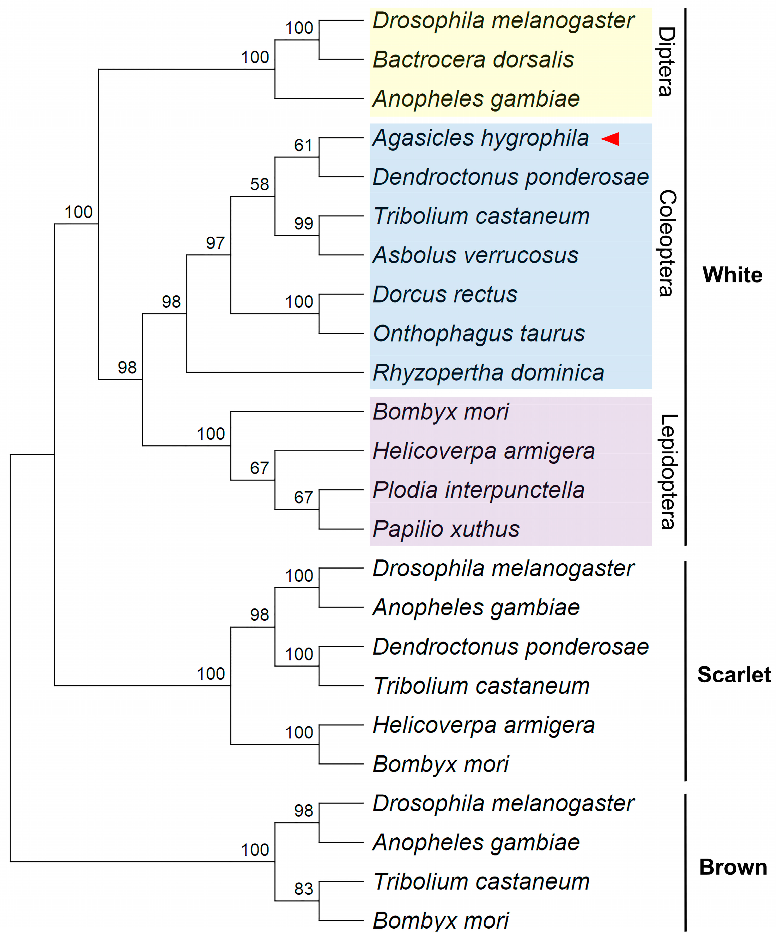
2.1. Phylogenetic Identification of the A. hygrophila White (AhW) Gene

A reciprocal TBLASTN search and a phylogenetic analysis were combined to identify the A. hygrophila white gene. The TBLASTN search of the A. hygrophila transcriptome dataset using the D. melanogaster white protein (Dm-w, GenBank accession # CAA26716.2) as the query found that the best hit was A. hygrophila unigene # c3849/f1p0/2312 (e value = 0, total score = 627, and query coverage = 88%), which shared an amino acid sequence identity of 53% with the D. melanogaster white protein gene. The amino acid similarity of the D. melanogaster white protein gene to other A. hygrophila unigenes was less than 30%. A Blastp search of the D. melanogaster gene dataset, with the translated A. hygrophila unigene # c3849/f1p0/2312 as the query, revealed that the best hit for A. hygrophila unigene # c3849/f1p0/2312 was the D. melanogaster white protein (Table 1; e value = 0, total score = 602, query coverage = 100%, and identity = 49.35%). Such a one-to-one orthologous relationship between the D. melanogaster white gene and A. hygrophila unigene # c3849/f1p0/2312 was consistent with our phylogenic analysis, which clearly showed that A. hygrophila unigene # c3849/f1p0/2312 clustered with other previously identified insect white protein genes from the orders Coleoptera, Diptera, and Lepidoptera, and that it was more distantly related to the scarlet and brown protein genes (Figure 1). These data confirm that A. hygrophila unigene # c3849/f1p0/2312 represents the transcript of the A. hygrophila white gene (AhW).
The full-length cDNA sequence of AhW was amplified with the primers AhW-F (located upstream of the putative start codon initiator) and AhW-R (located downstream of the putative stop codon) (Table S1) and then submitted to GenBank (accession number OR123872). Alignment of the obtained 2309 bp AhW cDNA with the unpublished A. hygrophila genome (personal communication with Dr. FangHao Wan) showed that the genomic DNA sequence of AhW is over 4917 bp long and comprises 12 exons and 11 introns (Figure 2A). The translated protein sequence of the AhW cDNA is 669 amino acids in length and exhibits the typical characteristics of the ABC transporter G subfamily, which include a C-terminal transmembrane domain (TMD) and a nucleotide-binding domain (NBD) composed of a Walker A motif, an ABC signature motif, a Walker B motif, a Q loop, a D loop, and an H loop (Figure 2B).

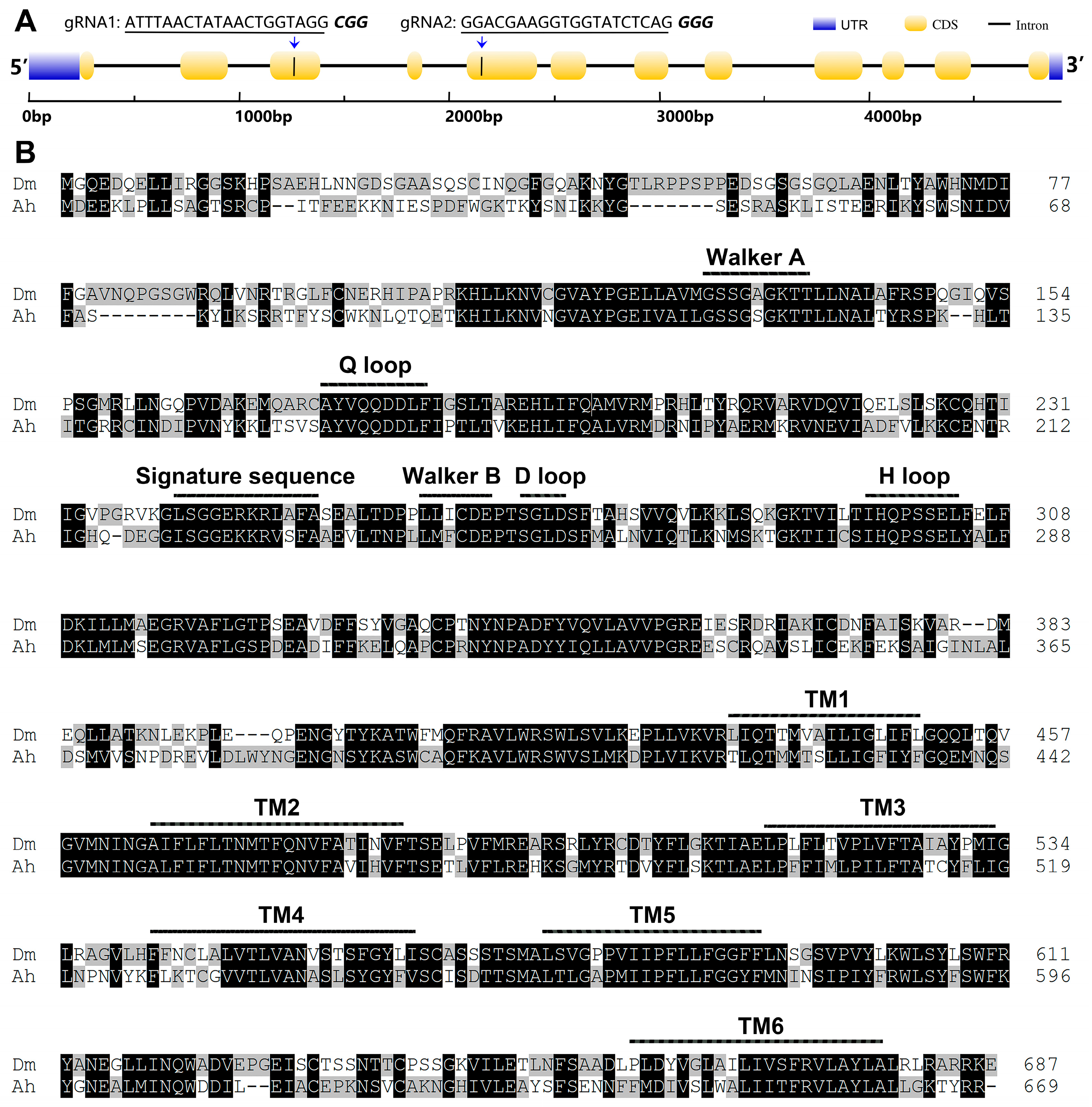
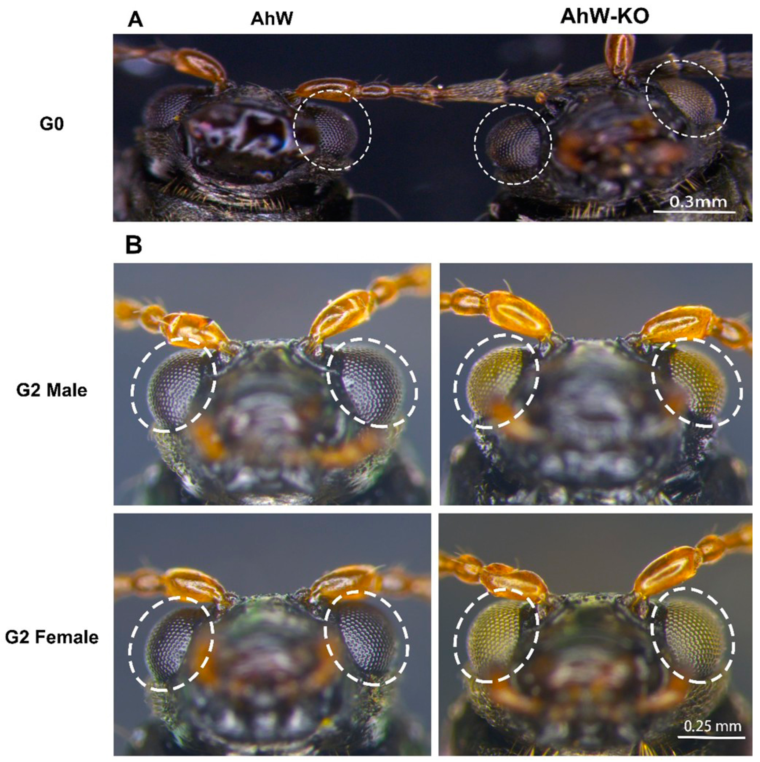
2.2. Creation of an AhW Knockout White-Eyed Strain

sgRNA1 and sgRNA2 are respectively located in exons 3 and 5 of the AhW. A total of 300 eggs (G0) were injected with RNP, consisting of Cas9 protein, sgRNA1, and sgRNA2. In the end, we obtained eight adults (three females and five males), with only one mutant female, which had two white and black chimera eyes (Figure 3A). The right eye of this G0 mutant female was mostly white, with a small number of black facets at the bottom edge, while its left eye was composed of three black faceted stripes and two white faceted stripes (Figure 3A).
To confirm that the AhW gene sequence was indeed successfully edited, we extracted the genomic DNA of the wild-type strain (AhW strain) beetle and the G0 white-eyed mutant and then amplified the sequences of the AhW editing sites. Electrophoretic results showed that the wild-type beetle’s AhW had a main band of 1818 bp. However, besides this main band, there were several shallow bands smaller than 1818 bp in the G0 white-eyed beetle (Figure 4).
To establish a homozygous white-eyed mutant strain, the G0 mutant female was allowed to mate with two wild-type black-eyed males. In the resultant G1 progeny, all 11 females had black eyes, whereas all males (8 in total) had white eyes. The eight white-eyed G1 males were allowed to mate with eight black-eyed G1 females in eight separate mating cages; all eight cages produced G2 progeny in which both females and males were ~50% white-eyed and ~50% black-eyed (a G2 mutant male and a female are shown in Figure 3B). This suggests that G0 AhW mutations responsible for white eyes in the AhW knockout strain (AhW-KO) are likely homozygous and fixed.

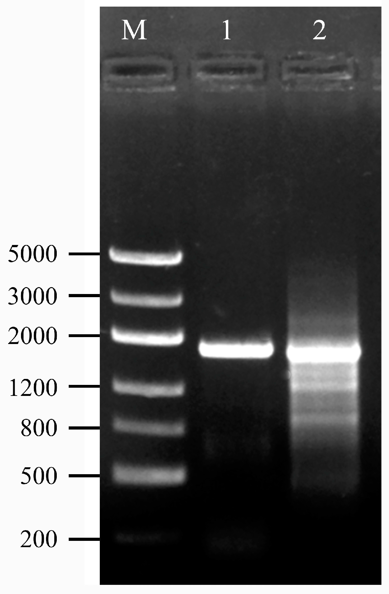
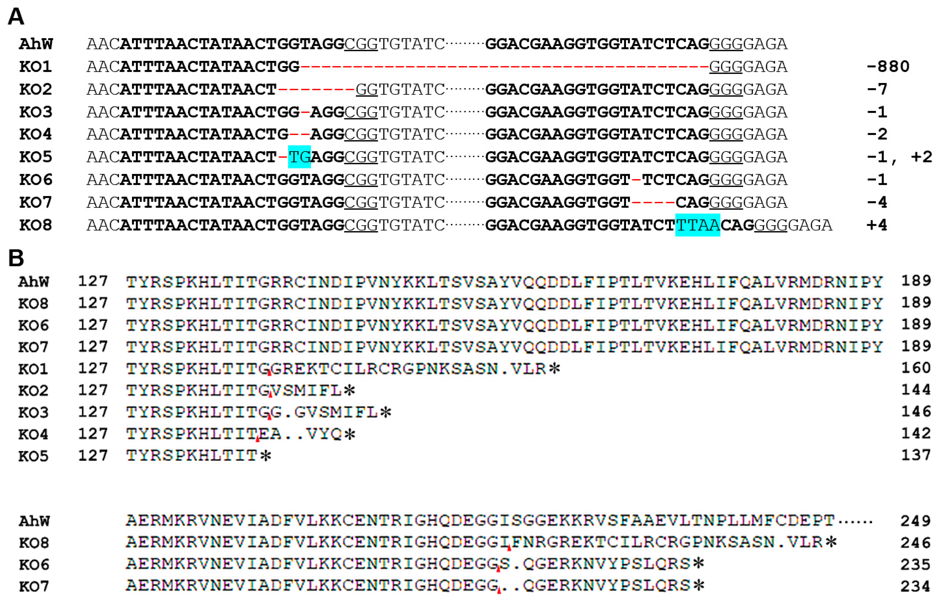
2.3. Mutations in the Two sgRNA Target Sites in AhW gDNA from the AhW-KO Strain

To identify the mutations corresponding to the two sgRNA target sites located in exons 3 and 5 of Ahw (Figure 2A), we used the genomic DNA samples individually extracted from one hind leg of each of the eight G1 white-eyed males as the templates to PCR-amplify the regions flanking the sgRNA target sites (Figure S2) using the gene-specific primers AhW-F1 and AhW-R1 (Table S1). Direct sequencing of the PCR products from each of the eight G1 white-eyed males showed that all eight PCR products had one unique disruptive mutation (Figure 5A). These included one 800 bp deletion spanning from the sgRNA1 target site to the sgRNA2 target site, one −1 bp/+2 bp indel and three deletions (−7, −1, and −2 bp) corresponding to the sgRNA1 target site, and two −1 and −4 bp deletions and one +4 bp insertion corresponding to the sgRNA2 target site (Figure 5A). The −1 bp/+2 bp indel in AhW-KO5 directly truncated the AhW protein with a premature stop codon, whereas all the other seven CRISPR/Cas9-induced mutations disrupted the AhW coding sequence by first shifting the reading frame, followed by the introduction of premature stop codons at various positions (Figure 5B).

2.4. Inheritance of Cas9-Introduced White Eye Mutations

Since all the males of the G1 generation obtained by mating with the wild type exhibited white eyes, while all the females exhibited black eyes, we speculate that AhW recessive inheritance of X chromosome. To test this speculation, two reciprocal single-pair crosses between the wild-type AhW strain and the AhW-KO strain (1 ♀AhW × 1 ♂AhW-KO; 1 ♂AhW × 1 ♀AhW-KO) were made to yield G1 and G1′ progeny, respectively, and all males and females of both G1 and G1′ hybrids were allowed to self-cross to produce G2 and G2′ generations. All the G1 hybrids and G2 progeny were individually examined for their sex and eye color phenotype. As shown in Table 2, all G1 hybrids, regardless of sex, had black eyes, while 50% of the G1′ hybrids had white eyes, and we observed only white eyes for males and black eyes for females. In addition, the black/white eye ratio of the self-inbred G2 offspring was 2.92:1.0 (no difference from 3:1), with all females and about half of the males having black eyes and the other half of the males having white eyes. For comparison, the black/white eye ratio was 1:1 for the self-inbred G2′ generation, regardless of sex (Table 2). Such an inheritance pattern demonstrates that the Cas9-introduced white eye mutation is an X-linked recessive trait that affects males more than females.

2.5. Time Course of AhW Expression and Blackening of Eyes

The compound eyes of the AhW strain were colorless and transparent on the day of pupation. They turned light red on the second day, gradually turned black on the third and fourth days, and remained black during the rest of the pupal stage (Figure 6) and throughout adult life (Figure 3B). In contrast, the eyes of the AhW-KO mutant strain were colorless throughout the pupal and adult stages (Figure 6). Similar to the time course for eye blackening, in the AhW strain, the AhW expression level was not different in the first three days of the pupal stage but rapidly reached a peak on the fourth day. It then dropped to its lowest level on the fifth day (Figure 7).

3. Discussion

Because eye color traits are easy to observe, do not require any professional equipment, and are free of fluorescent proteins, they are excellent markers for identifying germline transformation and gene editing events in insects. We identified the A. hygrophila homolog of the white gene, which has been previously reported as important in ommochrome synthesis for some fly species and the red flour beetle (T. castaneum). We characterized the mutant phenotype of this gene in A. hygrophila using the CRISPR/Cas9 system. By injecting the cas9 protein/gRNA complex into early A. hygrophila embryos, we achieved high rates of germline transformation. Our work validates that the CRISPR/Cas9 system is highly active in A. hygrophila for generating site-specific mutations. The ommochrome synthesis pathway of the model species T. castaneum, whose eye pigmentation is solely determined by ommochromes, has been well studied. There are three key genes involved in the ommochrome synthesis pathway: (1) the tryptophan oxidase gene vermilion (ver), which oxidizes the upstream component tryptophan to formylkynurenine; (2) ABC transporters such as White, which transfer 3-hydroxykynurenine into pigment granules; and (3) Cardinal, which auto-oxidizes 3-hydroxykynurenine to xanthommatin. Any functional abnormality in these genes leads to white eye phenotypes [17,22,23].
In addition to its involvement in ommochrome synthesis, White may have other functions. Knockout of White was lethal in Helicoverpa armigera and Oncopeltus fasciatus [18,24], and led to complete failure of copulation in Drosophila suzukii [25]. These phenotypes may arise from a loss of function, but it cannot be ruled out that they may be caused by cas9 off-targeting.
We injected Cas9 protein/gRNA into A. hygrophila embryos to achieve gene editing. This method is currently used for gene editing in most insects, which requires eggs to be injected individually. Due to the physical damage caused by injection, many eggs die during embryogenesis, and a large number of eggs must be injected to obtain a sufficient number of viable transformed adults. This greatly increases the work required. Recently, it has been reported that injection of the Cas9 protein/gRNA into the female hemocoel causes G1 eggs to undergo gene editing, and this strategy has also been shown to be effective in other beetles [22,26,27]. This approach has several advantages. First, injecting adults is significantly easier than injecting eggs. Second, adult injection is less likely to cause mechanical damage resulting in mortality. Third, since each A. hygrophila female can lay 20–30 eggs at a time, injecting a small number of females could produce a large number of transformed offspring, thereby greatly reducing the workload. The feasibility and effectiveness of this alternative injection process in A. hygrophila should be confirmed in future research.
The white gene and white mutants identified in this study are ideal materials for gene function research. As transgenic materials obtained from studies of Drosophila [14,15,16], the white gene and white mutants can be used as selectable markers for RMCE systems and provide useful genetic materials for the study of A. hygrophila. In addition, the 3xP3-EGFP is another transformation marker used in a variety of insects, including those in the order Coleoptera. However, the fluorescence of the eye is very weak in the wild type and is not easy to observe, but it is quite bright and easy to screen for in light-colored mutants, such as those with white or red eyes [28,29]. Therefore, the AhW-KO white eye line obtained in this study can be used for genetic engineering, and 3xP3-EGFP can be used as a screening marker. In fact, the white mutant (w1118) is the material most used to generate P-element insertion lines, including the widely-used GAL4/UAS system in Drosophila studies [30].
Because they do not contain foreign genes, gene editing products are subject to limited regulatory supervision. We have established a highly efficient gene knockout system in A. hygrophila, and this system and AhW mutants should be beneficial for the study of molecular interactions between weeds and their natural enemies as biological control agents, as well as for developing new strains with better ecological adaptability and control effects.

4. Materials and Methods

4.1. Insects

The A. hygrophila individuals used in this work were originally collected near the South China Agricultural University (Guangzhou, China), and a colony was reared in a growth chamber at 25–28 °C, with 85 ± 5% rh and a 14:10 L:D photoperiod. The larvae and adults were fed with fresh A. philoxeroides foliage, which was initially collected from Yuhuan County, Zhejiang Province, and thereafter grown for this use in a greenhouse at Shanxi Agricultural University.

4.2. Sequence Amplification and Analysis

Total RNA was isolated from A. hygrophila adults using RNAiso Plus (Takara, Kusatsu, Japan) according to the manufacturer’s protocol. cDNA was synthesized from 1 µg of total RNA with random primers using M-MLV Reverse Transcriptase (Promega, Madison, WI, USA). The open reading frames (ORFs) of AhW were amplified using Phanta Super-Fidelity DNA Polymerase (Vazyme, Beijing, China) with the primer of AhW-F/R (Table S1). The PCR products were subcloned using an M5 HiPer pTOPO-Blunt Cloning Kit (Mei5bio, Beijing, China) and sequenced by Sangon (Shanghai, China). To analyze the evolutionary relationship between AhW and eye pigment synthesis genes, the protein sequences of the white, scarlet, and brown were used to construct an evolutionary tree. The phylogenetic tree was constructed using the Neighbor-Joining method with MEGA (ver. 11.0.13) [31]. Numbers in the tree indicate bootstrap values (1000 replicates). DeepTMHMM (Transmembrane Helices Hidden Markov Models) was used for transmembrane prediction (https://dtu.biolib.com/DeepTMHMM) (16 March 2020).

4.3. sgRNA Design and Synthesis

The sgRNAs (single-guide RNAs) were designed using CRISPOR (http://crispor.tefor.net/crispor.py) (8 April 2020) [32]. To avoid the possibility of off-targeting, we performed a local blast (BlastN) comparison of the designed gRNAs with the A. hygrophila transcriptome [33] and the non-published genome to ensure that there were no consecutive 8-base matches. Mismatches 8 to 14 bp upstream of the PAM cause off-targeting [34].
sgRNAs with high scores and specificity were selected. The sgRNAs were prepared according to Kistler et al. [35]. The sgRNA templates were synthesized using template-free PCR with two oligos. The forward primer (Table S1) comprised a T7 promoter, an sgRNA sequence, and part of the sgRNA scaffold. The reverse primer contained a partial sgRNA scaffold. The sgRNA templates were amplified with Phanta Super-Fidelity DNA Polymerase (Vazyme), and the PCR products were purified using the E.Z.N.A Gel Extraction Kit (Omega, Biel/Bienne, Switzerland). Then, we used the purified products for in vitro transcription and purification using the T7 Ribomax Express RNAi System (Promega), following the manufacturer’s instructions. The sgRNAs were determined using a NanoDrop 2000 spectrophotometer (Thermo Fisher, Waltham, MA, USA), diluted to 3 µg/µL, and stored at −80 °C until use.

4.4. Embryonic Microinjection

The Cas9/sgRNA complex was produced by mixing 300 ng/μL sgRNA, 300 ng/μL Cas9 protein (NEB), 0.1% Phenol Red, and 1× NEBuffer r3.1; the mixture was incubated for 10 min at room temperature to form RNPs, and then the tubes containing the mixture were kept on ice. The eggs of A. philoxeroides, which were laid within the previous 30 min on fresh leaf discs, were used for microinjection using the FemtoJet 4× microinjection system (Eppendorf, Pune, Maharashtra). Since the eggs of the A. hygrophila were laid in two neat rows on the leaves of A. philoxeroides (Figure S1), there was no need to align the eggs neatly to facilitate microinjection. The needles used in this procedure were prepared from glass capillaries (1.0 mm on the outside and 0.5 mm on the inside; VitalSense Scientific Instruments, Wuhan, China) using a P-97 micropipette puller (Sutter Instrument, Novato, CA, USA; P: 500, heat: 500, pull: 60, vel: 75, time: 90). The needles were inserted from the posterior of the eggs to minimize mechanical damage, and the injection was completed within 30 min. The injected eggs were then placed in glass Petri dishes covered with moist filter papers and kept in a growth chamber.

4.5. Mutagenesis Analysis

To select germline-transmitted mutations, emerged G0 adults showing white eyes were screened. Then, G0 progeny were mated with the wild type individually to acquire G1 progeny. Genomic DNA was extracted from single beetles by homogenizing the beetles in 50 µL of the squishing buffer (25 mM NaCl; 1 mM EDTA; 10 mM Tris-HCl, pH 8.2; and 200 µg/mL of proteinase K). After heating to 37 °C for 30 min, followed by inactivation at 95 °C for 2 min, the PCR products spanning all sgRNA target sites were amplified using specific primers (Table S1). PCR was performed using Phanta Super-Fidelity DNA Polymerase (Vazyme). The PCR conditions consisted of 95 °C for 3 min, followed by 35 cycles of 95 °C for 15 s, 55 °C for 15 s, and 72 °C for 2 min 20 s, a final extension step at 72 °C for 5 min, and holding at 4 °C. The products were gel-purified for direct sequencing at Sangon Biotech (Shanghai, China) to determine the exact indel type.

4.6. Reverse Transcription Quantitative PCR (RT-qPCR)

To investigate the spatiotemporal distribution of the white gene, total RNA was isolated from pupae (1–5 days old) using RNAiso Plus (Takara). The RNA samples (1 µg each) were used to synthesize the first-strand cDNA using the HiScript III 1st Strand cDNA Synthesis Kit (+gDNA wiper) (Vazyme, Nanjing, China). Reverse transcription quantitative PCR (RT-qPCR) was carried out using ABI 7500 (Applied Biosystems, Foster City, CA, USA) with the ChamQ Universal SYBR qPCR Master Mix (Vazyme). Amplifications were performed in 20 µL reaction volumes consisting of 10 µL of the mix, 0.8 µL of each primer (10 µM), 7.4 µL of nuclease-free water, and 1 µL of cDNA (0.05 µg/µL). The PCR conditions consisted of 95 °C for 3 min, followed by 42 cycles of 95 °C for 10 s, 55 °C for 15 s, and 72 °C for 30 s. The melt curves were used to assess the specificity of amplification. The ribosomal protein gene S18 (Rps18) was used as an internal reference.

4.7. Eye Pigment Observation

Eye pigment observation of the adults and pupae from the first to the fourth day was performed under a Leica M205C stereomicroscope (Leica Microsystems, Wetzlar, Germany) with bright-field filters. To prevent them from moving under the microscope, the adults were frozen with liquid nitrogen, but the pupae were not treated.

4.8. Statistical Analysis

One-way ANOVA, followed by Tukey’s honest significant difference (HSD) test at p < 0.05, was used to detect significant differences among the means of pupal durations across the treatments using SPSS 22.0 software (IBM, Armonk, NY, USA).

Supplementary Materials

The following supporting information can be downloaded at: https://www.mdpi.com/article/10.3390/ijms26104586/s1.

Author Contributions

Conceptualization and methodology, R.M. and X.L.; investigation, L.F. and J.H.; resources, P.L., Z.R. and Y.W.; software, P.L., J.S. and Z.R.; data curation, L.F.; validation, J.Y. and Y.W.; formal analysis, J.Y., Y.W. and D.J.; visualization, P.L., J.S. and Z.R.; supervision, D.J.; writing—original draft preparation, L.F., J.H. and X.L.; writing—review and editing, L.F.; project administration, J.Y., Y.W. and D.J.; funding acquisition, R.M., D.J. and J.H. All authors have read and agreed to the published version of the manuscript.

Funding

This study was supported by the special fund for Science and Technology Innovation Teams of Shanxi Province (Grant No. 202304051001006), the Fundamental Research Program of Shanxi Province (Grant No. 20210302123386), the Science and Technology Innovation Fund of Shanxi Agricultural University (Grant No. 2016YJ03), and the Cultivation and Innovation Program for Scientific Research, College of Plant Protection, Shanxi Agricultural University (Grant No. ZBXY23A-10).

Institutional Review Board Statement

Not applicable.

Informed Consent Statement

Not applicable.

Data Availability Statement

The original contributions presented in this study are included in the article/Supplementary Materials. Further inquiries can be directed to the corresponding author.

Acknowledgments

We would like to thank FangHao Wan (Agricultural Genomics Institute at Shenzhen, Chinese Academy of Agricultural Sciences) for providing the unpublished genome sequence of AhWhite.

Conflicts of Interest

The authors declare that there are no conflicts of interest.

References

  1. Sander, J.D.; Joung, J.K. CRISPR-Cas systems for editing, regulating and targeting genomes. Nat. Biotechnol. 2014, 32, 347–355. [Google Scholar] [CrossRef]
  2. Cong, L.; Ran, F.A.; Cox, D.; Lin, S.; Barretto, R.; Habib, N.; Hsu, P.D.; Wu, X.; Jiang, W.; Marraffini, L.A.; et al. Multiplex genome engineering using CRISPR/Cas systems. Science 2013, 339, 819–823. [Google Scholar] [CrossRef]
  3. Wang, J.Y.; Doudna, J.A. CRISPR technology: A decade of genome editing is only the beginning. Science 2023, 379, eadd8643. [Google Scholar] [CrossRef] [PubMed]
  4. Gilles, A.F.; Schinko, J.B.; Averof, M. Efficient CRISPR-mediated gene targeting and transgene replacement in the beetle Tribolium castaneum. Development 2015, 142, 2832–2839. [Google Scholar] [CrossRef] [PubMed]
  5. Gui, S.; Taning, C.N.T.; Wei, D.; Smagghe, G. First report on CRISPR/Cas9-targeted mutagenesis in the Colorado potato beetle, Leptinotarsa decemlineata. J. Insect Physiol. 2020, 121, 104013. [Google Scholar] [CrossRef]
  6. Chu, F.C.; Wu, P.S.; Pinzi, S.; Grubbs, N.; Lorenzen, M.D. Microinjection of western corn rootworm, Diabrotica virgifera virgifera, embryos for germline transformation, or CRISPR/Cas9 genome editing. J. Vis. Exp. 2018, 27, 57497. [Google Scholar] [CrossRef]
  7. Wu, M.M.; Chen, X.; Xu, Q.X.; Zang, L.S.; Wang, S.; Li, M.; Xiao, D. Melanin synthesis pathway Interruption: CRISPR/Cas9-mediated knockout of dopa decarboxylase (DDC) in Harmonia axyridis (Coleoptera: Coccinellidae). J. Insect Sci. 2022, 22, 1–9. [Google Scholar] [CrossRef] [PubMed]
  8. Mackenzie, S.M.; Brooker, M.R.; Gill, T.R.; Cox, G.B.; Howells, A.J.; Ewart, G.D. Mutations in the white gene of Drosophila melanogaster affecting ABC transporters that determine eye colouration. Biochim. Biophys. Acta 1999, 1419, 173–185. [Google Scholar] [CrossRef]
  9. Mackenzie, S.M.; Howells, A.J.; Cox, G.B.; Ewart, G.D. Sub-cellular localisation of the white/scarlet ABC transporter to pigment granule membranes within the compound eye of Drosophila melanogaster. Genetica 2000, 108, 239–252. [Google Scholar] [CrossRef]
  10. Ewart, G.D.; Howells, A.J. ABC transporters involved in transport of eye pigment precursors in Drosophila melanogaster. Methods Enzymol. 1998, 292, 213–224. [Google Scholar] [CrossRef]
  11. Dreesen, T.D.; Johnson, D.H.; Henikoff, S. The brown protein of Drosophila melanogaster is similar to the white protein and to components of active transport complexes. Mol. Cell. Biol. 1988, 8, 5206–5215. [Google Scholar] [CrossRef] [PubMed]
  12. Howells, A.J. Isolation and biochemical analysis of a temperature-sensitive scarlet eye color mutant of Drosophila melanogaster. Biochem. Genet. 1979, 17, 149–158. [Google Scholar] [CrossRef] [PubMed]
  13. Sun, Y.H.; Tsai, C.J.; Green, M.M.; Chao, J.L.; Yu, C.T.; Jaw, T.J.; Yeh, J.Y.; Bolshakov, V.N. White as a reporter gene to detect transcriptional silencers specifying position-specific gene expression during Drosophila melanogaster eye development. Genetics 1995, 141, 1075–1086. [Google Scholar] [CrossRef] [PubMed]
  14. Siegal, M.L.; Hartl, D.L. Application of Cre/loxP in Drosophila. Site-specific recombination and transgene coplacement. Methods Mol. Biol. 2000, 136, 487–495. [Google Scholar] [CrossRef]
  15. Voutev, R.; Mann, R.S. Robust ΦC31-mediated genome engineering in Drosophila melanogaster using minimal attP/attB phage sites. G3 2018, 8, 1399–1402. [Google Scholar] [CrossRef]
  16. Oberstein, A.; Pare, A.; Kaplan, L.; Small, S. Site-specific transgenesis by Cre-mediated recombination in Drosophila. Nat. Methods 2005, 2, 583–585. [Google Scholar] [CrossRef]
  17. Grubbs, N.; Haas, S.; Beeman, R.W.; Lorenzen, M.D. The ABCs of eye color in Tribolium castaneum: Orthologs of the Drosophila white, scarlet, and brown Genes. Genetics 2015, 199, 749–759. [Google Scholar] [CrossRef]
  18. Khan, S.A.; Reichelt, M.; Heckel, D.G. Functional analysis of the ABCs of eye color in Helicoverpa armigera with CRISPR/Cas9-induced mutations. Sci. Rep. 2017, 7, 40025. [Google Scholar] [CrossRef]
  19. Xue, W.H.; Xu, N.; Yuan, X.B.; Chen, H.H.; Zhang, J.L.; Fu, S.J.; Zhang, C.X.; Xu, H.J. CRISPR/Cas9-mediated knockout of two eye pigmentation genes in the brown planthopper, Nilaparvata lugens (Hemiptera: Delphacidae). Insect Biochem. Mol. Biol. 2018, 93, 19–26. [Google Scholar] [CrossRef]
  20. Shi, M.; Fu, J. Alligator Weed Alternanthera philoxeroides (Mart.) Griseb. In Biological Invasions and Its Management in China; Wan, F., Jiang, M., Zhan, A., Eds.; Springer: Singapore, 2017; Volume 2, pp. 163–173. [Google Scholar]
  21. Zhang, H.; Liu, Y.; Jin, J.; Zhou, Z.; Guo, J. Identification and characterization of the vitellogenin receptor gene and its role in reproduction in the alligatorweed flea beetle, Agasicles hygrophila. Front. Physiol. 2019, 10, 969. [Google Scholar] [CrossRef]
  22. Shirai, Y.; Piulachs, M.D.; Belles, X.; Daimon, T. DIPA-CRISPR is a simple and accessible method for insect gene editing. Cell Rep. Methods 2022, 2, 100215. [Google Scholar] [CrossRef] [PubMed]
  23. Adrianos, S.; Lorenzen, M.; Oppert, B. Metabolic pathway interruption: CRISPR/Cas9-mediated knockout of tryptophan 2,3-dioxygenase in Tribolium castaneum. J. Insect Physiol. 2018, 107, 104–109. [Google Scholar] [CrossRef]
  24. Reding, K.; Lê, M.; Pick, L. Genome editing of the vermilion locus generates a visible eye color marker for Oncopeltus fasciatus. Sci. Rep. 2023, 13, 4188. [Google Scholar] [CrossRef]
  25. Yan, Y.; Ziemek, J.; Schetelig, M.F. CRISPR/Cas9 mediated disruption of the white gene leads to pigmentation deficiency and copulation failure in Drosophila suzukii. J. Insect Physiol. 2020, 126, 104091. [Google Scholar] [CrossRef]
  26. Chaverra-Rodriguez, D.; Macias, V.M.; Hughes, G.L.; Pujhari, S.; Suzuki, Y.; Peterson, D.R.; Kim, D.; McKeand, S.; Rasgon, J.L. Targeted delivery of CRISPR-Cas9 ribonucleoprotein into arthropod ovaries for heritable germline gene editing. Nat. Commun. 2018, 9, 3008. [Google Scholar] [CrossRef] [PubMed]
  27. Shirai, Y.; Daimon, T. Mutations in cardinal are responsible for the red-1 and peach eye color mutants of the red flour beetle Tribolium castaneum. Biochem. Biophys. Res. Commun. 2020, 529, 372–378. [Google Scholar] [CrossRef]
  28. Heryanto, C.; Mazo-Vargas, A.; Martin, A. Efficient hyperactive piggyBac transgenesis in Plodia pantry moths. Front. Genome Ed. 2022, 4, 1074888. [Google Scholar] [CrossRef] [PubMed]
  29. Berghammer, A.J.; Klingler, M.A.; Wimmer, E. A universal marker for transgenic insects. Nature 1999, 402, 370–371. [Google Scholar] [CrossRef] [PubMed]
  30. Duffy, J.B. Gal4 system in Drosophila: A fly geneticist’s swiss army knife. Genesis 2002, 34, 1–15. [Google Scholar] [CrossRef]
  31. Kumar, S.; Stecher, G.; Li, M.; Knyaz, C.; Tamura, K. MEGA X: Molecular evolutionary genetics analysis across computing platforms. Mol. Biol. Evol. 2018, 35, 1547–1549. [Google Scholar] [CrossRef]
  32. Concordet, J.-P.; Haeussler, M. CRISPOR: Intuitive guide selection for CRISPR/Cas9 genome editing experiments and screens. Nucleic Acids Res. 2018, 46, 242–245. [Google Scholar] [CrossRef] [PubMed]
  33. Jia, D.; Wang, Y.; Liu, Y.; Hu, J.; Guo, Y.; Gao, L.; Ma, R. SMRT sequencing of full-length transcriptome of flea beetle Agasicles hygrophila (Selman and Vogt). Sci. Rep. 2018, 8, 2197. [Google Scholar] [CrossRef] [PubMed]
  34. Hsu, P.D.; Scott, D.A.; Weinstein, J.A.; Ran, F.A.; Konermann, S.; Agarwala, V.; Li, Y.; Fine, E.J.; Wu, X.; Shalem, O.; et al. DNA targeting specificity of RNA-guided Cas9 nucleases. Nat. Biotechnol. 2013, 31, 827–832. [Google Scholar] [CrossRef] [PubMed]
  35. Kistler, K.E.; Vosshall, L.B.; Matthews, B.J. Genome engineering with CRISPR-Cas9 in the mosquito Aedes aegypti. Cell Rep. 2015, 11, 51–60. [Google Scholar] [CrossRef]
Figure 1. Phylogenetic analysis of the white, scarlet, and brown genes among insects. The tree was constructed based on the deduced amino acid sequences using the Neighbor-Joining method. The numbers in the tree indicate the bootstrap values (1000 replicates). The sequences were retrieved from the GenBank database, and their accession numbers are shown in Supplementary Table S2.
Ijms 26 04586 g001
Figure 2. Schematic diagram of the gene structure, protein transmembrane region, and conserved domain of AhW. (A) The genome of AhW consists of 12 exons (yellow boxes) spanning 4917 bp. The crRNA targets, gRNA1 and gRNA2, are located in exons 3 and 5, respectively, and are shown with underlining and protospacer adjacent motifs (PAMs) (in bold italics). (B) Comparative alignments of the white protein sequences of D. melanogaster and A. hygrophila: (1) TM1-TM6 are 6 transmembrane helices; (2) the Walker B motif (hhhhDE): IMFCDE for beetle and LLICDE for fly; (3) the D loop (SALD): SGLD for both insects; (4) the Q loop: AYVQQDDLFi for both insects; and (5) the H loop: IHQPSSEL for both insects. Dm for D. melanogaster, Ah for A. hygrophila.
Ijms 26 04586 g002
Figure 3. Photographs of wild-type AhW and AhW-KO mutant eyes. Somatic CRISPR edits of G0 adults (A) and germline-based G2 progeny (B).
Ijms 26 04586 g003
Figure 4. Electrophoresis of AhW editing site sequences. The genomic DNA of wild-type and G0 white-eye beetles was extracted, and then the sequences of the AhW editing sites were amplified and electrophoresized. M, DNA marker; lane 1, AhW of wild type; lane 2, AhW-KO of G0 white-eyed mutant.
Ijms 26 04586 g004
Figure 5. CRISPR-edited AhW-KO sequences of mutants. (A) Multiple types of deletions and/or additions cause frameshifts. The sgRNA target sequence is labeled in bold in the WT reference sequence, with PAMs underlined. Blue backgrounds indicate insertions, and red short horizontal lines indicate deletions; the long solid red line indicates a large missing fragment. (B) Protein sequences of the AhW-KO mutants: KO1-KO8 are AhW knockout mutants 1–8, where the red arrows indicate a frameshift mutation site. Asterisks (*) mark the stop codons, and ellipses stand for subsequent amino acid sequences that are not listed.
Ijms 26 04586 g005
Figure 6. Photographs of pupal eyes of AhW strain beetles and AhW-KO mutants. The ellipses mark the eye regions.
Ijms 26 04586 g006
Figure 7. Expression profiles of AhW gene in pupae aged 1–5 days. Data analysis was based on one-way ANOVA and Tukey’s honest significant difference (HSD) tests, Values in parentheses indicate 95% confidence intervals. A–F: significant difference, p < 0.05. Each developmental stage was examined with three biological replicates.
Ijms 26 04586 g007
Table 1. Protein sequence search results for AhW with D. melanogaster.
GeneTotal ScoreQuery CoveryE ValueIdentity
Dm-white694100%049.35%
Dm-scarlet39985%5 × 10−13036.12%
Dm-brown25086%2 × 10−7327.26%
Table 2. Two single-pair reciprocal crosses and the black/white eye ratios of the male and female progeny in the G1 and G2 generations.
GenerationsParentsTotal Adult ProgenyFemale ProgenyMale ProgenyFemales with Black EyesFemales with White EyesMales with Black EyesMales with White EyesBlack/White Ratio for All ProgenyBlack/White Ratio for FemalesBlack/White Ratio for Males
G1 hybrid1 ♀AhW × 1 ♂AhW-KO8344394403901.0:01.0:01.0:0
G1′ hybrid1 ♀AhW-KO × 1 ♂AhW7238343800341.1:11:00:1.0
Self-inbred G244 ♀G1 × 39 ♂ G148623225423201301242.92:11:01.05:1.0
Self-inbred G2′38 ♀G1′ × 34 ♂ G1′433226207115111108991.06:1.01.04:1.01.09:1.0
Disclaimer/Publisher’s Note: The statements, opinions and data contained in all publications are solely those of the individual author(s) and contributor(s) and not of MDPI and/or the editor(s). MDPI and/or the editor(s) disclaim responsibility for any injury to people or property resulting from any ideas, methods, instructions or products referred to in the content.

