Article

Characterization of the Antiproliferative and Antimetastatic Properties of Centrapalus pauciflorus Meroterpenoid Centrapalus Coumarin F

1
Institute of Pharmacodynamics and Biopharmacy, University of Szeged, 6720 Szeged, Hungary
2
College of Medicine, University of Duhok, Duhok 42001, Kurdistan Region, Iraq
3
Institute of Pharmacognosy, University of Szeged, 6720 Szeged, Hungary
4
Laboratory of Functional Genomics, Core Facility, HUN-REN Biological Research Centre, 6726 Szeged, Hungary
5
Department of Internal Medicine, Hematology Centre, University of Szeged, 6720 Szeged, Hungary
6
HUN-REN-USZ Biologically Active Natural Products Research Group, University of Szeged, 6720 Szeged, Hungary
7
Interdisciplinary Centre of Natural Products, University of Szeged, 6720 Szeged, Hungary
*
Author to whom correspondence should be addressed.
Int. J. Mol. Sci. 2025, 26(10), 4489; https://doi.org/10.3390/ijms26104489
Submission received: 16 April 2025 / Revised: 4 May 2025 / Accepted: 6 May 2025 / Published: 8 May 2025

Abstract

:
The current study examined the in vitro antineoplastic potentials of centrapalus coumarin F (CCF) obtained from aerial parts of Centrapalus pauciflorus (Willd.) H.Rob. (Asteraceae). Cytotoxic activity was tested against a panel of human adherent cancer cell lines, including breast, cervical, and oropharyngeal cancer and glioblastoma. Cell cycle analyses using flow cytometry and Hoechst 33258-propidium iodide (HOPI) fluorescent double staining were used to describe the proapoptotic property of CCF. Wound healing assessment and the Boyden chamber assay were performed to characterize the antimetastatic action of the compound. The firefly luciferase assay was applied to clarify the action of CCF on estrogenic receptors. CCF demonstrated remarkable selective growth inhibition against the HPV-18-positive human cervical cancer cell line HeLa (IC50 = 2.28 µM). The compound elicited crucial markers of apoptosis, inhibited the migration and invasion capacity of HeLa cells, and demonstrated an antiestrogenic property. Our current data indicate that the meroterpenoid scaffold presented here displays remarkable antiproliferative and antimetastatic effects on HeLa cells and can be considered a valuable model for designing further analogs targeting cervical carcinoma.

1. Introduction

Cancer is a diverse group of diseases that occur in various tissues and organs of the body due to a series of genetic and environmental alterations [1]. Its mortality rate consistently places the group as the second cause of death after cardiovascular diseases [2]. In addition to surgery and radiation, chemotherapy has been considered the golden option for the clinical management of various types of cancers; however, high toxicity and multidrug resistance remain the main obstacles [3,4,5]. Due to this, greater attention is being paid to the development of other classes of therapeutic anticancer agents that exert exceptional anticancer effects with low toxicity, as well as acting through different mechanisms of actions, such as alkylating agents, antimetabolites, hormones, antagonists, and even natural products [6,7].
Natural compounds represent a vital source of anticancer agents in drug discovery. Approximately 80% of anticancer agents are estimated to be derived from natural products [8]. Meroterpenoids are terpenoid-containing natural hybrid products with versatile structural architectures and impressive pharmacological properties. The non-terpenoid moiety of the molecules may have a polyketide, shikimate, or alkaloid origin [9]. Monoterpenoid-coupled coumarins represent a minor group of meroterpenoids that are rare compounds in plants that usually occur together with structurally related monoterpenoid chromones in the genera of the family Asteraceae [10,11]. The biological activity of coumarin–monoterpene adducts has currently barely been explored; however, simple coumarins are abundantly found in many dietary sources, such as in fruits (e.g., Aegle marmeleos L., Rutaceae), seeds (e.g., Calophyllum inophyllum L., Calophyllaceae), roots (e.g., Ferulago campestris (Besser) Grec., Apiaceae), and leaves (e.g., Murraya paniculata L., Rutaceae) [12]. With regard to anticancer potential, coumarin and its derivatives have been shown to possess anticancer activity against various cancers, mainly prostate, kidney, breast, lung, and colon cancer, leukemia, and malignant melanoma [13,14,15]. Coumarin derivatives exert their potential effects by easily interacting with various receptors and enzymes such as kinase, aromatase, sulfatase, and monocarboxylate transporters [16]. Scopoletin, one of the most abundant coumarins, has been found to inhibit the growth of cervical cancer cells due to the induction of apoptotic cell death and cell cycle arrest and inhibit cell invasion [17].
A previous study conducted by our group investigated the antiproliferative activities of thirteen undescribed monoterpene-fused 5-methylcoumarins together with seven known compounds isolated from the aerial parts of Centrapalus pauciflorus (Willd.) H.Rob. (Asteraceae) [18]. They were tested against a panel of human gynecological cancer cell lines, including breast (MCF-7 and MDA-MB-231), cervical (HeLa and SiHa), and ovary (A2780) cancer cells, using the MTT assay. One of the compounds tested, Centrapalus coumarin F (CCF), exerted substantial growth inhibition against the HPV-18-positive human cervical cancer cell line (HeLa); its calculated IC50 value was 2.28 µM [18]. Based on the above literature overview, we assume that the coumarin scaffold may be a valuable model for designing more analogs targeting carcinomas. The present study aimed to explore the pharmacological properties of CCF, including its antiproliferative and antimetastatic actions on HeLa cells.

2. Results

2.1. Antiproliferative Properties of CCF

The in vitro antiproliferative activities of the investigated natural product (Figure 1) were previously tested against a panel of human gynecological cancer cell lines [18]. Since the compound exhibited considerable effects, especially against HPV-18-positive cervical carcinoma cells (HeLa, IC50 = 2.28 μM), further human cancer cell lines were employed in its investigation (Table 1).
These included breast (T47D) and cervical (C33A) cell lines, as well as additional cells isolated from non-gynecological malignancies: two oropharyngeal (UPCI-SCC-131, UPCI-SCC-154) cell lines and a glioblastoma (U-87) cell line. The test compound showed moderate inhibition against cervical (C33A) and breast cancer (T47D) cell lines, generating IC50 values of 15.69 and 25.55 μM, respectively. The test compound moderately inhibited the proliferation of the glioblastoma cells, and the two oropharyngeal cell lines were even less sensitive. Therefore, HeLa cells remained the most sensitive cell line, in which CCF elicited the lowest calculated IC50 value. Non-cancerous mouse fibroblast cells (NIH/3T3) were also used to obtain results regarding CCF tumor selectivity. The natural product tested in our study moderately inhibited the proliferation of fibroblasts; a more than ten-fold higher IC50 value was calculated compared to that of HeLa cells. Based on these results, our additional experiments were carried out using HPV-18-positive cervical cancer cells.

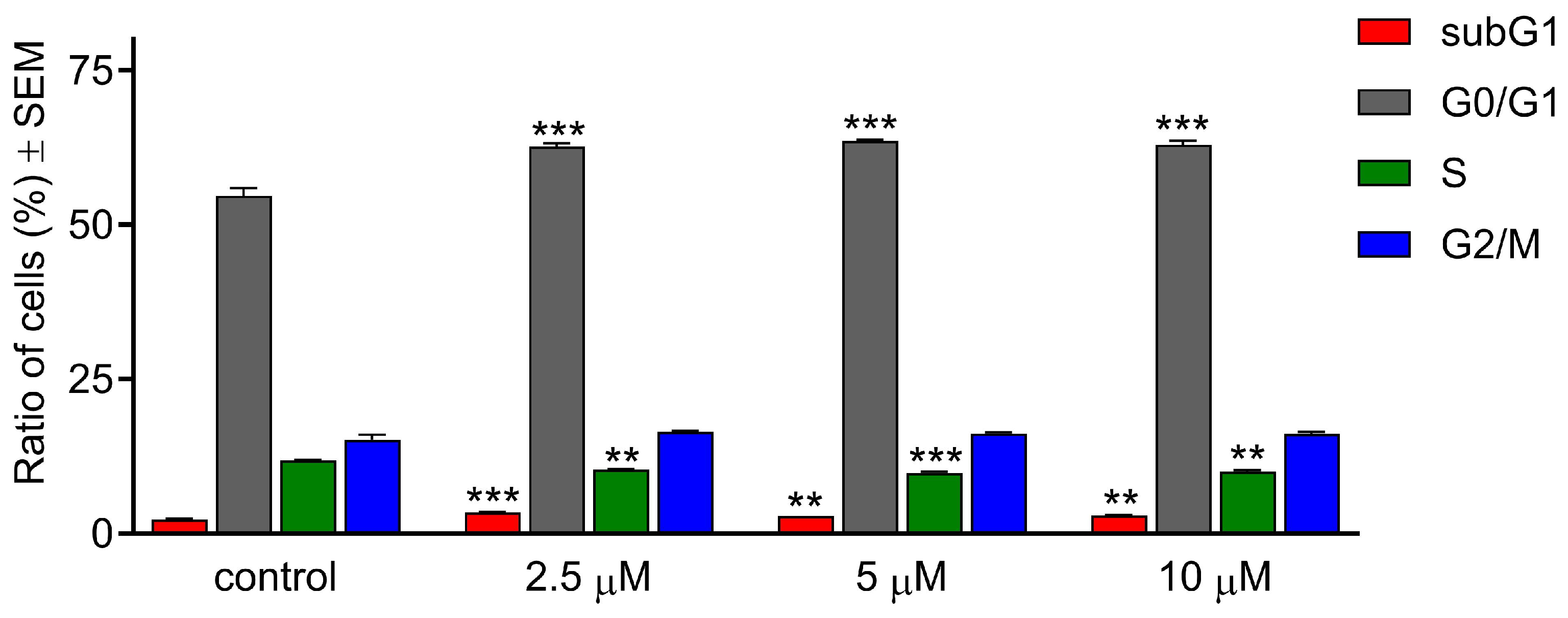
2.2. CCF Induced G0/G1 Cell Cycle Arrest

Cell cycle analysis was performed using flow cytometry to approach the mechanism of the antiproliferative action of CFF. HeLa cells were treated with selected concentrations (2.5, 5, and 10 µM) and incubated for 24 h (Figure 2).
The cell cycle distribution showed a statistically significant increase in the G0/G1 cell ratio at the expense of the S phase cell population, indicating a blockade in the G0/G1 transition. A noticeable elevation was also detected in the subG1 population in the treated groups compared to the control. However, no notable changes were observed in the population of G2/M cells.

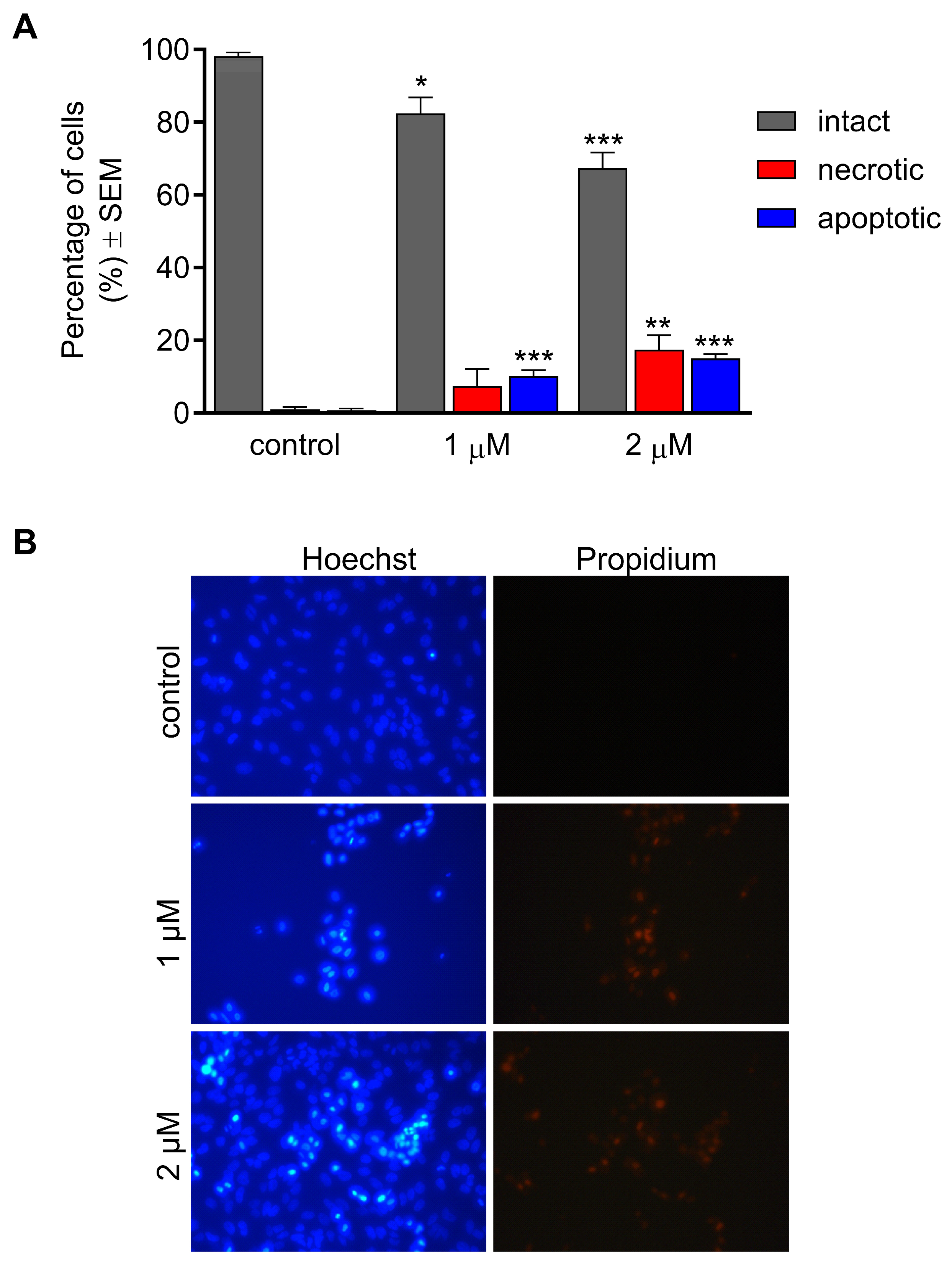
2.3. CCF Induced Morphological Changes

Hoechst 33258 and propidium iodide (HOPI) fluorescence double staining was performed to investigate the ability of CCF to induce morphological changes. HeLa cells were treated with two concentrations (1 and 2 µM) and incubated for 24 h. A significant concentration-dependent decrease in the intact cell population was observed after exposure (Figure 3).
Cell populations that emit blue fluorescence from accumulated Hoechst 33258 were significantly elevated at both applied concentrations, indicating treatment-related nuclear condensation. Red fluorescence is a consequence of propidium-positive nuclear staining, which is regarded as a marker of severe cell membrane damage. This damage can be attributed to the development of necrotic cell death. The portion of necrotic cells with damaged membranes was significantly higher after exposure to 2 μM CCF.

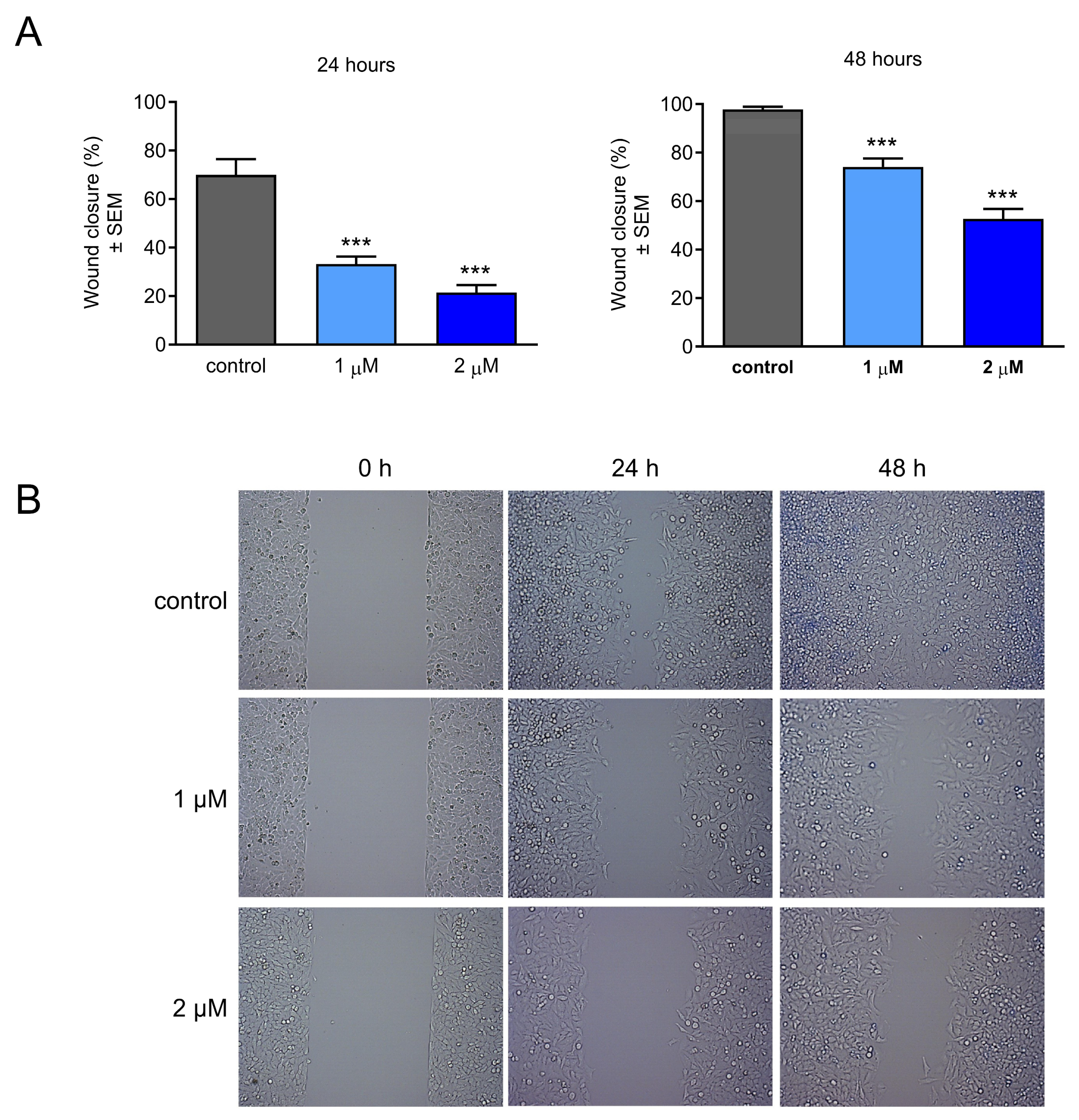
2.4. Effect of CCF on Cancer Cell Migration

Besides the direct antiproliferative action of CCF, its antimetastatic properties could also be perspectivistic. Therefore, a wound-healing assay was performed to determine the effects of the test compound on cell migration. For this purpose, HeLa cells were treated with two different concentrations (1 and 2 µM) and incubated for 24 or 48 h. The results showed significant concentration-dependent antimigratory actions after both treatment durations, even at the lower concentration (Figure 4).

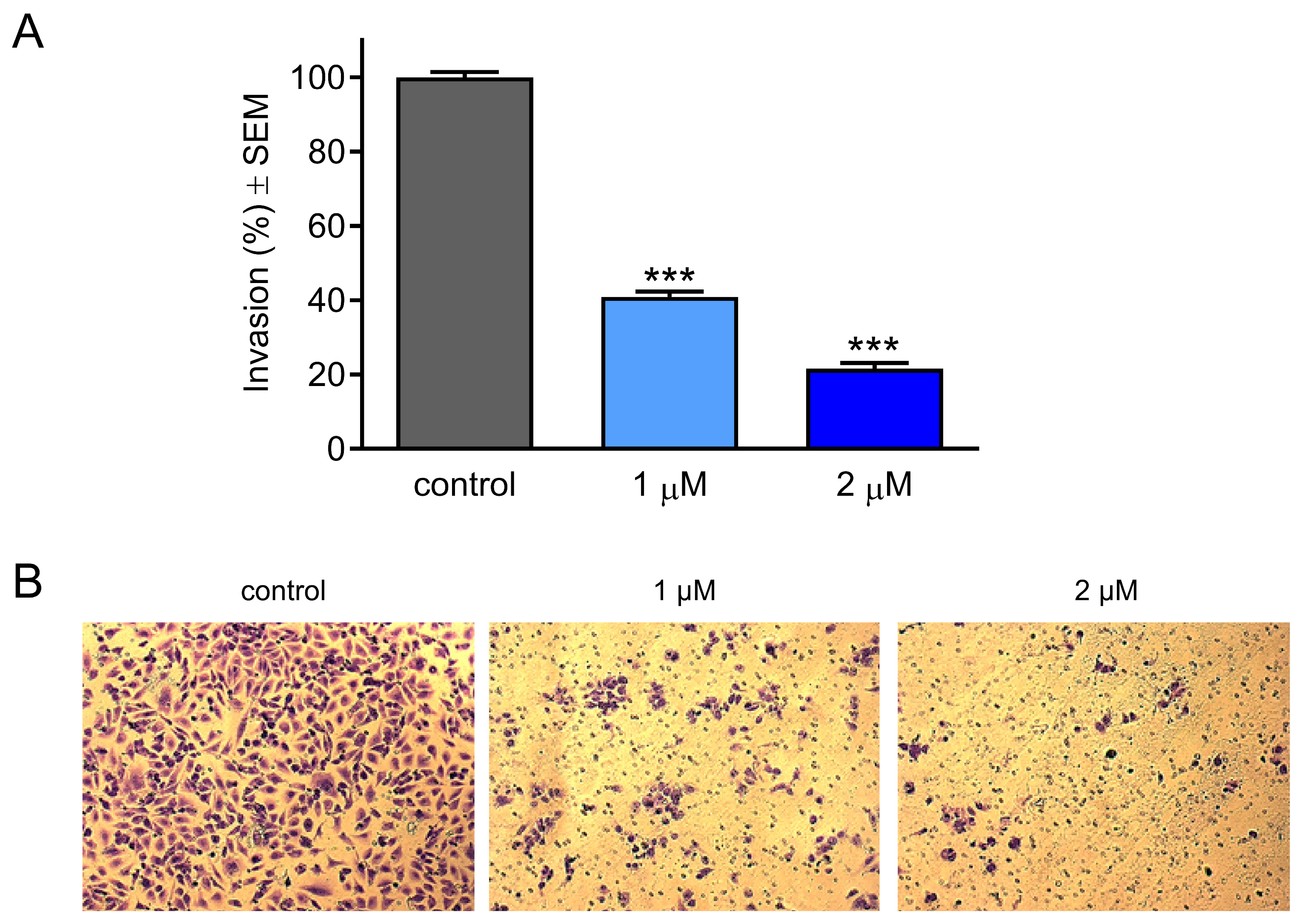
2.5. Effect of CCF on Cancer Cell Invasion

A Boyden chamber assay was performed to explore the anti-invasive capacity of the test compound. HeLa cells were treated with 1 or 2 µM of CCF and incubated for 24 h under previously described culture conditions. Based on the results obtained, CCF exerted a statistically significant concentration-dependent decrease in the percentage of invaded cells compared to the untreated control condition (Figure 5).

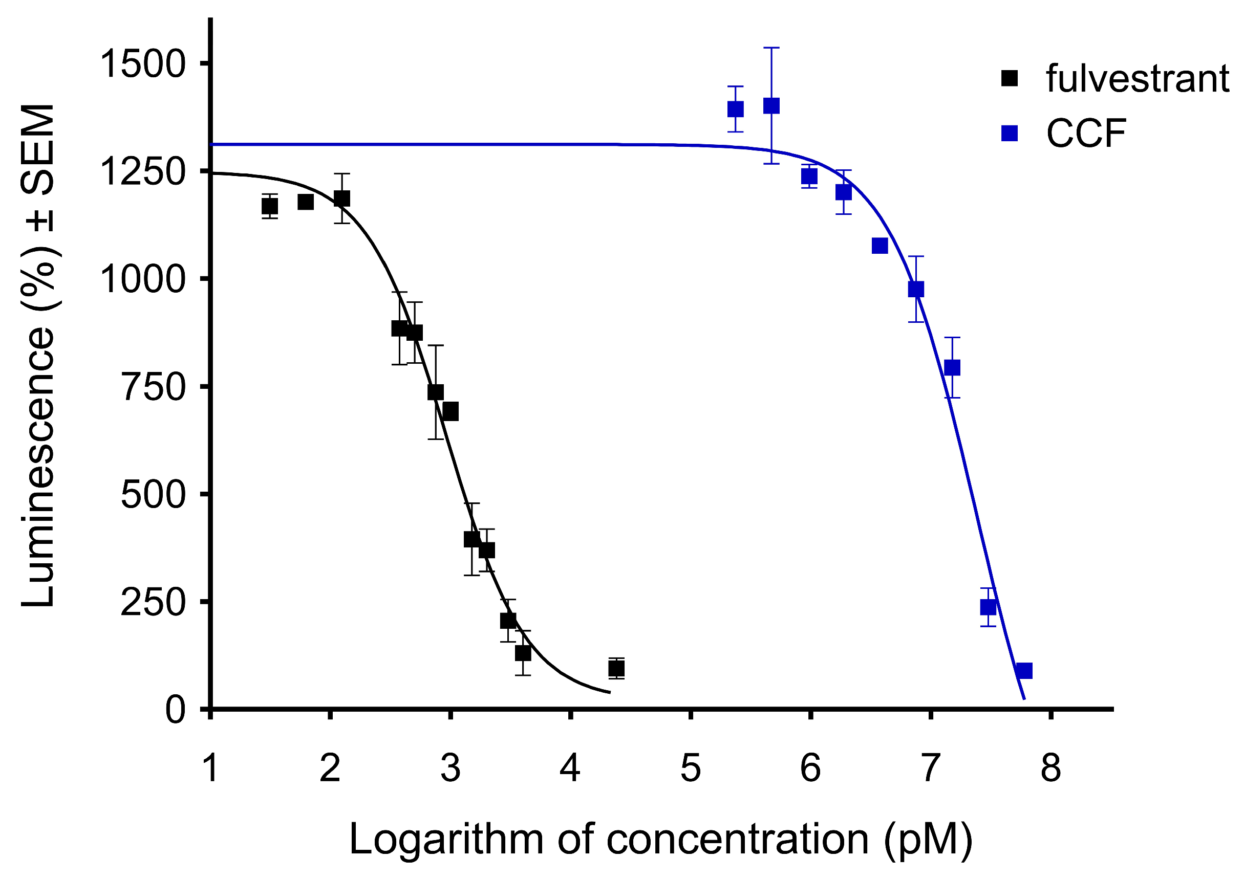
2.6. CCF Exerted an Antiestrogenic Effect

A cancer cell line transfected with an estrogen-responsive firefly luciferase reporter gene (T47D-KBluc) was applied to characterize the action of CCF on the estrogen receptor. The receptors of the transfected cells were stimulated with 40 pM 17β-estradiol, while fulvestrant, a selective estrogen receptor degrader, was used as a reference agent. According to the results, CCF can inhibit the action of 17β-estradiol, though this effect requires a substantially higher concentration than in the case of fulvestrant (Figure 6). The calculated IC50 values for the reference agent and the CCF are 0.922 nM and 23.97 μM, respectively. Therefore, CCF exhibits antiestrogenic properties at concentrations approximately ten times higher than its antiproliferative and antimetastatic activities against HeLa cells.

3. Discussion

Data from the previous literature demonstrated that naturally derived coumarins can provide a valuable base for designing more potent compounds that induce anticancer activities [19,20,21,22,23]. Moreover, coumarins are widely applied in hybridization approaches to generate novel anticancer drug candidates. They have been coupled to a variety of heterocyclic rings, as well as other natural products, including chalcones, artemisinin, and different monoterpenes [24,25]. Our present study aimed to investigate the antiproliferative and antimetastatic potentials of the coumarin–monoterpene adduct CCF and to approach its mechanism of antiproliferative action. Our group recently reported CCF as a previously unknown natural product, and its antiproliferative properties were tested against a panel of human gynecological cancer cell lines [18]. Since it proved to be a promising compound, we attempted to further investigate its antiproliferative properties by testing it against further malignant cell lines with different receptor expression and phenotype identification, including breast (T47D), cervical (C33A), and oropharyngeal (UPCI-SCC-131 and UPCI-SCC-154) cancer cells and glioblastoma (U-87). Among these newly included cell lines, the test compound moderately inhibited only papillomavirus-negative cervical cancer cells (C33A). Consistently, other cell lines did not show any notable activity. The cancer selectivity of the molecule was additionally tested by determining its cell growth-inhibitory action against non-cancerous NIH/3T3 cells. A viability assay against mouse fibroblasts cannot be considered to provide a fully reliable description of the tolerability of an anticancer drug candidate. However, the more than tenfold difference in IC50 values can be regarded as promising in terms of general toxicity.
The HPV-18-positive HeLa cell line was selected for the further evaluation of CCF based on previously confirmed outstanding growth inhibition. Since identifying the mechanism of action of anticancer agents is essential in pharmacodynamic research, we investigated the influence of CCF on cell cycle analysis using flow cytometry. Since the assayed events, including cell cycle disturbance or membrane damage, are expected within a doubling time, shorter CCF exposures were applied.
The test compound induced the arrest of the cell cycle in G0/G1 at the expense of the S cell population. These alterations in cell cycle behavior are consistent with previous studies, indicating that the G0/G1 cell cycle arrest is closely associated with the antiproliferative activity of the tested substances, including natural products [26,27,28]. This was further evidenced by measuring cell cycle-dependent proteins, which showed the down-regulation of cyclin-dependent kinase 4 (CDK4) and increased cyclin D1 [26,28]. Based on this concept, we assume that these changes in the cell cycle distribution induced by CCF may reflect its potent antiproliferative activity. Programmed cell death modalities, including apoptosis, constitute a crucial physiological process that restricts the expansion of cell proliferation, either to maintain homeostasis or to remove undesirable cells with sustained DNA damage [29]. The molecular mechanism of apoptosis is well understood and requires the extensive activation of caspase proteases through extrinsic or intrinsic pathways [30,31]. In cancer research, antineoplastic agents that are capable of induced apoptosis are currently receiving significant attention because one of the hallmarks by which cancer cells escape immune surveillance is through the evasion of apoptosis [32]. In that context and based on the noticeable elevation in the subG1 cell population in the cell cycle, we evaluated the morphological changes induced by our test compound using HOPI fluorescent double staining. The CCF-induced morphological changes were characterized by a concentration-dependent reduction in the percentage of intact cells with a significant elevation in the populations of necrotic and apoptotic cells. Based on these findings, it can be concluded that CCF has the ability to induce morphological changes represented by necrosis and apoptosis and may possess proapoptotic potential. However, identifying the precise mechanism of apoptotic induction still requires further investigation. Our data are supported by previous studies confirming the proapoptotic effects of various natural and semisynthetic coumarins that induce apoptosis in different cancer cell lines, including acute myeloid leukemia, breast and colorectal, hepatic, and pancreatic cancers [33,34,35,36].
Metastatic potency is considered one of the hallmarks of cancer. It involves disseminating cells from primary tumor sites to distant organs such as the lungs, liver, and brain [37]. This process requires a series of cellular events including migration, invasion, dissemination, and escape from immune surveillance. Due to the complexity of the tumor microenvironment, the molecular characterization of metastasis is not well understood [38,39]. Because the generation of secondary tumors involves a complex interplay between genetic and epigenetic factors, numerous investigations have aimed to develop therapeutic agents that target metastasis, thus decreasing the mortality rate and improving the efficacy of therapeutic antineoplastic agents [40,41,42]. Since migration and invasion are critical factors in the metastatic process, our objective was to investigate the antimetastatic effects of CCF using wound healing and Boyden chamber assays, respectively. On the basis of the obtained data, the migration and invasion of HeLa cells were substantially inhibited by our test compound in a concentration-dependent manner. Furthermore, these actions were elicited considerably below its antiproliferative IC50 value, indicating that they could be independent of the inhibition of cell growth. It can be concluded that CCF exhibits a remarkable antimetastatic action against HeLa cells.
Estrogens exert various physiological functions, from regulating reproductive functions to modulating bone density and cholesterol mobilization [43]. However, chronic exposure to exogenous estrogens can increase the risk of developing various types of gynecological cancer, mainly breast, ovarian, and cervical carcinomas [44,45,46]. Therefore, the estrogen-antagonistic behavior of a directly acting anticancer drug candidate can contribute to its overall pharmacological relevance. This multiple mechanism can be exploited for the treatment of hormone-dependent malignancies. Since coumarins are known for their antiestrogenic action, it was rational to investigate CCF for this activity [47]. The estrogenic behavior of CCF was characterized by testing it using the genetically modified T47D cell line expressing an estrogen-responsive element (T47D-KBluc). CCF displayed moderate antagonistic effects on the applied system, decreasing the action of 17β-estradiol. Our findings are consistent with previous reports on the estrogenic profiles of various natural and semisynthetic coumarins. It has been highlighted that, in addition to their potent cytotoxic effects against breast cancer cell lines, coumarins of different chemical backgrounds have exhibited strong estrogen receptor alpha antagonistic effects, making them suitable candidates for developing anticancer agents with multiple sites of action [47,48,49,50].

4. Materials and Methods
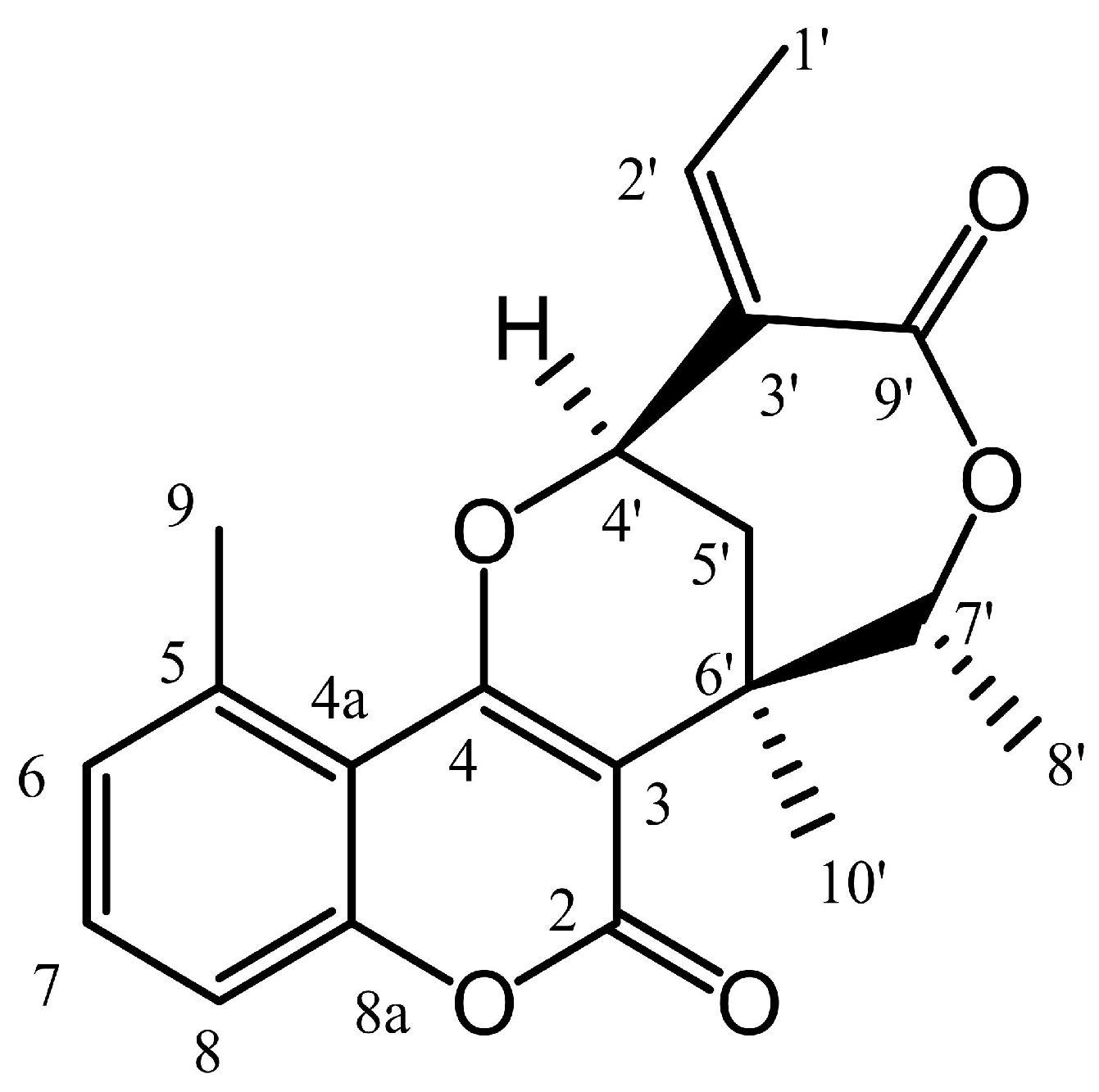
4.1. Test Compound

Centrapalus coumarin F (CCF) represents a naturally derived coumarin–monoterpene-based meroterpenoid isolated from the aerial parts of Centrapalus pauciflorus (Asteraceae). Its isolation process and the structure elucidation were previously described [18].

4.2. Tumor Cell Lines and Culture

All cell lines were purchased from the European Collection of Cell Cultures (ECCAC, Salisbury, UK) except for C33A (American Tissue Culture Collection, Manassas, VA, USA) and oropharyngeal cell lines (German Collection of Microorganisms and Cell Cultures GmbH, Braunschweig, Germany). In this study, a panel of human cancer cell lines was utilized, including breast (T47D), cervical (C33A and HeLa), and oropharyngeal (UPCI-SCC-131 and UPCI-SCC-154) cancer cells, as well as glioblastoma (U-87). The cancer selectivity of CCF was tested on NIH/3T3 non-cancerous fibroblasts isolated from mouse embryos. Culture media and supplements were purchased from Capricorn Scientific Ltd. (Ebsdorfergrund, Germany), while other chemicals used in the experiments, unless indicated otherwise, were obtained from Merck Life Science Ltd. (Budapest, Hungary). All cells (except for glioblastoma) were maintained in Eagle’s minimum essential medium (EMEM) supplemented with 10% fetal bovine serum, 1% non-essential amino acids, and 1% antibiotic–antimycotic mixture at 37 °C in humidified air containing 5% CO2. Regarding glioblastoma, cells were kept in a special medium containing the above supplements in addition to a 1% sodium-pyruvate solution and 1% L-glutamine.

4.3. Antiproliferative Assay

The cytotoxic effect of CCF against cancer cell lines was tested using the standard MTT assay. The experiments were repeated on a non-cancerous mouse fibroblast cell line (NIH/3T3) to determine tumor selectivity. Cells were seeded in 96-well plates at a density of 5000 cells/well, except for C33A, seeded at 10,000 cells/well, and incubated overnight. The cells were then treated with the desired concentrations of CCF and incubated for 72 h under standard culture conditions. Following the incubation period, 20 µL of MTT solution [3-(4,5-dimethylthiazol-2-yl)-2,5-diphenyltetrazolium bromide] was added to each well and incubated for 4 h. The supernatants were gently removed, and the precipitated blue formazan crystals were dissolved by adding 100 µL of dimethylsulfoxide (DMSO, Reanal, Budapest, Hungary) to each well, followed by shaking for 60 min. Finally, the absorbance values were recorded by a microplate reader (SPECTROstar Nano, BMG Labtech GmbH, Offenburg, Germany) at 545 nm. In the first step, the action of the compound was assayed at two concentrations (10 and 30 μM), and if a cell growth inhibition of greater than 50% was obtained at 30 μM, the assay was repeated with a set of dilutions. Sigmoidal concentration–response curves were fitted, and IC50 values were calculated using GraphPad Prism 5.01 software (GraphPad Software, San Diego, CA, USA). Untreated cells were used as a control condition. All experiments were repeated at least twice using five wells for each condition.

4.4. Cell Cycle Analysis

Cell cycle analysis was performed by flow cytometry to obtain information on the mechanism of the antiproliferative effect of CCF, as previously described [51]. Initially, HeLa cells were seeded into 24-well plates at 50,000 cells/well density and incubated overnight for proper cell attachment. The cells were then treated with 2.5, 5, or 10 µM CCF and incubated for 24 h. The harvested cells were centrifuged at 1400 rpm for 5 min at room temperature. The pellets were then resuspended and stained with 10 μg/mL propidium iodide (PI), 0.1% Triton-X, 10 μg/mL RNase A, and 0.1% sodium citrate dissolved in phosphate-buffered saline (PBS, Capricorn Scientific) for 30 min in a dark place at room temperature. The measurement of the DNA content of cells was performed using a FACSCalibur flow cytometer (BD Biosciences, Franklin Lakes, NJ, USA). Cell debris and aggregates were excluded by appropriate gating. Data were analyzed using ModFit LT 3.3.11 software (Verity Software House, Topsham, ME, USA), where untreated cells were considered a control.

4.5. Hoechst and Propidium Iodide Fluorescent Double Staining

Cell morphology and membrane integrity changes were investigated using Hoechst 33258 and propidium iodide fluorescent double staining (HOPI). HeLa cells were seeded in 12-well plates at a 50,000 cell/well density and incubated overnight. The cells were then treated with 1 and 2 µM concentrations of the test compound and incubated for 24 h. The medium was gently removed from each well, and 1 mL of fresh medium containing the formulated staining solution (5 mg/mL Hoechst and 1 mg/mL PI) was added and incubated for 90 min in a dark place at room temperature. Finally, at least ten pictures were taken per each condition using a Nikon Eclipse TS100 fluorescence microscope (Nikon Instruments Europe, Amstelveen, The Netherlands) equipped with appropriate optical blocks for Hoechst 33258 (excitation, 360/40 nm bandpass filter; emission, 460/50 nm bandpass filter; and 400 nm dichromatic mirror) and propidium iodide (excitation, 500/20 nm bandpass filter; emission, 520 nm longpass filter; and 515 nm dichromatic mirror). The images obtained using a CCD camera (QImaging MicroPublisher Color RTV5.0, Teledyne Photometrics, Tucson, AZ, USA) were analyzed with ImageJ 1.54d software (National Institute of Health, Bethesda, MD, USA), and the ratio of intact, necrotic, and apoptotic cell populations was determined for each condition.

4.6. Cell Migration Assay

The migration ability of cells after treatment was measured using a wound-healing assay. HeLa cells were seeded in a 24-well plate at a density of 50,000 cells/well and maintained in a serum-reduced medium (2% FBS), and implanted in special silicone inserts (Ibidi GmbH, Gräfelfing, Germany) as an in vitro model of wound induction. After overnight incubation, the inserts were gently removed and the cells were washed with PBS, treated with the desired concentrations (1 and 2 µM), and incubated for 24 and 48 h, respectively. The percentage of wound closure was determined for each sample after 0, 24, and 48 h after exposure using ImageJ 1.54d software (National Institute of Health, Bethesda, MD, USA).

4.7. Cell Invasion Assay

Boyden chambers (Corning, Corning, NY, USA), representing in vitro models of the extracellular environment, were used to determine the anti-invasive effects of HeLa cells upon treatment with the desired compound. While the lower parts of the chambers were supplemented with 10% FBS medium as a chemo-attractant, cells were supplemented in serum-free medium containing the desired concentrations (1 and 2 µM). They were added to the prehydrated upper part of the chambers. After 24 h of incubation, the supernatants and non-invading cells were carefully removed with a cotton swab, and the membranes were then washed with PBS twice. Subsequently, the samples were fixed in ice-cold 96% ethanol (Reanal, Budapest, Hungary) and left for around 20 min for proper fixation. Samples were then stained with 1% crystal violet dye for 30 min in a dark place at room temperature. Finally, proper images (at least five per insert) were taken using a Nikon Eclipse TS100 microscope. Invading cells were counted and expressed as a percentage.

4.8. Antiestrogenic Activity

T47D cells modified to express an estrogen-responsive luciferase (Luc) reporter gene (T47D-KBluc, American Tissue Culture Collection, Manassas, VA, USA) were used to describe the possible antiestrogenic activity of CCF [52]. Cells were maintained in phenol red-free RPMI-1640 medium completed with 10% FBS and penicillin-streptomycin antibiotics. Cells were seeded on 96-well white flat-bottom plates (Greiner Bio-One, Mosonmagyaróvár, Hungary) at 50,000/well density and attached for 48 h. Then, a medium containing 10% charcoal dextran-treated FBS was applied for another 72 h. Finally, cells were treated with CCF in the concentration range of 0.47–60 µM in the presence of 40 pM 17β-estradiol, and fulvestrant was applied as a reference compound. After incubation for 24 h, the medium was removed, and 30 µL of One-Glo firefly luciferase reagent (Promega, Madison, WI, USA) was added to each well. The luminescence signal was determined (FLUOstar Optima, BMG Labtech, Ortenberg, Germany) after 3 min of incubation at room temperature according to the manufacturer’s protocol. Sigmoidal curves representing luciferase enzyme activity were fitted using GraphPad Prism version 5.01 (GraphPad, San Diego, CA, USA).

4.9. Statistical Analysis

Data from all cell-based experiments were statistically analyzed using variance analysis followed by the Dunnett post-test. Data were expressed as mean values ± standard error of the mean (SEM) using GraphPad Prism version 5.01 (GraphPad, Sandiego, CA, USA). The calculated p-values show statistical differences between study groups: *, **, and *** indicate p < 0.05, p < 0.01, and p < 0.001, respectively, compared to untreated control samples.

5. Conclusions

In conclusion, recently isolated from Centrapalus pauciflorus CCF demonstrated outstanding antiproliferative activity against the HVP-18-positive human cervical cancer cell line (HeLa) and considerable tumor selectivity against non-cancerous cells. It elicited G0/G1 cell cycle arrest, and the hypodiploid cell population cumulated. The apoptosis-inducing property of CCF was confirmed by identifying the nuclear condensation morphological apoptotic marker using fluorescent staining. It also displayed potent antiestrogen behavior, as evidenced by the luciferase reporter assay. Based on these findings, Centrapalus coumarin F may be regarded as a promising candidate and a starting structure for designing innovative anticancer compounds.

Author Contributions

Conceptualization, D.R., J.H. and I.Z.; methodology, G.J.S.; formal analysis, G.J.S.; investigation, H.A., S.R., M.B.S., P.G. and E.S.; writing—original draft preparation, H.A.; writing—review and editing, J.H. and I.Z.; supervision, I.Z. All authors have read and agreed to the published version of the manuscript.

Funding

This research was funded by the Ministry of Culture and Innovation of Hungary from the National Research, Development, and Innovation Fund, grant number TKP2021-EGA-32, and the Hungarian National Science and Research Fund (OTKA), grant numbers 143690 and 135845. The APC was funded by the Open Access Fund of the University of Szeged, grant number 7704.

Institutional Review Board Statement

Not applicable.

Informed Consent Statement

Not applicable.

Data Availability Statement

The data presented in this study are available upon request from the corresponding author (I.Z.).

Conflicts of Interest

The authors declare no conflicts of interest.

Abbreviations

The following abbreviations are used in this manuscript:
CCFCentrapalus coumarin F
DMSODimethylsulfoxide
FBSFetal bovine serum
HOHoechst 33258
HPVHuman papillomavirus
MTT3-(4,5-dimethylthiazol-2-yl)-2,5-diphenyltetrazolium bromide
PBSPhosphate-buffered saline
PIPropidium iodide
SEMStandard error of the mean

References

  1. Roy, P.S.; Saikia, B.J. Cancer and cure: A critical analysis. Indian J. Cancer 2016, 53, 441–442. [Google Scholar] [CrossRef] [PubMed]
  2. Miranda-Filho, A.; Bray, F.; Charvat, H.; Rajaraman, S.; Soerjomataram, I. The world cancer patient population (WCPP): An updated standard for international comparisons of population-based survival. Cancer Epidemiol. 2020, 69, 101802. [Google Scholar] [CrossRef] [PubMed]
  3. Knezevic, C.E.; Clarke, W. Cancer chemotherapy: The case for therapeutic drug monitoring. Ther. Drug Monit. 2020, 42, 6–19. [Google Scholar] [CrossRef] [PubMed]
  4. Spencer, S.H.; Menard, S.M.; Labedz, M.Z.; Krueger, C.D.; Sarna, K.V. Enteral tube administration of oral chemotherapy drugs. J. Oncol. Pharm. Pract. 2020, 26, 703–717. [Google Scholar] [CrossRef]
  5. Zraik, I.M.; Hess-Busch, Y. Management von Nebenwirkungen der Chemotherapie und deren Langzeitfolgen. Urologe 2021, 60, 862–871. [Google Scholar] [CrossRef]
  6. Chau, C.H.; Steeg, P.S.; Figg, W.D. Antibody-drug conjugates for cancer. Lancet 2019, 394, 793–804. [Google Scholar] [CrossRef]
  7. Ciarimboli, G. Anticancer platinum drugs update. Biomolecules 2021, 11, 1637. [Google Scholar] [CrossRef]
  8. Efferth, T.; Saeed, M.E.M.; Kadioglu, O.; Seo, E.J.; Shirooie, S.; Mbaveng, A.T.; Nabavi, S.M.; Kuete, V. Collateral sensitivity of natural products in drug-resistant cancer cells. Biotechnol. Adv. 2020, 38, 107342. [Google Scholar] [CrossRef]
  9. Nazir, M.; Saleem, M.; Tousif, M.I.; Anwar, M.A.; Surup, F.; Ali, I.; Wang, D.; Mamadalieva, N.Z.; Alshammari, E.; Ashour, M.L.; et al. Meroterpenoids: A comprehensive update insight on structural diversity and biology. Biomolecules 2021, 11, 957. [Google Scholar] [CrossRef]
  10. Ahmed, M.; Jakupovic, J.; Bohlmann, F.; Mungai, M.G. A 5-methylcoumarin and glaucolides from Bothriocline-amplifolia. Phytochemistry 1991, 30, 2807–2808. [Google Scholar] [CrossRef]
  11. Mahmoud, A.A.; Ahmed, A.A.; Iinuma, M.; Tanaka, T. Further monoterpene 5-methylcoumarins and an acetophenone derivative from. Phytochemistry 1998, 48, 543–546. [Google Scholar] [CrossRef]
  12. Thakur, A.; Singla, R.; Jaitak, V. Coumarins as anticancer agents: A review on synthetic strategies, mechanism of action and SAR studies. Eur. J. Med. Chem. 2015, 101, 476–495. [Google Scholar] [CrossRef] [PubMed]
  13. Bhattarai, N.; Kumbhar, A.A.; Pokharel, Y.R.; Yadav, P.N. Anticancer potential of coumarin and its derivatives. Mini Rev. Med. Chem. 2021, 21, 2996–3029. [Google Scholar] [CrossRef] [PubMed]
  14. Lacy, A.; O’Kennedy, R. Studies on coumarins and coumarin-related compounds to determine their therapeutic role in the treatment of cancer. Curr. Pharm. Des. 2004, 10, 3797–3811. [Google Scholar] [CrossRef]
  15. Zhang, L.; Xu, Z. Coumarin-containing hybrids and their anticancer activities. Eur. J. Med. Chem. 2019, 181, 111587. [Google Scholar] [CrossRef]
  16. Singh, H.; Singh, J.V.; Bhagat, K.; Gulati, H.K.; Sanduja, M.; Kumar, N.; Kinarivala, N.; Sharma, S. Rational approaches, design strategies, structure activity relationship and mechanistic insights for therapeutic coumarin hybrids. Bioorg. Med. Chem. 2019, 27, 3477–3510. [Google Scholar] [CrossRef]
  17. Tian, Q.; Wang, L.; Sun, X.; Zeng, F.; Pan, Q.; Xue, M. Scopoletin exerts anticancer effects on human cervical cancer cell lines by triggering apoptosis, cell cycle arrest, inhibition of cell invasion and PI3K/AKT signalling pathway. J. BUON 2019, 24, 997–1002. [Google Scholar]
  18. Saidu, M.B.; Krstic, G.; Todorovic, N.; Berkecz, R.; Ali, H.; Zupko, I.; Hohmann, J.; Redei, D. Monoterpenoid 5-methylcoumarins from Centrapalus pauciflorus with antiproliferative activity. Arab. J. Chem. 2023, 16, 104777. [Google Scholar] [CrossRef]
  19. Huang, L.; Feng, Z.L.; Wang, Y.T.; Lin, L.G. Anticancer carbazole alkaloids and coumarins from Clausena plants: A review. Chin. J. Nat. Med. 2017, 15, 881–888. [Google Scholar] [CrossRef]
  20. Kostova, I. Studying plant-derived coumarins for their pharmacological and therapeutic properties as potential anticancer drugs. Expert Opin. Drug Discov. 2007, 2, 1605–1618. [Google Scholar] [CrossRef]
  21. Ma, Q.; Jiang, J.G.; Yuan, X.; Qiu, K.; Zhu, W. Comparative antitumor and anti-inflammatory effects of flavonoids, saponins, polysaccharides, essential oil, coumarin and alkaloids from Cirsium japonicum DC. Food Chem. Toxicol. 2019, 125, 422–429. [Google Scholar] [CrossRef] [PubMed]
  22. Song, X.F.; Fan, J.; Liu, L.; Liu, X.F.; Gao, F. Coumarin derivatives with anticancer activities: An update. Arch. Pharm. 2020, 353, e2000025. [Google Scholar] [CrossRef] [PubMed]
  23. Moi, D.; Carradori, S.; Gallorini, M.; Mencarelli, N.; Deplano, A.; Angeli, A.; Vittorio, S.; Supuran, C.T.; Onnis, V. Investigation on human carbonic anhydrase IX and XII inhibitory activity and A549 antiproliferative activity of a new class of coumarinamides. Pharmaceuticals 2025, 18, 372. [Google Scholar] [CrossRef] [PubMed]
  24. Zengin Kurt, B.; Celebi, G.; Ozturk Civelek, D.; Angeli, A.; Akdemir, A.; Sonmez, F.; Supuran, C.T. Tail-approach-based design and synthesis of coumarin-monoterpenes as carbonic anhydrase inhibitors and anticancer agents. ACS Omega 2023, 8, 5787–5807. [Google Scholar] [CrossRef]
  25. Koley, M.; Han, J.; Soloshonok, V.A.; Mojumder, S.; Javahershenas, R.; Makarem, A. Latest developments in coumarin-based anticancer agents: Mechanism of action and structure–activity relationship studies. RSC Med. Chem. 2024, 15, 10–54. [Google Scholar] [CrossRef]
  26. Lepley, D.M.; Pelling, J.C. Induction of p21/WAF1 and G1 cell-cycle arrest by the chemopreventive agent apigenin. Mol. Carcinog. 1997, 19, 74–82. [Google Scholar] [CrossRef]
  27. Liu, Q.; Cao, Y.; Zhou, P.; Gui, S.; Wu, X.; Xia, Y.; Tu, J. Panduratin A inhibits cell proliferation by inducing G0/G1 phase cell cycle arrest and induces apoptosis in breast cancer cells. Biomol. Ther. 2018, 26, 328–334. [Google Scholar] [CrossRef]
  28. Noori, S.; Hassan, Z.M. Tehranolide inhibits proliferation of MCF-7 human breast cancer cells by inducing G0/G1 arrest and apoptosis. Free Radic. Biol. Med. 2012, 52, 1987–1999. [Google Scholar] [CrossRef]
  29. Morana, O.; Wood, W.; Gregory, C.D. The apoptosis paradox in cancer. Int. J. Mol. Sci. 2022, 23, 1328. [Google Scholar] [CrossRef]
  30. Elmore, S. Apoptosis: A review of programmed cell death. Toxicol. Pathol. 2007, 35, 495–516. [Google Scholar] [CrossRef]
  31. Pistritto, G.; Trisciuoglio, D.; Ceci, C.; Garufi, A.; D’Orazi, G. Apoptosis as anticancer mechanism: Function and dysfunction of its modulators and targeted therapeutic strategies. Aging 2016, 8, 603–619. [Google Scholar] [CrossRef] [PubMed]
  32. Goldar, S.; Khaniani, M.S.; Derakhshan, S.M.; Baradaran, B. Molecular mechanisms of apoptosis and roles in cancer development and treatment. Asian Pac. J. Cancer Prev. 2015, 16, 2129–2144. [Google Scholar] [CrossRef] [PubMed]
  33. Huang, Q.; Wang, L.; Ran, Q.; Wang, J.; Wang, C.; He, H.; Li, L.; Qi, H. Notopterol-induced apoptosis and differentiation in human acute myeloid leukemia HL-60 cells. Drug Des. Devel. Ther. 2019, 13, 1927–1940. [Google Scholar] [CrossRef] [PubMed]
  34. Luo, G.; Muyaba, M.; Lyu, W.; Tang, Z.; Zhao, R.; Xu, Q.; You, Q.; Xiang, H. Design, synthesis and biological evaluation of novel 3-substituted 4-anilino-coumarin derivatives as antitumor agents. Bioorg. Med. Chem. Lett. 2017, 27, 867–874. [Google Scholar] [CrossRef]
  35. Yan, G.; Luo, J.; Han, X.; Zhang, W.; Pu, C.; Zhou, M.; Zhong, X.; Hou, X.; Hou, M.Z.; Li, R. Design, synthesis and biological evaluation of 4, 6-coumarin derivatives as anti- cancer and apoptosis-inducing agents. Anticancer Agents Med. Chem. 2021, 21, 2351–2367. [Google Scholar] [CrossRef]
  36. Zhu, H.; Ying, S.; Zhou, B.; Hu, X.; Liang, X.; Li, W.; Wang, D.; Jin, H.; Pan, Y. Design, synthesis, and evaluation of novel coumarin-dithiocarbamate derivatives (IDs) as anti-colorectal cancer agents. J. Enzyme Inhib. Med. Chem. 2021, 36, 593–604. [Google Scholar] [CrossRef]
  37. Suhail, Y.; Cain, M.P.; Vanaja, K.; Kurywchak, P.A.; Levchenko, A.; Kalluri, R.; Kshitiz. Systems biology of cancer metastasis. Cell Syst. 2019, 9, 109–127. [Google Scholar] [CrossRef]
  38. Clarke, R.; Kraikivski, P.; Jones, B.C.; Sevigny, C.M.; Sengupta, S.; Wang, Y. A systems biology approach to discovering pathway signaling dysregulation in metastasis. Cancer Metastasis Rev. 2020, 39, 903–918. [Google Scholar] [CrossRef]
  39. Seyfried, T.N.; Huysentruyt, L.C. On the origin of cancer metastasis. Crit. Rev. Oncog. 2013, 18, 43–73. [Google Scholar] [CrossRef]
  40. Ganesh, K.; Massague, J. Targeting metastatic cancer. Nat. Med. 2021, 27, 34–44. [Google Scholar] [CrossRef]
  41. Scheele, C.; Maynard, C.; van Rheenen, J. Intravital insights into heterogeneity, metastasis, and therapy responses. Trends Cancer. 2016, 2, 205–216. [Google Scholar] [CrossRef] [PubMed]
  42. Stoletov, K.; Beatty, P.H.; Lewis, J.D. Novel therapeutic targets for cancer metastasis. Expert Rev. Anticancer Ther. 2020, 20, 97–109. [Google Scholar] [CrossRef] [PubMed]
  43. Liang, J.; Shang, Y. Estrogen and cancer. Annu. Rev. Physiol. 2013, 75, 225–240. [Google Scholar] [CrossRef] [PubMed]
  44. Chen, G.G.; Zeng, Q.; Tse, G.M. Estrogen and its receptors in cancer. Med. Res. Rev. 2008, 28, 954–974. [Google Scholar] [CrossRef]
  45. Germain, D. Estrogen carcinogenesis in breast cancer. Endocrinol. Metab. Clin. N. Am. 2011, 40, 473–484. [Google Scholar] [CrossRef]
  46. Zhao, C.; Dahlman-Wright, K.; Gustafsson, J.A. Estrogen receptor b: An overview and update. Nucl. Recept. Signal. 2008, 6, e003. [Google Scholar] [CrossRef]
  47. Mihovic, N.; Tomasevic, N.; Matic, S.; Mitrovic, M.M.; Kostic, D.A.; Sabatino, M.; Antonini, L.; Ragno, R.; Mladenovic, M. Human estrogen receptor α antagonists. Part 1: 3-D QSAR-driven rational design of innovative coumarin-related antiestrogens as breast cancer suppressants through structure-based and ligand-based etudies. J. Chem. Inf. Model. 2021, 61, 5028–5053. [Google Scholar] [CrossRef]
  48. Bai, C.; Ren, S.; Wu, S.; Zhu, M.; Luo, G.; Xiang, H. Design and synthesis of novel benzothiophene analogs as selective estrogen receptor covalent antagonists against breast cancer. Eur. J. Med. Chem. 2021, 221, 113543. [Google Scholar] [CrossRef]
  49. Kurtanovic, N.; Tomasevic, N.; Matic, S.; Mitrovic, M.M.; Kostic, D.A.; Sabatino, M.; Antonini, L.; Ragno, R.; Mladenovic, M. Human estrogen receptor alpha antagonists, part 2: Synthesis driven by rational design, in vitro antiproliferative, and in vivo anticancer evaluation of innovative coumarin-related antiestrogens as breast cancer suppressants. Eur. J. Med. Chem. 2022, 227, 113869. [Google Scholar] [CrossRef]
  50. Selvaraj, J.; John, J.B.A.; Joghee, N.M.; Antony, J.; Wadhwani, A.; Natarajan, J. Coumarin-fatty acid conjugates as potential ERα/AKT-1 antagonists for ER positive breast cancer. Anticancer Agents Med. Chem. 2020, 20, 437–449. [Google Scholar] [CrossRef]
  51. Kotogány, E.; Balog, J.A.; Nagy, L.I.; Alföldi, R.; Bertagnolo, V.; Brugnoli, F.; Demjén, A.; Kovács, A.K.; Batár, P.; Mezei, G.; et al. Imidazo[1,2-b]pyrazole-7-carboxamide derivative induces differentiation-coupled apoptosis of immature myeloid cells such as acute myeloid leukemia and myeloid-derived suppressor cells. Int. J. Mol. Sci. 2020, 21, 5135. [Google Scholar] [CrossRef]
  52. Wilson, V.S.; Bobseine, K.; Gray, L.E., Jr. Development and characterization of a cell line that stably expresses an estrogen-responsive luciferase reporter for the detection of estrogen receptor agonist and antagonists. Toxicol. Sci. 2004, 81, 69–77. [Google Scholar] [CrossRef]
Figure 1. Chemical structure of centrapalus coumarin F (CCF).
Ijms 26 04489 g001
Figure 2. Cell cycle distribution of HeLa cells after 24 h of treatment with CCF. Results from three independent experiments carried out in triplicate. ** and *** indicate significance at p < 0.01 and p < 0.001, respectively.
Ijms 26 04489 g002
Figure 3. Proportion of intact, apoptotic, and necrotic HeLa cells after 24 h of CCF treatment (A) and representative images, magnification: 40× (B). Data from two independent experiments performed in triplicate. *, **, and *** indicate significance at p < 0.05, p < 0.01, and p < 0.001, respectively.
Ijms 26 04489 g003
Figure 4. Antimigratory action of CCF on HeLa cells after 24 and 48 h of treatment (A) and representative pictures (B). Results from three independent experiments carried out in triplicate. *** indicates significance at p < 0.001.
Ijms 26 04489 g004
Figure 5. Effect of CCF on the invasive capacity of HeLa cells after 24 h of treatment (A) and representative images, magnification: 10× (B). Results from three independent experiments carried out in triplicate. *** indicates significance at p < 0.001.
Ijms 26 04489 g005
Figure 6. The antiestrogenic effect of CCF determined by employing a luciferase reporter gene assay. Results from two independent experiments carried out in triplicate.
Ijms 26 04489 g006
Table 1. The antiproliferative properties of CCF against the applied cell lines.
Cell LineGrowth Inhibition (% ± SEM)Calculated IC50 Value (μM)Cisplatin IC50 (μM)
10 μM30 μM
HeLa 169.48 ± 1.3094.60 ± 0.552.2812.43
SiHa 120.83 ± 1.6840.84 ± 1.49n.d. 24.29
C33A37.32 ± 1.8965.91 ± 0.6715.69
MCF7 157.26 ± 1.0569.71 ± 1.016.595.78
MDA-MB-231 1<20 326.23 ± 1.38n.d.10.17
T47D35.65 ± 1.5353.06 ± 0.8825.55
A2780 137.22 ± 3.0267.96 ± 2.0115.411.30
UPCI-SCC-13124.50 ± 3.2138.01 ± 2.87n.d.
UPCI-SCC-15432.61 ± 2.2052.21 ± 1.1436.37
U-8722.25 ± 3.0262.60 ± 1.9621.54
NIH/3T328.59 ± 2.1851.38 ± 1.4628.52
1 Data from reference [18]. Cisplatin was included as a reference compound. 2 IC50 values are not determined when less than 50% growth inhibition was obtained at 30 μM. 3 An inhibition value less than 20% is considered negligible and is not given numerically.
Disclaimer/Publisher’s Note: The statements, opinions and data contained in all publications are solely those of the individual author(s) and contributor(s) and not of MDPI and/or the editor(s). MDPI and/or the editor(s) disclaim responsibility for any injury to people or property resulting from any ideas, methods, instructions or products referred to in the content.

