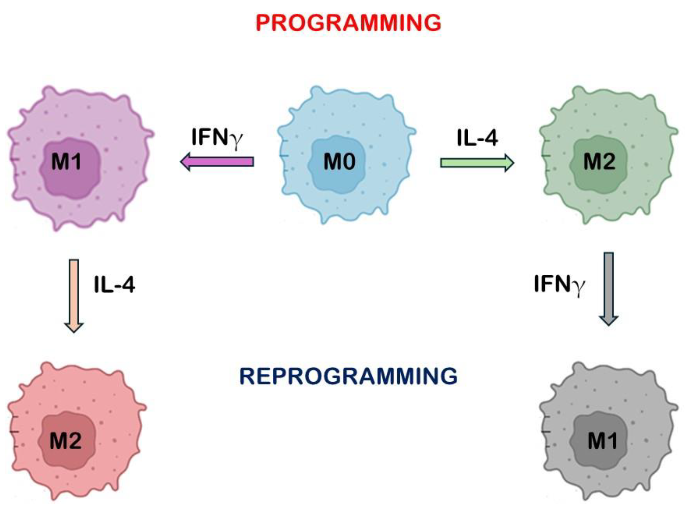
Macrophage Polarization: Learning to Manage It 3.0
Funding
Conflicts of Interest
References
- Ross, E.A.; Devitt, A.; Johnson, J.R. Macrophages: The good, the bad, and the gluttony. Front. Immunol. 2021, 12, 708186. [Google Scholar] [CrossRef] [PubMed]
- Nasir, I.; McGuinness, C.; Poh, A.R.; Ernst, M.; Darcy, P.K.; Britt, K.L. Tumor Macrophage Functional Heterogeneity Can Inform the Development of Novel Cancer Therapies. Trends Immunol. 2023, 44, 971–985. [Google Scholar] [CrossRef] [PubMed]
- Tcyganov, E.; Mastio, J.; Chen, E.; Gabrilovich, D.I. Plasticity of Myeloid-Derived Suppressor Cells in Cancer. Curr. Opin. Immunol. 2018, 51, 76–82. [Google Scholar] [CrossRef]
- Kwak, T.; Wang, F.; Deng, H.; Condamine, T.; Kumar, V.; Perego, M.; Kossenkov, A.; Montaner, L.J.; Xu, X.; Xu, W.; et al. Distinct Populations of Immune-Suppressive Macrophages Differentiate from Monocytic Myeloid-Derived Suppressor Cells in Cancer. Cell Rep. 2020, 33, 108571. [Google Scholar] [CrossRef] [PubMed]
- Lin, Y.; Xu, J.; Lan, H. Tumor-Associated Macrophages in Tumor Metastasis: Biological Roles and Clinical Therapeutic Applications. J. Hematol. Oncol. 2019, 12, 76. [Google Scholar] [CrossRef] [PubMed]
- Pittet, M.J.; Michielin, O.; Migliorini, D. Clinical Relevance of Tumor-Associated Macrophages. Nat. Rev. Clin. Oncol. 2022, 19, 402–421. [Google Scholar] [CrossRef]
- Duluc, D.; Delneste, Y.; Tan, F.; Moles, M.-P.; Grimaud, L.; Lenoir, J.; Preisser, L.; Anegon, I.; Catala, L.; Ifrah, N.; et al. Tumor associated leukemia inhibitory factor and IL-6 skew monocyte differentiation into tumor-associated macrophage-like cells. Blood 2007, 110, 4319–4330. [Google Scholar] [CrossRef] [PubMed]
- Liu, C.; Wang, Y.; Li, L.; He, D.; Chi, J.; Li, Q.; Wu, Y.; Zhao, Y.; Zhang, S.; Wang, L.; et al. Engineered Extracellular Vesicles and Their Mimetics for Cancer Immunotherapy. J. Control. Release 2022, 349, 679–698. [Google Scholar] [CrossRef] [PubMed]
- Sturniolo, I.; Váróczy, C.; Regdon, Z.; Mázló, A.; Muzsai, S.; Bácsi, A.; Intili, G.; Hegedus, C.; Boothby, M.R.; Holechek, J.; et al. PARP14 Contributes to the Development of the Tumor-Associated Macrophage Phenotype. Int. J. Mol. Sci. 2024, 25, 3601. [Google Scholar] [CrossRef] [PubMed]
- Cutter, S.; Wright, M.D.; Reynolds, N.P.; Binge, K.J. Towards using 3D cellular cultures to model the activation and diverse functions of macrophages. Biochem. Soc. Trans. 2023, 51, 387–401. [Google Scholar] [CrossRef]
- Korotkaja, K.; Jansons, J.; Spunde, K.; Rudevica, Z.; Zajakina, A. Establishment and Characterization of Free-Floating 3D Macrophage Programming Model in the Presence of Cancer Cell Spheroids. Int. J. Mol. Sci. 2023, 24, 10763. [Google Scholar] [CrossRef]
- Sazinsky, S.; Zafari, M.; Klebanov, B.; Ritter, J.; Nguyen, P.A.; Phennicie, R.T.; Wahle, J.; Kauffman, K.J.; Razlog, M.; Manfra, D.; et al. Antibodies Targeting Human or Mouse VSIG4 Repolarize Tumor-Associated Macrophages Providing the Potential of Potent and Specific Clinical Anti-Tumor Response Induced across Multiple Cancer Types. Int. J. Mol. Sci. 2024, 25, 6160. [Google Scholar] [CrossRef]
- Jung, K.; Jeon, Y.K.; Jeong, D.H.; Byun, J.M.; Bogen, B.; Choi, I. VSIG4-expressing tumor-associated macrophages impair anti-tumor immunity. Biochem. Biophys. Res. Commun. 2022, 628, 18–24. [Google Scholar] [CrossRef] [PubMed]
- Liao, Y.; Guo, S.; Chen, Y.; Cao, D.; Xu, H.; Yang, C.; Fei, L.; Ni, B.; Ruan, Z. VSIG4 expression on macrophages facilitates lung cancer development. Lab. Investig. 2014, 94, 706–715. [Google Scholar] [CrossRef] [PubMed]
- Roh, J.; Jeon, Y.; Lee, A.-N.; Lee, S.M.; Kim, Y.; Sung, C.O.; Park, C.-J.; Hong, J.Y.; Yoon, D.H.; Suh, C.; et al. The immune checkpoint molecule V-set Ig domain-containing 4 is an independent prognostic factor for multiple myeloma. Oncotarget 2017, 8, 58122–58132. [Google Scholar] [CrossRef] [PubMed]
- Xu, T.; Jiang, Y.; Yan, Y.; Wang, H.; Lu, C.; Xu, H.; Li, W.; Fu, D.; Lu, Y.; Chen, J. VSIG4 is highly expressed and correlated with poor prognosis of high-grade glioma patients. Am. J. Transl. Res. 2015, 7, 1172–1180. [Google Scholar] [PubMed]
- Lampiasi, N. New Strategies for Macrophage Re-Education in Cancer: An Update. Int. J. Mol. Sci. 2024, 25, 3414. [Google Scholar] [CrossRef] [PubMed]
- Liu, Y.; Liang, S.; Jiang, D.; Gao, T.; Fang, Y.; Fu, S.; Guan, L.; Zhang, Z.; Mu, W.; Chu, Q.; et al. Manipulation of TAMs functions to facilitate the immune therapy effects of immune checkpoint antibodies. J. Control. Release 2021, 336, 621–634. [Google Scholar] [CrossRef] [PubMed]
- Li, Z.; Liu, H.; Xie, Q.; Yin, G. Macrophage involvement in idiopathic inflammatory myopathy: Pathogenic mechanisms and therapeutic prospects. J. Inflamm. 2024, 21, 48. [Google Scholar] [CrossRef] [PubMed]
- Yang, X.; Li, J.; Xu, C.; Zhang, G.; Che, X.; Yang, J. Potential mechanisms of rheumatoid arthritis therapy: Focus on macrophage. polarization. Int. Immunopharmacol. 2024, 142 Pt A, 113058. [Google Scholar] [CrossRef]
- Ma, Y.; Sun, Y.; Guo, H.; Yang, R. Tumor-associated macrophages in bladder cancer: Roles and targeted therapeutic strategies. Int. Immunopharmacol. 2024, 15, 1418131. [Google Scholar] [CrossRef] [PubMed]
- Sedighzadeh, S.S.; Khoshbin, A.P.; Razi, S.; Keshavarz-Fathi, M.; Rezaei, N. A narrative review of tumor-associated macrophages in lung cancer: Regulation of macrophage polarization and therapeutic implications. Transl. Lung Cancer Res. 2021, 10, 1889–1916. [Google Scholar] [CrossRef]
- Jiang, S.; Yang, M.; Li, M. Emerging Roles of Lysophosphatidic Acid in Macrophages and Inflammatory Diseases. Int. J. Mol. Sci. 2023, 24, 12524. [Google Scholar] [CrossRef] [PubMed]
- Georas, S.N.; Berdyshev, E.; Hubbard, W.; Gorshkova, I.A.; Usatyuk, P.V.; Saatian, B.; Myers, A.C.; Williams, M.A.; Xiao, H.Q.; Liu, M.; et al. Lysophosphatidic acid is detectable in human bronchoalveolar fluids at baseline and increased after segmental allergen challenge. Clin. Exp. Allergy 2007, 37, 311–322. [Google Scholar] [CrossRef] [PubMed]
- Aoki, J.; Inoue, A.; Okudaira, S. Two pathways for lysophosphatidic acid production. Biochim. Biophys. Acta 2008, 1781, 513–518. [Google Scholar] [CrossRef] [PubMed]
- Benesch, M.G.K.; MacIntyre, I.T.K.; McMullen, T.P.W.; Brindley, D.N. Coming of Age for Autotaxin and Lysophosphatidate Signaling: Clinical Applications for Preventing, Detecting and Targeting Tumor-Promoting Inflammation. Cancers 2018, 10, 73. [Google Scholar] [CrossRef] [PubMed]
- Shi, X.; Chen, Y.; Shi, M.; Gao, F.; Huang, L.; Wang, W. The novel molecular mechanism of pulmonary fibrosis: Insight into lipid metabolism from reanalysis of single-cell RNA-seq databases. Lipids Health Dis. 2024, 23, 98. [Google Scholar] [CrossRef] [PubMed]
- Volkmann, E.R.; Denton, C.P.; Kolb, M.; Wijsenbeek-Lourens, M.S.; Emson, C.; Hudson, K.; Amatucci, A.J.; Distler, O.; Allanore, Y.; Khanna, D. Lysophosphatidic acid receptor 1 inhibition: A potential treatment target for pulmonary fibrosis. Eur. Respir. Rev. 2024, 33, 240015. [Google Scholar] [CrossRef] [PubMed]
- Barnes, P.J. Alveolar macrophages as orchestrators of COPD. COPD J. Chronic Obstr. Pulm. Dis. 2004, 1, 59–70. [Google Scholar] [CrossRef] [PubMed]
- Baltazar-García, E.A.; Vargas-Guerrero, B.; Gasca-Lozano, L.E.; Gurrola-Díaz, C.M. Molecular changes underlying pulmonary emphysema and chronic bronchitis in Chronic Obstructive Pulmonary Disease: An updated review. Histol. Histopathol. 2024, 39, 805–816. [Google Scholar] [PubMed]
- Kim, G.D.; Lim, E.Y.; Shin, H.S. Macrophage Polarization and Functions in Pathogenesis of Chronic Obstructive Pulmonary Disease. Int. J. Mol. Sci. 2024, 25, 5631. [Google Scholar] [CrossRef] [PubMed]
- Weigert, A.; Olesch, C.; Brüne, B. Sphingosine-1-Phosphate and Macrophage Biology-How the Sphinx Tames the Big Eater. Front. Immunol. 2019, 10, 1706. [Google Scholar] [CrossRef]
- Tran, H.B.; Jersmann, H.; Truong, T.T.; Hamon, R.; Roscioli, E.; Ween, M.; Pitman, M.R.; Pitson, S.M.; Hodge, G.; Reynolds, P.N.; et al. Disrupted epithelial/macrophage crosstalk via Spinster homologue 2-mediated S1P signaling may drive defective macrophage phagocytic function in COPD. PLoS ONE 2017, 12, e0179577. [Google Scholar] [CrossRef] [PubMed]
- Walton, E.L. Microbes are off the menu: Defective macrophage phagocytosis in COPD. Biomed. J. 2017, 40, 301–304. [Google Scholar] [CrossRef]
- Papi, A.; Bellettato, C.M.; Braccioni, F.; Romagnoli, M.; Casolari, P.; Caramori, G.; Fabbri, L.M.; Johnston, S.L. Infections and airway inflammation in chronic obstructive pulmonary disease severe exacerbations. Am. J. Respir. Crit. Care Med. 2006, 173, 1114–1121. [Google Scholar] [CrossRef] [PubMed]
- Patel, I.S.; Seemungal, T.A.; Wilks, M.; Lloyd-Owen, S.J.; Donaldson, G.C.; Wedzicha, J.A. Relationship between bacterial colonisation and the frequency, character, and severity of COPD exacerbations. Thorax 2002, 57, 759–764. [Google Scholar] [CrossRef] [PubMed]
- Singh, R.; Belchamber, K.B.R.; Fenwick, P.S.; Chana, K.; Donaldson, G.; Wedzicha, J.A.; Barnes, P.J.; Donnelly, L.E.; COPDMAP Consortium. Defective monocyte-derived macrophage phagocytosis is associated with exacerbation frequency in COPD. Respir. Res. 2021, 22, 113. [Google Scholar] [CrossRef] [PubMed]
- Lu, W.; Aarsand, R.; Schotte, K.; Han, J.; Lebedeva, E.; Tsoy, E.; Maglakelidze, N.; Soriano, J.B.; Bill, W.; Halpin, D.M.G. Tobacco and COPD: Presenting the World Health Organization (WHO) Tobacco Knowledge Summary. Respir. Res. 2024, 25, 338. [Google Scholar] [CrossRef] [PubMed]
- Zhan, L.; Luo, S.; Wang, H.; Wang, J.; Pan, X.; Lin, Y.; Jin, B.; Liang, Y.; Peng, C. Nicotine-induced transient activation of monocytes facilitates immunosuppressive macrophage polarization that restrains T Helper 17 cell expansion. Inflammation 2024. [Google Scholar] [CrossRef]
- Wang, H.; Yu, M.; Ochani, M.; Amella, C.A.; Tanovic, M.; Susarla, S.; Li, J.H.; Wang, H.; Yang, H.; Ulloa, L.; et al. Nicotinic Acetylcholine Receptor Alpha7 Subunit Is an Essential Regulator of Inflammation. Nature 2003, 421, 384–388. [Google Scholar] [CrossRef] [PubMed]
- Wu, X.J.; Yan, X.T.; Yang, X.M.; Zhang, Y.; Wang, H.Y.; Luo, H.; Fang, Q.; Li, H.; Li, X.Y.; Chen, K.; et al. GTS-21 ameliorates polymicrobial sepsis-induced hepatic injury by modulating autophagy through α7nAchRs in mice. Cytokine 2020, 128, 155019. [Google Scholar] [CrossRef]
- Roa-Vidal, N.; Rodríguez-Aponte, A.S.; Lasalde-Dominicci, J.A.; Capó-Vélez, C.M.; Delgado-Vélez, M. Cholinergic Polarization of Human Macrophages. Int. J. Mol. Sci. 2023, 24, 15732. [Google Scholar] [CrossRef] [PubMed]


Disclaimer/Publisher’s Note: The statements, opinions and data contained in all publications are solely those of the individual author(s) and contributor(s) and not of MDPI and/or the editor(s). MDPI and/or the editor(s) disclaim responsibility for any injury to people or property resulting from any ideas, methods, instructions or products referred to in the content. |
© 2025 by the author. Licensee MDPI, Basel, Switzerland. This article is an open access article distributed under the terms and conditions of the Creative Commons Attribution (CC BY) license (https://creativecommons.org/licenses/by/4.0/).
Share and Cite
Lampiasi, N. Macrophage Polarization: Learning to Manage It 3.0. Int. J. Mol. Sci. 2025, 26, 311. https://doi.org/10.3390/ijms26010311
Lampiasi N. Macrophage Polarization: Learning to Manage It 3.0. International Journal of Molecular Sciences. 2025; 26(1):311. https://doi.org/10.3390/ijms26010311
Chicago/Turabian StyleLampiasi, Nadia. 2025. "Macrophage Polarization: Learning to Manage It 3.0" International Journal of Molecular Sciences 26, no. 1: 311. https://doi.org/10.3390/ijms26010311
APA StyleLampiasi, N. (2025). Macrophage Polarization: Learning to Manage It 3.0. International Journal of Molecular Sciences, 26(1), 311. https://doi.org/10.3390/ijms26010311


