Article

Anti-Inflammatory 8-Shogaol Mediates Apoptosis by Inducing Oxidative Stress and Sensitizes Radioresistance in Gastric Cancer

1
Department of Biopharmaceutical Engineering, Dongguk University-WISE, Gyeongju 38066, Republic of Korea
2
Immunotherapy Research Center, Korea Research Institute of Bioscience and Biotechnology, Yuseong-gu, Daejeon 34141, Republic of Korea
*
Author to whom correspondence should be addressed.
Int. J. Mol. Sci. 2025, 26(1), 173; https://doi.org/10.3390/ijms26010173
Submission received: 9 December 2024 / Revised: 21 December 2024 / Accepted: 27 December 2024 / Published: 28 December 2024
(This article belongs to the Section Molecular Oncology)

Abstract

:
Radiotherapy is a powerful tumor therapeutic strategy for gastric cancer patients. However, radioresistance is a major obstacle to kill cancer cells. Ginger (Zingiber officinale Roscoe) exerts a potential function in various cancers and is a noble combined therapy to overcome radioresistance in gastric cancer radiotherapy. In this study, we suggested that 8-shogaol, a monomethoxybenzene compound extracted from Zingiber officinale Roscoe, has an anti-cancer and anti-inflammatory activity. In lipopolysaccharide (LPS)-induced inflammatory murine models in vivo and in vitro, 8-shogaol suppressed LPS-mediated cytokine production, including COX-2, TNFα, IL-6, and IL-1β. In xenograft mouse models of AGS gastric cancer cell lines, 8-shogaol reduced tumor volume. In gastric cancer cell lines AGS and NCI-N87, 8-shogaol reduced cell viability and increased caspase-3 activity and cytotoxicity LDH. However, combined with Z-VAD-FMK, 8-shogaol blocked caspase-dependent apoptotic cell death. 8-Shogaol induced intracellular reactive oxygen species (ROS) production, intracellular calcium (Ca2+) release, and endoplasmic reticulum (ER) stress response via the PERK-CHOP signaling pathway. Thapsigargin (TG), an ER stressor, mediated synergistic apoptosis and cell death in 8-shogaol-treated AGS and NCI-N87 cell lines. Nevertheless, loss of PERK or CHOP function suppressed ER-stress-induced apoptosis and cell death in 8-shogaol-treated AGS and NCI-N87 cell lines. 8-Shogaol-induced NADPH oxidase 4 (NOX4) activation is related to ROS generation. However, NOX4 knockdown and ROS inhibitors DPI or NAC blocked ER-stress-induced apoptosis by suppressing the inhibition of cell viability and the enhance of caspase-3 activity, intracellular ROS activity, and cytotoxicity LDH in 8-shogaol-treated AGS and NCI-N87 cell lines. Radioresistant gastric cancer models (AGSR and NCI-N87R) were developed and combined with 8-shogaol and radiation (2 Gy) to overcome radioresistance via the upregulation of N-cadherin and vimentin and the downregulation of E-cadherin. Therefore, these results indicated that 8-shogaol is a novel combined therapeutic strategy in gastric cancer radiotherapy.

1. Introduction

Globally, gastric cancer has been continuously diagnosed and reported to be a leading the high incidence rate of both women and men [1]. The tumor therapeutic direction for gastric cancer includes chemotherapy, immunotherapy, surgery, and radiotherapy [2]. Radiation therapy (radiotherapy) is the most potential tumor therapy to kill gastric cancer using particles and high-energy waves [3].
Radiotherapy exposes high-energy radiation from various sources like gamma rays, protons, and X-rays to kill cancer cells [4]. In the tumor microenvironment (TME), radioresistance is acquired by diverse factors such as the gene alterations, DNA repair system, cell cycle arrest, and autophagy [5]. In the TME, altered epithelial–mesenchymal transition (EMT) markers confer to tumor radioresistance [6]. Natural radiosensitizers such as curcumin, resveratrol, and paclitaxel are effective for sensitizing radioresistance and inducing the synergistic effect of radiotherapy [7].
8-Shogaol is a monomethoxybenzene and natural compound extracted from ginger (Zingiber officinale Roscoe) [8]. Gingerol’s bioactive molecules are gingerols such as 6-gingerol, 8-gingerol, and 10-gingerol and shogaol such as 6-shogaol 8-shogaol, and 10-shogaol and the dehydrated forms of shogaol have various bioactive properties, including anti-viral, anti-bacterial, anti-inflammatory, anti-tumor, and anti-allergenic efficacy [9]. Recent reports have indicated that 8-shogaol is a monomethoxybenzene of phenols and that 8-shogaol’s anti-inflammatory efficacy is mediated by the inhibition of p-nuclear factor kappa-light-chain-enhancer of activated B cells (NF-κB) and inflammatory cytokines, such as nitric oxide synthase (iNOS), cyclooxygenase-2 (COX-2), tumor necrosis factor α (TNFα), interleukin-6 (IL-6), interleukin-1β (IL-1β), and nitric oxide (NO) [10]. 8-Shogaol has also been shown to induce caspase-dependent apoptosis in human leukemia cells by producing ROS and releasing mitochondrial cytochrome c [11].
The ER is the key Ca2+ storage organelle, and the failure of protein folding capacity and the cytosolic release of Ca2+ mediates ER stress responses, inducing to the activation of the unfolded protein response (UPR) pathway [12]. The excessive or prolonged ER stress signaling pathways then regulate three UPR transmembrane sensors: activating transcription factor 6 (ATF6), PKR-like ER kinase (PERK), and inositol requiring enzyme 1α (IRE1α) [13]. The ER chaperone protein GRP78 (BIP) displaces and disrupts these proteins to modulate the interplay between unfolded proteins and the three UPR sensor proteins [14]. The activation of ER stress induces the pro-cell survival signaling pathway, but prolonged or excessive ER stress can mediate to apoptosis and cell death [15]. In addition, excessive ROS accumulation mediates oxidative stress and ER stress by producing ROS and Ca2+, leading to apoptosis and cell death [16]. NADPH oxidases (Noxs), particularly NOX4, confer significantly to ROS production and regulated ER stress signaling pathways [17].
In this study, we demonstrated 8-shogaol’s role in modulating anti-cancer and anti-inflammatory efficacy mediated by NOX4-induced ER stress signaling pathways and apoptosis in gastric cancer. In particular, we identified 8-shogaol’s effect to overcome radioresistant gastric cancer cells to radiotherapy.

2. Results

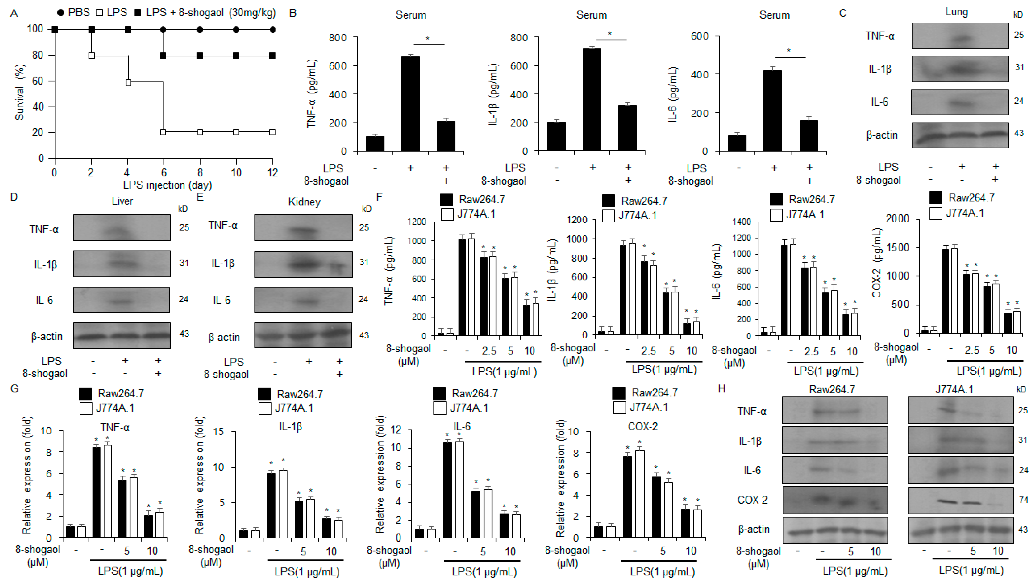
2.1. 8-Shogaol Inhibited Inflammation in LPS-Treated Macrophages and a Sepsis Mouse Model

To determine the anti-inflammatory effects of 8-shogaol, the LPS-induced sepsis mouse model was developed. Compared to the LPS group, the LPS + 8-shogaol group showed approximately a fourfold enhancement in survival rate (Figure 1A). We next demonstrated the protein levels of inflammatory cytokines, including IL-6, IL-1β, and TNF-α, via ELISA and Western blot. 8-Shogaol treatment notably reduced the levels of TNF-α, IL-6, and IL-1β in the kidney, lung, liver, and serum of the treated mice (Figure 1B–E). Furthermore, LPS-treated macrophages (Raw264.7 and J774.1) were used to confirm the anti-inflammatory effects of 8-shogaol. As identified by qRT-PCR, ELISA, and Western blot, 8-shogaol treatment dramatically downregulated the protein levels of inflammatory cytokines, including COX-2, IL-1β, IL-6, and TNF-α, in a dose-dependent manner (Figure 1F–H). This showed that 8-shogaol treatment effectively prevents the LPS-induced production of inflammatory cytokines in macrophages.

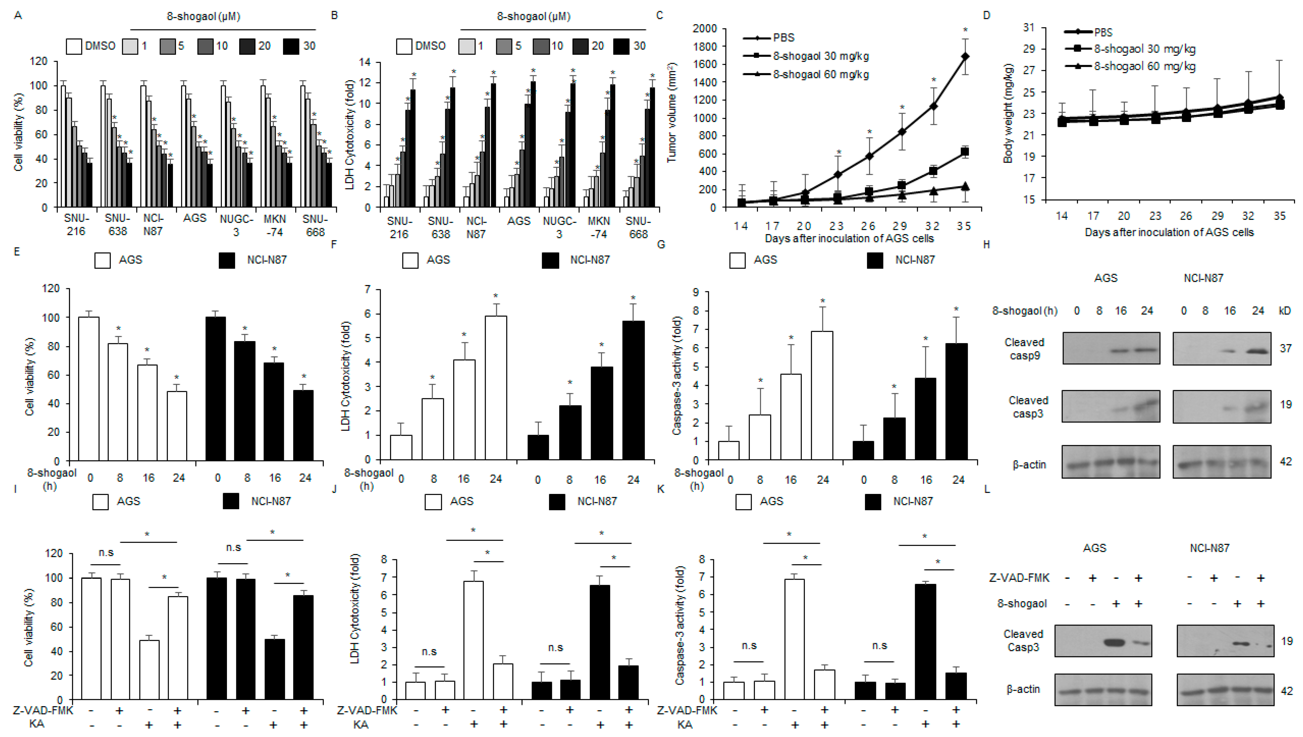
2.2. 8-Shogaol Induced Caspase-Dependent Apoptosis in Gastric Cancer Cells

The in vitro anti-cancer efficacy of 8-shogaol were investigated against gastric cancer cells (SNU-216, SNU-638, NCI-N87, AGS, NUGC-3, MKN-74, and SNU-668) at varying doses of 8-shogaol (1, 5, 10, 20, and 30 µM) (Figure 2A,B). The in vivo anti-tumor effects of 8-shogaol were evaluated using the AGS tumor mouse model. 8-Shogaol treatment (30 mg/kg and 60 mg/kg) significantly inhibited tumor growth compared to the control group (Figure 2C) without causing body weight loss (Figure 2D). The in vitro anti-tumor efficacy of 8-shogaol was assessed in a time-dependent manner (0, 8, 16, and 24 h; 10 µM). AGS and NCI-N87 cells were treated with 8-shogaol and subjected to LDH cytotoxicity, WST-1, and colorimetric caspase-3 activity assays. 8-Shogaol treatment indicated increased colorimetric caspase-3 activity and cytotoxicity in a time-dependent manner (Figure 2E–G). Western blot analysis also showed that 8-shogaol mediates time-dependent cleavage of caspase-3 and -9 (Figure 2H). To investigate whether 8-shogaol treatment induces apoptosis through the caspase-dependent pathway, NCI-N87 and AGS cells were co-treated with Z-VAD-FMK, a cell-permeant pan-caspase inhibitor. The 50 μM Z-VAD-FMK treatment alone did not significantly affect cytotoxicity, caspase-3 activity, or cell viability. At 10 µM 8-shogaol, however, cytotoxicity and caspase-3 activity increased. When 50 μM Z-VAD-FMK was co-treated with 10 µM 8-shogaol, cytotoxicity and colorimetric caspase-3 activity decreased significantly, leading to an enhancement in cell viability (Figure 2I–K). Western blot analysis indicated that this co-treatment reduced the level of cleaved caspase-3 compared to 8-shogaol treatment alone (Figure 2L). These results indicated that 8-shogaol suppresses the proliferation of gastric cancer cells via the caspase-related signaling pathway.

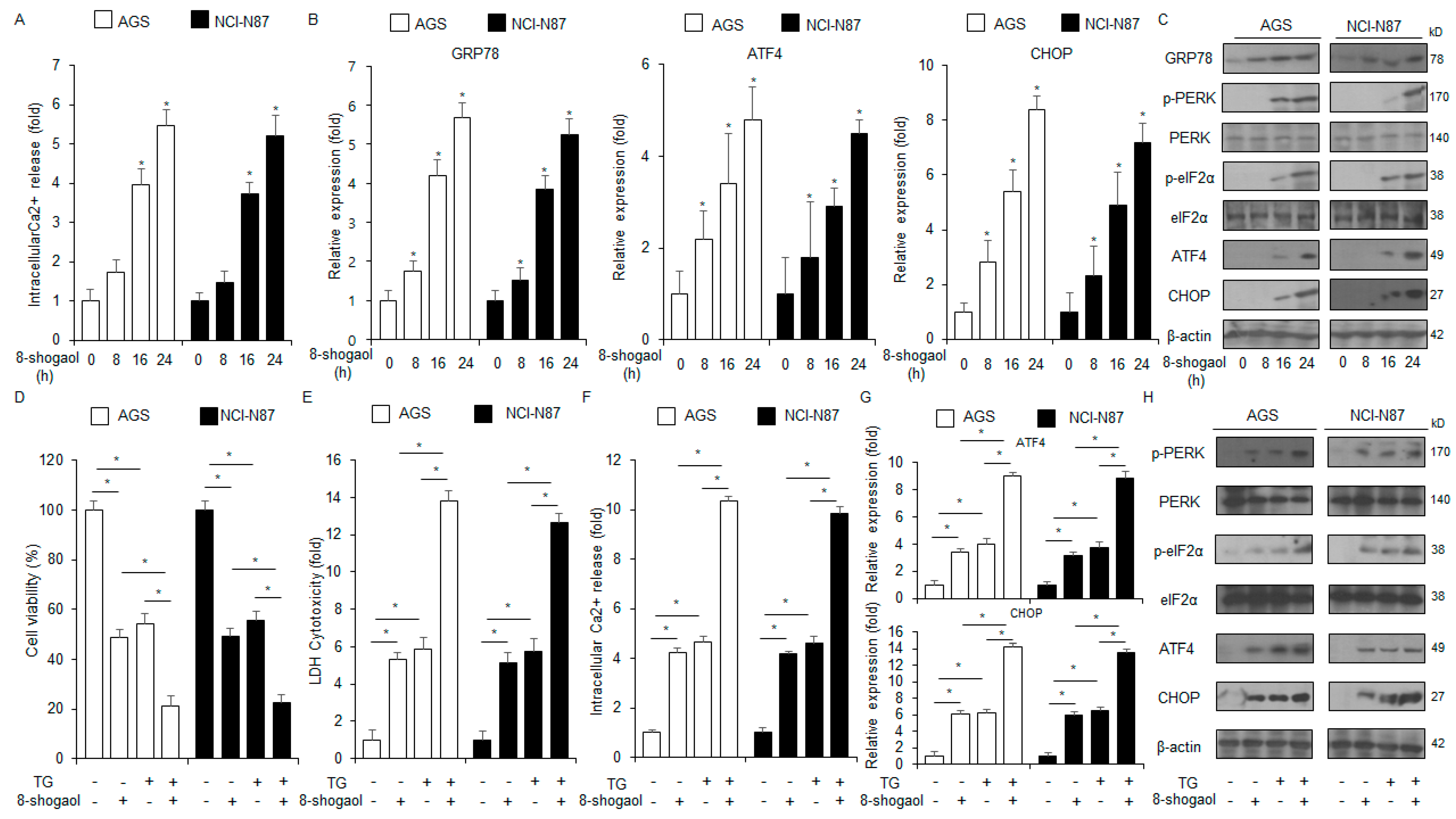
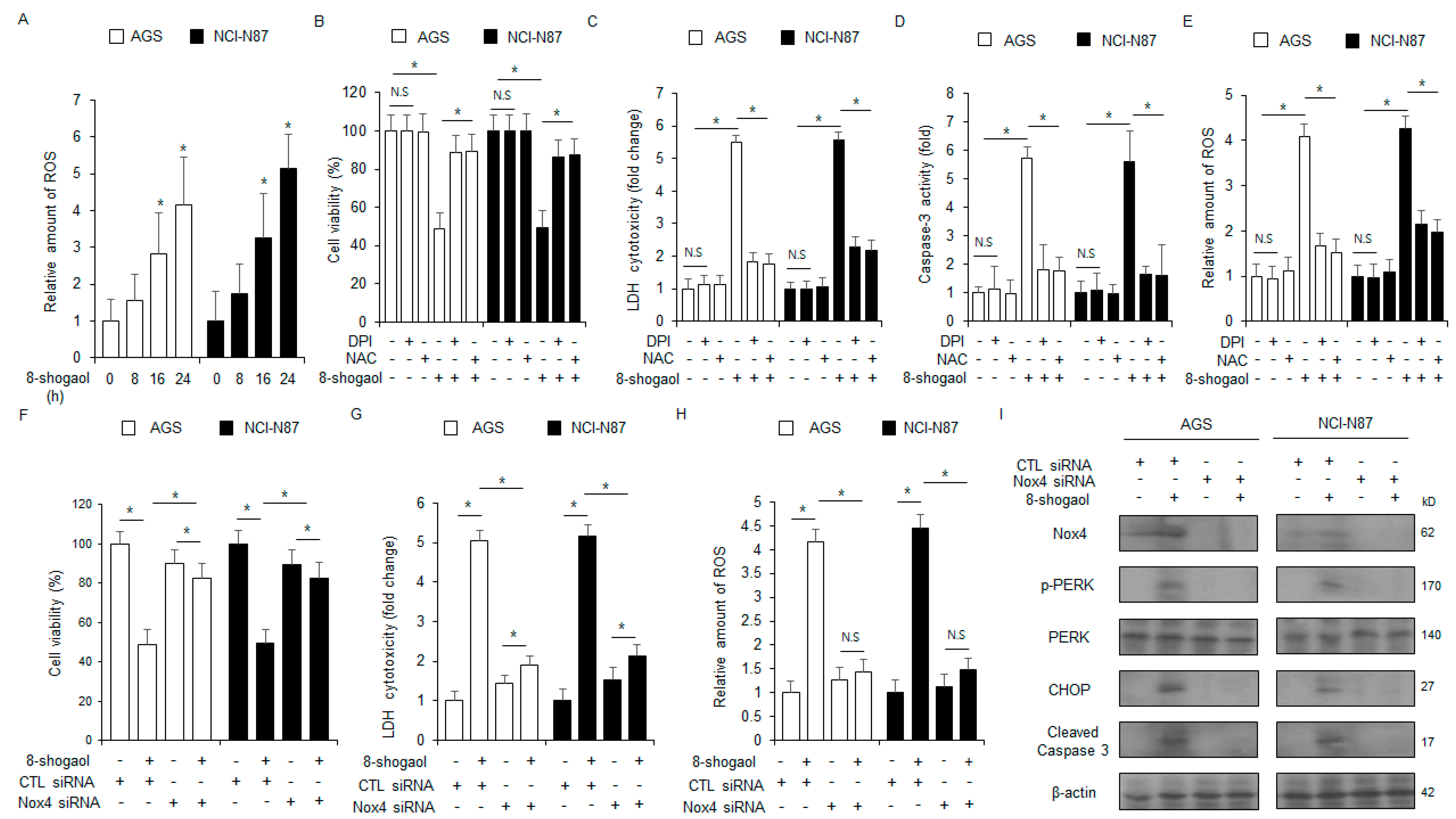
2.3. 8-Shogaol-Induced ER Stress Mediated Apoptosis in Gastric Cancer Cells

Ca2+ regulates immune evasion, cell proliferation, cell migration, survival, cell cycle arrest, and death [18]. Excessive Ca2+ efflux from the ER lumen into the cytosol induces ER stress, leading to apoptosis [19]. Ca2+ assay showed that 8-shogaol treatment increased Ca2+ production in NCI-N87 and AGS cells in a time-dependent manner (Figure 3A). Moreover, qRT-PCR analysis of these cells indicated that 8-shogaol treatment enhanced the mRNA levels of CHOP, GRP78, and ATF4 in a time-dependent manner; Western blot analysis indicated that the protein levels of p-eIF2α, p-PERK, CHOP, ATF4, and GRP78 increased with 8-shogaol treatment (Figure 3B,C). To investigate whether ER stress is associated with 8-shogaol-mediated apoptosis, the cells were co-treated with 8-shogaol and the ER stressor TG. This co-treatment synergistically enhanced the intracellular Ca2+ release and cytotoxicity (Figure 3D–F). In addition, the levels of CHOP, ATF4, p-eIF2α, and p-PERK also significantly enhanced following the treatment (Figure 3G,H).

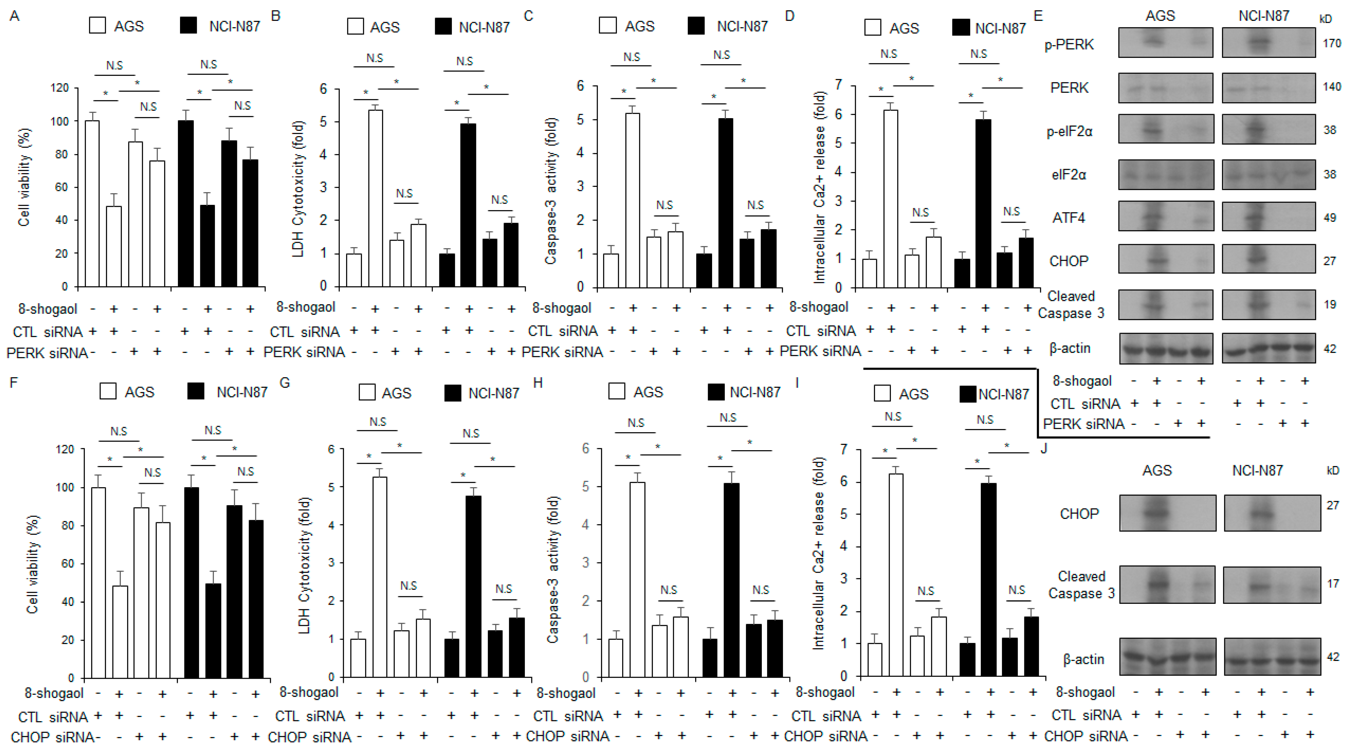
2.4. PERK or CHOP Silencing Inhibited 8-Shogaol-Induced Apoptosis in Gastric Cancer Cells

To assess whether 8-shogaol-induced apoptosis is dependent on PERK or CHOP, NCI-N87 and AGS cells were transfected with siRNAs for the corresponding genes. PERK silencing reduced 8-shogaol-induced increase in caspase-3 activity, intracellular Ca2+ activity, and cytotoxicity (Figure 4A–D). Western blot analysis showed that p-PERK, p-eIF2α, cleaved caspase-3, ATF4, and CHOP levels were also reduced after PERK silencing in 8-shogaol-treated NCI-N87 and AGS cells (Figure 4E). CHOP silencing also decreased 8-shogaol-mediated increases in caspase-3 activity, intracellular Ca2+ activity, and cytotoxicity (Figure 4F–I). Western blot analysis showed that CHOP knockdown decreased the levels of cleaved caspase-3, as well as CHOP, in 8-shogaol-treated NCI-N87 and AGS cells (Figure 4J). These findings identified that 8-shogaol-mediated apoptosis in gastric cancer cells is mediated by ER stress.

2.5. Nox4 Silencing Inhibited 8-Shogaol-Induced Apoptosis in Gastric Cancer Cells

We next investigated the impact of 8-shogaol treatment on intracellular ROS release. 8-Shogaol treatment increased the level of ROS production in gastric cancer cells over varying treatment times (Figure 5A). Next, NCI-N87 and AGS cells were treated with ROS inhibitors (DPI or NAC) in the presence of 8-shogaol. This co-treatment with 8-shogaol-mediated enhancements in cytotoxicity, intracellular ROS generation, and caspase-3 activity (Figure 5B–E). Next, the cells were transfected with a NOX4-specific siRNA and treated with 8-shogaol. NOX4 silencing decreased the enhancements in intracellular ROS and cytotoxicity induced by 8-shogaol treatment (Figure 5F–H). Western blot analysis showed that NOX4 silencing also suppressed the enhancements in the levels of CHOP, cleaved caspase-3, p-PERK, and NOX4 (Figure 5I). These results showed that NOX4 confers to 8-shogaol-mediated intracellular ROS release and apoptosis in gastric cancer cells.

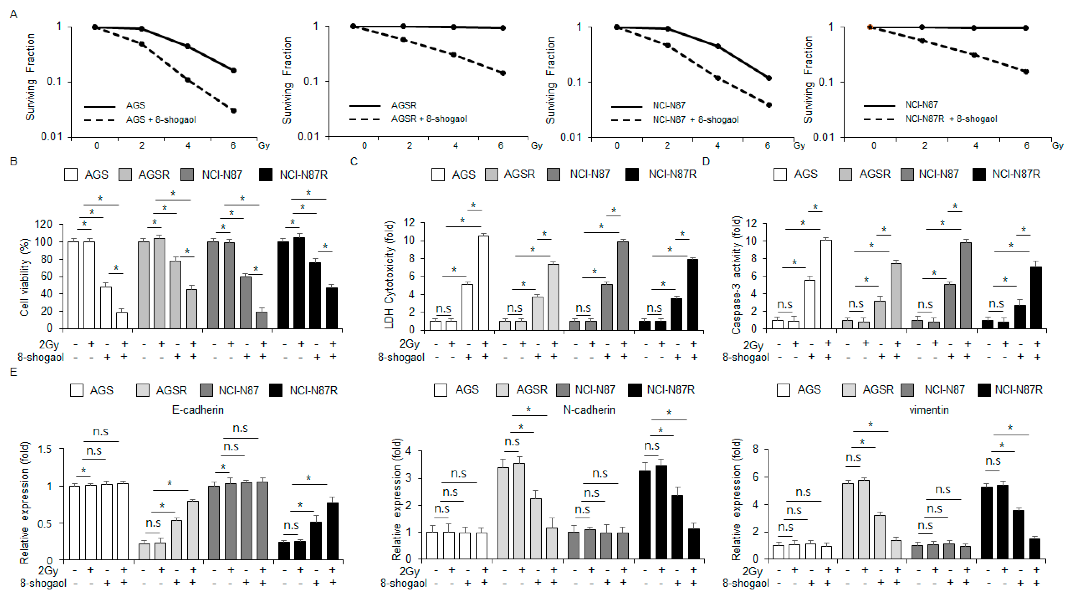
2.6. 8-Shogaol Sensitized Radio-Resistant Gastric Cancer Cells to Radiotherapy by Modulating EMT Markers

Although radiotherapy is a potential tumor therapeutic strategy to sensitize gastric cancer cells, it frequently causes radioresistance [20]. Next, we assessed whether 8-shogaol overcomes radio-resistant NCI-N87R and AGSR cells using a clonogenic cell assay. 8-Shogaol treatment was synergized with varying radiation intensities (2, 4, and 6 Gy) to enhance cytotoxic effects against NCI-N87R, AGSR, NCI-N87, and AGS cells (Figure 6A). In NCI-N87 and AGS cells, 8-shogaol enhanced caspase-3 activity and cytotoxicity. The 2 Gy radiation combined with 8-shogaol further enhanced these properties and significantly decreased cell viability, with radiation alone having no significant effects (Figure 6B–D). In NCI-N87R and AGSR cells, 8-shogaol treatment again increased cytotoxicity and caspase-3 activity, leading to synergistic anti-cancer effects when combined with 2 Gy radiation; radiation alone had no significant effects in these cells (Figure 6B–D). To investigate whether this combination regulates the EMT phenomenon, the radio-resistant gastric cancer cells were subjected to qRT-PCR analysis. Both 8-shogaol alone and co-treatment with 2 Gy radiation increased the mRNA level of E-cadherin and decreased the mRNA levels of vimentin and N-cadherin in NCI-N87R and AGSR cells. On the other hand, the non-resistant cells showed no significant changes in these mRNA levels (Figure 6E). These results suggested that combining 8-shogaol with radiation may be an effective strategy for treating radio-resistant gastric cancer.

3. Discussion

Recent studies have reported that natural compounds, including phenols, flavonoids, terpenoids, and alkaloids, display powerful anti-tumor efficacy [21,22]. In various cancer cell types, these compounds often mediate intracellular ROS and Ca2+ release, thereby causing oxidative-stress-driven apoptosis [23,24]. In this study, we explored 8-shogaol’s anti-tumor effects and ability to sensitize radio-resistant breast cancer cells to radiotherapy. Our findings identified that 8-shogaol’s anti-tumor efficacy induce apoptosis in gastric cancer cells both in vitro and in vivo, highlighting its clinical potential to inhibit tumor growth [25]. By co-treating Z-VAD-FMK and 8-shogaol, we identified that 8-shogaol-mediated apoptosis in gastric cancer cells is dependent on caspase activity. Moreover, 8-shogaol increased ER stress in these cells, thereby activating the downstream ER stress sensors, IRE1α, PERK, and ATF6, inducing apoptotic cell death [26]. Upon UPR induction, the ER chaperone protein GRP78 dissociated from these sensors mediated the phosphorylation of eIF2α and PERK [27]. It has been identified that p-eIF2α mediates the activation of cytosolic ATF4. The translocated nuclear ATF4 binds to the promoter of nuclear CHOP and induces its expression [28]. 8-Shogaol was found to induce the ER stress signaling pathway by promoting the generation of ROS and cytosolic Ca2+. This process ultimately mediated apoptosis through the PERK-ATF4-CHOP signaling pathway. Silencing PERK or CHOP in these cells led to the suppression of 8-shogaol-caused apoptosis. When 8-shogaol was co-treated with TG, synergistic apoptosis was mediated via the PERK-ATF4-CHOP axis in gastric cancer cells. In this phenomenon, NOX4 was demonstrated as a regulator of ROS generation promoted by 8-shogaol treatment. The activation of NOX4 was found to mediate ER-stress-induced apoptosis by promoting ROS and intracellular Ca2+ release in 8-shogaol-treated cells. NOX4 silencing or ROS inhibitor (NAC or DPI) treatment inhibited the increase in ROS, caspase-3 activity, cytotoxicity, and the PERK-ATF4-CHOP signaling pathway in 8-shogaol-treated gastric cancer cells. Although radiotherapy is a primary cancer treatment option, tumor cells often develop radioresistance after exposure, reducing the effectiveness of therapy [29]. Recent reports have indicated that natural compounds, including withaferin A, celastrol, ursolic acid, zerumbone, C-phycocyanin, emodin, flacopiridol, and berberine, are effective radiotherapy sensitizers with minimal side effects [30,31]. The EMT regulatory proteins have been related to the development of radioresistance, chemoresistance, and hypoxic environments [32]. The development of anti-cancer agents that can sensitize radio-resistant cancer cells has the possibility to increase the effectiveness of radiotherapy. When radiation was combined with 8-shogaol, the radio-resistant NCI-N87R and AGSR cells effectively overcame radiotherapy. This phenomenon was indeed identified to be dependent on the change of EMT-regulatory proteins, such as N-cadherin, vimentin, and E-cadherin.
Various natural compounds have powerful anti-tumor effects in many cancer types [33]. Kaempferol has been shown to mediate apoptosis by promoting the cleavage of caspase-3 and G2/M phase cell cycle arrest in human gastric cells [34]. The monomeric compound alternol has been found to promote apoptosis and ROS-dependent ER stress via the PERK-ATF4-CHOP axis and the IRE1α–XBP1-CHOP axis in prostate cancer cells, PC-3, 22RV1, and C4-2, where the NF-κB inhibitor Imoxin and SN50 treatment were able to suppress alternol-mediated ER stress and apoptosis [35]. Moreover, catechol has been identified to display anti-tumor efficacy and the ability to sensitize radio-resistant cells through the change of EMT markers such as vimentin, Snail, and E-cadherin in pancreatic cancer cells [36]. Analogously to these natural compounds, we identified that 8-shogaol induces apoptosis in gastric cancer cells through the PERK-ATF4-CHOP axis. Importantly, 8-shogaol was demonstrated to powerfully sensitize radio-resistant cells by regulating the aforementioned EMT-related proteins, including vimentin, N-cadherin, and E-cadherin. Recently, it has been demonstrated that 8-shogaol induces caspase-3-dependent apoptosis via the activation of ROS-mediated ER stress in human oral cells [37].
ROS release and mitochondrial dysfunction induce apoptosis by activating the PERK-ATF4-CHOP axis in many cancers [38]. Gossypol has been shown to induce apoptosis in human pancreatic cancer cells BxPC-3 and MIA PaCa-2 cells through the CHOP-DR5 axis [39]. NOXs are potential enzymes that produce ROS and serve as important characteristics of cell metabolism in cancer [40]. The seven transmembrane NOX family, including dual oxidases 1 and 2 and NOX1-5, generate superoxide anion radicals and play potential roles in various cancers [41]. NOX4-mediated ROS generation induces apoptotic cell death and plays a potential role in regulating cell survival and death [42]. Cannabidiol has been found to induce NOX4-mediated release of mitochondrial ROS and promote caspase-dependent apoptosis through the activation of ER stress signaling pathway and the inhibition of the mTOR signaling pathway in breast cancer cells [43]. Similarly, we identified that 8-shogaol mediates apoptosis via the PERK-ATF4-CHOP axis driven by NOX4-caused release of ROS. Silencing NOX4 or ROS inhibitors (DPI or NAC) treatment inhibited the increases in ROS, cytotoxicity, and caspase-3 activity in 8-shogaol-treated gastric cancer cells, meaning NOX4 as the regulator of 8-shogaol-mediated ROS production.
Radiotherapy utilizes high-energy beams to kill cancer cells, but gastric cancer patients often acquire radioresistance [44]. Therefore, strategies to overcome resistant cancer cells to radiation are significantly needed [45]. Radiation treatment decreases oxygen levels in the TME, leading to a hypoxic condition that mediates radioresistance by modulating EMT [46]. In this process, the epithelial cell regulatory protein E-cadherin is downregulated, while mesenchymal cell regulatory proteins such as vimentin and N-cadherin are upregulated [47]. In radio-resistant NCI-N87R and AGSR cells, 8-shogaol combined with radiation blocked these phenomena, suppressing radioresistance. Ultimately, 8-shogaol combined with radiation significantly increased ER stress and apoptosis by modulating EMT-related proteins in radio-resistant gastric cancer cells

4. Materials and Methods

4.1. Reagents

8-Shogaol (PHL83910), N-acetylcysteine (NAC), diphenyleneiodonium (DPI), lipopolysaccharide (LPS; L4391), Z-VAD-FMK, and TG (T9033) were purchased from Sigma-Aldrich (St. Louis, MO, USA).

4.2. Cell Culture

Human gastric cancer cell line (SNU-216, SNU-638, NCI-N87, AGS, NUGC-3, MKN-74, and SNU-668) was purchased from the American Type Culture Collection (ATCC, Manassas, VA, USA) and the Korean Cell Line Bank (Seoul, Republic of Korea) and then cultured at 37 °C under 5% CO2 in Dulbecco’s modified Eagle’s medium (DMEM; Welgene, Gyeongsan-si, Gyeongsangbuk-do, Republic of Korea) supplemented with 10% fetal bovine serum (FBS; Welgene) and 1% penicillin–streptomycin (PS; Welgene).

4.3. Cell Viability and Proliferation Assays

To identify the effect of 8-shogaol on the viability of treated cells, the WST-1 assay (Roche Applied Science, Indianapolis, IN, USA) was conducted in both treatment time- and concentration-dependent manners (8-shogaol; 1, 5, 10, 20, and 30 µM; 24 h). Gastric cancer cells were seeded and cultured in a 96-well cell culture plate (1 × 10⁴ cells/well). Following the manufacturer’s protocol, the absorbance at 450 nm was measured from each well using a microplate reader (Molecular Devices, San Jose, CA, USA).

4.4. Lactate Dehydrogenase (LDH) Cytotoxicity Assay

To determine the cytotoxicity of 8-shogaol against breast cancer cells, LDH cytotoxicity assay (Abcam, Cambridge, MA, USA) was conducted in both treatment time- and concentration-dependent manners. Gastric cancer cells were seeded and cultured in a 96-well cell culture plate (1 × 10⁴ cells/well). Following the manufacturer’s protocol, the absorbance at 490 nm was measured from each well using a microplate reader.

4.5. Colorimetric Caspase-3 Activity Assay

To identify 8-shogaol’s impact on caspase-3 activity in gastric cancer cells, a colorimetric caspase-3 activity assay (Abcam) was conducted in both treatment time- and concentration-dependent manners. Gastric cancer cells were seeded and cultured in a 96-well cell culture plate (1 × 10⁴ cells/well). Following the manufacturer’s protocol, the absorbance at 490 nm was measured from each well using a microplate reader.

4.6. Intracellular Ca2+ Assay

To identify the level of the intracellular Ca2+ in gastric cancer cells treated with 8-shogaol, an intracellular Ca2+ assay (Abcam) was carried out in both treatment time- and concentration-dependent manners. Gastric cancer cells were seeded and cultured in a 96-well cell culture plate (1 × 10⁴ cells/well). Following the manufacturer’s protocol, the fluorescence at 575 nm was measured from each well using FilterMax F5 (Molecular Devices).

4.7. Intracellular ROS Assay

To identify the level of ROS in gastric cancer cells treated with 8-shogaol, an intracellular ROS assay (Abcam) was carried out in both treatment time- and concentration-dependent manners. Gastric cancer cells were seeded and cultured in a 96-well cell culture plate (1 × 10⁴ cells/well). Following the manufacturer’s protocol, the fluorescence at 605 nm (excitation at 520 nm) was measured from each well using FilterMax F5.

4.8. Radio-Resistant NCI-N87R and AGSR Cell Lines

NCI-N87 and AGS cells were cultured in 60 mm culture plates. The cells were irradiated at 4 Gy daily for 90 days to establish radio-resistant NCI-N87R and AGSR cell lines.

4.9. Radiotherapy

NCI-N87, AGS, NCI-N87R, and AGSR cells were cultured in 60 mm culture plates at 37 °C under 5% CO2. The cells were irradiated using an irradiator with a cesium-137 source (Atomic Energy of Canada, Ltd., Mississauga, ON, Canada).

4.10. Clonogenic Cell Assays

NCI-N87, AGS, NCI-N87R, and AGSR cells were cultured in 60 mm culture plates at 37 °C under 5% CO2 for colony formation. The colonies were stained with 0.5% crystal violet solution (Sigma-Aldrich, St. Louis, MO, USA).

4.11. Transfection Assay

Small interfering RNAs (siRNAs) for CHOP (Bioneer, Daejeon, Republic of Korea), Nox4 (Santacruz, Dallas, TX, USA), and PERK (Santacruz) were used for gene-silencing experiments. NCI-N87 and AGS cells were first seeded in 6-well culture plates. Following the manufacturer’s protocol, the cells were transfected with siRNAs (30 nmol/mL). Lipofectamine 2000 (Invitrogen, Carlsbad, CA, USA) was used as the transfection agent for siRNAs.

4.12. Protein and RNA Purification

Gastric cancer cells were cultured in 100 mm culture plates at 37 °C under 5% CO2. Following the manufacturer’s protocol, total RNA and protein were isolated with Trizol reagent (Invitrogen) and extracted using the radio-immunoprecipitation assay (RIPA) lysis buffer (Thermo Fisher Scientific, Waltham, MA, USA).

4.13. Quantitative Reverse Transcription Polymerase Chain Reaction (qRT PCR) and Western Blot Analyses

For qRT-PCR assay, all reactions were performed in triplicate. The primers were adopted from a study by Kim et al. and purchased from Bioneer (Daejeon, Republic of Korea) [48]. To determine relative gene expression levels, the 2−ΔΔCt method was used. To identify protein expression levels, Western blot analysis was performed. After separating the proteins via SDS-PAGE, they were transferred to PVDF membranes, which were blocked with 5% skim milk and incubated with primary antibodies. The primary antibodies used were as follows: cleaved caspase-9 (Cell Signaling, Danvers, MA, USA), CHOP (Cell Signaling), ATF4 (Cell Signaling), p-PERK (Thr980) (Cell Signaling), cleaved caspase-3 (Cell Signaling), p-eIF2ɑ (Ser51) (Cell Signaling), PERK (Cell Signaling), eIF2α (Santa Cruz, CA, USA), GRP78 (Santa Cruz), β-actin (Santa Cruz), and Nox4 (Proteintech, Rosamond, IL, USA). HRP-conjugated secondary antibodies were used with an anti-rabbit IgG HRP-linked antibody (Santa Cruz) and m-IgGK BP-HRP-linked antibody (Santa Cruz). The membranes were visualized using a chemiluminescent substrate for HRP (MilliporeSigma, Burlington, MA, USA).

4.14. Tumor and Inflammation Mouse Models

All mouse models were stabilized using female mice. Five-week-old mice were obtained from OrientBio, Inc. (Daejeon, Republic of Korea), and acclimated in a sterile room for one week on an NIH-7 open formula diet. The mice were then divided randomly into three groups. All mouse experiments were conducted in accordance with the guidelines of the Kyung-Hee University Animal Care and Use Committee. The AGS tumor model was established by subcutaneously injecting 1 × 10⁷ cells suspended in PBS into the right dorsal flanks of athymic BALB/c nude mice (nu/nu). When the average tumor volume reached 200 mm3, the mice were randomly divided into three groups (n = 10): control (PBS), 30 mg/kg 8-shogaol, and 60 mg/kg 8-shogaol. The specified doses of 8-shogaol or PBS were administered by intraperitoneal (i.p.) injection twice weekly. Tumor volume (mm3) was calculated using the following formula: (L × W2)/2, where L is the length and W is the width of the tumor (mm3). The anti-inflammatory effects of 8-shogaol were assessed using the LPS-induced inflammation model. The mice were randomly divided into three groups: PBS, LPS, and LPS + 8-shogaol. The groups receiving LPS were administered 20 mg/kg of LPS by i.p. injection. The LPS + 8-shogaol group was administered with 30 mg/kg of 8-shogaol by i.p. injection twice weekly. The survival rate was monitored for 12 days following LPS injection, and blood and tissues were collected for analysis.

4.15. Cytokine Levels

Raw264.7 and J774.1 cells were seeded in 96-well culture plates (1 × 10⁴ cells/well). The cells were treated with 1 μg/mL LPS in the absence or presence of 8-shogaol (0, 2.5, 5, and 10 μM; 24 h) for 24 h. The protein levels of IL-6, IL-1β, and TNF-α were assessed via enzyme-linked immunosorbent assay (ELISA) Following the manufacturer’s protocol: IL-6 (DY-406; R&D Systems, Minneapolis, MN, USA), IL-1β (DY-401; R&D Systems), and TNF-α (DY-410; R&D Systems).

4.16. Statistical Analysis

All experiments were conducted at least three times. Statistical significance was analyzed using analysis of variance (ANOVA) and Student’s t-test, with a p-value < 0.05 considered statistically significant.

5. Conclusions

In conclusion, we demonstrated 8-shogaol’s anti-cancer and anti-inflammatory properties both in vitro and in vivo. 8-Shogaol treatment was shown to mediate apoptosis through the PERK-ATF4-CHOP axis. Moreover, the treatment increased the production of ROS and intracellular Ca2+ and upregulated NOX4 in gastric cancer cells. When 8-shogaol was combined with radiation, radioresistance was effectively sensitized via the EMT change in NCI-N87R and AGSR cells.

Author Contributions

Designed, performed, and supervised the experiments: T.W.K. and H.G.L. Analyzed the data: T.W.K. Wrote the paper: T.W.K. All authors have read and agreed to the published version of the manuscript.

Funding

This work was supported by the National Research Foundation (NRF) and the National Research Council of Science and Technology (NST) funded by the Korean government (RS-2023-00237729 and CAP21022-000). This work was founded by KRIBB Research Initiative Program (1711196163).

Institutional Review Board Statement

All procedures were carried out in accordance with the National Institutes of Health Guide for the Care and Use of Laboratory Animals and approved by the Institutional Animal Care and Use Committee (IACUC) at the Kyung Hee University (KHSASP-22-583).

Informed Consent Statement

Not applicable.

Data Availability Statement

Data are contained within the article.

Conflicts of Interest

The authors declare no biomedical financial interests or competing financial interests.

References

  1. Thrift, A.P.; Wenker, T.N.; El-Serag, H.B. Global burden of gastric cancer: Epidemiological treands, risk factors, screening and prevention. Nat. Rev. Clin. Oncol. 2023, 20, 338–349. [Google Scholar] [CrossRef]
  2. Guan, W.L.; He, Y.; Xu, R.H. Gastric cancer treatment: Recent progress and future perspectives. J. Hematol. Oncol. 2023, 16, 57. [Google Scholar] [CrossRef]
  3. Chong, I.Y.; Chau, I. Is there still a place for radiotherapy in gastric cancer? Curr. Opin. Pharmacol. 2023, 68, 102325. [Google Scholar] [CrossRef]
  4. Baskar, P.; Lee, K.A.; Yeo, R.; Yeoh, K.W. Cancer and radiation therapy: Current advances and future directions. Int. J. Med. Sci. 2012, 9, 193–199. [Google Scholar] [CrossRef]
  5. Pagliari, F.; Jansen, J.; Knoll, J.; Hanley, R.; Seco, J.; Tirinato, L. Cancer radioresistance is characterized by a differential lipid droplet content along the cell cycle. Cell Div. 2024, 19, 14. [Google Scholar] [CrossRef] [PubMed]
  6. Chang, L.; Graham, P.H.; Hao, J.; Ni, J.; Bucci, J.; Cozzi, P.J.; Kearsley, J.H.; Li, Y. Acquisition of epithelial-mesenchymal transition and cancer stem cell phenotypes is associated with activation of the PI3K/Akt/mTOR pathway in prostate cancer radioresistance. Cell Death Dis. 2013, 4, e875. [Google Scholar] [CrossRef] [PubMed]
  7. Komorowska, D.; Radzik, T.; Kalenik, S.; Rodacka, A. Natural radiosensitizers in radiotherapy: Cancer treatment by combining ionizing radiation with resveratrol. Int. J. Mol. Sci. 2022, 23, 10627. [Google Scholar] [CrossRef]
  8. Cheng, X.L.; Ruan, Y.L.; Dai, J.Y.; Fan, H.Z.; Ling, J.Y.; Chen, J.; Lu, W.G.; Gao, X.J.; Cao, P. 8-shogaol derived from dietary ginger alleviated acute and inflammatory pain by targeting TRPV1. Phytomedicine 2024, 128, 1555000. [Google Scholar] [CrossRef] [PubMed]
  9. Maghraby, Y.R.; Labib, R.M.; Sobeh, M.; Farag, M.A. Gingerols and shogaols: A multi-faceted review of their extraction, formulation, and analysis in drugs and biofluids to maximize their nutraceutical and pharmaceutical applications. Food Chem. X 2023, 20, 100947. [Google Scholar] [CrossRef] [PubMed]
  10. Kim, H.R.; Noh, E.M.; Kim, S.Y. Anti-inflammatory effect and signaling mechanism of 8-shogaoland 10-shogaol in a dextran sodium sulfate-induced colitis mouse model. Heliyon 2023, 9, e12778. [Google Scholar] [CrossRef] [PubMed]
  11. Shieh, P.C.; Chen, Y.O.; Kuo, D.H.; Chen, F.A.; Tsai, M.L.; Chang, I.S.; Wu, H.; Sang, S.; Ho, C.T.; Pan, M.H. Induction of apoptosis by 8-shogaol via reactive oxygen species generation, glutathione depletion, and caspase activation in human leukemia cells. J. Agric. Food Chem. 2010, 58, 3847–3854. [Google Scholar] [CrossRef]
  12. Groenendyk, J.; Michalak, M. Interplay between calcium and endoplasmic reticulum stress. Cell Calcium 2023, 113, 102753. [Google Scholar] [CrossRef] [PubMed]
  13. Sundaram, A.; Appathurai, S.; Plumb, R.; Mariappan, M. Dynamic changes in complexes of IRE1α, PERK, ATF6α during endoplasmic reticulum stress. Mol. Biol. Cell 2018, 29, 1376–1388. [Google Scholar] [CrossRef] [PubMed]
  14. Walter, P.; Ron, D. The unfolded protein response: From stress pathway to homeostatic regulation. Science 2011, 334, 1081–1086. [Google Scholar] [CrossRef]
  15. Spencer, B.G.; Finnie, J.W. The role of endoplasmic reticulum stress in cell survival and death. J. Comp. Pathol. 2020, 181, 86–91. [Google Scholar] [CrossRef] [PubMed]
  16. Cao, S.S.; Kaufman, R.J. Endoplasmic reticulum stress and oxidative stress in cell fate decision and human disease. Antioxid. Redox Signal 2014, 21, 396–413. [Google Scholar] [CrossRef]
  17. Santos, C.X.; Nabeebaccus, A.A.; Shah, A.M.; Camargo, L.L.; Filho, S.V.; Lopes, L.R. Endoplasmic reticulum stress and nox-mediated reactive oxygen species signaling in the peripheral vasculature: Potential role in hypertension. Antioxid. Redox Signal 2014, 20, 121–134. [Google Scholar] [CrossRef] [PubMed]
  18. Moon, D.O. Calcium’s role in orchestrating cancer apoptosis: Mitochondrial-centric perspective. Int. J. Mol. Sci. 2023, 24, 8982. [Google Scholar] [CrossRef] [PubMed]
  19. Rao, R.V.; Ellerby, H.M.; Bredesen, D.E. Coupling endoplasmic reticulum stress to the cell death program. Cell Death Differ. 2004, 11, 372–380. [Google Scholar] [CrossRef] [PubMed]
  20. Wang, M.; Huang, K.; Fan, X.; Wang, J.; Jin, Y.; Zheng, Z.J. Access to radiotherapy in improving gastric cancer care quality and equality. Commun Med. 2024, 4, 225. [Google Scholar] [CrossRef]
  21. Nan, Y.; Su, H.; Zhou, B.; Liu, S. The function of natural compounds in important anticancer mechanisms. Front. Oncol. 2023, 12, 1049888. [Google Scholar] [CrossRef] [PubMed]
  22. Wang, J.; Jiang, Y.F. Natural compounds as anticancer agents: Experimental evidence. World J. Exp. Med. 2012, 2, 45–57. [Google Scholar] [CrossRef] [PubMed]
  23. Muchtaridi, M.; Az-Zahra, F.; Wongso, H.; Setyawati, L.U.; Novitasari, D.; Ikram, E.H.K. Molecular mechanism of natural food antioxidants to regulate ROS in treating cancer: A review. Antioxidants 2024, 13, 207. [Google Scholar] [CrossRef]
  24. Redza-Dutordoir, M.; Averill-Bates, D. Activation of apoptosis signaling pathways by reactive oxygen species. Biochim. Biophys. Acta 2016, 12, 2977–2992. [Google Scholar] [CrossRef] [PubMed]
  25. Sang, S.; Hong, J.; Wu, H.; Liu, J.; Yang, C.S.; Pan, M.H.; Badmaev, V.; Ho, C.T. Increased growth inhibitory effects on human cancer cells and anti-inflammatory potency of shogaols from Zingiber officinale relative to gingerols. J. Agric. Food Chem. 2009, 57, 10645–10650. [Google Scholar] [CrossRef] [PubMed]
  26. Szegezdi, E.; Logue, S.E.; Gorman, A.M.; Samali, A. Mediators of endoplasmic reticulum stress-induced apoptosis. EMBO Rep. 2006, 7, 880–885. [Google Scholar] [CrossRef]
  27. Gardner, B.M.; Pincus, D.; Gotthardt, K.; Gallagher, C.M.; Walter, P. Endoplasmic reticulum stress sensing in the unfolded protein response. Cold Spring Harb. Perspect. Biol. 2013, 5, a013169. [Google Scholar] [CrossRef] [PubMed]
  28. Rozpedek, W.; Pytel, D.; Mucha, B.; Leszczyńska, H.; Alan Diehl, J.; Majsterek, I. The role of the PERK/eIF2α/ATF4/CHOP signaling pathway in tumor progression during endoplasmic reticulum stress. Curr. Mol. Med. 2016, 16, 533–544. [Google Scholar] [CrossRef] [PubMed]
  29. Wu, Y.; Song, Y.; Wang, R.; Wang, T. Molecular mechanisms of tumor resistance to radiotherapy. Mol. Cancer 2023, 22, 96. [Google Scholar] [CrossRef] [PubMed]
  30. Calvaruso, M.; Pucci, G.; Musso, R.; Bravatà, V.; Cammarata, F.P.; Russo, G.; Forte, G.I.; Minafra, L. Nutraceutical compounds as sensitizers for cancer treatment in radiation therapy. Int. J. Mol. Sci. 2019, 20, 5267. [Google Scholar] [CrossRef] [PubMed]
  31. Kefayat, A.; Ghahremani, F.; Safavi, A.; Hajiaghababa, A.; Moshtaghian, J. C-phycocyanin: A natural product with radiosensitizing property for enhancement of colon cancer radiation therapy efficacy through inhibition of COX-2 expression. Sci. Rep. 2019, 9, 19161. [Google Scholar] [CrossRef]
  32. Xiong, H.; Nie, X.; Zou, Y.; Gong, C.; Li, Y.; Wu, H.; Qiu, H.; Yang, L.; Zhuang, L.; Zhang, P.; et al. Twist1 enhances hypoxia induced radioresistance in cervical cancer cells by promoting nuclear EGFR localization. J. Cancer 2017, 8, 345–353. [Google Scholar] [CrossRef]
  33. Nobili, S.; Lippi, D.; Witort, E.; Donnini, M.; Bausi, L.; Mini, E.; Capaccioli, S. Natural compounds for cancer treatment and prevention. Pharmacol. Res. 2009, 59, 365–378. [Google Scholar] [CrossRef] [PubMed]
  34. Song, H.; Bao, J.; Wei, Y.; Chen, Y.; Mao, X.; Li, J.; Yang, Z.; Xue, Y. Kaempferol inhibits gastric cancer tumor growth: An in vitro and in vivo study. Oncol. Rep. 2015, 33, 868–874. [Google Scholar] [CrossRef] [PubMed]
  35. Liu, W.; He, C.; Li, C.; Ye, S.; Zhao, J.; Zhu, C.; Wang, X.; Ma, Q.; Li, B. Natural compound alternol actives multiple endoplasmic reticulum stress-responding pathways contributing to cell death. Front. Pharmacol. 2024, 15, 1397116. [Google Scholar] [CrossRef] [PubMed]
  36. Moon, J.Y.; Ediriweera, M.K.; Ryu, J.Y.; Kim, H.Y.; Kim Cho, S. Catechol enhances chemo- and radio-sensitivity by targeting AMPK-Hippo signaling in pancreatic cancer cells. Oncol. Rep. 2021, 45, 1133–1141. [Google Scholar] [CrossRef] [PubMed]
  37. Kim, H.J.; Shin, J.A.; Lee, Y.G.; Jin, B.H.; Lee, W.W.; Lee, Y.S.; Choi, S.J.; Han, J.M.; Ahn, M.H.; Kim, J.H.; et al. Zingiber officinale promotes autophagy and apoptosis in human oral cancer through the C/EBP homologous protein. Cancer Sci. 2024, 115, 2701–2717. [Google Scholar] [CrossRef] [PubMed]
  38. Gu, S.; Chen, C.; Jiang, X.; Zhang, Z. ROS-mediated endoplasmic reticulum stress and mitochondrial dysfunction underlie apoptosis inducedby resveratrol and arsenic trioxide in A549 cells. Chem. Biol. Interact. 2016, 245, 100–109. [Google Scholar] [CrossRef] [PubMed]
  39. Lee, S.; Hong, E.; Jo, E.; Kim, Z.H.; Yim, K.J.; Woo, S.H.; Choi, Y.S.; Jang, H.J. Gossypol induces apoptosis of human pancreatic cancer cells via CHOP/endoplasmic reticulum stress signaling pathway. J. Microbiol. Biotechnol. 2022, 32, 645–656. [Google Scholar] [CrossRef]
  40. Tuy, K.; Rickenbacker, L.; Hjelmeland, A.B. Reactive oxygen species produced by altered tumor metabolism impacts cancer stem cell maintenance. Redox Biol. 2021, 44, 101953. [Google Scholar] [CrossRef]
  41. Fujii, J.; Homma, T.; Osaki, T. Superoxide radicals in the execution of cell death. Antioxidants 2022, 11, 501. [Google Scholar] [CrossRef]
  42. Seo, S.U.; Kim, T.H.; Kim, D.E.; Min, K.J.; Kwon, T.K. NOX4-mediated ROS production induces apoptotic cell death via down-regulation of c-FLIP and Mcl-1 expression in combined treatment thioridazine and curcumin. Redox Biol. 2017, 13, 608–622. [Google Scholar] [CrossRef] [PubMed]
  43. Shrivastava, A.; Kuzontkoski, P.M.; Groopman, J.E.; Prasad, A. Cannabidiol induces programmed cell death in breast cancer cells by coordinating the cross-talk between apoptosis and autophagy. Mol. Cancer Ther. 2011, 10, 1161–1172. [Google Scholar] [CrossRef] [PubMed]
  44. Li, Y.; Yang, X.; Yao, P.; Shen, W.; Wu, Y.; Ye, Z.; Zhao, K.; Chen, H.; Cao, J.; Xing, C. B7-H3 increases the radioresistance of gastric cancer cells through regulating baseline levels of cell autophagy. Am. J. Transl. Res. 2019, 11, 4438–4449. [Google Scholar] [PubMed]
  45. Poon, D.J.J.; Tay, L.M.; Ho, D.; Chua, M.L.K.; Chow, E.K.H.; Yeo, E.L.L. Improving the therapeutic ratio of radiotherapy against radioresistant cancers: Leveraging on novel artificial intelligence-based approaches for drug combination discovery. Cancer Lett. 2021, 511, 56–67. [Google Scholar] [CrossRef] [PubMed]
  46. Chen, Z.; Han, F.; Du, Y.; Shi, H.; Zhou, W. Hypoxic microenvironment in cancer: Molecular mechanisms and therapeutic interventions. Signal Transduct. Target. Ther. 2023, 8, 70. [Google Scholar] [CrossRef] [PubMed]
  47. Theys, J.; Jutten, B.; Habets, R.; Paesmans, K.; Groot, A.J.; Lambin, P.; Wouters, B.G.; Lammering, G.; Vooijs, M. E-cadherin loss associated with EMT promotes radioresistance in human tumor cells. Radiother. Oncol. 2011, 99, 392–397. [Google Scholar] [CrossRef] [PubMed]
  48. Kim, T.W.; Lee, H.G. 6-Shogaol overcomes gefitinib resistance via ER stress in ovarian cancer cells. Int. J. Mol. Sci. 2023, 24, 2639. [Google Scholar] [CrossRef]
Figure 1. The impact of 8-shogaol on the mRNA and protein levels of inflammatory cytokines in LPS-treated macrophages. (A) C57BL/6 mice were administered LPS (20 mg/kg) via i.p. injection. The treatment group received 8-shogaol (30 mg/kg) via i.p. injection. The survival rate of all groups (n = 10) was analyzed daily for 12 days following LPS injection. (BE) The protein levels of IL-1β, IL-6, and TNF-α in the serum, lung, liver, and kidney of the treated mice, as assessed by ELISA and Western blot. (FH) The mRNA and protein levels of IL-1β, IL-6, and TNF-α in LPS (1 µg/mL)-treated Raw264.7 and J774.1 cells in the presence or absence of 8-shogaol (0, 2.5, 5, and 10 µM; 24 h), as assessed by ELISA, Western blot, and qRT-PCR. β-Actin was used to normalize the relative mRNA and protein levels. *, p < 0.05. All experiments were conducted three times.
Ijms 26 00173 g001
Figure 2. The in vitro and in vivo anti-cancer effects of 8-shogaol. (A,B) LDH and WST-1 assays were conducted for cells (SNU-216, SNU-638, NCI-N87, AGS, NUGC-3, MKN-74, and SNU-668) treated with varying doses of 8-shogaol (0, 1, 5, 10, 20, and 30 µM; 24 h). (C,D) The AGS tumor model was established by injecting 1 × 10⁷ cells into the right dorsal flank of nude mice (n = 10 per group). 8-Shogaol (30 and 60 mg/kg) was administered (i.p. injection) twice weekly; *, p < 0.05. The body weights of the treated mice were measured twice weekly. (EH) NCI-N87 and AGS cells were treated with 8-shogaol for varying durations (0, 8, 16, and 24 h; 10 µM) and subjected to caspase-3, LDH cytotoxicity, and WST-1 assays. Western blot analysis of cleaved caspase-9 and -3 was also conducted following 8-shogaol treatment for the indicated durations; *, p < 0.05. β-actin was used as the loading control. (IL) NCI-N87 and AGS cells were pre-treated with Z-VAD-FMK (50 μM) for 4 h before 8-shogaol treatment (10 µM, 24 h). Caspase-3 activity, LDH cytotoxicity, and WST-1 assays were performed; *, p < 0.05; n.s, no significance. Western blot analysis was carried out to identify the level of cleaved caspase-3. β-actin was used as the loading control.
Ijms 26 00173 g002
Figure 3. 8-Shogaol induced the generation of cytosolic Ca2+ and apoptosis. (A) NCI-N87 and AGS cells were treated with 8-shogaol for varying durations (0, 8, 16, and 24 h; 10 µM) and were subjected to Ca2+ assay; *, p < 0.05. (B) The mRNA levels of CHOP, ATF4, and GRP78 were analyzed by qRT-PCR. β-Actin was used as the loading control. (C) NCI-N87 and AGS cells were treated with 8-shogaol for varying durations (0, 8, 16, and 24 h; 100 µM). Western blot analysis was carried out for the proteins associated with the ER stress signaling pathway: CHOP, ATF4, GRP78, p-eIF2α, and p-PERK. β-Actin was used as the loading control. (DF) NCI-N87 and AGS cells were treated with 3 μM TG and 10 µM 8-shogaol for 24 h. LDH cytotoxicity, intracellular Ca2+, and cell viability assays were carried out; *, p < 0.05. (G,H) The mRNA levels of CHOP and ATF4 were analyzed by qRT-PCR. Western blot analysis was carried out to identify the protein levels of p-eIF2α, p-PERK, CHOP, and ATF4 in NCI-N87 and AGS cells treated with 10 µM 8-shogaol and 3 µM TG for 24 h. β-Actin was used as the loading control.
Ijms 26 00173 g003
Figure 4. PERK silencing suppressed 8-shogaol-mediated apoptosis in gastric cancer cells. (AE) PERK siRNA was transfected into NCI-N87 and AGS cells. The cells were then treated with 10 µM 8-shogaol for 24 h. Cytosolic Ca2+, caspase-3 activity, LDH cytotoxicity, and WST-1 assays were carried out; *, p < 0.05; N.S, no significance. Western blot analysis was carried out to identify the protein levels of cleaved caspase-3, CHOP, ATF4, p-eIF2α, and p-PERK in NCI-N87 and AGS cells treated with 10 µM 8-shogaol for 24 h. β-Actin was used as the loading control. (FJ) CHOP siRNA was transfected into NCI-N87 and AGS cells. The cells were then treated with 10 µM 8-shogaol for 24 h. Cytosolic Ca2+, caspase-3 activity, LDH cytotoxicity, and WST-1 assays were carried out; *, p < 0.05; N.S, no significance. Western blot analysis was carried out to identify the protein levels of cleaved caspase-3, as well as CHOP, in NCI-N87 and AGS cells treated with 10 µM 8-shogaol for 24 h. β-Actin was used as the loading control.
Ijms 26 00173 g004
Figure 5. NOX4 silencing suppresses ROS-mediated ER stress and apoptosis in 8-shogaol-treated gastric cancer. (A) NCI-N87 and AGS cells were treated with 10 µM 8-shogaol for the indicated durations and subjected to intracellular ROS assay DCFDA; *, p < 0.05. (BE) NCI-N87 and AGS cells were treated with 10 µM 8-shogaol, 1 µM DPI, and 100 µM NAC for 24 h. Caspase-3 activity, LDH cytotoxicity, intracellular ROS, and WST-1 assays were carried out; *, p < 0.05; N.S, no significance. (FI) NOX4 siRNA was transfected into NCI-N87 and AGS cells and then treated with 10 µM 8-shogaol for 24 h. Cytosolic ROS, WST-1, and LDH cytotoxicity assays were carried out; *, p < 0.05; N.S, no significance. Western blot analysis was carried out to identify the protein levels of cleaved caspase-3, CHOP, NOX4, PERK, and p-PERK in NCI-N87 and AGS cells treated with 10 µM 8-shogaol for 24 h. β-Actin was used as the loading control.
Ijms 26 00173 g005
Figure 6. Radiation combined with 8-shogaol overcame radioresistance in gastric cancer cells. (A) Colony formation analysis was carried out for NCI-N87, AGS, NCI-N87R, and AGSR cells following radiation at varying intensities (0, 2, 4, and 6 Gy) and/or 8-shogaol treatment. The cell survival rate was quantified. (BD) NCI-N87, AGS, NCI-N87R, and AGSR cells treated with 10 µM 8-shogaol and 2 Gy radiation for 24 h were subjected to caspase-3 activity, WST-1, and LDH cytotoxicity assays; *, p < 0.05; n.s, no significance. (E) The mRNA levels of vimentin, N-cadherin, and E-cadherin were analyzed by qRT-PCR in NCI-N87, AGS, NCI-N87R, and AGSR cells treated with 10 µM 8-shogaol and 2 Gy radiation for 24 h; *, p < 0.05; n.s, no significance. β-actin was used as the loading control.
Ijms 26 00173 g006
Disclaimer/Publisher’s Note: The statements, opinions and data contained in all publications are solely those of the individual author(s) and contributor(s) and not of MDPI and/or the editor(s). MDPI and/or the editor(s) disclaim responsibility for any injury to people or property resulting from any ideas, methods, instructions or products referred to in the content.

